9 minute read
MEDBULLETINS
from Issue 40
MEDBULLETIN

Advertisement
PROLONGING LIFE
NUTRITIONAL COMPOUNDS TO INCREASE LONGEVITY
GENE REPAIR THE HUMAN CODE
ANALYZING ANCIENT DNA USING NOVEL TECHNIQUES
MEERA CHOPRA
Biological aging is the result of a complex interplay between multiple factors, one of which is mitochondrial dysfunction. The current hypothesis is that as we age, mutations in mitochondrial DNA accumulate, resulting in the overproduction of reactive oxygen species (ROS).1 These reactive molecules oxidize mitochondrial DNA, proteins, and lipids, resulting in impaired mitochondrial function, which further perpetuates this cycle of damage.²
To prevent the overproduction of ROS, cells can degrade dysfunctional mitochondria in a process known as mitophagy. Although this mechanism is not completely known, it is thought to function similarly to macroautophagy, in which cellular products are degraded after being engulfed in a phagosome and fused with a lysosome.³ In mice, rates of neurological mitophagy decrease by 70% from 3 to 21 months of life, meaning that aging is related to the decreased capacity to break down dysfunctional mitochondria.⁴ Researchers have accordingly generated a new hypothesis about aging —rather than being the result of mitochondrial dysfunction due to increased ROS production, aging may be caused by a decreased capacity to degrade dysfunctional mitochondria.⁴
Upon further research into nutritional anti-aging compounds, a molecule found in walnuts, Urolithin A (UA), has been shown to increase mitophagy capacity.⁵ In a study on Caenorhabditis elegans, administration of UA increased gene expression of sequences coding for proteins involved in mitophagy, resulting in a 45% increased lifespan compared to control.⁶ UA was also found to upregulate gene expression for sequences related to mitophagy or mitochondrial biogenesis in human skeletal muscle, demonstrating an ability to improve cellular health.⁷
Although more research must be done to further elucidate the long-term impact of UA on human longevity, this molecule shows incredible promise as a tool to delay the effects of aging. MEERA CHOPRA
Sequencing DNA of ancient Homo sapiens gives important archaeological and anthropological insight into human evolution. Researchers sequenced the first ancient genome in 2010, discovered from a 4000-year old hair sample of an early settler of Greenland.¹ Based on the genetic difference between this ancient individual and current groups, phylogenetic trees and dispersal routes can be created, allowing for a more comprehensive understanding of the ancestry of modern populations.²
Challenges associated with sequencing the genome of ancient humans have prevented rapid developments in the field of ancient DNA (aDNA) analysis. Since fossils are left in the environment for many years prior to being collected, they may experience degradation, chemical contamination, or colonization by microorganisms.² These conditions result in low percentages of endogenous DNA in fossil samples, creating difficulties for geneticists trying to identify sequences belonging to ancient populations. As a result, a recent study by Suchan et al. sought to determine if hybridization capture using Restriction Associated DNA (hyRAD) probes could be used to capture gene sequences of interest when samples are contaminated.³ This procedure involves using restriction enzymes to cleave DNA samples of the target species, then using the digested fragments as probes in aDNA samples.³ To determine the effectiveness of hyRAD, analysis was performed on bone and tooth samples from the 10,000 BC to the 400 CE. For samples with low (0.34%) target DNA content, the hyRAD method was able to increase enrichment of the target sequence by a factor of 146. However, for samples with larger (>30%) target DNA content, hyRAD only slightly increased enrichment.³
hyRAD’s effectiveness in low-content samples suggests that this technique may be used to identify DNA sequences in samples that were previously unable to be analyzed. This advancement in genetic technology may pave the way to a better understanding of human ancestry.
1. Sun N, Youle RJ, Finkel T. The mitochondrial basis of aging. Mol Cell. 2016;61(5):654-66. Available from: doi:10.1016/j. molcel.2016.01.028. 2. Stefanatos R, Sanz A. The role of mitochondrial ROS in the aging brain. FEBS Lett. 2018;592(5):743-58. Available from: doi:10.1002/1873-3468.12902. 3. Chen G, Kroemer G, Kepp O. Mitophagy: An emerging role in aging and age-associated diseases. Front Cell Dev Biol. 2020;8:200. Available from: doi:10.3389/fcell.2020.00200. 4. Sun N, Yun J, Liu J, Malide D, Liu C, Rovira II, et al. Measuring in vivo mitophagy. Mol Cell. 2015;60(4):685-96. Available from: doi:10.1016/j.molcel.2015.10.009. 5. D’Amico D, Andreux PA, Valdés P, Singh A, Rinsch C, Auwerx J. Impact of the natural compound Urolithin A on health, disease, and aging. Trends Mol Med. 2021. Available from: doi:10.1016/j.molmed.2021.04.009. 6. Ryu D, Mouchiroud L, Andreux PA, Katsyuba E, Moullan N, Nicolet-dit-Félix AA, et al. Urolithin A induces mitophagy and prolongs lifespan in C. elegans and increases muscle function in rodents. Nat Med. 2016;22(8):879-88. Available from: doi:10.1038/ nm.4132. 7. Andreux PA, Blanco-Bose W, Ryu D, Burdet F, Ibberson M, Aebischer P, et al. The mitophagy activator urolithin A is safe and induces a molecular signature of improved mitochondrial and cellular health in humans. Nat Metab. 2019;1(6):595-603. Available from: doi:10.1038/s42255-019-0073-4. 1. Rasmussen M, Li Y, Lindgreen S, Pedersen JS, Albrechtsen A, Moltke I, et al. Ancient human genome sequence of an extinct Palaeo-Eskimo. Nature. 2010;463(7282):757-62. Available from: doi:10.1038/nature08835. 2. Slatkin M, Racimo F. Ancient DNA and human history. Proc Natl Acad Sci USA. 2016;113(23):6380-7. Available from: doi:10.1073/pnas.1524306113. 3. Suchan T, Kusliy MA, Khan N, Chauvey L, Tonasso-Calvière L, Schiavinato S, et al. Performance and automation of ancient DNA capture with RNA hyRAD probes. Mol Ecol Resour. 2021. Available from: doi:10.1111/1755-0998.13518.

MYOCARDITIS

SURAJ BANSAL
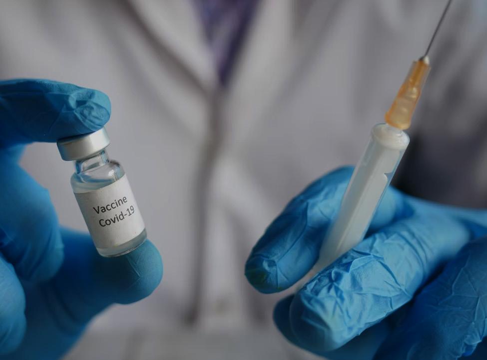
Myocarditis is characterized as a heterogeneous inflammatory disease with diverse etiologies and therapeutic responses.¹ Although normally caused by viral infections, recent cases of inflammatory responses to vaccine exposure have temporally associated hypersensitivity myocarditis with the mRNA COVID-19 vaccination.² Increased attention to myocarditis as a potential adverse complication of immunization is warranted.
An investigation by Fox et al. uncovered a greater diffuse distribution of CD68+ cells in the hearts of COVID-19 patients compared to matched control or other myocarditis patients. Their findings indicate that COVID-19-infected hearts contain diffusely infiltrative cells of monocyte or macrophage lineage instead of lymphocytes.³ A concurrent study by Bozkurt et al. suggests that interstitial cells, pericytes, and macrophages in the myocardium contain SARS-CoV-2 RNA by in situ hybridization. This may contribute to capillary endothelial cell or microvascular dysfunction and individual cell necrosis. Thus, macrophages, which mediate both local and systemic responses to viral infections, may activate apoptotic attack complexes to kill nearby myocytes.² Collectively, these studies suggest that COVID-19 incites a derivative of myocarditis that is unlike typical lymphocytic myocarditis.
Furthermore, a case series by Montgomery et al. recently identified 23 men (aged 20 to 51 years) diagnosed with myocarditis after vaccination.¹ These previously healthy patients presented with acute onset of marked chest pains and elevated cardiac troponin levels within four days of receiving an mRNA COVID-19 vaccine. Among eight patients who underwent cardiac magnetic resonance imaging (MRI), findings were consistent with the clinical diagnosis of myocarditis. Additional testing did not identify other etiologies for myocarditis, such as acute COVID-19, ischemic injury, or underlying autoimmune conditions. Cardiac symptoms resolved within seven days for 16 people.⁵
These findings cumulatively highlight the potential for rare adverse events, including cardiac injury, following COVID-19 vaccination.⁵ However, concerns regarding rare adverse complications following immunization should not diminish overall confidence in mRNA COVID-19 vaccines.
1. Mayo Clinic. Myocarditis [Internet]. 2021. Available from: https://www.mayoclinic.org/diseases-conditions/myocarditis/ symptoms-causes/syc-20352539 [cited 2021 Sep 18]. 2. Canadian Paediatric Society. Clinical guidance for youth with myocarditis and pericarditis following mRNA COVID-19 vaccination: Canadian Paediatric Society [Internet]. 2021. Available from: https://www.cps.ca/en/documents/position/clinicalguidance-for-youth-with-myocarditis-and-pericarditis [cited 2021 Sep 18]. 3. Fox SE, Falgout L, Heide RSV. Covid-19 myocarditis: Quantitative analysis of the inflammatory infiltrate and a proposed mechanism. Cardiovasc Pathol. 2021;54:107361. Available from: doi:10.1016/j.carpath.2021.107361. 4. Bozkurt B, Kamat I, Hotez PJ. Myocarditis with Covid-19 mRNA vaccines. Circulation. 2021;144(6):471-84. Available from: doi:10.1161/CIRCULATIONAHA.121.056135. 5. Montgomery J, Ryan M, Engler R. Myocarditis following immunization with mRNA COVID-19 vaccines in members of the US military. JAMA Cardiol. 2021;6(10):1202-6. Available from: doi:10.1001/jamacardio.2021.2833.
HUMAN TOUCH
INTRICATE MOLECULAR PATHWAYS UNDERLIE SIMPLE HUMAN SENSES
SURAJ BANSAL
The Nobel Prize in physiology or medicine was recently awarded to Drs. David Julius and Ardem Patapoutian for their parallel investigations into skin receptors for temperature and touch. Julius’ extensive classification of TRP proteins and Patapoutian’s identification of pressure-sensitive proteins provide complementary insight into understanding mechanobiology.¹
Julius et al. uncovered the TRPV1 receptor for capsaicin, an active component of chillies which elicits a burning sensation by stimulating the false presence of heat.¹ The TRPV1 receptor is a temperature-sensitive ion channel receptor in cell membranes that opens in response to high temperatures.² These receptors are abundant in pain pathways, making them important targets in the development of analgesics without the drawbacks of traditional opioid treatments.³
Independently, Julius and Patapoutian identified a similar ion receptor termed TRPM8, which opens in response to cold temperatures. Their investigations exploited the activity of these receptors in response to menthol, which triggers their opening. This discovery subsequently sparked a series of investigations into similar ion channels that allow for multiple different temperatures to be sensed.⁴
To identify touch receptors, Patapoutian and his colleagues used pressure-sensitive cells responsible for producing an electrical signal when prodded with a micropipette. Using this information, they identified 270 genes that could encode for touch receptor proteins. By systematically deactivating individual genes, they uncovered PIEZO1, a force-activated cation channel triggered by mechanical pressure and proprioception.⁵ This led to the discovery of another mechanosensitive ion channel, PIEZO2, which is present in sensory neurons and responsible for detecting physiological processes like blood pressure, bladder control, and breathing.⁶
These discoveries uncovered important physiological understandings for converting external stimuli into nerve signals at the molecular level. Taken together, these findings are promising for informing treatment development for neuropathic pain and associated conditions.
1. Cooney E, Molteni M. Scientists win Nobel in medicine for how we sense temperature and touch [Internet]. 2021. Available from: https://www.statnews.com/2021/10/04/nobel-prize-medicine-2021-temperature-touch-receptors [cited 2021 Oct 26]. 2. Le Page M. Medicine Nobel awarded for explaining how we sense heat and touch [Internet]. 2021. Available from: https:// www.newscientist.com/article/2292196-medicine-nobel-awarded-for-explaining-how-we-sense-heat-and-touch [cited 2021 Oct 26]. 3. Durrani J. Science behind sense of touch and temperature wins medicine Nobel prize [Internet]. 2021. Available from: https:// www.chemistryworld.com/news/science-behind-sense-of-touch-and-temperature-wins-medicine-nobel-prize/4014503. article [cited 2021 Oct 26]. 4. Jara-Oseguera A, Simon SA, Rosenbaum T. TRPV1: On the road to pain relief. Curr Mol Pharmacol. 2021;1(3):255-69. Available from: doi:10.2174/1874467210801030255. 5. Lewis T. 2021 medicine Nobel prize winner explains the importance of sensing touch [Internet]. 2021. Available from: https:// www.scientificamerican.com/article/2021-medicine-nobel-prize-winner-explains-the-importance-of-sensing-touch [cited 2021 Oct 26]. 6. Resnick B. Our amazing sense of touch, explained by a Nobel laureate [Internet]. 2021. Available from: https://www.vox.com/ science-and-health/22710533/nobel-prize-2021-ardem-patapoutian-touch [cited 2021 Oct 26]. 7. Oldach L. Nobel prize in physiology or medicine recognizes discoveries of ion channels [Internet]. 2021. Available from: https:// www.asbmb.org/asbmb-today/people/100421/2021-nobel-in-medicine [cited 2021 Oct 26].


