13 minute read
SPINAL SURGERY
from Issue 40
CURRENT LIMITATIONS AND FUTURE DIRECTIONS CURRENT LIMITATIONS AND FUTURE DIRECTIONS
ARTISTS: ARIM YOO & YOOHYUN PARK CRITICAL REVIEW
Advertisement
doi: 10.35493/medu.40.14 JUSTIN PHUNG
Bachelor of Health Sciences (Honours), Class of 2024, McMaster University phungj3@mcmaster.ca
ABSTRACT Surgical robotics have been introduced in a number of disciplines, with the aim of minimizing tissue disruption, reducing operating personnel radiation exposure, and improving dexterity and efficiency relative to human operation. In spinal surgery, robotic systems are relatively novel, applied to date largely for the placement of pedicle screw instrumentation. Only a few robotic systems have been approved for spinal surgery, and there remain significant barriers to the widespread implementation of surgical robotic techniques. This review provides an overview of robotic systems in spinal surgery and identifies current limitations that must be addressed before clinical use, including clinical merit relative to freehand navigation systems, steep learning curves, and unclear cost-effectiveness.
CONTEXT
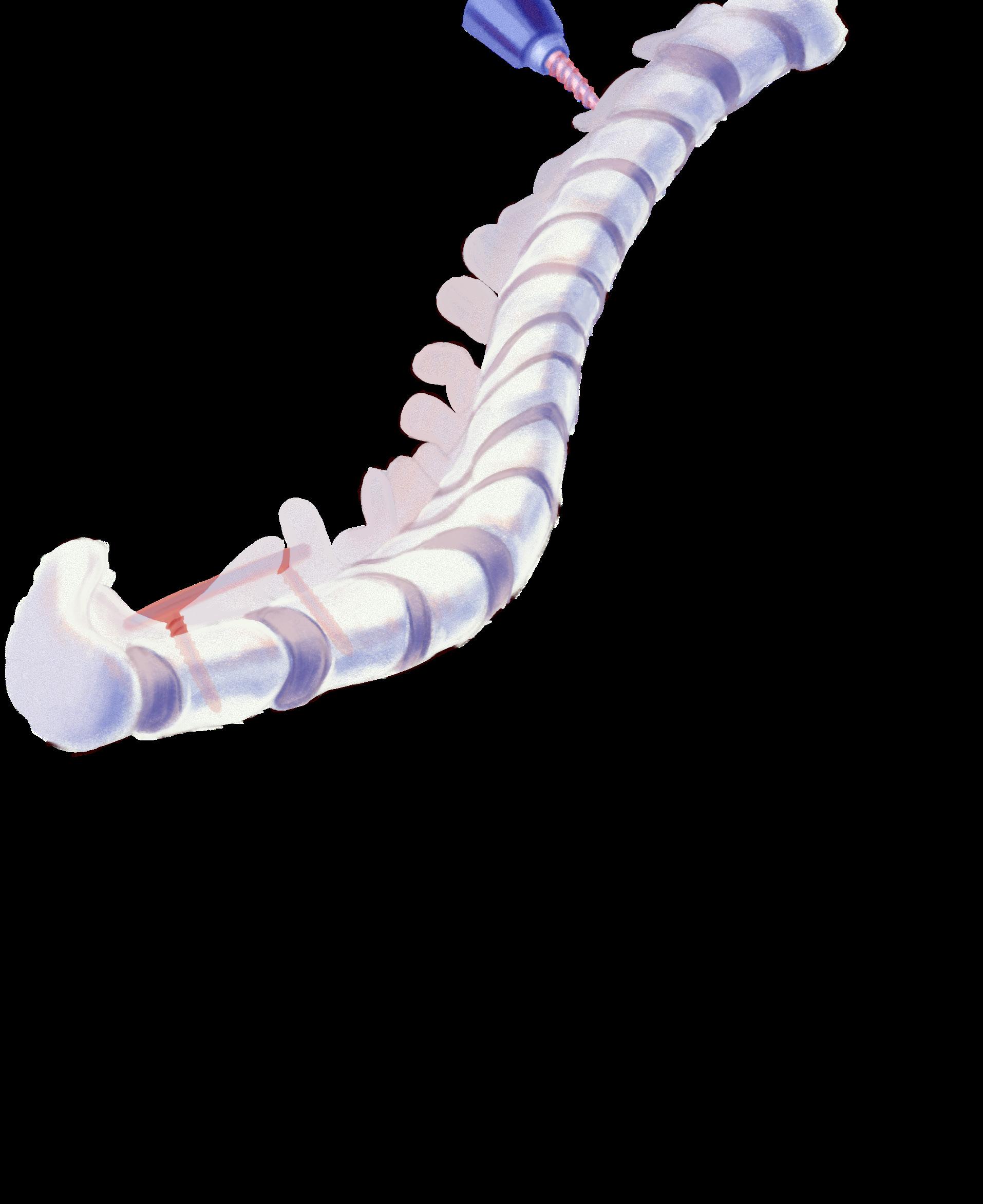
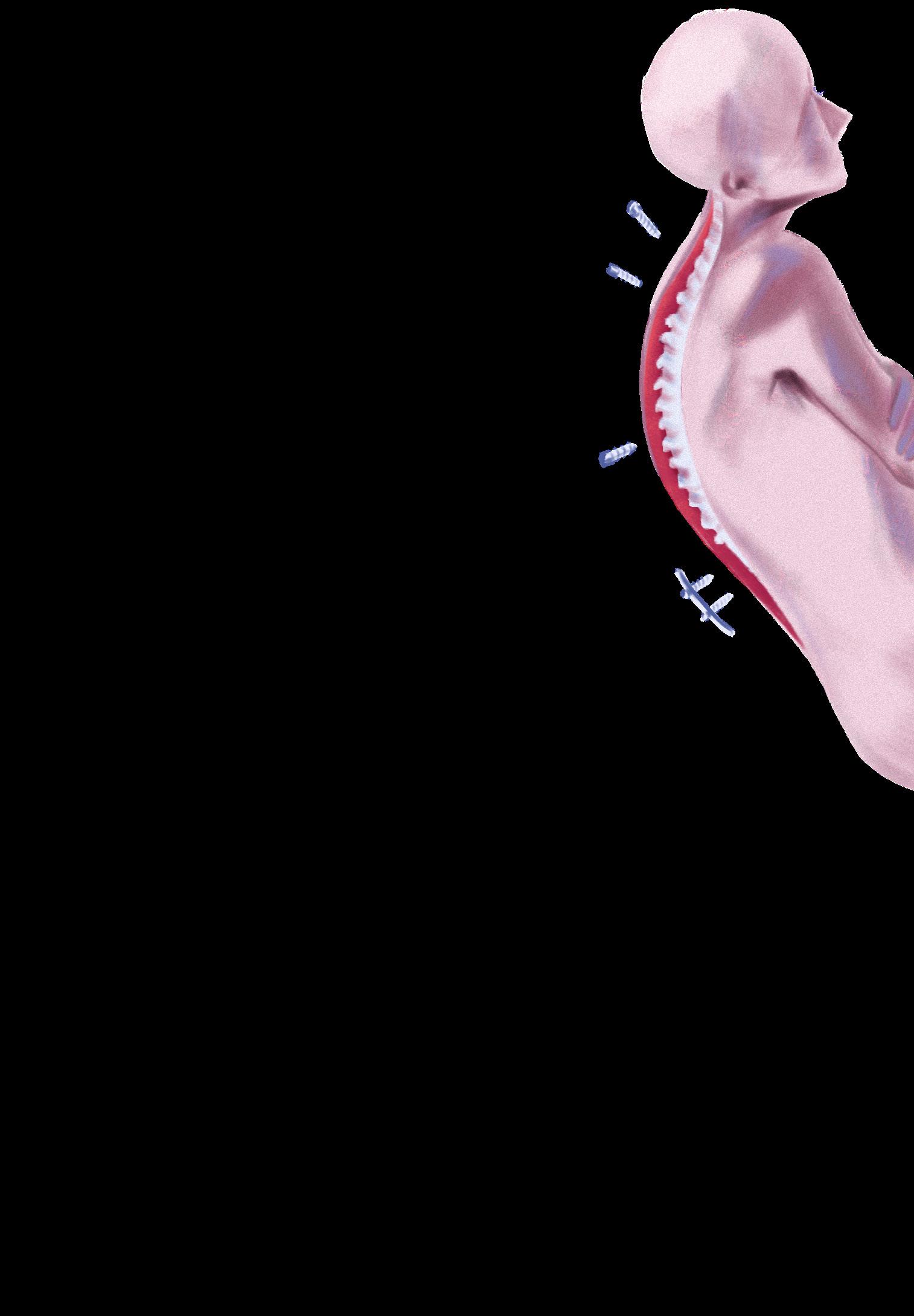
With aging populations, studies from the US and England have demonstrated a higher prevalence of degenerative spinal disorders, resulting in greater demands for lumbar spinal surgery.1,2 Spinal surgery is associated with higher rates of complications compared to other orthopaedic procedures; thus, robotics could significantly impact future spinal surgeries by improving safety and producing consistent results.3 Currently, the primary application of robotics is for pedicle screw insertion.3
Pedicle screw instrumentation connected to rod constructs in the thoracolumbar spine is the most commonly used technique, widely applied for degenerative, traumatic, neoplastic, and deformative spinal disorders.4,5 As such, this review will focus on pedicle screw placement with robotic guidance. Traditionally, pedicle screw fixation was conducted with the freehand technique.6 In expert hands, rates of successful placement were as high as 80-90%, though screw malposition can result in severe and potentially permanent clinical sequelae. Malpositioned screws may be associated with long-term poor construct strength and accelerated degeneration of adjacent spinal segments.7 Over the past two decades, intra-operative 2D-fluoroscopy navigation was introduced into spinal surgery to improve pedicle screw placement accuracy. In these procedures, pre-operative computed tomography (CT) scans were reconstructed to generate a 3D model of the spine.8 Despite the critical role of fluoroscopy in spinal navigation, operating room (OR) staff and the patient are exposed to significant harmful ionizing radiation.3,6 Thus, radiation exposure and operating time are important to consider. Modern image-guidance systems using CT scans have significantly reduced radiation exposure to OR staff. Intra-operative CT or 3D-fluoroscopy systems, in particular, allow real-time imaging of the patient in the surgical prone position (as opposed to supine positioning for a pre-operative CT), eliminating errors due to motion between pre- and intra-operative positioning.9 Intra-operative CT scan-based navigation has resulted in improved pedicle screw placement accuracy compared to 2D-fluoroscopy techniques.10,11 Robot-assisted (RA) spinal surgery allows the physician to insert surgical instruments and screws with the use of a rigid robotic arm and drill guide effector.12 RA spinal surgeries have therefore developed alongside navigation techniques including 2D-fluoroscopy, 3D-fluoroscopy, pre-operative CT, and intra-operative CT.13 In most cases, RA pedicle screw placement and novel imaging navigation systems have resulted in improved accuracy and efficiency of the tedious procedure, reducing human variation, fatigue, and radiation exposure for OR personnel.12,14-18

EVALUATING EFFECTIVENESS RA pedicle screw insertion has resulted in higher accuracy, safety, and feasibility of the procedure compared with its conventional alternatives. Two meta-analyses demonstrated that RA pedicle screw insertion operations had lower neurological complication rates and reduced risk of pedicle perforation compared to the freehand placement technique.6 Lieberman et al. also noted RA accuracy rates between 94.5-99%, even in cases involving severe deformity or revision surgeries due to congenital malformations, degenerative disorders, destructive tumors, and trauma.19 It must be noted, however, that no study to date has demonstrated improved screw placement accuracy with robotic guidance vs. freehand navigation guidance, with most prospective series citing accuracy rates in the 90%+ range for both techniques.20 Some current robotic systems include ROSA Spine, the Excelsius GPS, the TiRobot, and the Mazor X.21 To classify pedicle screw accuracy, the Gertzbein and Robbins system is frequently used, with grades A or B considered clinically acceptable and C or D considered unacceptable, often requiring immediate or delayed revision.22 The original model of ROSA Spine attained combined accu-


racy rates of 96.3%, 97.3%, and 98.3% with A and B grades.21 Similarly, the Excelsius GPS attained high levels of accuracy in two retrospective studies. Jain et al. noted 100% of the 66 reviewed post-operative CT screws were categorized as grade A or B, with no major screw-related complications from 643 total screws placed.23 Elswick et al. reported 97.6% grades A and B for their Excelsius GPS study involving 125 screws.24 Finally, a randomized prospective trial comparing the TiRobot with free-hand screw placement had similar conclusions, with 95.3% of scores classifying as grade A and 98.7% as combined grades A and B.14 Evidently, many available robot systems are effective and accurate.
CURRENT PRACTICAL LIMITATIONS Radiation exposure is one concern that must be addressed in the field of robotic spine surgery. In several studies, the RA technique was reported to provide a 40-70% reduction in intra-operative radiation exposure rates for the patient, the surgeon, and the OR personnel.19 The RA technique reduced the average radiation exposure time to 34 seconds, compared to the freehand fluoroscopic technique with an average exposure time of 77 seconds.19 Nevertheless, an average radiation exposure time of 34 seconds is still substantial, and in current use, navigation with intra-operative CT or 3D-fluoroscopy systems is associated with insignificant radiation exposure to OR personnel as they step out of the room during the scans, whereas robotic guidance requires several orthogonal single X-ray shots in the OR for registration.25,26 Given that pedicle screw insertions are common procedures, the cumulative radiation exposure over one’s lifetime can be carcinogenic. Future studies should investigate methods to facilitate the reduction of radiation exposure for subsequent RA systems. Furthermore, the surgical learning curve could limit widespread clinical application as RA software can be cumbersome and unintuitive.6 However, there have been several studies indicating that screw placement accuracy improves as more procedures are performed. Schatlo et al. reported that the rate of misplaced screws significantly decreased after surgeons completed 25 RA surgeries, while Hu et al. reported similar findings, where screw placement success rate increased after a surgeon’s first 30 patients.27,28 To address this learning curve, practicing under the supervision of an RA-experienced surgeon and engaging in wet lab or cadaver training prior to surgery in patients is suggested.3 The lack of research on clinical outcomes and cost-effectiveness also holds back patient and hospital decisions to undergo RA surgery, specifically due to the novelty of these systems and their questionable benefits relative to freehand navigation.29 Although evidence suggests intra-operative benefits, little is known about post-operative complications and duration of hospital stay.29 These factors influence patient perspective and can sway their decision to opt for RA surgery. Furthermore, barriers in assessing cost-effectiveness hinder RA spinal surgeries. One particular study by Menger et al. reported savings of $608 546 USD in a year associated with 557 RA thoracolumbar cases.30 However, the cost-effectiveness of the robot cannot be applied to smaller healthcare settings. Further reviews on post-operative outcomes and cost-effectiveness of RA spine surgery are necessary for the progression of robots into clinical settings.
FUTURE PROSPECTS With digital optics continuing to advance in the next decade, high-resolution imaging may serve as an alternative to intra-operative fluoroscopy. 7D Surgical, a Toronto-based company, offers a navigation system that utilizes digital stereoscopic topographical referencing and matches it with pre-operative CT, resulting in rapid image registration and elimination of intra-operative radiation exposure.21 Augmented reality (AR) is also a visualization technique to explore in spinal surgery, although it must currently remain with freehand actuation. Augmedics Xvision System is an AR platform that provides live 3D navigational feedback, reducing radiation exposure to OR personnel.21 Similarly, ImmersiveTouch and MagicLeap provide surgeons with intra-operative virtual headsets that allow real-time 3D visualization during the surgery, mitigating differences between the surgical environment and 2D intra-operative imaging.31 Elmi-Terander et al. achieved an accuracy of 94.1% using an AR surgical navigation system, while Alaraj et al. reported that ImmersiveTouch allowed trainee surgeons
to place screws precisely.32,33 Although accuracy and radiation exposure are critical to assess, future research should evaluate operative time, clinical outcomes, and intra- and post-operative complications.
CONCLUSION RA spinal surgery could potentially revolutionize the surgical field. Although clinical implementation is feasible, further research must be conducted to clarify the benefits and drawbacks of robotic spinal surgery. Overall, the evidence demonstrates that RA pedicle screw insertion is more accurate than its freehand counterpart, with several models achieving high GR grades. However, radiation exposure to OR personnel and the learning curve that surgeons face when adapting to new technology remain concerning. Further reviews on cost-effectiveness and clinical outcomes should be conducted to inform hospitals and patients of the benefits, especially as novel high-resolution imaging systems develop alongside RA spinal surgery. Comprehensive reports and evaluations in future publications are essential to pave the way for robotics in the field of spinal surgery.
REVIEWED BY: DR. DAIPAYAN GUHA (MD, PHD, FRCSC)
Dr. Daipayan (Deep) Guha is a spinal neurosurgeon at Hamilton Health Sciences and an Assistant Professor of neurosurgery at McMaster University. Some of his research interests include emerging imaging technologies for spinal surgery, the applications of augmented reality (AR) in Neurosurgery, and intra-operative three-dimensional spinal navigation.

1. Pannell WC, Savin DD, Scott TP, Wang JC, Daubs MD. Trends in the surgical treatment of lumbar spine disease in the United States. Spine J. 2015;15(8):1719–27. Available from: doi:10.1016/j.spinee.2013.10.014. 2. Sivasubramaniam V, Patel HC, Ozdemir BA, Papadopoulos MC. Trends in hospital admissions and surgical procedures for degenerative lumbar spine disease in England: A 15-year time-series study. BMJ Open. 2015;5(12):e009011. Available from: doi:10.1136/bmjopen-2015-009011. 3. Garg B, Mehta N, Malhotra R. Robotic spine surgery: Ushering in a new era. J Clin Orthop Trauma. 2020;11(5):753–60. Available from: doi:10.1016/j. jcot.2020.04.034. 4. Verma K, Boniello A, Rihn J. Emerging techniques for posterior fixation of the lumbar spine. J Am Acad Orthop Surg. 2016;24(6):357–64. Available from: doi:10.5435/JAAOS-D-14-00378. 5. Gaines RWJ. The use of pedicle-screw internal fixation for the operative treatment of spinal disorders. J Bone Joint Surg Am. 2000;82(10):1458-76. Available from: doi:10.2106/00004623-200010000-00013. 6. Vo CD, Jiang B, Azad TD, Crawford NR, Bydon A, Theodore N. Robotic spine surgery: Current state in minimally invasive surgery. Global Spine J. 2020;10(2 Suppl):34S-40S. Available from: doi:10.1177/2192568219878131. 7. Verma R, Krishan S, Haendlmayer K, Mohsen A. Functional outcome of computer- assisted spinal pedicle screw placement: A systematic review and meta-analysis of 23 studies including 5,992 pedicle screws. Eur Spine J. 2010;19(3):370-5. Available from: doi:10.1007/s00586- 009-1258-4. 8. Weiner JA, McCarthy MH, Swiatek P, Louie PK, Qureshi SA. Narrative review of intraoperative image guidance for transforaminal lumbar interbody fusion. Ann Transl Med. 2021;9(1):89. Available from: doi:10.21037/atm-20-1971. 9. Guha D, Jakubovic R, Gupta S, Fehlings MG, Mainprize TG, Yee A et al. Intraoperative error propagation in 3-dimensional spinal navigation from nonsegmental registration: A prospective cadaveric and clinical study. Global Spine J. 2019;9(5):512-20. Available from: doi:10.1177/2192568218804556. 10. Wood M, Mannion R. A comparison of CT-based navigation techniques for minimally invasive lumbar pedicle screw placement. J Spinal Disord Tech. 2011;24(1):E1-5. Available from: doi:10.1097/BSD.0b013e3181d534b8. 11. Silbermann J, Riese F, Allam Y, Reichert T, Koeppert H, Gutberlet M. Computer tomography assessment of pedicle screw placement in lumbar and sacral spine: Comparison between free-hand and O-arm based navigation techniques. Eur Spine J. 2011;20(6):875–81. Available from: doi:10.1007/ s00586-010-1683-4. 12. Chen H-Y, Xiao X-Y, Chen C-W, Chou H-K, Sung C-Y, Lin FH et al. A spine robotic-assisted navigation system for pedicle screw placement. J Vis Exp. 2020;(159):e60924. Available from: doi:10.3791/60924. 13. Amr AN, Giese A, Kantelhardt SR. Navigation and robot-aided surgery in the spine: Historical review and state of the art. Robot Surg. 2014;1:19–26. Available from: doi:10.2147/RSRR.S54390. 14. Han X, Tian W, Liu Y, Liu B, He D, Sun Y, et al. Safety and accuracy of robotassisted versus fluoroscopy-assisted pedicle screw insertion in thoracolumbar spinal surgery: A prospective randomized controlled trial. J Neurosurg Spine. 2019;30(5):615–22. Available from: doi:10.3171/2018.10.SPINE18487. 15. Hyun S-J, Kim K-J, Jahng T-A, Kim H-J. Minimally invasive robotic versus open fluoroscopic-guided spinal instrumented fusions: A randomized controlled trial. Spine. 2017;42(6):353–8. Available from: doi:10.1097/ BRS.0000000000001778. 16. Kantelhardt SR, Martinez R, Baerwinkel S, Burger R, Giese A, Rohde V. Perioperative course and accuracy of screw positioning in conventional, open robotic-guided and percutaneous robotic-guided, pedicle screw placement. Eur Spine J. 2011;20(6):860–8. Available from: doi:10.1007/s00586-0111729-2. 17. Keric N, Eum DJ, Afghanyar F, Rachwal-Czyzewicz I, Renovanz M, Conrad J, et al. Evaluation of surgical strategy of conventional vs. percutaneous robotassisted spinal trans-pedicular instrumentation in spondylodiscitis. J Robot Surg. 2017;11(1):17–25. Available from: doi:10.1007/s11701-016-05975. 18. Stüer C, Ringel F, Stoffel M, Reinke A, Behr M, Meyer B. Robotic technology in spine surgery: Current applications and future developments. Acta Neurochir Suppl. 2011;109:241–5. Available from: doi:10.1007/978-3-211-996515_38. 19. Lieberman IH, Kisinde S, Hesselbacher S. Robotic-assisted pedicle screw placement during spine surgery. JBJS Essent Surg Tech. 2020;10(2):e0020. Available from: doi:10.2106/JBJS.ST.19.00020. 20. Peng YN, Tsai LC, Hsu HC, Kao CH. Accuracy of robot-assisted versus conventional freehand pedicle screw placement in spine surgery: A systematic review and meta-analysis of randomized controlled trials. Ann Transl Med. 2020;8(13):824. Available from: doi:10.21037/atm-20-1106. 21. Huang M, Tetreault TA, Vaishnav A, York PJ, Staub BN. The current state of navigation in robotic spine surgery. Ann Transl Med. 2021;9(1):86. Available from: doi:10.21037/atm-2020-ioi-07. 22. Vardiman AB, Wallace DJ, Booher GA, Crawford NR, Riggleman JR, Greeley SL, et al. Does the accuracy of pedicle screw placement differ between the attending surgeon and resident in navigated robotic-assisted minimally invasive spine surgery? J Robot Surg. 2020;14(4):567–72. Available from: doi:10.1007/s11701-019-01019-9. 23. Jain D, Manning J, Lord E, Protopsaltis T, Kim Y, Buckland AJ, et al. Initial single-institution experience with a novel robotic-navigation system for thoracolumbar pedicle screw and pelvic screw placement with 643 screws. Int J Spine Surg. 2019;13(5):459–63. Available from: doi:10.14444/6060. 24. Elswick CM, Strong MJ, Joseph JR, Saadeh Y, Oppenlander M, Park P. Robotic-assisted spinal surgery: Current generation instrumentation and new applications. Neurosurg Clin N Am. 2020;31(1):103–10. Available from: doi:10.1016/j.nec.2019.08.012. 25. Mendelsohn D, Strelzow J, Dea N, Ford NL, Batke J, Pennington A, et al. Patient and surgeon radiation exposure during spinal instrumentation using intraoperative computed tomography-based navigation. Spine J. 2016;16(3):343-54. Available from: doi:10.1016/j.spinee.2015.11.020. 26. Klingler JH, Scholz C, Krüger MT, Naseri Y, Volz F, Hohenhaus M, et al. Radiation exposure in minimally invasive lumbar fusion surgery: A randomized controlled trial comparing conventional fluoroscopy and 3D fluoroscopybased navigation. Spine. 2021;46(1):1-8. Available from: doi:10.1097/ BRS.0000000000003685. 27. Hu X, Lieberman IH. What is the learning curve for robotic-assisted pedicle screw placement in spine surgery? Clin Orthop Relat Res. 2014;472(6):1839–44. Available from: doi:10.1007/s11999-013-3291-1. 28. Schatlo B, Martinez R, Alaid A, von Eckardstein K, Akhavan-Sigari R, Hahn A, et al. Unskilled unawareness and the learning curve in robotic spine surgery. Acta Neurochir (Wien). 2015;157(10):1819–23. Available from: doi:10.1007/ s00701-015-2535-0. 29. Huang J, Li Y, Huang L. Spine surgical robotics: Review of the current application and disadvantages for future perspectives. J Robot Surg. 2020;14(1):11–6. Available from: doi:10.1007/s11701-019-00983-6. 30. Menger RP, Savardekar AR, Farokhi F, Sin A. A cost-effectiveness analysis of the integration of robotic spine technology in spine surgery. Neurospine. 2018;15(3):216–24. Available from: doi:10.14245/ns.1836082.041. 31. McDonnell JM, Ahern DP, Ó Doinn T, Gibbons D, Rodrigues KN, Birch N, et al. Surgeon proficiency in robot-assisted spine surgery. Bone Joint J. 2020;102B(5):568–72. Available from: doi:10.1302/0301-620X.102B5.BJJ-20191392.R2. 32. Elmi-Terander A, Burström G, Nachabe R, Skulason H, Pedersen K, Fagerlund M, et al. Pedicle screw placement using augmented reality surgical navigation with intraoperative 3D imaging. Spine. 2019;44(7):517–25. Available from: doi:10.1097/BRS.0000000000002876. 33. Alaraj A, Lemole MG, Finkle JH, Yudkowsky R, Wallace A, Luciano C, et al. Virtual reality training in neurosurgery: Review of current status and future applications. Surg Neurol Int. 2011;2:52. Available from: doi:10.4103/21527806.80117.


