


This column, brought to you by Merck Animal Health, features insightful answers from leading minds.
The current development of sequenc ing technology has broadened the scope of infectious disease monitor ing. Genetic sequencing is a powerful tool for understanding the significance of antigenic drift in the equine influenza virus (EIV) and subsequently determining how well an equine influenza vaccine might protect against a par ticular influenza virus. To help shed some light on this subject, Dr. Kyuyoung Lee discusses his recently published article, “Genome-in formed characterization of antigenic drift in the hemagglutinin gene of equine influenza strains circulating in the United States from 2012-2017.”
Antigenic drift of EIV is evolutionary bi ology at its best—for survival, a species must constantly adapt, evolve and rapidly reproduce in the face of competing species. This “Red Queen hypothesis," as it’s been termed, helps explain that in order to keep one strain of EIV spreading in a horse popu lation, the strain should compete with many other novel variants each season. In this pro cess, transmissibility or survival against the host's immunity are key factors in determin ing the compatibility of each strain. If one strain dominantly spreads in one season, the dominant strain will reproduce many novel variants rooted on its own genetic character istic. These novel descendants will compete
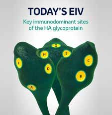
again in the next season. As EIV continu ously repeats this process, the EIV strains accumulate mutations in key antigenic sites, such as the hemagglutinin (HA) or neur aminidase gene, which can possibly affect their transmissibility or survival against the host's immunity. We call this antigenic drift: the continuous accumulation of genetic mutations over time in key antigenic sites through the natural selection.

Many studies of influenza virus revealed multiple key antigenic sites in the HA gene. One significant study is Woodward et al (2015),1 which mapped key antigenic sites in the HA protein structure of EIV H3N8. The study found that even though EIV vaccines were widely used in the global horse popula tion, EIV epidemics occurred seasonally, and
the epidemic EIV strains had unique genetic differences in key antigenic sites in the HA gene compared with vaccine strains. Consid ering that antigenic drift selects novel EIV strains that are able to avoid hosts’ immune response (either naturally infected or vaccine induced), we can infer that EIV strains with genetic changes in specific key antigenic sites in the HA gene would have higher compat ibility than other strains.
Conventionally, antigenic modifications of EIV strains were evaluated by serologi cal tests, such as the Haemagglutination test comparing vaccine and wild type strains. Current sequencing technology allows us to collect nucleotide sequences of pathogens much faster and easier. Thanks to this de velopment, our study collected nucleotide sequences of the HA gene of EIV strains in samples provided by equine veterinary prac titioners from EIV-infected U.S. horses from 2012-2017. We concluded that antigenic drift of EIV was the most probable cause of the in creased incidence of influenza vaccine failure in the U.S., as opposed to the introduction of foreign EIV strains.
Continued monitoring through genetic se quencing of dominant wild EIV strains is an essential practice for not only understanding what’s circulating but, as important, develop ing vaccine strains to combat these novel EIV variants infecting horses.
For more information about antigenic drift and vaccine efficacy, visit https://www.merckanimal-health-usa.com/species/equine/ prestige-equine-vaccines
“How does antigenic drift impact equine influenza vaccine efficacy?”Copyright © 2022 Merck & Co., Inc., Rahway, NJ, USA and its affiliates. All rights reserved.
The number of organisms linked to placen titis in mares is increasing, according to Margo L. Macpherson, DVM, MS, DACT, a professor at the University of Florida, College of Veterinary medicine. This is especially true with focal mucoid placentitis.
“There are some very interesting new bugs out there,” she said, and they include both gram-positive and gram-negative organisms. In addition to the wellknown Streptococcus zooepidemicus or Escherichia coli; Amycolatopsis, Crosiella equi, Aspergillus, Steno trophomonas maltophilia, Mycobacterium goodii, Pas teurella pneumotropical and Pantoea agglomerans are also being seen. The 2 gram-negative organisms that are very unusual—Pasteurella pneumotropica and Pantoea agglomerans—are both organisms that reside in soil, water and plant material.
“They are just commensal organisms [P. pneumo tropica and P. agglomerans] that are out there that are being identified as causing focal mucoid placentitis in these mares.

“That's a little bit hard for me to wrap my head around when I get a diagnosis, and I don't recognize the organism when so many of the cases have been identified as being Streptococcus or E. coli,” she admit ted during a presentation at the BEVA Congress 2022. In addition, some mares suffer from both ascending and focal mucoid placentitis.
“The goal of all of us as veterinarians is to address
equine placentitis in whatever form we have and take it from the terrible infection to the live foal,” she said, “but that’s a difficult journey because gathering infor mation about this condition has been slow. Part of the problem, of course, is the gestational length of a foal. It takes a long time to see the out come in any studies of pregnant mares with placentitis.”
But there is work that has enabled a better understanding of the disease, and that is looking for more treatment options. Ascending placentitis, for instance, begins as a bacterial infection that migrates and infiltrates the chorioallantois causes a tre mendous inflammatory reaction that releases many different cytokines.
“We know that within the tissues of the pla centa, the uterus, in the fetal fluids, on the umbilicus of that foal and the fetus, we're seeing an upregulation of inflammatory cytokines, predominantly interleu kin 6 [IL-6] and IL-1beta,” she said.
“This information is very important for us to un derstand. There is a tremendous upregulation and dysregulation in the immune response,” she said.
“This is not just a bacterial infection [that we treat]
By Marie Rosenthal MS
and then we go on our merry way.”
Studies suggest that there are 3 components to an ascending pla centitis that involves the inflam mation that occurs at the placenta, endometrial activation and then cervical remodeling.
When the bacteria infiltrate the chorioallantois, they initi ate a reaction that stimulates the formation of toll-like receptors, which stir up the inflammatory re sponse, and send leukocytes into the placental tissues, which affects progestin production, turning off progestins that “keep the uterus quiet” and turning on those that initiate parturition.
“Our basic treatment approach has always been to try to address the bacteria,” she said, “so, provide some sort of an antimicrobial to address the inflam mation and cause immunomodulation by some sort of an anti-inflammatory and to keep the uterus quiet by somehow counteracting the prostaglandins that are produced.”
Unfortunately, the list of antibiotics that penetrate fetal membranes is short, she admitted: penicillin, gen tamicin and trimethoprim-sulfamethoxazole (TMPSMX) penetrate the fetal membranes and can be detect ed in the fetal fluids at concentrations able to inhibit S. zoepidemicus and E. coli. Enrofloxacin and doxycycline also appear to reach the fetal fluids in healthy mares but have not been tested to see if they can be effective in treating placentitis.
Ceftiofur crystalline-free acid and ceftiofur so dium have been tested in pregnant mares, but do not pass the fetal membranes to enter the fetal fluids. “We don't consider those to be adequate therapeutics for mares with placentitis,” she said.
“Of those that have been tested for treatment of placentitis, it’s an even shorter list,” she said.
An anti-inflammatory is often prescribed to ad dress the cytokine cascade, but that is a pretty short list, too: flunixin meglumine, firocoxib and pentoxifylline. Flunixin meglumine, was not detected in fetal fluids of pregnant mares, and it inhibits both COX 1 and 2, but COX 1 is important for normal homeostatic mecha nisms, so it does not appear useful in placentitis.
Pentoxifylline does pass the fetal membrane and attains therapeutic concentration in the fetal fluids and tissues of the fetal membranes as well as the fetus.
Firocoxib is a potent COX 2 inhibitor, and in a
study done by Dr. Macpherson’s group, in which they infected pregnant pony mares with an ag gressive organism that causes pla centitis and compared them with a normal group of pregnant mares, treating them with firocoxib.
“It’s a little bit of a mixed bag. Prostaglandins were inhibited in tissues as well as fetal fluids in mares that were administered firocoxib. So, we've seen some evidence to support some of our treatment and that's what makes me feel good at the end of the day,” Dr. Macpherson said.
But that wasn’t the endpoint of the study. All the foals died in the placentitis group. However, she said the placentitis was an aggressive form of the disease.
“Combined therapies are where we've put the em phasis on in terms of treating mares with placentitis,” she said. Scott Bailey, DVM, while at the University of Florida showed that if you treat mares as soon as disease was detected with TMP-SMX, pentoxifylline and altrenogest until the mares delivered foals, the foal survival rate was encouraging.
However, outside of a clinical study, the results of the combination have been inconsistent. And they are not entirely sure why. However, it could be the organ isms are resistant to the antibiotic.
It is important to remember that even if a foal is born alive, it will need supportive care, she reminded. She talked about 1 neonatal foal that was almost eutha nized but responded to the Madigan squeeze.
“I was very close to euthanizing it, and the Madi gan squeeze is why it's alive today.
“But my take-home message truly is that you can not assume that the treatment's going to deliver that live foal and it's going to be normal at birth. It will require supportive care in some fashion or another. Typically, we just assume that if it's been in a bacte rial infection in that environment, it will need anti microbials and anti-inflammatories and so forth,” Dr. Macpherson said.
She said there is a lot of exciting work going on in placentitis, and she hopes that more options will be available shortly. Of particular note are several stud ies recently published by the group at the University of Kentucky, which is helping us to better understand the disease process. In turn, this information will help us select effective treatments for placentitis. MeV
Even if a foal survives, it will need supportive care, often antibiotics and antiinflammatories.
For more than 30 years, Adequan® i.m. (polysulfated glycosaminoglycan) has been administered millions of times1 to treat degenerative joint disease, and with good reason. From day one, it’s been the only FDA-Approved equine PSGAG joint treatment available, and the only one proven to.2, 3
Reduce inflammation
Restore synovial joint lubrication
Repair joint cartilage
Reverse the disease cycle
When you start with it early and stay with it as needed, horses may enjoy greater mobility over a lifetime.2, 4, 5 Discover if Adequan is the right choice. Visit adequan.com/Ordering-Information to find a distributor and place an order today.

BRIEF SUMMARY: Prior to use please consult the product insert, a summary of which follows: CAUTION: Federal law restricts this drug to use by or on the order of a licensed veterinarian. INDICATIONS: Adequan® i.m. is recommended for the intramuscular treatment of non-infectious degenerative and/or traumatic joint dysfunction and associated lameness of the carpal and hock joints in horses. CONTRAINDICATIONS: There are no known contraindications to the use of intramuscular Polysulfated Glycosaminoglycan. WARNINGS: Do not use in horses intended for human consumption. Not for use in humans. Keep this and all medications out of the reach of children. PRECAUTIONS: The safe use of Adequan® i.m. in horses used for breeding purposes, during pregnancy, or in lactating mares has not been evaluated. For customer care, or to obtain product information, visit www.adequan.com. To report an adverse event please contact American Regent, Inc. at 1-888-354-4857 or email pv@americanregent.com. Please see Full Prescribing Information at www.adequan.com.
www.adequan.com
1 Data on file.
2 Adequan® i.m. Package Insert, Rev 1/19.
3 Burba DJ, Collier MA, DeBault LE, Hanson-Painton O, Thompson HC, Holder CL: In vivo kinetic study on uptake and distribution of intramuscular tritium-labeled polysulfated glycosaminoglycan in equine body fluid compartments and articular cartilage in an osteochondral defect model. J Equine Vet Sci 1993; 13: 696-703.

4 Kim DY, Taylor HW, Moore RM, Paulsen DB, Cho DY. Articular chondrocyte apoptosis in equine osteoarthritis. The Veterinary Journal 2003; 166: 52-57.
5 McIlwraith CW, Frisbie DD, Kawcak CE, van Weeren PR. Joint Disease in the Horse.St. Louis, MO: Elsevier, 2016; 33-48.
All trademarks are the property of American Regent, Inc.
© 2021, American Regent, Inc.
PP-AI-US-0629 05/2021
A conventional in vitro fertilization (IVF) technique for horses was developed by Katrin Hin richs, DVM, PhD, DACT, and colleagues at New Bolton Center.
The 3 mares—into which resultant embryos were transferred—each carried healthy foals to term.

IVF in horses has been frustrating, according to Dr. Hinrichs, who is the Harry Werner Endowed Professor of Equine Medicine, chair of the Depart ment of Clinical Studies-New Bolton Center. One she and her team have been trying to tackle for more than 3 decades.
“When we put horse sperm with eggs, they don’t even try to penetrate them. They just swim happily about, ignoring the egg, leaving us with a zero-fertil ization rate,” said Dr. Hinrichs, who is also a profes sor of reproduction in the University of Pennsylva nia School of Veterinary Medicine.
Dr. Hinrichs and others have developed tech niques to produce embryos using intracytoplasmic sperm injection (ICSI), a method of fertilization that requires the technically challenging injection of a single sperm into a single oocyte aided by a highpower microscope and manipulation equipment.
 By Katherine Unger Baillie


However, supporting sperm to achieve “true” IVF—in which sperm incubated in a Petri dish fertilize an oocyte without fur ther manipulation, as they would naturally inside a mare—proved elusive.
Until now.
“The demand for assisted re productive technologies like IVF is getting larger and larger in the horse-breeding community,” Dr. Hinrichs said.
“The approach we’ve devel oped would allow more veterinary practices to offer IVF, as it doesn’t require the expensive equipment and training needed to do it the way it’s done now—by injecting each sperm into each egg. But for me the fun part is just nailing this down. I’ve been a horse person all my life, and for decades we have tried to figure out why this doesn’t work in horses. And now we have a repeatable method that does work, so we can explore the why.”
One of the most successful forms of IVF in hors es to date, is ICSI, using a tiny needle to pick up a sole sperm and inject it into an oocyte. In the early 2000s, Dr. Hinrichs, then at Texas A&M University, increased the efficiency of that procedure and devel oped methods to culture the resulting embryo in the laboratory until it could be easily transferred with out surgery to a recipient mare. By around 2009, Dr. Hinrichs’ clinic offered this, and specialized facilities around the world continue to do so.
Still, Dr. Hinrichs kept pursuing the development of a simplified, conventional IVF procedure. After her lab had devoted considerable energy to studying oocytes, around 2011 she turned attention to sperm. For sperm to fertilize an egg it must undergo a se ries of physiological changes in a process known as capacitation.
In 2019, a researcher in Dr. Hinrichs’ lab, Matheus Felix, now chief embryologist in the Penn Equine Assisted Reproduction Laboratory, began investigat ing how long it takes for horse sperm to capacitate and what conditions support that process.
The team had gathered clues that sperm from horses might need more time than that of other spe cies, such as mice, to fertilize eggs. So, they tried a longer-than-normal incubation.
“Horse sperm are finicky and like to die in cul ture,” Dr. Hinrichs said, “but we had done some pre vious work that suggested factors that could prolong
their life during incubation.”
When Dr. Felix employed a complex medium for incubating them, which contains the com pounds penicillamine, hypotau rine and epinephrine (PHE), the team finally found a way to keep sperm alive in culture for more than a few hours.
“He tried to culture the sperm overnight under these condi tions, and by gosh it worked,” Dr. Hinrichs said. “The sperm were alive the next day, which is a tri umph.”
When Dr. Felix tried again, incubating the sperm overnight and then adding an oocyte, he documented signs of fertilization. “Because typical results in the horse are zero, this 1 fertilized oocyte was a sign that the process could work, and we were off on our journey to develop the procedure,” Dr. Hinrichs said.
Using the fledgling procedure as the basis for op timizing equine IVF, the research team found that pre-incubating the sperm for 22 hours in the PHE medium, then co-incubating it with oocytes for 3 hours, led to the greatest efficiency, including a 74% rate of production of blastocysts; 3 were transferred to mares that are part of the PennVet research herd. Three healthy foals were born as a result.
The technique had a fertilization rate 90% fertil ization rate.
There is still room to improve on the methods, ac cording to Dr. Hinrichs. The approach only worked well with fresh sperm. Frozen sperm, which is the most practical method for clinical IVF, did not result in impressive fertilization rates. And the PHE me dium is cumbersome to make, meaning slight varia tions could compromise the procedure’s success.
“For the first time, we have a method that works, and we can use it as a basis to explore what it is that makes it work and what variations are possible: how to make the procedure simpler and more applicable to practice,” Dr. Hinrichs explained. MeV
The article was originally posted on the PennVet website. It has been edited for style and content. https://www.vet.upenn.edu/about/news-room/newsstories/news-story-detail/making-true-equine-ivf-areproducible-success
This approach may allow more veterinary practices to offer IVF, as it does not require expensive equipment or training.
Running an equine veterinary practice is both a passion and a business. While caring for horses is your priority, you also need to get paid for the expertise you provide. The CareCredit healthcare credit card can help meet the nancial needs of your business and your clients.

CareCredit is a exible payment solution that comes with budget-friendly nancing options that allow your clients to pay over time for their horse’s care. And you get paid within two business days, so you can practice medicine instead of chasing A/R.
Enroll for free today. Call 844-812-8111 and mention code MEV2022VA. O er ends December 31, 2022.
Insulin dysregulation is a central component of equine metabolic syndrome, and it is closely associ ated with laminitis in equids of all stripes, including ponies and donkeys.

20%
Determining the degree of a horse’s insulin dys regulation can serve as an important foundation for dietary changes and medical interventions that may prove to be inconvenient for some owners. However, the current testing protocols have certain limitations that could muddy the waters.
Currently, the diagnosis of insulin dysregulation in the field involves testing for hyperinsulinemia and insulin resistance, with hyperinsulinemia assessed by basal testing of serum insulin concentration. Ad ditionally, dynamic testing includes an oral sugar and oral glucose test, which also encompasses the enteroinsular axis.
Insulin resistance is typically assessed in the field with a 2-step insulin tolerance test.
There are certainly limitations to these current testing protocols. Particularly, basal testing for hy perinsulinemia that does not include the enteroinsu lar axis. It also has limited reported sensitivity.
Further, varying cutoffs are reported in the litera ture for the oral sugar tests, with different doses of corn syrup and different time points.
To further evaluate these common testing protocols and narrow down the appropriate cutoffs, my col
leagues and I created a study looking at the oral sugar test and the 2-step insulin tolerance test for insulin dysregulation testing in ponies.

We also wanted to explore the relationship be tween insulin resistance, basal hyperinsulinemia, and dynamic hyperinsulinemia, and evaluate the 2 tests for their association with laminitis. The hypoth esis was that the characteristics of the oral sugar test and the 2-step insulin tolerance tests would [clarify] the association with laminitis through updated diag nostic cutoff values.

The team recruited 146 client-owned ponies for the study. As part of the initial clinical assessment, they evaluated the ponies for adiposity—both re gional and generalized—and determined the ponies’ cresty neck and body-condition scores. Current lam initis was evaluated using the modified Obel scoring system, with laminitis defined as a score >1.
We performed 2 farm visits on consecutive days. On the first day we used the insulin tolerance test to evaluate for insulin resistance, and on the following day—after an overnight fast—we performed the oral sugar test with a corn syrup dose of 0.45 mL/kg.
Insulin resistance was defined as a glucose level that was <50% of baseline. Basal hyperinsulinemia was defined using cutoffs of >20 μIU/mL or >50 μIU/ mL, and dynamic hyperinsulinemia was defined us ing cutoffs of >45 μIU/mL or >60 μIU/mL at 60- and 90-minutes post-administration of corn syrup.
Basal hyperinsulinemia was present in 16% of ponies using the 20 μIU/mL cutoff, and 10% had basal hy perinsulinemia at 50 μIU/mL.
We had 79 ponies that were diagnosed as insulin resistant based on the 2-step insulin tolerance test.
On the oral sugar test, dynamic hyperinsulinemia was present in 42% of ponies at 60 minutes and 45% at 90 minutes using the 45 μIU/mL cutoff. Almost 60 ponies (38%) were positive at both times. Four ponies were positive at 60 minutes but not at 90 min utes, and 9 ponies were positive at 90 minutes but not at 60 minutes.
For the 60 μIU/mL cutoff, 36% were positive at 60 minutes, 38% were positive at 90 minutes, and 32% were positive at both times. Six ponies were positive
DHI >45 μIU/mL (60 minutes) 80% 74%
DHI >60 μIU/mL at (60 minutes) 69% 79%
DHI >45 μIU/mL (90 min) 76% 70%
DHI >60 μIU/mL (90 min) 66% 75%
at 60 minutes but not 90 minutes, and 9 ponies were positive at 90 minutes but not 60 minutes.
For the oral sugar test, we had 46 ponies [32%] that were posi tive for all the diagnostic criteria in the study and at all time points.
To evaluate the diagnostic overlap, the team combined the results from both tests.
There were 18 ponies that were positive with all the diagnostic crite ria. There were 22 ponies that were only insulin resistant, and there were no ponies that had basal hy perinsulinemia that were positive on the other tests, and there were 39 ponies that were positive on the insulin tolerance test that also had dynamic hyperinsulinemia.”
The team then evaluated the ROC curves to gauge the sensitivity and specificity of the tests with lamini tis as the outcome. Results were mixed.
VARIABLE OPTIMAL
BHI >3.6 μIU/mL 76% 54%
DHI 60-min >30.4 μIU/mL 95% 63%
DHI 90-mi >43.5 μIU/mL 80% 69%
2-step ITT % glucose reduction <44.5% 80% 69%
the cutoff was slightly reduced at >30 μIU/mL. With the 2-step in sulin tolerance test, we found an optimal percent glucose reduc tion of less than 44.5% of base line, which is similar to the 50% cutoff that we tend to use clini cally.
In this population, we found that it wasn’t possible to capture all the insulin dysregulated indi viduals based on 1 test. Dynamic hyperinsulinemia did have im proved results when compared with basal testing, which has been reported in the literature.
Decreasing cutoffs for dynamic hyperinsulinemia—particularly with the oral sugar test—would reduce specificity, but the result would increase the number of false positives.
For basal hyperinsulinemia, the specificity was acceptable (94% at >20 μIU cutoff; 99% at >50 μIU), but the sensitivity was quite lower, at 38% and 29% at both cutoffs, respectively.
For dynamic hyperinsulinemia, the results were more promising (see Table 1).
Looking at the 2-step insulin tolerance test as a measure of insulin resistance, we had acceptable sensitivity [82%] that was an improvement over the dynamic testing of hyperinsulinemia, but a reduced specificity at 58%.
There was no association between measures of adiposity and laminitis.
Optimal ROC cut-offs were determined using the Youden Index with laminitis as the outcome (see Table 2).
With dynamic hyperinsulinemia at 60 minutes,
Clinically, we tend to test ponies for insulin dysregulation or endocrinopathic laminitis that are overweight by putting them on a diet and re ducing non-structural carbohydrates, for example,” she said. “That’s probably more important than increasing the false negatives.”
The study showed slightly improved testing char acteristics at 60 minutes vs. 90 minutes based on the ROC curves. MeV
Brianna L. Clark, BVSc (Hons), MANZCVS, is an equine internal medicine resident at the University of Queensland, Australia.
Paul Basilio is a regular contributor to the Modern Equine Vet. He did not participate in the study discussed here.
We know horse people because we’re horse people. And like you, the love and respect we have for horses is unconditional. Everything we do is for their benefit. If we do right by the horse, we’ll never do wrong.

 By Jessica Cook
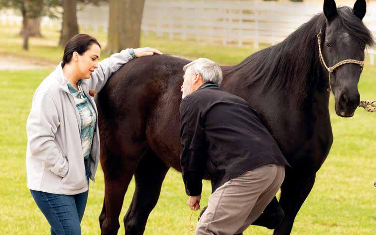
Every practice will have its own protocols for com mon procedures. This ar ticle will describe—step by step—how we perform an ultrasound guided in jection of a palmar metacarpal soft-tissue injury of the forelimb using an extracellular matrix. The procedure varies minimally depending upon the reparative ther apy chosen by the client.

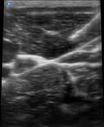

We most commonly use the extracellular matrix, MatriStem MicroMatrix, which is an off-the-shelf urinary bladder matrix available in 100 mg and 200 mg vials for reconstitution with normal saline. Other orthobiologics include platelet rich plasma (PRP), which can be processed on-site with any number of concentration centrifuges and stem cells. Stem cells add time and expense to the procedure due to the need to harvest marrow or fat to be cultured for con centration within a laboratory.
Our practice cares for several steeplechase-racing patients, which are generally running 2 plus miles
over jumps. As you can imagine tendon and suspen sory injuries commonly occur under this amount of stress. Performance horses in multiple other disci plines make up the rest of our patients and soft tis sue injuries can occur in any stress or strain sport, or in acute incidents from pasture. Most owners and trainers elect to rehabilitate these injuries, and the horses often return to their sport. Given the number of cases we have seen of this nature over the years, we have created a standard protocol, but always allow for variations that would depend on client concerns and the patient's behavior.
Once the horse is appropriately sedated; the ar eas for regional analgesia are prepared first. This will permit a patient to stand completely still during the injection procedure and allow the veterinarian to focus completely on needle placement. If the lesion being treated starts proximal to zone 2, we typically perform median and ulnar nerve analgesia using ul trasound guidance. If zone 2 or more distal, a proxi mal metacarpal block will suffice.
The primary reason to have your analgesia distant from your procedure is to prevent the microbubbles from inhibiting your visibility since they will be re flective on your ultrasound image.
When performing median and ulnar nerve an algesia the nerves are first located with ultrasonog


raphy in case of anatomic varia tions and then sites are clipped and surgically scrubbed. A probe cover is used for each step in the procedure to prevent contamina tion, and a generous dousing of alcohol is applied to clear away scrub and promote ample contact for visualizing the nerves while perfusing the analgesia around them.
The palmar metacarpal area is clipped and a surgical scrub with betadine is performed. Allow extra lateral or medial access de pendent on which approach you will be placing the needles from. The probe will be on the palmar aspect since the needles will have to be perpendicular to the image to visualize their re flection. When using a urinary bladder matrix, the scrub is rinsed with normal saline and not alcohol. The matrix being collagen-based could potentially be denuded by the alcohol.
A 100-mg or 200-mg vial of matrix is selected de pending on the length of the structure. For example, a suspensory branch will require far less product than a superficial flexor tendon lesion that extends its full length. Once reconstituted with normal sa line, the matrix is drawn into multiple syringes with
There is no specific timing for healing and although regenerative therapies can improve the quality of healing, it does not always expedite them.
Except for confined structures, most tendon and ligament lesions don’t cause excessive lameness, and it’s important to convey this to clients, so they do not use soundness as a gauge of healing progression.
There is always an optimal management of any patient with an injury, but confinement and controlled work can be limited by client schedules, finances, or horse behavior. It’s always best to work with the client to create a rehabilitation plan that is beneficial and realistic for each case.
approximately 0.5 mL. Whether it is structural fibers or fibrin within a lesion, there is not al ways communication through its entire length. Starting distally, each needle is visualized within the lesion and the product is in jected. Intermittently dousing the area with saline will continue to promote good ultrasound visibil ity during the procedure.
Once the procedure is com plete, use saline-soaked gauze to clear any small amounts of blood from the injection sites. A sterile bandage is applied to the limb us ing either Xeroform or non-stick gauze pads. There is an expectation of minimal heat and swelling, so an IV nonsteroidal anti-inflammatory is given at the time of treatment and then an oral NSAID for 3 days following. Any significant heat, swelling or lameness should be closely monitored.
The sterile bandage is removed the following day and a clean standing bandage with alcohol brace is used for the next 10 to 14 days. Strict stall rest is rec ommended during the initial rehabilitation and— depending on severity of injury—a work program is planned out to be instituted by the horse owner or a professional rehabilitation facility.
A serial ultrasonography is usually performed 6 to 8 weeks from the day of the injection to monitor healing and direct any increase in exercise or per mission to turn out. MeV
This year marks 20 years of Jessica working with Cooper Williams, VMD, at Equine Sports Medicine of Maryland. After horse showing for several years in the hunter/jumpers, she was hired by the practice and trained by Dr. Williams. She has a special fondness for the racehorses within the practice but enjoys getting to know the wide variety of patients. Being a technician, Jess relishes the opportunity to learn and aid in diagnostic imaging. In 2018 she submitted a case study paper to the American Association of Equine Veterinary Technicians and was selected to have the paper printed in the proceedings, as well as present the case at the San Francisco meeting for the organization. When not keeping up with the doctors on the road, or working in the office, Jess enjoys spending as much time as possible outside with her rescue dog, Pilot, hiking and gardening.
Serial ultrasonography is often performed 6 to 8 weeks after the injection to monitor healing.

The prognosis for survival in Thoroughbred foals with a single joint septic arthritis was high at 93%, and owners and trainers can expect them to race again, but their total earnings might be lower than their siblings that did not have the condition, ac cording to a recent study.
Previous studies have shown that the prognosis af ter hematogenous septic arthritis among foal athletes is variable. However, many of the cases studied were very complex: having multiple joint involvement and comorbidities that contributed to survival. In addi tion, they tended to study a diverse breed of horses.
The researchers wanted to know what the progno sis would be among racing Thoroughbred foals with a single joint arthritis that was presumed hematoge nous in origin. The foals, which were 6 months of age or younger, did not have systemic sepsis or other seri ous comorbidities. They compared them with a group of maternal siblings without joint septic arthritis.
In this retrospective study, the researchers col lected data from in-patient records from 2009 to 2016—looking at diagnostic tests, therapeutic regi mens, final diagnosis and outcome.
In addition, they obtained racing records look ing for number of starts, age at first race, number of wins, places, shows and total winnings.
They included 95 cases, of which 93% survived to discharge. The researchers found that the last mea sured synovial cell count prior to hospital discharge or euthanasia was an indicator of poor prognosis for survival to discharge. In cases that did not respond to treatment, the last synovial cell count prior to eu thanasia was higher than the final measured synovial cell count in cases that were still alive and discharged from the hospital.
The Cornell University College of Veterinary Medi cine launched a new Certificate in Veterinary Busi ness and Management, providing veterinary stu dents an opportunity to pursue advanced training in business and management disciplines.
MeV
Total winnings per career were the only statisti cally significant racing performance variable be tween cases and paired controls (incidence rate ratio 0.7, P=0.05, 95% CI: 0.5–0.99).
For more information:
Whisenant KD, Ruggles Aj, Stefanovski D, et al. Prognosis for survival to discharge and racing performance in foals treated for single joint septic arthritis (2009-2016). Equine Vet J. 2022 Oct. 9. https://doi.org/10.1111/evj.13892 https://beva.onlinelibrary.wiley.com/doi/10.1111/ evj.13892
The program was created by Jorge Colón, DVM, MBA. The certificate program is designed to give graduates the skills needed to succeed in any veteri nary field.
The certificate program consists of 8 elective courses in 6 focus areas: financial literacy, profes sional development, financial management, orga nizational management, personal development and entrepreneurship.
The creation of this certificate program will help Cornell graduates maximize their future success. MeV
For more information, contact the CVBE at cvm-cvbe@cornell.edu.
Every veterinarian knows that turnover is high in their chosen field.
Ann Stacey Cordivano, DVM, wagers it is worse for equine practice. “You have probably all now heard the statistic that we lose 50% of practitio ners within the first 5 years,” said Dr. Cordivano, the owner of Clay Creek Equine Veterinary Services in Penn sylvania.
That’s a big problem for practice owners, she said at the 68th Annual AAEP Convention. “Every time we lose a staff member, we're look ing at a $100,000 off of our bottom line,” she said. If that staff member is a key associate veterinarian, that’s going to cost the practice up to $300,000.
At the AAEP meeting, she talked about changing the culture of a practice to place an emphasis on psy chological safety:
• creating a work climate where candor is encour aged and allowed, • where mistakes are forgiven,
• where asking for help is not a sign of weakness, and • where questioning the current norms can occur without fear of retribution.

While at Harvard, Amy Edmondson, PhD, looked at high-performing teams in human medi cine and found that the highest performing teams had the highest number of mistakes, which surprised her, because one would expect them to have the few est mistakes. What Dr. Edmondson found was that these teams were not committing more mistakes and medical errors than other teams. They were just admitting to more mistakes, allowing these teams “to change their processes, work together to create better ways to prevent mistakes and increase overall productivity,” Dr. Cordivano explained.
“Considering that veterinary medicine is an everchanging landscape and equine practice in particu lar is getting flipped on its head right now, I think we can all agree that innovation and collaboration is needed like never before,” Dr. Cordivano said.
“The research shows that once a culture of psy chological safety has been established, team mem bers begin to feel more engaged, more innovative and more productive,” she added, and it is being practiced by businesses of all types and sizes.
Some characteristics that Google found in high-performing teams in cluded “clear goals, dependable col leagues, personally meaningful work, and the fact that the work was making a difference,” she said. “But what they found—without question on every single high-performing team—was the presence of psychological safety.”
In a safe culture, mistakes and failures are reported sooner, which allows management to intervene and correct the problems sooner.
Dr. Cordivano took an informal poll on the Equine Vet-to-Vet Group, a closed chat room for equine veteri narians. She asked whether anyone had worked in a place where they had been punished or penalized for admitting a mistake or suggesting a new idea. Just more than 300 people responded, and 67% of those people said they had been punished in some way for admitting a mistake or coming up with something new for the practice.
“Considering that we want to keep as many people in equine practice as possible, and we want our prac tices to be as profitable as possible to attract amazing new talent,” that was a disheartening response to her.
Practices are busy and many are already under staffed. Where can a practice owner or manager even start changing the culture? Dr. Cordivano said that recognizing the need is a good place to start.
“Just recognizing that you may need to increase the safety in your practice is the number one place to start. By being approachable and accessible, by being inclusive in decision making and having the ability to admit mistakes and admit that you don't know ev erything is a great place to start,” she said.
“Mostly what our teams need from us as leaders is the ability to be vulnerable. That may sound like a soft word to some of you,” she said. “It may sound scary to others of you like me, but the ability to be the boss that doesn't always know it all and doesn't always have all the answers but is willing to include the team in finding the right path forward is the best way to start increasing psychological safety in your practice. Demonstrating engagement with our team members is a key priority,” she said.
“I am certainly not an expert on psychological safety, but I did discover the topic a couple of years ago as I prepared to hire my first associate. I find it vital to equine practice,” she said.






Preserving and restoring natural habitats could prevent pathogen spillover from wildlife into domesticated ani mals and humans, according to 2 companion studies.
When bats experience loss of winter habitat and food shortages in their natural settings, their populations splinter, and they excrete more virus. When populations break up, bats move closer to people.
The data, which combines multiple datasets over 25 years, includes information about bat behavior, distri butions, reproduction and food availability, records of climate, habitat loss and environmental conditions. The study predicts when Hendra virus—which causes respi ratory and or neurological signs in horses—spills over from fruit bats to horses and then people.
The researchers found that in years when food was abundant in their natural habitats during winter months, bats emptied out of agricultural areas to feed in native forests, and away from people.
The second study reveals ecological conditions when bats excrete more or less virus.
While previous research showed correlations be tween habitat loss and occurrence of pathogen spillover, these studies reveal a mechanism for such events and provide a method to predict and prevent them.
“Right now, the world is focused on how we can stop the next pandemic,” said Raina Plowright, PhD, MS, BVSc, a professor in the Department of Public and Eco system Health at Cornell University, and senior author of both studies. “Unfortunately, preserving or restoring nature is rarely part of the discussion. We're hoping that this paper will bring prevention and nature-based solu tions to the forefront of the conversation.”
For the studies, the researchers developed datasets from 1996 to 2020 in subtropical Australia that described the locations and sizes of fruit bat populations, the land scapes where they foraged, climate and El Niño events, years when there were food shortages, bat reproductive rates, records of bat intakes into rehabilitation facilities, habitat loss in forests that provide nectar in winter, and years when flowering in existing winter forests occurred.
They discovered 2 factors driving spillover:
1. habitat loss pushing animals into agricultural areas and
2. climate-induced food shortages.
In years following an El Niño event, buds of trees that bats depend on for nectar failed to produce flowers in the subsequent winter, leading to a food shortage. Hu man destruction of forest habitat for farmland and urban development has left few forests that produce nectar for bats in winter.
Due to food scarcity, large populations of bats split into smaller groups and moved to agricultural and urban areas, where weedy species and fig, mango and shade trees offered shelter and reliable but less nutritious food sources than nectar.
When stressed from lack of food, few bats success fully reared their young. They also shed virus, possibly because they needed to conserve energy by directing it away from their immune systems. Also, the bats that had moved to novel winter habitats, such as agricul tural areas, shed more virus than bats in traditional winter habitats.
Pathogens may spread when urine and feces drop to the ground where horses are grazing, leading to Hendra virus infections. Horses act as an intermediary and oc casionally spread the virus to people.
To their surprise, Dr. Plowright and colleagues dis covered that when remaining stands of eucalyptus trees bloomed in winter, large numbers of bats flocked to these areas. During those flowering events, pathogen spillover completely ceased.
Since 2003, researchers have noticed a gradual dwin dling of large nomadic roosts in favor of many smaller roosts in agricultural and urban areas, a five-fold increase over the study period. Bats are less frequently returning in large numbers to their shrinking native habitats. This could be because forests that provide nectar in winter have been extensively cleared. MeV
This article was originally posted on the Cornell web site. It has been edited for style and content. https:// news.cornell.edu/stories/2022/11/prevent-next-pan demic-restore-wildlife-habitats
Eby P, Peel AJ, Hoegh A, et al. Pathogen spillover driven by rapid changes in bat ecology. Nature. 2022 Nov. 16. https://doi.org/10.1038/ s41586-022-05506-2
https://www.nature.com/articles/s41586-022-05506-2

DJ, Eby P, Madden W, et al. Ecological conditions predict the intensity of Hendra virus excretion over space and time from bat reservoir hosts. Ecol Lett. 2022 Oct 3. https://doi.org/10.1111/ele.14007
https://onlinelibrary.wiley.com/doi/10.1111/ele.14007


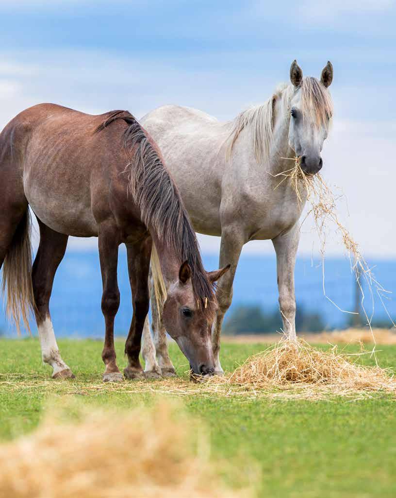


Although there have been studies conducted regarding the influence of stress on the horse and equine gastric ulcer syndrome, very few have focused on the influence of stress on total digestive health.
Pathophysiological stressors, such as parasite infesta tion, various bacterial pathogens, sand accumulation, enterolith formation, high-grain diets and rapid feed changes are recognized. However, little consideration has been given to the effect of psycho logical stressors on gastrointestinal (GI) health in the horse. Trailering, separation anxiety, isolation, high traffic areas and the demands of training and competition all contribute to gastric upset, inflammation and in extreme cases, colic.
Many times, this stress presents clinically as acute or chronic di arrhea, weight loss and poor condition, or behavior changes. Horses with these conditions are living in a state of constant digestive dis turbance and consequently are predisposed to suffer from acute or recurrent colic episodes. Even a small amount of added stress such as a change in temperature or low water consumption can provoke these individuals into a state of digestive distress or even colic.
Though a prevalent issue in the equine community, little research has been performed on stress and its biophysical influence on the horse with almost no research conducted on its effect on colonic function or general GI disfunction. Of the few studies conducted, many focused-on param eters such as heart rate, breathing and cortisol levels. Others examined the relationship between feeding patterns and reproductive efficiency or how air and road travel affect heart rate. One area of the GI tract and digestive condition that has been investigated is gastric ulcers (EGUS), as this area of the digestive tract is most easily accessed. Additionally, work has been done observing the influence of psychological stress such as training and showing on gastric ulcer formation.
Unlike the horse, the human digestive tract is more easily ac cessible, allowing for the use of many new technologies, including Positron Emission Tomography (PET) and Functional Magnetic Reso nance Imaging (FMRI) helping physicians to understand the pathology behind many GI diseases. These technologies have led to a shift in approach, from a reductionist model of the diseases and treatments
to a more integrative view focusing on altered motility, enhanced visceral sensitivity, and brain-gut dysregulation resulting in a relatively new term, Functional Gastrointestinal Disorders (FGID.) A new biopsy chosocial model, FGID in humans has been thoroughly researched resulting in a comprehensive table classification that is site specific.
Even though these new technologies are not available for the horse, veterinarians can use the components of FGID in humans to better understand equine digestive disfunction. There are several fac tors that comprise FGID in the human model—abnormal motility,
visceral hypersensitivity and inflammation, which can exacerbate the afore mentioned conditions. These elements are often observed in equine digestive disorders including diarrhea, colitis and even colic. The nervous system is important to understanding FGID in both humans and equines. The brain-gut interaction from the sympathetic and parasym pathetic nervous systems can directly influence the hindgut, which during time of stress can create a negative feedback loop. External and interoceptive neural connections elicit GI reactions, disrupting normal motility and secretions, as well as, creating an inflammatory response. However, this brain-gut cycle can work both ways with viscerotropic effects influencing pain perception, mood and behavior.
What would this FGID model look like in the horse? Both genetic and environmental factors can deeply influence the horse in its formative years, especially now when more young horses are com peting than ever before. These factors, combined with underlying physiology, can lead to motility disturbances and inflammation in the GI tract. Combine this physiology with psychosocial factors, such as general life stressors and psychological state, and the beginnings of FGID can be seen in changed behavior or symptoms such as diarrhea or weight loss affecting overall quality of life, general health and condition and trainability. If FGID progresses, more serious GI disorders such as colitis, gastric and colonic ulcers, or even colic may occur. The pathology of colonic disturbance is very familiar to equine veterinarians.
Intermittent meals, sand irritation, antibiotics, NSAIDs and large grain meals lead to a buildup of lactic acid, altered volatile fatty acid production, motility disturbance and the death of beneficial hindgut microflora. These negative side effects merge, leading to pathogenic bacterial overgrowth, a change in mucosal secretions and mucosal inflammation resulting in colitis. Underlying this pathology are the everyday stressors our horses endure; trailering, separation, restricted turn out and the stress of performance and training all feed into this cycle, intensifying the underlying GI disorder. This pathophysiologi cal cycle can greatly alter GI function, not only affecting performance
and overall condition but leading to various disorders including leaky gut syndrome, fecal water syndrome and in the most severe cases, systemic inflammatory response syndrome (SIRS), multiple organ dysfunction syndrome (MODS), and even death.
Many advancements have been made in equine care over the past 30 years, one of the outstanding advancements has been the development of effective anthelmintics. Before modern dewormers the most common cause of colic was due to parasite infection. This has become rare, but the number of colic cases has not decreased. Horses are now living more stressful lives in a confinement manage ment system and are competing at a younger age than ever before. Stress has now replaced parasites as the most common cause of colic. However, unlike parasite infestation, there is not a simple solu tion to this source of disease.
In many cases, it is impossible for the horse owner to implement the necessary changes to completely reduce the stressors in their horse’s life. Lack of real estate and the move of horse owners to more suburban areas limit the ability for the horse to live in its natural state, on pasture. Some changes can be made to their diet, such as, feed ing less concentrate and giving higher quality, free choice forage, as well as feeding more meals per day. Changing training and trailering routines may also help to reduce everyday stressors. However, these changes alone may not be enough to improve the horse’s stress level and mitigate its effect on their digestive health.
Changing management practices can be difficult to gain consen sus on and changes can be challenging or impossible to implement. Incorporating a high-quality digestive aid into the feeding program is the best or only solution for many to stabilize the negative effects caused by the plethora of stressors horses encounter on a regular basis. The best digestive aids can help to balance the microbial populations, reduce acid and increase pH, reduce inflammation, and stimulate the repair and regeneration of colonocytes, which in turn can provide a solution to biophysical stress-induced dysfunction that so much of our equine population battle.
Al though many people im mediately look at the limbs when a horse is not up to par, poor performance can be multifactorial, warned Frank M. Andrews, DVM, the clinical service chief of large animal medicine at Louisiana State University College of Veterinary Medicine.
And 1 of those factors is the gut, particularly gastric ulcers, he said during a presentation at the 67th Annual Conven tion of the AAEP, in Nashville, Tenn.
“Horses are basically the GI tract on 4 legs,” joked Dr. Andrews, whose research area is the GI tract. “So, they are hindgut fermenters. They eat a variety of feeds that we give them, which can contain a lot of carbohydrates. Horses weren't meant to be in stalls and eat high-grain diets. They were meant basically to be out on the pasture grazing and continuously eating and then storing feed for that next pasture.
“We've taken them out of that environment, and we need to look at the gastrointestinal tract when we're looking at poor
performance,” he said, “paying attention to how they are fed, how they are housed, and whether they are stressed is impor tant regarding performance. Because performance horses are fed high-carbohy drate diets, stabled and travel, instead of allowing them to graze, they are at risk for developing gastric ulcers,” he explained.
“We know that there are a lot of risk factors that are associated with gastric ulcers in horses,” said Dr. Andrews, who is also a professor of equine medicine at LSU, “such as stabling, certainly exercise, fasting periods that occur. And then 1 thing that we really don't think about too much, frequent traveling.
“The more the horses travel, the higher prevalence of gastric ulcers and a lot of horses that travel to competitions are in the trailer for long periods of time and certainly fed high-grain diets before and after the exercise periods.”
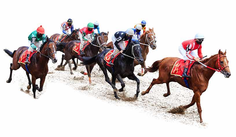
Human athletes can suffer gastroesophageal reflux, splashing of stomach acid up the esophagus. Horses also suffer from similar
BY MARIE ROSENTHAL MS"Paying attention to how they are fed, how they are housed, and whether they are stressed is important regarding performance."
—Dr. Frank M. Andrews
acid splashing while exercising, which is increased as the effort is expended.
Unfortunately, there is no clear way to tell if an ulcer is present except by scop ing the horse, he said. There are clinical signs that suggest a gastric ulcer might be present: reluctance to work, lack of energy or dullness, poor performance— slowing near the end of the race, sudden stops, reluctance to gallop—and abdomi nal discomfort. In addition, horses tend to lose a little weight because they don’t eat as well. Laboratory findings are not useful, although a horse might be mildly anemic.
The concern for owners, of course, is that a horse that is not performing is a horse that is not winning a purse. “Our owners are
looking for earnings,” he said, so scoping the horse could be worthwhile if an ulcer is suspected.
Management involves not only treat ment but changing some habits. Reduce the days and level of training. Allow ad lib forage and daily pasture turnout.
“I would suggest that using alfalfa hay or adding alfalfa hay to the diet can be important,” Dr. Andrews said. “Horses that are out in the pasture have a lower preva lence of gastric ulcers, however, that's not necessarily true in all pastures, especially when the pastures are of really poor quality.”
A course of omeprazole might be useful. Give the horse omeprazole for 2 weeks. “If you can rescope that horse in 2 weeks, you might find that the ulcers have healed, and you can save your clients 2 more weeks of omeprazole treatment at the normal dose, and then maybe taper the dose,” he said.
To make sure that the dose of omeprazole was appropriate, a veterinarian can aspirate gastric juice and measure the pH after dosing the omeprazole, as omeprazole should increase pH and reduce stomach acid.
Sometimes, medical management involves a multimedication regimen, which can be difficult for owners and trainers to do.
Owners might balk a bit though. “You have all these treatments and owners are going, ‘gosh, darn, when do I give all this stuff?
You got to have somebody at the barn all the time.” He suggested this schedule, which would fit in with the training of a racehorse. At 5 a.m., give the omeprazole before he’s fed. Exercise and then around 6 or 6:30, give the animal some hay and corn oil. At least 2 hours after the omeprazole, give the sucralfate, which is cytoprotective. At 10 a.m., give miso prostol. Both sucralfate and misoprostol must be given later, too.
Ulcer prevention using low dose omeprazole is effective so think about prevention. “Preventive treatment can make a difference and is effective in decreasing the number of gastric ulcers in our horses,” Dr. Andrews added. A recent metanalysis paper showed that prevention of ulcers using omeprazole is effective.
Management involves not only treatment but changing some habits, such as allowing ad lib forage and daily pasture turnout.
NSAIDs are ubiquitous. Despite the risk of adverse gastrointestinal and renal effects, more than 40% of equids are prescribed nonsteroidal anti-inflammatory drugs during veterinary treatment, according to a 2019 study in the Equine Veterinary Journal .
Although COX-2 inhibitors—or coxibs—were developed to reduce unwanted effects, traditional NSAIDs like flunixin meglumine and phenylbutazone are still the predominant choice in most clinics.
NSAIDs rely on inhibition of the COX pathways, which are respon sible for constitutive functions in gastroprotection, renal homeo stasis and platelet function (COX-1), and inducible functions, including pain, fever and inflammation (COX-2).
“Traditional NSAIDs are non-specific, in that they work both at the level of COX-2, which affects the therapeutic effects, but they also impact COX-1 production, which results in delayed mucosal
healing, increased intestinal permeability, right dorsal colitis, gas tric ulceration and kidney damage,” said Rebecca Bishop, DVM, MS, resident in equine surgery at the University of Illinois, College of Veterinary Medicine.
To avoid the negative effects, COX-2 specific inhibitors—such as firocoxib—were developed to specifically target the COX-2 pathway while avoiding the dangers involved in COX-1 inhibition.
“However, recent studies have shown that COX-2 pathways also play a role in renal blood flow in states of hypoperfusion, as well as gastric ulcer repair,” Dr. Bishop said at the 67th Annual AAEP Convention in Nashville. “This led my team to wonder if there could be adverse lower intestinal effects in horses, which are also seen in human medicine with coxib administration.”
Prior research on the subject showed no difference in clini
cal signs of NSAID-associated colitis in horses administered firocoxib vs. flunixin or phenylbutazone. However, monitoring for subclinical colonic inflammation rarely was reported in those studies.

Dr. Bishop’s team created a model to evaluate the 2 classes of drugs and their impact on colonic inflammation in healthy horses.
The prospective study involved 12 healthy adult horses from the University of Illinois teaching hospital herd in a con trolled but non-randomized block design.
The first treatment consisted of flunixin meglumine via IV cath eter at 1.1 mg/kg every 12 hours for 5 days. Following a 6-month washout period, firocoxib was administered to the same horses to achieve a rapid, steady state concentration. The dosage followed current recommendations of a single 0.3 mg/kg dose orally fol lowed by 0.1 mg/kg orally every 24 hours.
Each horse received omeprazole at 1 mg/kg orally every 24
hours, to mitigate effects of gastric ulcer ation for each treatment arm.
Edema of the large colon was sub jectively evaluated on a score of 0-to-2 on transabdominal ultrasonography, and maximal colon thickness was evaluated via a single ultrasonographic measurement.


Blood samples were collected for a complete blood count and biochemistry to assess markers of colonic and renal injury.
The primary finding of the study was a significant increase in colon wall thickness and colonic edema following treatment with firocoxib, but not flunixin.
“Eleven of 12 horses had subjective evidence of colonic inflammation or edema after receiving firocoxib, compared with 1 horse following flunixin,” Dr. Bishop said.
There was also a statistically significant increase in total pro
BAUltrasonographic images were obtained of the right dorsal colon before and after treatment. Subjective assessments were made of colonic edema, which was scored on a scale of 0-to-2 where 0 was no edema and 2 was severe edema, and the wall thickness was measured, with the maximum thickness recorded at the time of each examination. The + symbol marks the boundaries of wall measurement in each image. (A) Before treatment (day 0), edema score zero, maximum thickness 3.6 mm. (B) After treatment (day 7), edema score 2, maximum thickness 8.2 mm
More that 40% of equids are prescribed NSAIDs during veterinary treatment, but they can have GI and renal effects.
tein following firocoxib and a decrease in protein following flunixin.
“That was surprising, especially given that decreases in total protein are typi cally associated with colon inflammation and protein-losing enteropathy,” she said. “There was no difference in packed-cell volume pre- and post-treatment in the 2 groups, so changes in total protein cannot be attributed to hydration status.”
For creatinine, there was a signifi cant increase following both treatments, but there was no significant difference between the groups.
“Creatinine could be attributed to sub clinical kidney injury or dehydration, but there was no evidence of clinical dehydra tion in the study horses,” she explained.
There was also a significant decrease in phosphorus following firocoxib, but not flunixin. Hypophosphatemia can be associ ated with either renal injury or colitis due to decreased absorption or increased loss of phosphorus.
“Overall, we did not see any clinically significant changes in the biochemical parameters,” Dr. Bishop added. “They all remained within normal limits, even though there were statistically significant differences over time.”
Overall, the findings suggest that subclinical colonic inflammation occurred following firocoxib, but not flunixin, as evi denced by increased colon wall thickness and more frequent colonic edema.
“Horses were administered omeprazole in both treatment groups to mitigate effects of gastric ulcer ation and to satisfy the IACUC [Institutional Animal Care and Use Committee] protocol, However, a recent study found that there was seeming interaction between omeprazole and phenylbu tazone, in which horses administered the 2 drugs concurrently had increased incidence of lower intestinal complications, which included impactions and signs of colic,” she said. “The relation
ship between NSAIDs and omeprazole warrants further investigation, and we’re not sure how that may have impacted our results.”
Dr. Bishop also noted that the avail ability of firocoxib during the study period necessitated the use of an oral formulation of the drug.
“We assume that the drug should have been absorbed before it reached the level of the large intestine, but there’s a pos sibility that the differing routes of adminis tration altered the effect of the medication on the lower intestine,” she said.
Although the sample size was small and consisted of healthy horses, the findings do point to subclini cal colon inflammation following firocoxib administration.
“COX-2 selective NSAIDs may carry a risk of subclinical colitis,” Dr. Bishop concluded. “Although [coxibs] are still regarded as the safer option for gastrointestinal health if an NSAID must be administered, it is important to remember that no medication is without risk of side effects.”
Duz M, et al. Proportion of nonsteroidal anti-inflammatory drug prescription in equine practice. Equine Vet J. 2019;51(2):147-153.
https://beva.onlinelibrary.wiley.com/doi/10.1111/evj.12997
Cook VL, Meyer CT, Campbell NB, et al. Effect of firocoxib or flunixin meglumine on recovery of ischemic-injured equine jejunum. Am J Vet Res. 2009;70:992-1000. https://avmajournals.avma.org/view/journals/ajvr/70/8/ajvr.70.8.992.xml
Cook VL, Blikslager AT. The use of nonsteroidal anti-inflammatory drugs in critically ill horses. J Vet Emerg Crit Care. 2015;25:76-88. https://onlinelibrary.wiley.com/doi/10.1111/vec.12271
K. Morrissey N, R. Bellenger C, T. Ryan M, et al. Cyclooxygenase-2 mRNA expression in equine nonglandular and glandular gastric mucosal biopsy specimens obtained before and after induction of gastric ulceration via intermittent feed deprivation. Am J Vet Res. 2010;71:1312-1320. https://avmajournals.avma.org/view/journals/ajvr/71/11/ajvr.71.11.1312.xml
Ricord M, et al. Impact of concurrent treatment with omeprazole on phenylbutazone-induced equine gastric ulcer syndrome (EGUS). Equine Vet J. 2021;53(2):356-363. https://beva.onlinelibrary.wiley.com/doi/10.1111/evj.1332 3

Traditional NSAIDs are non-specific in that they affect both the COX-1 and COX-2 pathways.


