
Arm
With
DISCLAIMER: The content in this digital issue is for general informational purposes
PercyBo Publishing Media LLC makes no representations or warranties of any kind about the completeness, accuracy, timeliness, reliability or suitability of any of the information, including content or advertisements, contained in any of its digital content and expressly disclaims liability of any errors or omissions that may be presented within its content. PercyBo Publishing Media LLC reserves the right to alter or correct any content without any obligations. Furthermore, PercyBo disclaims any and all liability for any direct, indirect, or other damages arising from the use or misuse of the information presented in
content. The views expressed in its digital content are those of sources and authors
necessarily reflect the opinion or policy of PercyBo. The content is for veterinary professionals. ALL RIGHTS RESERVED. Reproduction in whole or in part without

do

AAEP to NRHA: Reconsider Sedative Rule
The American Association of Equine Practitioners (AAEP) asked the National Reining Horse Associa tion (NRHA) to reconsider its recent policy allowing the use of compounded romifidine for sedative ef fects 30 minutes before a competi tion. The AAEP said the medica tion could have a negative impact on the health and welfare of both horse and rider.
him/herself and the rider.”
The FDA has not approved romifidine in any form in the United States, so the medication can only be ob tained through a compounding pharmacy, she added.
Romifidine
About 80% of horses given romifidine still show signs of sedation at 30 minutes after administration, ac cording to AAEP President Emma Read, DVM, MVSc, DACVS, who signed the letter to the NRHA.

“Ataxia may still be present up to 1 hour after ad ministration, and diminished response to painful stimuli and other signs of sedation may persist up to 2 hours in some horses,” she wrote.
She said that allowing the horse into the ring after 30 minutes of receiving romifidine “presents a safety and welfare risk to both the horse and rider, includ ing masking a lameness that could become worse with riding, or results in the horse tripping and injuring
“Legal drug compounding re quires a valid Veterinary Client/ Patient Relationship (VCPR). A compounded medication can only be used when there is not an equivalent FDA-approved medi cation available. Off label (extra label) medication is the use of an FDA-approved product for a non-equine species that is used (off label) in the horse.
“It should be noted that the use of compounded drugs in a highly regulated competitive environment should be discouraged due to the variability of thera peutic levels and detection times,” she said.
“For the safety of the discipline’s equine and human athletes, we urge NRHA members and leadership to reconsider this recent change allowing for the use of compounded romifidine. We do not believe it is in the best interest of the horse,” Dr. Read wrote. MeV
ARM BREEDERS
With Knowledge to Control Genetic Conditions
By Marie Rosenthal MSTwo novel, and relatively rare genetic diseases—equine familial isolated hypoparathy roidism (EFIH) and equine ju venile degenerative axonopathy (EJDA)—are devastating for af fected foals, requiring euthanasia because there is no treatment for either one.
Owners are incentivized to understand genetic issues.
However, the birth of an af fected foal doesn’t necessarily have to affect the breeding potential of the sire and dam. Owners can use current knowledge about the genetic causes be hind these conditions to “breed smart,” and avoid producing sick foals, explained Carrie J. Finno, DVM, PhD, DACVIM, a geneticist and the director of the Center for Equine Health at UC Davis School of Veterinary Medicine.
Specific animals are chosen for breeding based on desirable traits, such as speed, conformation and endurance.
“Those carriers were selected for breeding for a reason.” Dr. Finno explained at the ACVIM Forum 2022.
“Owners can keep those stallions, and their de sirable traits, in the breeding pool by using genetic tools to make informed breeding decisions.”
The 2 diseases she discussed are both inherited as autosomal recessive diseases, meaning the foal must have 2 copies of the genetic variant, one from each parent, to develop the disease. Since genetic tests for these diseases are now available, breeders can screen the stallion and mare(s) and avoid breeding 2 car riers.
“Stallion owners are very educated. They are in centivized to understand the underlying genetic
causes to extend the breeding ca reers for their stallions,” she said.
Equine Familial Isolated Hypoparathyroidism
Dr. Finno, along with her gradu ate students and colleagues at multiple institutions, has been working to find the cause of EFIH, which was previously called idiopathic hypocalcemia, a fatal condition that affects very young Thoroughbred foals.

Although relatively rare, anyone who works with Thoroughbreds should keep this disease on their ra dar.
Affected foals typically begin to show clinical signs between 16 hours and 5 weeks of age when they present with low levels of calcium in the blood that lead to involuntary muscle contractions, stiffness that affects their gaits, seizures, fevers and fast heart rates. Parathyroid hormone (PTH), which is normal ly released by the body to increase the calcium levels, will test as inappropriately normal (should be high due to the low calcium) or low.
In the neonatal intensive care unit, these foals can be supplemented with calcium and improve. How ever, as soon as the supplemental calcium is discon tinued, the clinical signs reappear, and the foal often dies or is euthanized.
“If you give these foals calcium, they will look normal. You can get their levels up, they will buck, they will play,” said Dr. Finno. “But the minute that calcium drops again, you're right back where you started. It’s not something that can really be man aged medically.”
Therefore, the best approach is to avoid produc
BREEDERS

ing affected foals in the first place.
After considerable investiga tion, the multidisciplinary team found a nonsense variant in the Rap Guanin Nucleotide Exchange Factor 5 (RAPGEF5) gene, which plays a role in developmental pro cesses and signaling in early em bryonic development and is high ly expressed in parathyroid tissue.
Understanding the genetics behind some diseases allows smart breeding choices.
This variant was identified in 3 out of 82 Thoroughbreds that were genotyped. The researchers estimate an allele frequency of 1.80% in Thoroughbreds.
“This RAPGEF5 variant is highly associated with this disease,” Dr. Finno said. “There is now a genetic test available. The first carrier was identified in 1992, and fortunately it does not look like we've been se lecting for this, which is a good thing. However, we can now screen carriers and try to at least not have any of these affected foals.”
In cases where Thoroughbred foals are found de ceased, it is a good idea to test the stallion and mare to determine if they are carriers for the EFIH variant to avoid producing affected foals in the future.
Equine Juvenile Degenerative Axonopathy
According to Dr. Finno, the search for the cause of EJDA occurred right as the pandemic hit.
“The first call we received was in March 2020. A 3-week-old Quarter Horse filly presented to the vet erinarian with severe acute ataxia,” she said. “The pelvic limbs were more severely affected than the thoracic limbs. The problem had started the week before and progressed rapidly. This presentation is a common theme in all these foals—pelvic limb paresis, no cranial nerve deficits, and no history of trauma.”
Diagnostics revealed that the filly had a leukocy tosis characterized by a mature neutrophilia. She was hyperglycemic with an increased gamma-glutamyl transferase, and hyperbilirubinemic with increased sorbitol dehydrogenase. Cytology of the cerebrospi nal fluid was normal and there were no significant findings on her computed tomography (CT) scan.
The most obvious finding was the pelvic ataxia,
Dr. Finno said. However, another notable finding in these foals is that sometimes their tales are dis placed to the left.
“The team did an amazing job working this foal up and trying to figure out what was going on,” said Dr. Finno. ”"In the meantime, de spite intensive treatment, her condi tion worsened, and she had a hard time standing without assistance. In these cases, the pelvic lymph paresis just overtakes the whole situation. It starts at this ataxia and then they progress to this paresis.”
As they were working up this case, she received a call about a 2-week-old female Quarter Horse with the same clinical signs. Both fillies unfortunately died due to their condition.
As she was talking to the veterinarian of the sec ond case, he said, “‘We actually had a colt that we eu thanized a month ago that looked just like this, and he's related to that filly.”
To date, the researchers have performed genetic sequencing on five cases. The problem appears to be in a region of chromosome 11, but the exact variant has not been identified yet.
“There is this phenotype of an early onset, severe ataxic disease in Quarter Horse foals. It progresses very rapidly to recumbency,” said Dr. Finno. “I think that's important for owners to know. If you recog nize this, I don't think we should be spending a lot of money on these cases quite honestly, because we don't have a treatment for them. The lesions are re stricted to the spinal cord. It's a degenerative axo nopathy. It's likely autosomal recessive, and it looks like the causative variant is on chromosome 11.”
Dr. Finno and her colleagues recognize that pri vacy is an important issue when it comes to genetics research. All research samples receive an identifica tion code in place of the animal’s name to protect the owner’s privacy. Hopefully this encourages owners and breeders to support genetic research. For dis eases like EFIH, and soon EJDA, breeders can take steps to breed healthy offspring, extend the breeding careers of their stallions, and contribute to overall improvements in the health of their breeds. MeV
There’s nothing else like it.
For more than 30 years, Adequan® i.m. (polysulfated glycosaminoglycan) has been administered millions of times1 to treat degenerative joint disease, and with good reason. From day one, it’s been the only FDA-Approved equine PSGAG joint treatment available, and the only one proven to.2, 3

Reduce inflammation
Restore synovial joint lubrication
Repair joint cartilage
Reverse the disease process
When you start with it early and stay with it as needed, horses may enjoy greater mobility over a lifetime.2, 4, 5 Discover if Adequan is the right choice. Visit adequan.com/Ordering-Information to find a distributor and place an order today.

BRIEF SUMMARY: Prior to use please consult the product insert, a summary of which follows: CAUTION: Federal law restricts this drug to use by or on the order of a licensed veterinarian. INDICATIONS: Adequan® i.m. is recommended for the intramuscular treatment of non-infectious degenerative and/or traumatic joint dysfunction and associated lameness of the carpal and hock joints in horses. CONTRAINDICATIONS: There are no known contraindications to the use of intramuscular Polysulfated Glycosaminoglycan. WARNINGS: Do not use in horses intended for human consumption. Not for use in humans. Keep this and all medications out of the reach of children. PRECAUTIONS: The safe use of Adequan® i.m. in horses used for breeding purposes, during pregnancy, or in lactating mares has not been evaluated. For customer care, or to obtain product information, visit www.adequan.com. To report an adverse event please contact American Regent, Inc. at 1-888-354-4857 or email pv@americanregent.com.
Please see Full Prescribing Information at www.adequan.com
American
PP-AI-US-0629
the
www.adequan.com
Owner of a Broken Heart: Donkey Receives Pacemaker
By Melanie Greaver CordovaA 3-month-old miniature donkey named Nix under went cardiac surgery at Cornell University College of Veterinary Medicine to implant a pacemaker.
When Nix was just a few months old, she began hav ing collapsing episodes and overall lethargy. Joan Ayers, DVM, of Genesee Valley Veterinary Hospital, examined the miniature donkey on her farm in Canandaigua, New York, where Nix lives with her parents, June and Hoot, a miniature donkey named Charlie, and a herd of goats.
Dr. Ayers, in consultation with the owners and Bar bara Delvescovo, DVM, clinical fellow in the Section of Large Animal Medicine at Cornell, originally thought she might have epilepsy or have suffered some sort of traumatic injury to her neck or skull, but ruled these conditions out.
In February, Nix’s condition worsened. Nix was fall ing again, this time from a standing position, and she staggered even more while walking.
horses are relaxed but disappear with stress, excitement or exercise—or pathological, meaning they are abnor mal, dangerous and can cause poor performance. Nix’s arrhythmia was the latter.
“This is a pathological arrhythmia that we see pretty uncommonly in horses, but a little more occasionally in donkeys, and especially mini donkeys,” said Katharyn Mitchell, DVM, PhD, ACVIM (LAIM), assistant profes sor in the Section of Large Animal Medicine, who over saw Nix’s case at Cornell.
Although it’s more common to see this type of ar rhythmia in miniature donkeys, it is still rare. “Given the severity of the arrhythmia and the frequency of col lapse, medication will not be effective, so we only had the choice of placing a pacemaker or euthanasia, given the high risk of continued self-trauma,” Dr. Mitchell said.
Given Nix’s age and her lack of other underlying problems, she was a great candidate for a pacemaker, ac cording to Dr. Mitchell.
In a collaborative effort between Drs. Mitchell, Del vescovo from the large animal internal medicine service, Lawrence Santistevan, DVM, of the cardiology service, members of the large animal soft tissue surgery service, the anesthesia service and multiple hospital staff mem bers, the complicated procedure to implant Nix’s pace maker went well.

“There are a few mini donkeys around the world with pacemakers, but certainly it’s not common,” Mitchell said. ”It was great teamwork.”
Nix’s care team noticed immediate improvement after surgery—now the miniature donkey had enough blood flow to her brain to let her walk normally, with out any fainting episodes or lethargy. The pacemaker battery will need replacing after approximately seven to nine years, but if the device continues to work well, Nix will live a normal life. In the short-term, it is imperative that Nix remain calm and have limited exercise. If the pacemaker lead pulls out of the heart muscle or becomes infected, both would be hazardous to her health.
The Cornell veterinarians did an echocardiogram and diagnosed third-degree atrioventricular block—her atria and ventricles weren’t communicating, meaning her heart’s rhythm was very slow and irregular. There were frequent pauses of 20-30 seconds with no heart beats or blood flow to her body, enlarging her heart. Without getting enough blood to her brain or around her body for long periods, Nix exhibited the classic clini cal signs of this condition, including collapse, episodes of weakness and severe exercise intolerance.
Arrhythmias are divided into 2 categories, either physiological—meaning they happen normally when
“We will keep her calm for the first month to lower these risks, and if everything looks okay, then we will increase the pacemaker’s rate a little bit so she can get up some speed and play with her mum in the paddock,” Mitchell said.
At Nix’s recheck appointment this month, the pace maker was working well, and her heart had only a mild reaction to the pacemaker lead. MeV
Originally published on the Cornell website https://www. vet.cornell.edu/news/20220811/mini-donkey-gets-bigboost-pacemaker. It was edited for style.


Pleuropneumonia and a Surprise in the Oven Threaten the Life of Thoroughbred Mare
 By Rob Warren

Lillipad, an 11-year-old Thoroughbred mare who was la beled barren at auction and went unsold, was rescued by Jillian Fallon of Reno, Nevada.

The trailer ride to her new home was uneventful, but the horse started to deteriorate shortly after arrival. Ms. Fallon noticed that the mare’s respiration, heart rate and temperature were all elevated to dangerous levels. A local veterinarian performed an ultrasound and found fluid build-up in and around both lungs and diagnosed pleuropneumonia.
Medications administered onsite helped bring Lilli pad’s vital signs closer to normal levels, but it was clear she needed further treatment, so Ms. Fallon brought her to the UC Davis veterinary hospital. Lillipad would re main hospitalized for 3 months, culminating in a shock ing surprise.
She was treated extensively for severe pleuropneumo nia, including management with fluid drainage through multiple chest tubes and a thoracotomy, aggressive anti biotic therapy and intense supportive care by a dedicat ed team of students, technicians, residents, and faculty members in the Equine Internal Medicine Service.
Lillipad was not responding as well as expected, and
she lost a significant amount of weight—except in her abdomen. Her care team performed an ultrasound and found out this “barren” horse was pregnant.
They also found evidence of hemorrhaging into her abdomen because of the pregnancy. The Equine Repro duction Service found the foal to be of proper size with a good heart rate. However, Lillipad was still critically ill, and it was unknown how her illness and its complica tions would affect the continued growth and health of the unborn foal, as well as her ability to have a normal birth. The team found out that it was her fourth foaling, so the medical team felt more confident she could handle the birth if she and the baby were healthy enough.
Thankfully, Lillipad’s hemoabdomen stabilized quickly and her pleuropneumonia continued to slowly improve over the next 2 months, and she was transferred from the hospital’s Large Animal Clinic to the layup ser vices at the UC Davis Center for Equine Health for a few weeks before returning home. There, she was checked regularly regarding the progress of her pneumonia, tho racotomy incision sites, and the viability of her foal.
She was discharged in January 2022 with about 2 months remaining of her high-risk pregnancy. Because of these risks, Lillipad return to UC Davis for the birth.
Lillipad did very well at home, gaining weight and finally starting to act like a spirited mare with a zest for life. Ms. Fallon brought her to UC Davis for foal watch in March, where she continued to improve and was off all medications before the big day. On March 27, she ex perienced a quick, uncomplicated birth and delivered a healthy filly.
“Lillipad was a difficult case and involved the dedica tion of everyone on the Equine Internal Medicine Ser vice, as well as multiple technicians and students,” said Emily Berryhill, DVM. “She is a perfect example of the collaborative nature and extensiveness of our care at UC Davis. This success showcases that dedicated teamwork between our clinical staff, our clients, and our referring veterinarians can result in a positive outcome, even with many ups and downs in between. We are grateful to Ms. Fallon for entrusting us with Lillipad’s care.”
Lillipad and her filly, LP’s Sunny Miracle, or Mira for short, were able to return home a week later, where they are healthy and happy. MeV
This story originally ran on the UC Davis website https:// www.vetmed.ucdavis.edu/news/horse-survives-severecase-life-threatening-pneumonia. It has been edited for space and style.


CLINICAL PHARMACOLOGY
PHARMACOKINETICS
EFFECTIVENESS
Two
Diagnosing and Managing Respiratory Abnormalities in the Equine Athlete
 By Molly Cripe Birt, BS, RVT, VTS-EVN
During exer tion, the horse’s airflow will increase nearly
20 times than when at rest, which changes the shape and rigidity of the airway. For the equine athlete, particularly horses compet ing in high-speed events, the upper airway is vital for its performance and success. Three common abnormalities in the pharynx and larynx can affect that performance:

1. dorsal displacement of the soft palate,
2. laryngeal hemiplegia and
3. entrapped epiglottis.
These conditions occur more often in young Thoroughbreds or Standardbreds. However, Warmbloods and Draft horses are also affected more than other horses. Signs of abnormalities affecting the struc tures of the pharynx and larynx are expira tory or inspiratory noise, exercise intoler ance and poor performance.
Diagnosing Upper Airway Abnormalities
The primary presenting complaints of an upper airway abnormality include poor performance or exercise intolerance, com bined with an expiratory or inspiratory noise during respiration. Many presump tive diagnoses are made based on history alone and confirmed with a careful physi cal examination and the use of endoscope, ultrasonography and diagnostic imaging.
In all cases of poor performance or exercise intolerance, the veterinarian must rule out pulmonary, cardiac or neuromuscular dis ease, or lameness.
The physical examination should in clude assessing air flow through both nos trils, percussion on the sinuses, auscultation of lung sounds, a rebreathing examination and digital palpation of the larynx, which is done by facing the shoulder of the horse with its nose resting on the shoulder of the technician. The index fingers should then
palpate the larynx until they rest above the dorsal larynx. The left and right muscular processes are palpable in this position.
A “slap test” can also be performed to evaluate arytenoid function. Another tech nician can slap, with a flat hand, just ventral to the right or left withers; if the arytenoid is functioning normally the technician pal pating should feel the corresponding cri coarytenoideus dorsalis muscle contract. When the left side of the withers is tapped, the right arytenoid should move.
Flexible endoscopic examination will definitively diagnose an upper airway ab normality and has become a common tool in many practices. The horse must be restrained for its and the staff’s safety. Se dation alters the muscular function of the soft palate and larynx, so is contraindicated during the endoscopic examination—un less the patient is fractious. A nose twitch is preferred if the patient resists the passing of the scope. Stocks, if available, do offer more control because they limit movement.
With the veterinarian holding the hand piece and directing the exam, the techni cian passes the flexible endoscope by plac ing a hand on the bridge of the nose of the horse and lifting the alar fold of the false nostril. The other hand passes the endo scope into ventral meatus, directing slight ly medial. The endoscope should be passed deliberately to avoid unnecessarily agitat ing the horse, but without haste to avoid striking the ethmoid turbinate and causing epistaxis.
The veterinarian should be able to fully examine the nasal passages, pharynx and larynx to rule out any abnormalities. The nasal septum, meati, nasomaxillary sinus opening, ethmoid turbinate, pharyngeal lymphoid tissue, pharyngeal openings to the guttural pouch, and the guttural pouches can be seen. Using a flexible stylet passed through the biopsy channel, the soft palate, the epiglottis and aryepiglottic folds, arytenoids and the trachea can be seen.
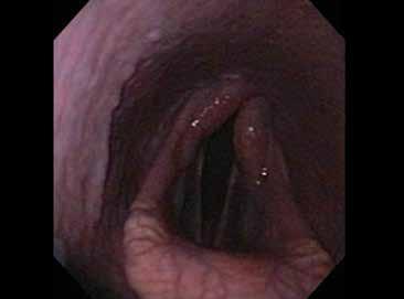
The normal endoscopic examination of the pharynx will reveal a soft palate posi
The


SHORT.


tioned ventrally to the epiglottis. The soft palate may intermittently displace dorsally, shortly return ing to its original position when the patient swallows. Pharyngeal hyperplasia is common in younger patients, but excessive inflamma tion is considered abnormal. The arytenoids should abduct with inspiration and adduct with swal lowing; both can be elicited by holding of the airway and intro ducing water into the airway.
Dynamic endoscopy may be indicated when no abnormality can be identified.
3-year-old racehorses were affect ed. One research study found that only 1.3% of 479 horses had DDSP during resting endoscopic exams.
Dynamic endoscopy may be indicated when no abnormality is identified, or when the extent of the problem must be examined more closely. Dynamic en doscopy is done using a high-speed treadmill or a tele metric endoscope. This allows the veterinarian to eval uate the soft palate, epiglottis and arytenoids during maximal exercise, which may reveal abnormalities that are not visible while at rest. If indicated, blood sampling before, during and after exercise can be performed to evaluated blood lactate, carbon dioxide and creatinine kinase levels.
Ultrasonography can be used to evaluate laryngeal and soft palate function and identify airway inflamma tion. Radiographs of the skull that focus on the throat latch are useful in identifying the cartilage structures of the larynx, the hyoid apparatus, guttural pouches and paranasal sinuses.
3 Common Upper Airway Abnormalities
Dorsal Displacement of the Soft Palate
Dorsal displacement of the soft palate (DDSP) occurs when the caudal free margin of the soft palate billows dorsally over the epiglottis, resulting in an airway ob struction. Mechanical causes include the presence of subepiglottic or palatal granulomas or cysts. DDSP is likely caused by excessive sternohyoideus and ster nothyroideus muscles contraction due to excitement, nervousness or head positioning, which results in the caudal movement of the larynx. This pulls the epiglot tis away from the soft palate. Another potential cause of DDSP is a neuromuscular deficit of the pharyngeal branch of the vagus nerve or intrinsic muscles.
DDSP decreases expiratory air flow, resulting in gurgling or “choking down” during exhalation as air becomes caught in the billowing soft palate, resulting in exercise intolerance and poor performance. Owners often complain the horse is “choking” or “swallowing its tongue” during exercise.
DDSP occurs more often in the exercising horse, and it has been suggested that 10% to 20% of the 2- to
A definitive diagnosis is made by history, physical and endoscopic examination. Endoscopic exami nation reveals a dorsally displaced soft palate that prevents visualiza tion of the epiglottis. It should be noted that DDSP is classified as either persistent or intermittent. Intermittent DDSP at rest might or might not worsen during exercise, and some cases will present with a nor mally positioned soft palate but with a history of exer cise intolerance Therefore, the practitioner may recom mend dynamic endoscopy to confirm DDSP during exercise. This confirmation is vital to proper treatment recommendations, as there are management changes that can be implemented prior to surgery.
The trainer may elect to change the head tack used during training and performances to alter head posi tion or prevent mouth opening. There are several op tions, including nosebands...its mouth, and W-bits and spoon bits to prevent caudal retraction of the tongue. Some trainers, particularly in the Standardbred circuit, will tie the tongue forward; however, research suggests that this practice is not as successful as trainers think. Finally, several companies sell a throat support device to position the larynx rostrally.
Pharyngitis and inflammatory diseases within the guttural pouch can cause neuromuscular deficits and can be treated with systemic anti-inflammatory medi cation, such as phenylbutazone. A pharyngeal spray comprised of glycerin, dimethyl sulfoxide, predniso lone and nitrofurazone can be administered topically through a long polyethylene catheter inserted into the nasal passaged to the pharynx.
Treatment over 30 days has been found to signifi cantly decrease inflammation. Overall, there is a 60% success rate with medical and non-surgical options.
However, with persistent DDSP diagnosed at rest or through dynamic endoscopy, there are several surgi cal treatment options that the veterinarian can recom mend, including thermal palatoplasty, which causes fibrosis and stiffening of the soft palate, which is done with a laser on the end of an endoscope.
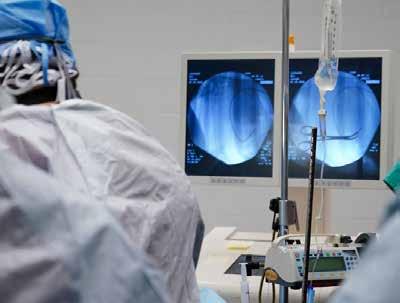
Thermal palatoplasty can be performed on a stand ing patient as well as under general anesthesia. For a standing procedure, the patient must be restrained properly and sedated adequately. The soft palate must be displaced dorsally over the epiglottis to perform ther mal palatoplasty, which can be achieved by inducing
swallowing. If DDSP cannot be maintained, broncho esophageal forceps can grasp the epiglottis and retract it caudally. Topical local anesthetic spray administered through an endoscopic sprayer, will desensitize the soft palate. Some surgeons prefer thermal palatoplasty with the patient under general anesthesia.
The endotracheal tube will automatically displace the soft palate dorsally, which removes one of the great est difficulties of the procedure in the standing patient. However, the patient’s anesthesia must be maintained using total IV anesthesia, as oxygen ventilating through the endotracheal tube will cause a catastrophic airway fire if exposed to the laser beam.
Overall, thermal palatoplasty is a simple surgical procedure with a good prognosis for return to activ ity. Postoperatively, a systemic anti-inflammatory is administered for two days. Topical pharyngeal spray containing glycerin, prednisolone, nitrofurazone and dimethyl sulfoxide is administered transnasally via a polyethylene catheter for up to 10 days. Postoperative dysphagia and coughing are common in the first 7 to 10 days and generally resolves.
Thermal palatoplasty is often combined with an other surgical procedure to achieve the best outcome possible. A standard myectomy of the sternohyoideus and sternothyroideus muscles will prevent the caudal retraction of the larynx. A sternothyroideus myotenec tomy and staphylectomy likely will not be performed with thermal palatoplasty; this procedure transects the sternothyroideus tendons and trims a small crescent of the caudal free margin of the soft palate.
The laryngeal tie forward, however, is considered a superior procedure for treating DDSP, particularly when combined with thermal palatoplasty. The larynx moves rostral and dorsal, and the basihyoid bone will move dorsal and caudal; hence the name “tie-foreward” surgery. Prior to surgery, systemic antimicrobials and anti-inflammatory drugs are administered.
Once induced under general anesthesia, the pa tient is intubated. Should thermal palatoplasty be performed, total IV anesthesia will be administered until the laser is no longer used. Once complete, inhal ant anesthesia can begin. The patient is positioned in dorsal recumbency with the head and neck extended fully. The ventral cervical and intermandibular regions are clipped and aseptically prepared. The incision will be located 2 cm caudal to the cricoid cartilage and 2 cm rostral to the basihyoid bone. The incision is made, and tissue dissected to expose the larynx. Heavy, nonabsorbable suture is placed in the caudal aspect of the wing of the thyroid cartilage.
The surgeons at Purdue University Large Animal Hospital have implemented the use of Arthrex Suture
FIG 1.
Buttons to distribute the pressure of the suture, pre venting it from tearing through the cartilage. Anoth er method is to pass the suture 2 to 4 times through the cartilage. Using a wire passer, the suture is passed around the basihyoid bone.
These steps are repeated on the contralateral side. Upon completion of the suture placement, the horse’s nose is tilted 90⁰ while the sutures are snugly tied. The surgeon confirms the cricoid cartilage in relation to the basihyoid bone, and once satisfied, the head is lowered into a semi-flexed position with a pad supporting the nose. Once the surgical site is closed, a bandage using elastic tape is placed to support the soft tissue and pre vent seroma formation.

Systemic antimicrobial and anti-inflammatory drugs are continued for several days. An important as pect of postoperative care is reducing the tension on the prosthetic sutures. Elevate feed and water to shoulder height to prevent undue stress on the neck. The same considerations must be made for pasturing postopera tive patients. After 2 weeks, regular feeding, water, turn out and training can resume.
Laryngeal Hemiplegia
Laryngeal hemiplegia is the result of variable amounts of paralysis associated with the arytenoid cartilage, with 95% occurring on the left side and 5% on the right side. Dynamic collapse of the affected arytenoid car tilage into the airway results in inspiratory noise and exercise intolerance.
Laryngeal hemiplegia or recurrent laryngeal neurop athy is considered idiopathic, although the loss of large,
myelinated nerve fibers in the left recurrent laryngeal nerve can cause neurogenic atrophy of the circo arytenoideus dorsalis muscle. This results in progressive loss of abduc tion and adduction function of the arytenoid.
Many owners report that clini cal signs developed acutely, but it is a slow, insidious progress until the muscles just fatigue. However, trau ma to the recurrent laryngeal nerve or guttural pouch mycosis, strangles and neoplastic tu mors have been reported to cause laryngeal hemiplegia.

Surgery for laryngeal hemiplegia may not be the best choice.
movement. Invasive surgery may not be the optimum choice for some equine athletes.
However, the noise associated with laryngeal hemiplegia is un desirable in the show arena, and disciplines that require an elevated head or an intense flexion of the neck, such as dressage or carriage driving, may exacerbate the inspi ratory noise.
This causes the exercise intolerance and poor per formance because of hypoxemia, hypercarbia and met abolic acidosis, and the classic “roaring” inspiratory noise resulting from air passing over the affected vocal cord and ventricle. Large breed horses and racehorses are most affected, although the highest incidence is seen in young Thoroughbreds. Often, laryngeal hemiplegia is discovered in the workup examinations for yearling sales, or as 2- and 3-year-old horses enter training.
A definitive diagnosis is made through history, physical examination and endoscopic examination.
Palpation of the larynx will reveal atrophy of the cri coarytenoideus dorsalis muscle. Endoscopic examina tion will reveal asymmetric and asynchronous abduc tion or complete paralysis of the arytenoids, which can be graded on a subjective scale. There are several treat ments for laryngeal hemiplegia, with laser ventriculo cordectomy and laryngoplasty being common surgical corrections currently performed.

The veterinarian will make treatment recommenda tions based upon the severity of the presenting com plaint, the horse’s work and the grade of laryngeal
In this instance, a ventriculo cordectomy, which removes the mucosal lining of the ventricle and part of the vocal fold, might be offered. It was once performed by a laryngotomy under general anesthesia. However, advances in laser ventriculocor dectomy via the nasal passage or oral cavity have largely eliminated the need for ventral laryngotomy. The sur geon uses the laser to ablate the ventricle and vocal cord. Laser ventriculocordecomy can be performed on a standing patient or under general anesthesia and is frequently combined with prosthetic laryngoplasty.
Standing ventriculocordectomy is achieved using proper restraint by an assistant with the head elevated. IV sedation, possibly with a combination of detomidine hydrochloride and butorphanol tartrate, is necessary to prevent horse movement during the procedure. Topical anesthesia is applied to the nasal passages, pharynx and larynx using an endoscopic sprayer.
Should the procedure be performed under general anesthesia, special precautions must be taken. Endotra cheal intubation must be delayed so that the vocal cords are visible, and laser safety considerations are respected. Once the horse has been induced and positioned in the proper recumbency, anesthesia is maintained with total intravenous anesthesia.
A flexible 600-micron laser fiber is passed through the biopsy channel of the endoscope. The surgeon may use a combination of non-contact and contact tech niques when performing the laser ventriculocordec tomy. Starting ventral to the vocal process, the vocal cord is ablated as the fiber is moved carefully over its surface. The ventricle is then ablated with the laser fiber in direct contact with the mucosa. Many surgeons pre fer specific settings for their practice.
Specialized instruments can be used to aid in the ventriculocordectomy. Bronchoesophageal forceps are passed through the nostril opposite the endoscope. Bronchoesophageal forceps grasp and evert the ven tricle, placing it in a position for excision using a laser fiber. Lower wattage is used for excision. Alternatively, some surgeons prefer the use of a custom-made roar ing burr, which is passed through the nostril opposite the endoscope. The burr is pressed into the ventricle and rotated with gentle traction placed to evert the ven tricle. Then the laser fiber is used for surgical excision.
Complications include intraoperative hemorrhage and iatrogenic damage to the vocal process and aryte noid. Postoperatively, the patient should receive phen ylbutazone intravenously or orally and dexamethasone intravenously. Laser ventriculocordectomy patients typically heal in 30 to 45 days.
A standalone ventriculocordectomy would not be sufficient to correct the problems associated with a grade III-C and IV laryngeal hemiplegia. A laryngo plasty is the recommended treatment for horses who experience exercise intolerance and inspiratory noise. Laryngoplasty is the placement of one or two prosthetic sutures between the cricoid and arytenoid cartilages with the intention of permanently retracting the affect ed arytenoid out of the airway—hence, the other name for the surgery; tie-back surgery. The surgical “art” of
the laryngoplasty is achieving laryngeal opening that is adequate for performance, but not so wide as to cause chronic aspiration of feed and saliva.


Preoperative nonsteroidal anti-inflammatory drugs and antimicrobials are administered prior to induction. Presuming the horse has a left-sided laryngeal hemiple gia, the horse is induced under general anesthesia and positioned in right lateral recumbency. The head and neck are extended fully. If a laser ventriculocordectomy is to be performed during general anesthesia, the pro cedure would begin at this point with safety consider ations in place that were described previously. The inci sion of the palatoplasty is 10 to 12 cm long, ventral and parallel to the linguofacial vein and begins 4 cm cranial to the ramus of the mandible.
The area of interest is clipped and aseptically pre pared. Using basic instrumentation, the skin and mus cle layers are incised and dissected until the lateral and dorsal aspects of the larynx are visualized. Strong re tractors, such as Saurbach, Richardson or Kelley retrac tors, expose the larynx.
Two large-caliber, non-absorbable sutures are placed through a trocar point needle and used for the laryngoplasty. Trocar point needles will not create large, traumatizing hole in the cartilage and will likely not break off within the cartilage. The needle and suture are passed through a notch in the cricoid cartilage and then advanced cranially to avoid piercing the trachea. At this point, an endoscope should be advanced caudally to the arytenoids and tilted dorsally to visualize the roof of the cricoid cartilage and trachea. This verifies that there is no suture piercing the airway, thus contaminat ing the prosthetic suture. Next, the suture is drawn un der the cranial aspect of the cricopharyngeus muscle. A similar, second suture is placed 1 cm caudal in the same fashion. The ends of the suture are tensioned, abduct
Teaching Points
Care and cleaning the endoscope is important to maintain the equipment and to prevent infections.
When the endoscopic examination is complete, the endoscope is dismantled.
As each endoscope is different, it is paramount that the technician refers to the manufacturer’s cleaning instructions. Basic cleaning procedures, however, include flushing a diluted enzymatic detergent through the chambers, brushing the chambers with a special bristle brush and rinsing with deionized water. Some endoscopes can be submerged in water so long as a waterproof cap is in place over the camera connection.
If the endoscope was used with surgical laser or if a suspected trauma occurred, the technician should leak test the endoscope.
Fill the inside of the endoscope with air, and air bubbles will stream from the defect if damage is present. This not only alerts the technician of damage, but also prevents water from leaking inside of the endoscope. Water contamination in an endoscope is catastrophic.
Once the endoscope is disinfected and rinsed, it should be connected to the air source so that any water droplets can be blown out of the chambers to prevent water from collecting and sitting in the chambers,
ing the arytenoid. The endoscope is pulled rostrally to visualize the degree of abduction. Once the desired de gree is reached, the surgeon will tie the sutures. Special instrumentation by Securos includes a tensioning de vice, crimps and crimper to achieve a very specific level of tensioning and securement; some surgeons will use this special instrumentation.
Also within the surgical procedure is the surgical debridement of the cricoarytenoid joint using curettes or ablation by the CO2 laser. These limit or completely prevent the movement of the joint associated with ary tenoid movement, enhancing the permanent abduction of the arytenoid and reducing post-operative prosthetic failure. The CO2 laser is prepared by setting the laser for a defocused beam, inserting the sterile laser hand piece and draping the articulating laser arm with sterile adhesive wraps. Surgeons at Purdue University Large Animal Hospital prefer setting the CO2 laser at a con tinuous wave for 15 watts with a 5-mm circular spot.
Once the cricoarytenoid joint is exposed and opened, the surgeon will position the laser hand piece three centimeters from the joint surface, which is then ablated. The entirety of the cricoarytenoid joint is not fully accessible with the CO2 laser; completion of fus ing the joint is achieved using a 2-0 curette. If a CO2 laser is not available, curettage of the joint is acceptable.
The incision is closed, and a bandage is placed over the throatlatch for recovery. Postoperative nonsteroidal anti-inflammatory drugs and antimicrobial therapy are continued. Hay and water should be lowered to prevent
aspiration. Thirty days of stall rest with light handwalking is recommended. By weeks 5 and 6, light, controlled exercise can begin. After 6 weeks, a return to training can resume. Complications of laryngoplasty include hemorrhage from a needle piercing through laryngeal veins, needle breakage in the cricoid or aryte noid cartilage, development of a large seroma over the surgical site, dysphagia and chronic cough, aspiration pneumonia, incisional infection, infected prosthetic and prosthetic failure.
Many horses who cough immediately postopera tively will resolve within seven to ten days. Coughing and dysphagia beyond ten days is considered abnormal and warrants a recheck examination. If the prosthetic laryngoplasty fails, the surgeon may elect for another laryngoplasty or would consider an arytenoidectomy. An arytenoidectomy performed via laryngotomy will fully remove the affected arytenoid. Despite the list of potential complications associated with laryngoplasty, research reports that nearly 50 to 70% of racehorses treated will have improved racing performances.
Entrapped Epiglottis
The epiglottis becomes entrapped when the ventral ly located aryepiglottic folds become dorsally mal positioned over the epiglottis. Entrapped epiglottis will cause respiratory noise, exercise intolerance and coughing. Nasal exudate has been described but is not as common. Some epiglottic entrapments are intermit tent and relieved by swallowing and, patients will show
minimal clinical signs. Other entrapments are persis tent; the longer the epiglottis remains entrapped, the more inflamed the tissue becomes, causing thickening and ulceration of the tissue. Epiglottic hypoplasia is a risk factor that contribute to persistent epiglottic en trapment. The most affected horses are Thoroughbreds and Standardbreds, often because of the effect on their performance. Some horses are unaffected by entrapped epiglottis and live normally.
Entrapped epiglottis is diagnosed by endoscopic examination, sometimes coincidentally as no clinical signs may be present. On endoscopic examination, the general shape of the epiglottis is visible, but the distinct margins and classic vascular pattern are obscured. The veterinarian might describe the membranes as being thin or thick, narrow or wide, ulcerated or nonulcer ated, with mild to extensive ulceration. Approximately 95% of cases have aryepiglottic folds that are described as persistent, thick and wide.
A laryngotomy with resection of redundant sub epi glottic tissue is a viable surgical option, but DDSP might develop because of altered epiglottic function. A concur rent staphylectomy might reduce the chance of DDSP from occurring after this surgery. A more modern, less invasive approach to managing entrapped epiglottis is the axial division of the aryepiglottic fold with a laser or epiglottic hook bistoury knife. 75% to 80% of patients can be treated in this method if the aryepiglottic fold is thin without inflammation. Patients with chronically thickened ulcerated entrapments are good candidates for resection of sub epiglottic tissue via a laryngotomy.
Axial division of the aryepiglottic folds is com pleted using a shielded, curved bistoury knife through a transoral or transnasal approach, or by using a la ser through an endoscope. The transoral approach is performed with the patient under general anesthesia. Transnasal axial division is completed in a standing pa tient that is properly restrained and sedated. The most dangerous complication from the transnasal approach is the iatrogenic laceration and damage to the soft pal ate, which can destroy the athletic animal’s career. The transoral approach requires a mouth speculum and is considered the safest, as it is nearly impossible to dam age the soft palate.
Laser axial division of the aryepiglottic fold is per
For more information:
formed standing. The patient must have proper re straint and sedation, as previously described. A flexible 600-micron laser fiber is passed through the biopsy channel of an endoscope. A topical anesthetic, such as Cetacaine, is sprayed over the epiglottis and aryepi glottic fold using an endoscopic sprayer. Surgeons at Purdue University Large Animal Hospital prefer the laser settings at 15-25 watts at three seconds on and one second off. The laser fiber is used in contact fash ion, incising layer by layer from caudal to rostral across the aryepiglottic fold. Swallowing by the patient will progressively retract the aryepiglottic folds. Once the division is complete, the patient is made to swallow to confirm the epiglottis will not become re-entrapped.
Complications from laser axial division of the ary epiglottic fold include iatrogenic damage to the tip of the epiglottis, the development of DDSP and prolonged sub epiglottic swelling. Postoperatively, the patient is administered a systemic anti-inflammatory drug and steroid for several days. Stall rest is ordered for 21 days with a slow return to work. Returning to training too soon will lead to prolonged edema and inflammation, thus reducing the healing time.
The technician working in an equine practice is in a unique position to assist with the workup and treat ment of equine athletes presenting with complaints of upper respiratory noise and poor performance. With a basic understanding of the bones, cartilage, tendons and muscles that are vital to the function of the upper respi ratory tract, the technician will have a better insight into the diagnosis and treatment methods of dorsal displace ment of the soft palate, laryngeal hemiplegia, entrapped epiglottis and a variety of other abnormalities.
Becoming experts in handling and using endoscop ic equipment and laser safety and surgical treatment methods can help make the technician an invaluable member of the veterinary team. MeV
About the Author
Molly Cripe Birt, BS, RVT, VTS-EVN, is a large animal surgery Technician, at the Purdue University Veterinary Teaching Hospital PURDUE, University College of Veterinary Medicine. She presented this information at the 2017 Fall Conference.
Auer J. A., Stick J. A. Equine Surgery, Third Edition. St. Louis: Saunders Elsevier, 2006.
Couetil, L., Hawkins J. Respiratory Diseases of the Horse: A problem-oriented approach to diagnosis & management. London: Manson, 2013.
Hawkins J. Advances in Equine Respiratory Surgery. Wiley-Blackwell, 2015;3-8.
Zoetis Foundation and the AVMF Offer New Scholarships for Vet Tech Students


A new scholarship program to support students in vet erinary technician programs was announced today by the Zoetis Foundation and the American Veterinary Medical Foundation (AVMF).

The Zoetis Foundation Veterinary Technician Schol arship program will provide $1,000 scholarships to up to 270 students. This program is funded by a grant from the Zoetis Foundation and will be managed by the AVMF.
Applicants must be enrolled fulltime in an AVMA-accredited veteri nary technology, veterinary nursing or animal health technology pro gram in the United States or Puer to Rico and be in good academic standing according to their institu tion's policies. To apply, eligible stu dents must visit VetVance.com. The application period is from Sept. 1 to Oct. 7, 2022. For more information about the scholarship program and criteria, please visit avma.org/scholarships.
cal Association. “Veterinary technicians are an inte gral part of veterinary medicine and our animal care teams, and these scholarships will allow veterinary technology students to cover some of the costs associ ated with their education.”
The program will provide $1,000 scholarships to up to 270 students.

The Zoetis Foundation supports communities and the people who care for animals, with a specific focus on advancing opportunities for veterinarians and farmers around the world. The Foundation’s grantmaking and strategic efforts help provide access to education and mental wellness resources, expand veterinary debt relief, sup port diversity and inclusion efforts, and enable thriving livelihoods by funding programs that help veterinary practices and livestock farmers adopt sustainable business practices. To learn more about the Foundation, please visit: www.zoetisfoundation.org.
“The Zoetis Foundation is thrilled to support the AVMF through a grant to recognize student leadership and promote diversity among future veterinary techni cians by helping to offset the significant costs associated with education,” said Jeannette Ferran Astorga, the ex ecutive vice president, Corporate Affairs, Communica tions and Sustainability at Zoetis and President of the Zoetis Foundation. “The new scholarships advance our commitments to veterinary education and support for people who care for animals.”
“We are honored for the opportunity to partner with the Zoetis Foundation in supporting our vet erinary technicians through the creation of the Zoetis Foundation Veterinary Technician Scholarship,” said Jose Arce, DVM, the Chair of the AVMF and imme diate past president of the American Veterinary Medi
The American Veterinary Medical Foundation (AVMF) is the charitable arm of the American Vet erinary Medical Association (AVMA), one of the old est and largest veterinary medical organizations in the world. For almost 60 years, the AVMF has been dedi cated to developing resources to advance the science and practice of veterinary medicine to improve animal and human health. Foundation programs and activities are designed to benefit veterinary medicine, promote animal welfare, and enhance research so that the pro fession is better prepared to deal with difficult problems facing animal health today and tomorrow. Charitable contributions and support to the Foundation assist veterinarians and the entire veterinary healthcare team help animals. Its funding priorities include Education, Disaster Relief, Crisis Support, Charitable Veterinary Care and Animal Health Research. MeV


Equine


