7 minute read
CLINICAL FEATURE
from JIDA
by Th!nk Media
Application of the new periodontal classification: Part 2
The second part of our series on the application of the 2017 World Workshop classification of periodontal and peri-implant diseases and conditions in daily practice presents two further clinical cases.
Introduction
The World Workshop on the Classification of Periodontal and Peri-implant Diseases and Conditions was convened in 2017 and resulted in the publication of a new classification system in 2018.1 This replaces the formerly used 1999 (Armitage) Classification.2 The complete Workshop proceedings are available to clinicians for free online via the European Federation of Periodontology (EFP) website.3 The new process for diagnosing and classifying cases of periodontitis incorporates staging and grading of each case.4 At its simplest, the stage represents an interpretation of periodontitis severity and the complexity of management of the case. The grade provides supplemental evidence on the historic rate of disease progression, and can help to identify cases where risk factors exist and/or where expected outcomes of therapy may be less favourable.5 Diagnostic decision trees may be of value to practitioners in applying the new classification in daily practice. The current series utilises the decision tree published by the British Society of Periodontology (BSP),6 as this arguably represents the simplest approach to classifying periodontitis cases.
CASE 1
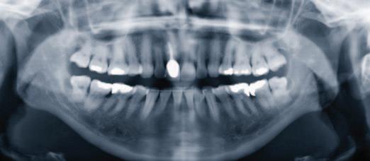
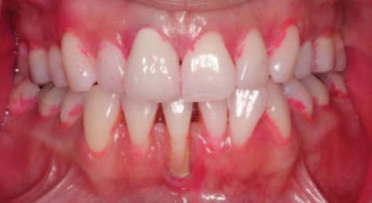
This case assimilates patient history, clinical and radiographic findings from a 54-year-old female patient who attended the Dublin Dental University Hospital (DDUH) for periodontal assessment, in order to establish a clinical case diagnosis (Figures 1 and 2). To assist readers in understanding the new classification system, the rationale for the clinical diagnosis is presented.
Dr Michael Nolan
Postgraduate student in periodontology, Dublin Dental University Hospital Dr Suha Aljudaibi


Postgraduate student in periodontology, Dublin Dental University Hospital FIGURE 1: Orthopantomogram (OPG) of patient taken at initial periodontal assessment.


FIGURE 2: Clinical photograph at initial presentation at DDUH.
Case presentation: patient history
Table 1: Overview of case presentation. Patient: 54-year-old female Presenting complaint: “I’m conscious of the gap on my upper right side”
Medical history:
No significant medical history Smoking status: Non-smoker Family history of periodontitis:No Other risk factors: No
Table 2: Summary of clinical findings. Visual assessment: Relatively good tissue tone and colour Probing pocket depths: 1-5mm Clinical attachment loss: 2-6mm Bleeding on probing: 35% Plaque control: Fair Tooth mobility: Nil Furcation involvement: Grade 1 mesial and distal 1.7 Tooth loss due to periodontitis: Nil – all lost to repeated restoration failure and peri-apical infection Other factors of relevance: Poorly adapted restorative margins
Dr Peter Harrison
Division of Restorative Dentistry & Periodontology, Dublin Dental University Hospital Dr Lewis Winning
Division of Restorative Dentistry & Periodontology, Dublin Dental University Hospital
RADIOGRAPHIC FINDINGS: Bone loss present: Yes Pattern of bone loss: Horizontal Severity of bone loss: 10-50% Distribution: Generalised
Clinical findings
What is the diagnosis using the new classification? The diagnosis in this case is: n generalised periodontitis; n Stage III, Grade B; and, n currently unstable.
How this diagnosis was reached n This is a periodontitis case as clinical attachment loss is present at ≥2 nonadjacent teeth. n This is a generalised periodontitis case as >30% of teeth are affected by attachment loss/bone loss. n Stage III was selected based on the site of greatest bone loss severity based on the radiographic assessment: approximately 50% radiographic bone loss at tooth 1.7 equating to the middle third of the root. n Grade B was selected based on calculation of the ratio of percentage bone loss at the worst-affected tooth divided by patient age. In this case, the ratio is >0.5 and <1 (50% [bone loss] ÷ 54 [age] =0.93). n The disease is currently unstable based on the presence of probing pocket depths (PPDs) ≥5mm. n Risk factor assessment: disease moderators were not present and the periodontal destruction was commensurate with the biofilm deposits present and level of oral hygiene. FIGURE 3 (ABOVE): Orthopantomogram (OPG) of patient taken at initial periodontal assessment.

FIGURE 4 (LEFT): Intra-oral periapical radiograph of mandibular anterior teeth.
FIGURE 5 (BELOW): Clinical photograph following plaque disclosure at initial presentation at DDUH.

CASE 2
This case assimilates patient history, and clinical and radiographic findings, from a 34-year-old female patient who attended the Dublin Dental University Hospital (DDUH) for periodontal assessment, in order to establish a clinical case diagnosis (Figures 3-5). To assist readers in understanding the new classification system, the rationale for the clinical diagnosis is presented.
Case presentation: patient history
Table 3: Overview of case presentation. Patient: 34-year-old female Presenting complaint: Receding gums, tooth sensitivity History of presenting complaint:Recession present since age 16; previous orthodontic treatment
Medical history:
No significant medical history Smoking status: Current smoker (15 cigarettes/day) Family history of periodontitis:No Table 4: Summary of clinical findings. Visual assessment: Thin gingival biotype, gingival recession evident (8mm at buccal 41) Probing pocket depths: Range 4-7mm Clinical attachment loss: Range 1-7mm Bleeding on probing: 23% Plaque control: Fair Tooth mobility: Grade I mobility at 4,1; 4,2; 3,1; 3,2 Furcation involvement: Class II 2.7 Tooth loss due to periodontitis:No Other factors of relevance: Iatrogenic factors (overhanging restoration 3,6 and 3,7)
RADIOGRAPHIC FINDINGS: Bone loss present: Yes Pattern of bone loss: Mainly horizontal with localised vertical components Severity of bone loss: Range 10-40% coronal third to mid third of the root
Distribution: Generalised (>30% teeth)
Clinical findings
What is the diagnosis using the new classification? The diagnoses in this case is: n generalised periodontitis; n Stage III, Grade C; n currently unstable; n risk factors: current smoker; and, n localised recession defect 4,1 (RT 2).
How this diagnosis was reached n This is a periodontitis case as clinical attachment loss is present at ≥2 nonadjacent teeth. n This is a generalised periodontitis case as >30% of teeth are affected by attachment loss/bone loss. n Stage III was selected based on the site of greatest bone loss severity (based on the radiographic assessment: approximately 40% radiographic bone loss at tooth 4.1, equating to the middle third of the root). n Grade C was chosen based on calculation of the ratio of percentage bone loss at the worst-affected tooth divided by patient age. In this case, the ratio is >1 (40% [bone loss] ÷ 34 [age] =1.18). n The disease is currently unstable based on the presence of probing pocket depths (PPDs) ≥5mm. n Risk factor assessment: the patient is a current smoker. n The present case contains additional subtlety in the presence of a notable gingival recession lesion at 4.1. Gingival recession is not specifically addressed in the simplified decision trees, where the focus is primarily on staging and grading of periodontitis. The recession lesion at 4.1 was classified using the system of recession type (RT) proposed by Cairo et al., (2011),7 which was adopted in the new classification.8 RT 2 describes a gingival recession lesion that is associated with interproximal attachment loss. In RT 2 cases, the interproximal attachment loss is less than or equal to the attachment loss seen at the buccal aspect.
References
1. Caton, J.G., Armitage, G., Berglundh, T., et al. A new classification scheme for periodontal and peri-implant diseases and conditions – introduction and key changes from the 1999 classification. J Clin Periodontol 2018; 45 (Suppl. 20): S1-S8. 2. Armitage, G.C. Development of a classification system for periodontal diseases and conditions. Ann Periodontol 1999; 4 (1): 1-6. 3. European Federation of Periodontology. New classification micro-site. [Internet]. [Accessed March 23, 2020]. https://www.efp.org/publications/projects/newclassification/index.html. 4. Tonetti, M.S., Greenwell, H. Kornman, K.S. Staging and grading of periodontitis: framework and proposal of a new classification and case definition. J Clin
Periodontol 2018; 45 (Suppl. 20): S149-S161. 5. Papapanou, P.N., Sanz, M., Budunelli, N., et al. Periodontitis: consensus report of workgroup 2 of the 2017 World Workshop on the Classification of Periodontal and
PeriImplant Diseases and Conditions. J Clin Periodontol 2018; 45 (Suppl. 20):
S162–S170. 6. British Society of Periodontology. Flowchart implementing the 2017
Classification. [Internet]. [Accessed March 23, 2020]. Available from: https://www.bsperio.org.uk/publications/downloads/111_153050_bsp-flowchartimplementing-the-2017-classification.pdf. 7. Cairo, F., Nieri, M., Cincinelli, S., Mervelt, J., Pagliaro, U. The interproximal clinical attachment level to classify gingival recessions and predict root coverage outcomes: an explorative and reliability study. J Clin Periodontol 2011; 38: 661-666. 8. Jepsen, S., Caton, J.G., Albandar, J.M., et al. Periodontal manifestations of systemic diseases and developmental and acquired conditions: consensus report of workgroup 3 of the 2017 World Workshop on the Classification of Periodontal and
PeriImplant Diseases and Conditions. J Clin Periodontol 2018; 45 (Suppl. 20): S219-
S229.


