23 minute read
PEER-REVIEWED
from JIDA
by Th!nk Media
Abstract Statement of the problem: The management of the deep carious lesion is a topic of keen interest to the dental profession with many and varied treatment modalities advocated in the scientific literature. Purpose of the study: This literature review proposes to summarise current consensus approaches and scientific thinking in this area. Some new treatment advances have been advocated in recent years and their efficacy is also examined. Topics and areas of interest are proposed for future research. Methods: The studies examined in this review were based on searches online in the PubMed, Embase, and Google Scholar search engines, and Cochrane reviews, and include systematic reviews and consensus papers, as well as observational studies, randomised controlled trials and meta-analyses. Conclusion: Problems exist in this area regarding precise definitions and measurement of deep carious lesions in practice, and standardisations of measurement do not currently exist. This is an area where further study and research would be welcome.
Journal of the Irish Dental Association 2020; 67 (1): 36-42
Introduction: dental caries, its sequalae and appropriate caries management
Dental caries is a disease process affecting dental hard tissues, caused by a shift in the normal oral microbiological biofilm balance to a more acidophilic, aciduric and cariogenic, biofilm consisting mainly but not exclusively of Streptococci mutans and lactobacilli. Frequent ingestion of fermentable carbohydrates encourages an environment of low pH within the biofilm, which favours the selective growth of cariogenic bacteria.1 A cumulative demineralisation pattern over time leads to dissolution of dental hard tissues and the formation of a carious lesion. Other factors such as fluoride ion concentration and salivary flow rate modify the caries process and are intimately involved in determining the likelihood of overall mineral loss and the rate at which this occurs. The operative treatment of the deep carious lesion (DCL) should: n aid biofilm control on a tooth surface; n protect the pulp-dentine complex and arrest the lesion activity by sealing the coronal part; and, n restore the function, form and aesthetics of the tooth.2
Dr Brenda Barrett
BDentSc MClinDent General practitioner Pembroke Dental Carlow Dr Michael O’Sullivan
Senior Lecturer/Consultant in Restorative Dentistry (Special Needs) Department of Restorative Dentistry and Periodontology Dublin Dental University Hospital Lincoln Place, Dublin 2
Corresponding author: Dr Brenda Barrett, Pembroke Dental, Pembroke, Carlow Town, Co. Carlow. T: 059-913 1667 E: bbarrett@pembrokedental.ie
Caries risk assessment
Correct diagnosis
Clinical judgement

Individualised treatment plan
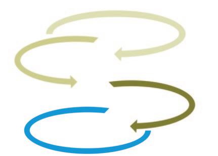
FIGURE 1: Sequential gathering of information leading to individualised patient treatment plan during caries management.
Maintaining pulpal vitality has a great impact on the lifetime prognosis of a tooth and also reduces the overall lifetime cost of retaining that tooth.3 Many studies have shown that sealing of carious lesions can lead to caries arrest so this is accepted as one of the guiding principles when restoring a DCL.4 Evidence-based research has encouraged a minimally invasive (MI) approach to the management of caries in the post-fluoride caries generation.2 This approach stresses a preventive philosophy, individualised risk assessments for patients, early detection of lesions, and efforts to remineralise non-cavitated lesions, with the provision of preventive care to minimise the need for operative intervention (Figure 1). When operative intervention is unequivocally required, the procedure used should be as minimally invasive as possible.5 With the above treatment principles in mind, the DCL should be treated with an MI treatment strategy, with the primary aim of preserving pulp vitality if possible and restoring the tooth to its original form so that normal biofilm control can be re-established.6
Precise terminology used in relation to the operative management of the deep carious lesion
In 2015, the International Caries Consensus Collaboration, comprising worldwide cariology experts, decided on consensus recommendations for terminology in relation to managing carious lesions.7 This terminology is used throughout this review. A carious lesion is a consequence of a disease process and its management involves intervention to arrest its progression by conversion of the lesion to a cleansable form. The size and depth of a carious lesion can be assessed clinically or radiographically, but there is currently no standard definition or measurement of the term DCL. According to this Consensus Collaboration, “deep lesions are defined as those radiographically involving the inner pulpal third or quarter of dentine or with clinically assessed risk of pulpal exposure” (Figure 2). The hardness of dentine is an indicator of the extent of caries in dentinal tissue. The International Caries Consensus Collaboration has defined the different clinical presentations of affected carious dentine.
Soft dentine Soft dentine will deform or deflect when a hard instrument is pressed onto it and can be easily scooped up (e.g., with a hand excavator), with little force being required.
Leathery dentine Leathery dentine does not deform when an instrument is pressed onto it and can still be easily lifted without much force being required. There may be little difference between leathery and firm dentine, with leathery being a transition on the spectrum between soft and firm dentine.
Firm dentine Firm dentine is physically resistant to hand excavation, and some pressure needs to be exerted through an instrument to lift it.
Hard dentine For hard dentine, a pushing force needs to be used with a hard instrument to engage the dentine, and only a sharp cutting edge or a bur will lift it. A scratchy sound or ‘cri dentinaire’ can be heard when a straight probe is taken across the dentine.
Cross-section of tooth with occlusal carious lesion Enlarged cross-section of carious lesion Dentine tubule Histological terms

FIGURE 2: Diagrammatic representation of the carious lesion.8 Necrotic zone
Contaminated zone
Demineralised zone
Translucent zone
Sound dentine
Tertiary dentine
Dentine: Clinical (tactile manifestations)
Soft dentine
(Leathery dentine)
Firm dentine
Hard dentine
Management of deep carious lesion

Caries risk assessment
Correct diagnosis
Clinical judgement
Preventive treatment plan • biofilm control/use of fluoride products • limitation of fermentable carbohydrates
• stepwise caries removal • selective caries removal to soft dentine
• minimally invasive endodontics • choice of pulpal wound dressing
Minimally invasive approach
Individualised treatment plan Preserve pulp vitality
FIGURE 3: Sequential, evidence-based management approach to DCLs.
Systematic individualised follow-up with focus on preventive efforts
The classical approach to caries removal: non-selective removal to firm/hard dentine or complete caries removal
Conventional or classical management of caries involves: n removal of all carious tissue (non-selective removal to hard dentine) even at the risk of pulpal exposure; n remaining dentine must be hard and firm, often tested by means of a tactile approach with a sharp excavator; and, n looking for the ‘chattering’ sound or cri dentinaire. The rationale for this extensive tissue removal is: n removal of all infected dentine and bacterial removal so that caries could be stopped from progressing further; n providing a firm base to the lesion so that restorative materials could be placed and retained adequately; and, n removing demineralised discoloured dentine. There is no evidence-based scientific rationale behind this approach, although it is practised worldwide and in some countries remains the overwhelming treatment of choice for DCLs.9
Inherent risks There are a number of risks to this approach, including: n high risk of pulpal exposure during complete caries removal; n can also be very destructive of tooth tissue, pushing the affected tooth further along the restorative cycle; and, n unnecessary reduction of residual dentine floor thickness above the pulpal tissue, which is critical to pulpal health. Non-selective removal to hard dentine or complete caries removal is now considered overtreatment and this approach is no longer recommended. Several recent systematic reviews agree with this consensus opinion.10-12
Stepwise management approach
This technique was introduced to manage DCLs with no signs or symptoms of irreversible pulpitis, but where pulpal exposure could be expected if complete caries removal was attempted (Figure 3).13 The outline operative procedure involves: n the outermost necrotic carious dentine is partially removed, leaving a soft layer of carious dentine over the pulpal floor; n the peripheries of the lesion are cleaned to hard dentine; n the tooth is then sealed with a provisional restoration to entomb any remaining bacteria in the carious dentine for several weeks to months, to
allow remineralisation of the carious dentine and the formation of tertiary dentine within the pulpal chamber; and, n when the tooth is definitively restored, the amount of carious dentine that requires removal is often lessened due to remineralisation and re-hardening of dentine. Upon re-entering the lesion, the remaining dentine is drier and harder, making it easier to remove without exposing pulpal tissue, indicating reduced lesion activity. The cultivable microflora in the lesion change before and after stepwise caries removal.13,14 At the first stage of caries removal, a mixed microbiota is found containing mainly lactobacilli, gram-positive and gram-negative rods, and streptococci. Lactobacilli and gram-positive rods dominate the colony-forming units. After re-entry, the overall colony numbers fall markedly, and the overall proportion of lactobacilli and gram-negative rods substantially reduces. The flora is dominated by Actinomyces naeslundii and Streptococci orallis, not typical of the cariogenic microbiota of DCLs.15 Stepwise caries removal involves sealing off residual caries from their source of fermentable dietary carbohydrates, thus encouraging arrest of the caries process. The provision of an adequate seal by the provisional restoration provided is integral to the success of this treatment. If an adequate peripheral seal is provided, the need for the re-entry second stage has been questioned.16 Several studies show higher success rates in terms of retaining pulp vitality long term when the stepwise technique is used in comparison to complete caries removal.17-19
Selective removal of caries is a similar approach to stepwise caries removal but is more conservative in nature. Its aim is to avoid pulpal exposure by restricting caries removal comfortably away from the pulp chamber. The operative plan is to clean carious dentine from the peripheral walls of the carious lesion but to leave caries in situ over the pulpal floor and place a definite restoration that seals the carious dentine. This takes place in one visit and no re-entry visit is envisaged. Selective removal of dentine can be further classified into two subsections:
1. Selective removal to firm dentine
This involves removing peripheral dentine around the cavity margins to
firm dentine but only excavating to leathery dentine over the pulpal floor. There is resistance to a hand excavator on the pulpal floor, but the peripheral margins are left hard (cri dentinaire) after removal of dentine is complete. This is the treatment of choice in shallow or moderately deep cavitated dentine lesions according to the International Caries Consensus Committee.
2. Selective removal to soft dentine
This is advocated as the treatment of choice in DCLs as it lessens the risk of physiological stress or exposure of pulpal tissue. Soft carious tissue is left over the pulpal tissues to avoid exposure, encouraging pulp health, while peripheral enamel and dentine are prepared to hard dentine, to allow an effective adhesive seal to be achieved by restoration placement.
Selective removal to soft dentine reduces the risk of pulp exposure in deep lesions significantly compared with non-selective removal to hard dentine or selective removal to firm dentine.7
Postulated concerns in relation to selective caries removal include the risk of residual caries progression, reduced fracture resistance, and possible higher incidence of long-term clinical restoration failure.
In a Cochrane review by Ricketts et al., exposure rates were shown to be significantly lower using a stepwise approach as opposed to complete caries removal. Complete caries removal involves a much higher risk of pulpal exposure in comparison to selective removal of carious dentine.20
Assuming that these pulpal exposures are treated mainly by direct pulp capping, which has a poorer success rate in cases of carious exposure, then it must be inferred that selective removal of dentine should be used routinely as it does not have any disadvantages compared to complete caries removal.21
Vital pulp therapy is the umbrella term for three types of procedure that are performed on vital carious exposures: direct pulp capping; partial/complete pulpotomy; and, full/partial pulpectomy. Direct pulp capping involves the placing of a medicament or wound dressing, commonly calcium hydroxide, over the pulp exposure. The success rate of direct pulp capping procedures using setting calcium hydroxide as measured by maintenance of pulpal vitality was 37% after five years and 13% after 10 years. Most failures happened slowly and asymptomatically over time, with the pulp becoming necrotic or calcifying.22 The low success rates of direct pulp capping using calcium hydroxide in cariously exposed teeth have led to controversy about the use of this technique. The introduction of newer biocompatible materials, such as mineral trioxide aggregate (MTA) and Biodentine (Septodont; Lancaster, PA, USA, and France), has sparked renewed interest with their promise of comparatively higher success rates. Full/partial pulpotomy is a well-established technique in primary teeth and has shown some success in permanent young molars using calcium hydroxide materials. In light of studies showing much higher success rates following partial pulpotomy techniques in carious teeth using MTA,23,24 the advent of MI endodontics may be approaching. Overall, in keeping with the philosophy of MI techniques, the maintenance of a vital pulp, even partially, has many advantages over full pulpectomy. The management of the inflamed pulp is trending to a more conservative approach and techniques such as direct pulp capping, full or partial pulpotomy in vital DCLs using newer materials such as MTA or similar are being revisited with some success. 25
Table 1: Properties of commonly used restorative materials.
Amalgam
Historical significance –long track record
Unaesthetic; patient concerns re appearance and safety
Minamata signals end of era in Europe
Ease of use, placement and finishing
Proven longevity
Good seal; corrosion products
Lack of adhesive properties Composite Glass ionomer
In use over 50 years
Aesthetic demand by patients
Technically more demanding
Longevity nearing equivalence of amalgam
Greater fracture resistance than amalgam in large restorations in some studies
Adhesive restoration In use over 50 years
Main use in permanent teeth is as provisional/ interim restoration
Usage mainly limited to primary teeth
Poor longevity and wear resistance
Release of fluoride ions
Chemical adhesion properties
Restoring the deep carious lesion
The choice of restorative material used depends on many factors, such as: n extent of lesion; n overall carious risk; n carious lesion activity; and, n individual patient conditions, e.g., dental crowding, saliva rate (Table 1). Increasingly, amalgam is becoming unacceptable to dental patients from an aesthetic viewpoint. Environmental concerns are also an issue, and the Minamata Treaty proposes the eventual phase out of amalgam in Europe by 2030 with increasing phase down of its use currently being introduced.26 Improvements in the composition of composite resin and bonding agents are leading to increased longevity of restorations.27 Most studies show dental amalgam to have superior longevity as a restoration in comparison to resin composite. However, some studies have shown that composite is now reaching near equivalence.28 Large amalgam restorations may show a higher fracture failure rate than the comparable composite resin restoration.29


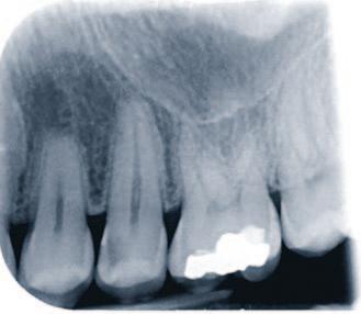
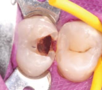
FIGURE 4: Clinical and radiographic appearance of a carious lesion – can we accurately describe how deep this lesion is? FIGURE 5: Clinical decisions made during caries removal have a direct bearing on the long-term outcome for this tooth, such as the caries management approach undertaken, pulpal treatment considerations and choice of restorative material.

The guiding principles of minimal intervention dentistry should still be practised when deciding to replace an existing restoration or re-intervene when defects are found in current restorations. Similarly, once the decision to re-intervene has been made, sound tooth tissues should be preserved during replacement to preserve pulpal health, reduce costs, and limit the subjective burden to the patient. Thus, resealing, refurbishing, repolishing, and repairing restorations should be performed whenever possible, and complete restoration replacement avoided.30 Repaired restorations have a clinically equivalent survival rate to those restorations that are completely replaced.31
Pulpal wound dressings
The purpose of pulp capping materials is to produce and maintain a bacterially impervious seal and physical barrier over the direct pulpal complex, thus reducing bacterial insult following pulpal exposure. Ideally, hard tissue barrier formation is also induced, resulting from pulpal activity. Ideally, these dressings should: n be non-toxic, biocompatible, antibacterial, and provide a long-term impervious seal over the wound; and, n provide an environment that encourages regeneration of the pulp-dentinal complex so that the self-reparative capacity of the pulp is optimised.32
Calcium hydroxide cement has been the gold standard material for many years. Its mode of action involves the production of hydroxyl ions in a high pH environment, inducing a superficial pulpal necrosis. This mild cytotoxicity stimulates pulpal cells to proliferate and differentiate, producing reparatory tertiary dentine.33 This tertiary dentine forms a calcific dentinal bridge and acts as a physical barrier to stop ingress of bacteria into the pulpal tissues. Unfortunately, there are some issues with calcium hydroxide, including: n it does not bond directly to and does not adequately seal the pulpal tissue exposure area; n it is soluble and degrades over time leaving voids or dead space, with microleakage under restorations; n the tertiary dentine produced has numerous tunnel defects and is irregular in production; and, n the current hypothesis is that bacterial ingress could occur through porous tertiary dentine and induce pulpal irritation, dystrophic calcification and potentially degenerative changes in the pulp.34
Some calcium silicate-based bioceramic materials, such as MTA, calciumenriched mixture (CEM) and Biodentine, have been introduced as alternatives in recent years. All are capable of inducing osteogenesis, dentinogenesis and cementogenesis, inducing hard tissue formation.35 MTA and Biodentine have been shown in vivo to produce thicker, more homogenous and complete reparative dentine bridges in comparison to calcium hydroxide. The main active compounds in these products are calcium hydroxide and a calcium silicate hydrate gel, which solidifies and forms an effective seal and barrier. Advantages of MTA include: n excellent sealing ability; n non-absorbable due to its low solubility; and, n high compressive strength.
Problems exist and include: n long setting time; n difficult handling properties; and, n staining and discolouration of the treated tooth.36,37
Biodentine is a tri-calcium silicate-based material used as a bioactive material and pulp-capping agent. It has: n a much shorter setting time than MTA (approximately 12 minutes); n when set, similar mechanical properties to dentine itself; and, n handling characteristics that are easier than MTA.
Research on the biocompatibility and dentinogenic capacity of Biodentine as compared to MTA is currently scarce but one recent study38 showed similar results between the two products. Its long-term efficacy needs further investigation.
Discussion
Figures 4 and 5 show general visual examples of the deep carious lesion. The treatment of the DCL is one of the most common scenarios in dentistry, but
some basic key definitions are still a matter of debate, such as: n the precise definition of a DCL; n the precise definition of the depth and size of a DCL; and, n as a rule, is a DCL cavitated?
These fundamental features of a DCL should be agreed and precisely defined so that future scientific studies can be undertaken with agreed definition parameters in place. Any results and conclusions from these studies would have a uniform scientific basis and their results and conclusions would be more universally accepted. This could translate more simply and quickly into true evidence-based dental operative practices. This literature review has shown that it is a period of great change but also great innovation in the management approach for DCLs. The dental profession is in a time of flux with evidence-based research and best practice not always in line with what happens in daily general dental practice. More wide-ranging, evidence-based research should ideally be undertaken to support these changes in the best interests of science and patients.
References
1. Marsh, P.D. Microbiology of dental plaque biofilms and their role in oral health and caries. Dent Clin North Am 2010; 54 (3): 441-454. 2. Kidd, E.A.M., Fejerskov, O. What constitutes dental caries? Histopathology of carious enamel and dentin related to the action of cariogenic biofilms. Journal of
Dental Research 2004; 83. 3. Fejerskov, O. Concepts of dental caries and their consequences for understanding the disease. Community Dent Oral Epidemiol 1997; 25: 5-12. 4. Oong, E.M., Griffin, S.O., Kohn, W.G., Gooch, B.F., Caufield, P.W. The effect of dental sealants on bacteria levels in caries lesions. J Am Dent Assoc 2008; 139 (3): 271-278. 5. Pitts, N.B., Longbottom, C., Fontana, M., Young, D.A., Wolff, M.S., Pitts,
N.B., et al.Defining dental caries for 2010 and beyond. Dent Clin North Am 2010; 54 (3): 423-440. 6. Tyas, M.J., Anusavice, K.J., Frencken, J.E., Mount, G.J. Minimal intervention dentistry – a review. FDI Commission Project 1-97. Int Dent J 2000; 50 (1): 1-12. 7. Innes, N.P.T., Frencken, J.E., Bjørndal, L., Maltz, M., Manton, D.J., Ricketts,
D., et al. Managing carious lesions: consensus recommendations on terminology.
Adv Dent Res 2016; 28 (2): 49-57. 8. Ogawa, K., Yamashita, Y., Fusayama, T. The ultrastructure and hardness of the transparent layer of human carious dentin. J Dent Res 1983; 62 (1): 7-10. 9. Schwendicke, F., Stangvaltaite, L., Holmgren, C., Maltz, M., Finet, M.,
Elhennawy, K., et al. Dentists’ attitudes and behaviour regarding deep carious lesion management: a multi-national survey. Clin Oral Investig 2017; 21 (1): 191-198. 10. Ricketts, D., Lamont, T., Innes, N., Kidd, E., Clarkson, J. Operative caries management in adults and children (Review). Cochrane Database Syst Rev 2013; (3) :1-52. 11. Thompson, V., Craig, R.G., Curro, F.A., Green, W.S., Ship, J.A. Treatment of deep carious lesions by complete excavation or partial removal: a critical review. J
Am Dent Assoc 2008; 139 (6): 705-712. 12. Schwendicke, F., Meyer-Lueckel, H., Dörfer, C., Paris, S. Failure of incompletely excavated teeth – a systematic review. Journal of Dentistry 2013; 41: 569-580. 13. Bjorndal, L., Larsen, T., Thylstrup, A. A clinical and microbiological study of deep carious lesions during stepwise excavation using long treatment intervals.
Caries Res 1997; 31 (6): 411-417. 14. Bjørndal, L. Indirect pulp therapy and stepwise excavation. Journal of
Endodontics 2008; 34: S29-S33. 15. Bjørndal, L., Larsen, T. Changes in the cultivable flora in deep carious lesions following a stepwise excavation procedure. Caries Res 2000; 34 (6): 502-508. 16. Maltz, M., de Oliveira, E.F., Fontanella, V., Bianchi, R. A clinical, microbiologic, and radiographic study of deep caries lesions after incomplete caries removal.
Quintessence Int 2002; 33 (2): 151-159. 17. Leksell, E., Ridell, K., Cvek, M., Mejare, I. Pulp exposure after stepwise versus direct complete excavation of deep carious lesions in young posterior permanent teeth. Endod Dent Traumatol 1996; 12 (4): 192-196. 18. Schwendicke, F., Frencken, J.E., Bjørndal, L., Maltz, M., Manton, D.J.,
Ricketts, D., et al. Managing carious lesions: consensus recommendations on carious tissue removal. Adv Dent Res 2016; 28 (2): 58-67. 19. Hoefler, V., Nagaoka, H., Miller, C.S. Long-term survival and vitality outcomes of permanent teeth following deep caries treatment with step-wise and partialcaries-removal: a systematic review. J Dent 2016; 54: 25-32. 20. Ricketts, D., Lamont, T., Innes, N.P.T., Kidd, E., Clarkson, J.E. Operative caries management in adults and children. Cochrane Database Syst Rev 2013; (3):
CD003808. 21. Schwendicke, F., Dörfer, C.E., Paris, S. Incomplete caries removal: a systematic review and meta-analysis. J Dent Res 2013; 92 (4): 306-314. 22. Asgary, S., Fazlyab, M., Sabbagh, S., Eghbal, M.J. Outcomes of different vital pulp therapy techniques on symptomatic permanent teeth: a case series. Iran
Endod J 2014; 9 (4): 295-300. 23. Barthel, C.R., Rosenkranz, B., Leuenberg, A., Roulet, J.F. Pulp capping of carious exposures: treatment outcome after 5 and 10 years: a retrospective study.
J Endod 2000; 26 (9): 525-8. 24. Matsuo, T., Nakanishi, T., Shimizu, H., Ebisu, S. A clinical study of direct pulp capping applied to carious-exposed pulps. J Endod 1996; 22 (10): 551-556. 25. Nowicka, A., Lipski, M., Parafiniuk, M., Sporniak-Tutak, K., Lichota, D.,
Kosierkiewicz, A., et al. Response of human dental pulp capped with biodentine and mineral trioxide aggregate. J Endod 2013; 39 (6): 743-747. 26. United Nations Environmental Programme. Conference of Plenipotentiaries on the Minamata Convention on Mercury. Kumamoto, Japan. In: Text of the
Minamata Convention on Mercury for Adoption by the Conference of
Plenipotentiaries 2013: 5-8. 27. Opdam, N.J.M., Bronkhorst, E.M., Loomans, B.A.C., Huysmans, M.C.D.N.J.M. 12-year survival of composite vs. amalgam restorations. J Dent Res 2010; 89 (10): 1063-1067. 28. Kovarik, R.E. Restoration of posterior teeth in clinical practice: evidence base for choosing amalgam versus composite. Dental Clinics of North America 2009; 3: 7176. 29. Opdam, N.J.M., Bronkhorst, E.M., Roeters, J.M., Loomans, B.A.C. A retrospective clinical study on longevity of posterior composite and amalgam restorations. Dent Mater 2007; 23 (1): 2-8 30. Green, D., Mackenzie, L., Banerjee, A. Minimally invasive long-term management of direct restorations: the “5 Rs”. Dent Update 2015; 42 (5): 413416, 419-421, 423-426. 31. Frencken, J.E., Peters, M.C., Manton, D.J., Leal, S.C., Gordan, V.V., Eden, E.
Minimal intervention dentistry for managing dental caries – a review: report of a
FDI task group. International Dental Journal 2012; 62: 223-243. 32. Komabayashi, T., Zhu, Q., Eberhart, R., Imai, Y. Current status of direct pulpcapping materials for permanent teeth. Dent Mater J 2016; 35 (1): 1-12.
33. Cox, C.F., Suzuki, S. Re-evaluating pulp protection: calcium hydroxide liners vs. cohesive hybridization. Journal of the American Dental Association (1939) 1994; 125: 823-831. 34. Cox, C.F., Sübay, R.K., Ostro, E., Suzuki, S., Suzuki, S.H. Tunnel defects in dentin bridges: their formation following direct pulp capping. Oper Dent 1996; 21 (1): 4-11. 35. Nosrat, A., Peimani, A., Asgary, S. A preliminary report on histological outcome of pulpotomy with endodontic biomaterials vs calcium hydroxide. Restor Dent
Endod 2013; 38 (4): 227. 36. Parirokh, M., Torabinejad, M. Mineral trioxide aggregate: a comprehensive literature review – part I: chemical, physical, and antibacterial properties. Journal of Endodontics 2010; 36: 16-27. 37. Ioannidis, K., Mistakidis, I., Beltes, P., Karagiannis, V. Spectrophotometric analysis of coronal discolouration induced by grey and white MTA. Int Endod J 2013; 46: 137-144. 38. Mozynska, J., Metlerski, M., Lipski, M., Nowicka, A. Tooth discoloration induced by different calcium silicate-based cements: a systematic review of in vitro studies. Journal of Endodontics 2017; 43: 1593-1601.
CPD questions
To claim CPD points, go to the MEMBERS’ SECTION of www.dentist.ie and answer the following questions:
1. According to the
International Caries
Consensus Committee, the treatment of choice in shallow or moderately deep cavitated dentine lesions is:
l A: Selective dentine removal to soft dentine
l B: Complete caries removal
l C: Selective dentine removal to firm dentine
CPD
2. The newer calcium silicate materials such as MTA and
Biodentine have significant advantages over the previous gold standard material, calcium hydroxide. These materials:
l A: Induce osteogenesis, dentinogenesis and cementogenesis
l B: Produce thicker, more homogenous and complete reparative dentine bridges in comparison to calcium hydroxide
l C: Consist of calcium hydroxide and a calcium silicate hydrate gel, which solidifies and forms an effective seal and barrier
l D: All of the above 3. Evidence-based research has encouraged a minimally invasive (MI) approach to the management of caries in the post-fluoride caries generation. What does a minimally invasive approach to caries management involve?
l A: Adoption of a preventive philosophy
l B: Designing individualised risk assessments for patients
l C: Early detection of carious lesions
l D: Efforts to remineralise noncavitated lesions
l E: When operative intervention is unequivocally required, the procedure used should be as minimally invasive as possible


