
7 minute read
ADVERTISING INDEX


A-dec 15
Belmont 9
BIDM 64
DMP 1
Directa 13
Directa 29
Durr 21
Edsic 44
FKG 31
HS Ortho 11 Henry Schein 17
NSK C 2
MANI 57
Promedica 23
Rolence 19
Scheu 24
Shining 3D 27
34 SIDC
January 19-21, 2023 RIYADH
SDI Pola 39
Septodont C 4
Total Core Bone 30
Total Core Laser 33
ULTRADENT 25
Voco Admira Fusion 2
Voco V Print C 3
Wild 3-4-5
45 49 58 60
Egyptian Ortho Society
January 26-28, 2023, LUXOR
AEEDC
February 7-9, 2023, DUBAI
LSE
March 10-11 2023, BEIRUT
IDS
March 14-18, 2023 COLOGNE
Mini Screw Assisted Limited Orthodontic Tooth Movement: About 2 Cases Reports
Abstract:
1: 4th year dental student, faculty of Dentistry, Beirut Arab University, Beirut Lebanon
2: Assistant Professor, Department of Orthodontics, Faculty of Dentistry Lebanese University Beirut Lebanon- Private Practice limited to orthodontics, Beirut Lebanon and Dubai UAE.
Orthodontic micro-implants changed the way we treat our patients, allowing clinicians to manage more difficult cases by pushing the envelope of tooth movement. Two case reports will be discussed in this paper highlighting the importance of using miniscrews to achieve critical tooth movement without patient cooperation and with limited orthodontic apparatus.
Introduction:
In 1983 Creekmore and Eklund1 used a small vitalium bone screw to intrude maxillary incisors, in 1997 Kanomi2 reported the use of 1.2mm Titanium screw to intrude mandibular anterior teeth. Costa3, Park4, and many authors5,6,7 succeeded in using titanium miniscrews as skeletal anchorage to treat various malocclusions without the need for patient cooperation.
Miniscrews come in various sizes and shapes, depending on the site of insertion and the type of auxiliaries used to connect the screw head to the teeth or orthodontic arch wire. Small miniscrews with a diameter ranging from 1.2 to 1.8mm and a length of 6 to 8mm are inserted between the roots of teeth allowing a wide range of orthodontic tooth movement8 but not limited to , distal movement of a single or a group of teeth, anchorage reinforcement in extraction cases , skeletal anchorage for the exposure and alignment of impacted teeth, uprighting of mesially inclined molars and premolars, intrusion of over erupted molars for the purpose of future prosthetic procedure.
Aim:
The aim of this paper is to highlight the importance of mini screws in achieving predictable and successful tooth movement that would have been otherwise merely impossible or extremely difficult to address.
Case 1:
A 24-year-old female presented to the orthodontic clinic with the chief complaint of “Presence of space in the posterior mandibular right region”.
An intra- oral examination revealed the absence of the mandibular right first molar, tilting and oblique inclination of the mandibular second and third molar.
The patient had skeletal and dental class I, a normal overbite an overjet, slight mandibular anterior crowding, rotation of lower right first and second premolars, impaction of upper left and right third molars, and mandibular left third molar.
The patient recalls an extraction that was carried out during childhood but couldn’t specify exactly if it was the mandibular right first molar.
A panoramic x-ray showed the absence of the right first mandibular molar, a 45 degrees inclination of the second and third mandibular right molars, a vertical bone defect at the old extraction site, with a large composite filling on a endodontically treated mandibular left first molar.


Treatment Options
The patient was presented with the following options to address her chief complaint.
Option 1: Space Opening
Extraction of the mandibular right third molar, orthodontic uprighting and distal movement of the mandibular second molar to open space for a prosthetic implant with a ceramic crown replacement of the extracted mandibular first molar. This option would require shorter orthodontic treatment time, limited follow up visits at the cost of the loss of an extra tooth( the third molar) and higher fees dedicated for the implant procedure in its surgical and prosthetic stages.
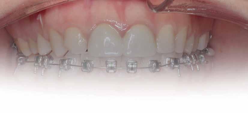
Option 2: Space Closure
Non extraction treatment, with mesial root movement and protraction of the mandibular second and third molars in order to close the residual space, create a solid contact between the mandibular second molar and second premolar without the need for prosthetic dental implant or crown. This option would require longer orthodontic treatment time with extensive follow up visits, however no extra cost is allocated for surgical and prosthetic procedures. (Figures 1 and 2)
After thorough explanation of the pros and cons of each option the patient opted for option 2.
Treatment Progress
February 2021:
Bonding: Genius Passive self ligation ( MEM Corporation, Sweden Taiwan) with 14 NiTi (Thermal ultra) engaged. First molar bondable tube with a built in long extended hook that reached the level of the furcation to apply the force close to the center of resistance of the second mandibular molar.
®
Articulating foil
No need for forceps. The foil is pre-mounted in a sturdy plastic frame.
Super thin foil.
The foil is only 8 micron thick and inked on both sides. Available in blue or red.
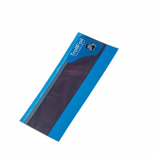
No ink on your fingers. The foil is covered on both sides at the grip.
Protective strip Easily removed before use.
The only articulating foil you will ever need!
TrollFoil
takes the guesswork out of occlusal adjustments.
TrollFoil can be used under a wide variety of clinical situations including wet or dry teeth, limited opening, limited vestibular space, gaggers, and metal and non-metalic restorations. You are able to verify occlusal contacts for both arches from one tooth to an entire quadrant using the mini or the original quadrant size. The double-sided foil is only 8 microns thick, and it has no problem marking wet surfaces, dry surfaces or highly polished surfaces such as cast gold or BruxZir.
For further information please contact your local dealer or Annelie Johansson, Area Sales Manager – Product specialist TrollDental x-ray, Finland, Baltic, East Europe, Greece, Turkey, MENA annelie.johansson@directadental.com www.directadental.com
April 2021:
- 18 NiTi Thermal ultra
June 2021:
- 18*25 NiTi Thermal ultra
July 2021:
- Short head Mini-screw 1.6 diameter, 8 mm length Titanium alloy (Absoanchor®) palced between the mandibular first Premolar and canine at the junction between attached and movable mucosa in a 45 degrees inclination.
- Lower wire: 16x25 stainless steel wire
- Connecting first molar hook to the micro-implant with a chain generating about 200g of force
- To avoid irritation of the cheek when in contact with the miniscrew head, a light cure silicone material was applied to cover the chain and the mini-screw all-together (Softflow®).
For almost 8 months, during the Covid pandemic, the patient was unable to present to the clinic. The work was resumed in March 2022. It was decided then to segment the appliance, including third molar to canine. The 16x25 SS wire was replaced with 17X25 S.S in the lower arch to increase the rigidity of the wire given the short span available.
June 2022:
- Patient presented to the clinic, with pain from the lower left side
- It was found out that the lower left first molar was vertically fractured and needed extraction
- Since the loss of the lower left first molar came at the later stages of the treatment and the patient didn’t want to go through another 1,5 years of orthodontic treatment to achieve space closure
- The patient decided to extract the affected molar and preserve the space for later implant placement
July 2022:
- A metallic ligature is placed from second molar to canine and a second one from third molar to mini screw
- Slight inter-proximal reduction (IPR) is performed between second molar and second premolar, to create a surface area contact and allow for further Protraction of the second and third molar.
- Debonding was done later in the session
- Fixed retainer was placed between the second molar and the second premolar to preserve the space needed for later implant placement (lower left side)
- Fixed retention was also needed on the lower right side to prevent relapse of space closure using a fixed 16x 22 SS wire bonded and contoured with flowable composite resin to the buccal side of teeth 48-47-45-44 since in adult space closure, chance of relapse is very high, this retention is kept in place for indefinite period of time.


Case Discussion
Both mandibular second and third right molars were mesialized and uprighted with more root movement taking place than the crown in a 2:1 ratio. Ensuring parallelism between the roots is a necessary step for long term stability of the achieved movement, nonetheless a fixed retention was undertaken . If maxillary right third molar shows any signs of passive eruption , the patient is instructed to extract it. A vacuum formed retainer was proposed during night time but unfortunately the patient declined.
Case 2:
A 33 year old male presented to the orthodontic clinic referred by his prosthodontist . His main concern was the overeruption of maxillary right and left second molar due to bilateral loss of second and third mandibular molars that rendered the placement of prosthodontic implant and crown not possible. The patient had a 4 unit bridge, to replace missing maxillary right first and second premolar, with a root canal treatment and crown done on maxillary right lateral incisor and left second premolar. The patient’s main condition was to reduce the orthodontic system to a minimum with no visible appliances due to the nature of his work in sales.


Treatment options
Two options were presented to the patient
Option 1
Endodontic treatment and crown preparation on both maxillary left and right molars to shorten the vertical dimensions of the crowns. This option would require extensive reduction of tooth material due to the fact that upon closure, maxillary second molars were in close proximity with mandibular retromolar area.
Option 2
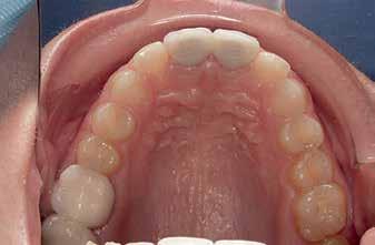
Placement of temporary anchorage devices (Miniscrew) in the buccal and palatal side of the maxillary second molars in order to orthodontically intrude those teeth and create the required biological space for implant and crown placement in the mandibular posterior molar region(Figure 3).
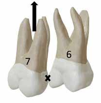
Interproximal reduction in the area between maxillary first and second molar is necessary to allow the intrusion of the second molar without being hindered by the proximal contact of the first molar.(figure 4)
Option 2 was the patient’s choice based on the fact that it saves tooth structure and integrity nonetheless he was informed about the possibility of failure of the miniscrews and the potential for discomfort due to contact between his tongue and the palatal miniscrews.
Treatment Progress
April 2021:
- Bonding of tubes on buccal and palatal surfaces of upper 2nd molars
- 2 Buccal (1.6 mm diameter 8mm length) infra-zygomatic mini screw on upper 2nd molars of each side were placed this high in order not to interfere with the intrusive movement.
- 2 Palatal (1.8 mm diameter 8 mm length) placed in the midpalatal suture and another halfway the distance between the left second molar and the mid-palatal suture .
- The mini-screws were placed in a way that the resultant forces on each side are 120-150 g of force leading to close to pure intrusion of 2nd molars .


