




Table
1
New
Honors and Awards
9 Kellogg photographers take top honors
10 Finding therapies in old ideas
16 Gerald D. Aurbach Award
18 Lifetime Service Award
19 David Musch recognized as fellow of AAAS
23 Precision naming for precision medicine
26 Yannis Paulus, M.D., named IEEE senior member
27 A life built around preventing vision loss in children
29 7th annual Kushner webinar
33 Sally Temple, Ph.D., named to National Academy of Medicine
Philanthropic Support
35 Kenneth H. Musson, M.D., and Patricia M. Musson give back
36 Marilyn and Richard Witham invest in the
2022 marks the 150th anniversary of the establishment of a Department of Ophthalmology at the University of Michigan. At the time of its founding in 1872, the department was only the fourth academic program in the nation devoted to vision science and eye care. From the start, we set a course to transform ophthalmology.
Among the Michigan milestones which changed the field of ophthalmology and the lives of patients:
• The U-M Heredity Clinic was established in 1941 by a faculty pioneer in human genetics and vision. In 1956 it formed the basis for the first dedicated medical school genetics department in the country.
• Beginning in the 1960s, basic science work conducted in our original Vision Research Laboratory helped form the foundation of knowledge about mechanisms of color vision and color blindness.
• In 1995 the human gene encoding the retinal pigment epithelium protein RPE65 was cloned at Kellogg, and in 1997 was linked to Leber congenital amaurosis. The disease has been treated successfully with FDA-approved gene therapy since 2017.
• Pioneering work in ophthalmology and endocrinology by U-M faculty on the repurposing of teprotumumab led to the first FDA-approved treatment for thyroid eye disease in 2020.


• Results from the Collaborative Initial Glaucoma Treatment Study (CIGTS), a multi-center NIH clinical trial led by Kellogg faculty and staff, demonstrated that intensive treatment for early glaucoma can preserve vision and that greater patient adherence with medical therapy is indeed associated with better outcomes in glaucoma.
There is no better way to honor our past than to celebrate the people and ideas which continue to propel Kellogg forward. This year’s Annual Report shares the efforts of our faculty, learners, staff and alumni to transform the scope and methods of education and learning, create and validate new treatments and therapeutic approaches, personalize individual patient care, and preserve and improve the eye health of our communities.
The innovation in this year’s report—from biomedical engineering to fresh training approaches to community models of eye care—is cultivated by collaboration and teamwork. Our team approach reflects the longstanding model of cooperation at Kellogg and across Michigan. That cooperation will be bolstered as we welcome the 15th President of the University of Michigan and renowned vision researcher, Dr. Santa Ono, to our department.
It is with deep appreciation and admiration of our alumni, faculty, learners, staff, friends and supporters—your team effort makes all we do possible—that I invite you to learn about some of this year’s accomplishments in the 2022 Annual Report. We hope that you share our excitement to see where 2023 and the changes to come will lead us all.
Paul P. Lee, M.D., J.D.Improving the vision health of underserved populations has been a key focus at Kellogg. Those facing the greatest risk of vision loss from diseases like glaucoma and diabetic retinopathy tend to be the least likely to have access to an eye care provider. And far too many people suffer from poor vision because of a lack of access to basics like vision screening and affordable glasses.
An ambitious pilot intervention now being evaluated in two southeast Michigan communities is addressing these issues. Launched in 2019, MI SIGHT (Michigan Screening & Intervention for Glaucoma & Eye Health Through Telemedicine) is funded through 2024 by a grant from the Centers for Disease Control and Prevention (CDC).
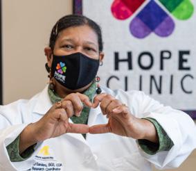
Both clinical data and anecdotal evidence indicate that the program’s combination of telehealth and community-based medicine is an effective model to deliver screening and timely followup care to patients at elevated risk of vision loss from undetected or untreated eye disease or from just needing glasses.
Rich and Kathy Sistek learned about MI SIGHT soon after its launch through a neighborhood online forum. “There was a post about a new eyecare program in the area offering free vision exams and accepting new patients.” Kathy recalls. The Sisteks phoned and arranged back-toback appointments at the Hope Clinic in nearby Ypsilanti.
Detailed intake interviews preceded their eye exams. “The technician, Londa, was wonderful,” says Rich. Although neither Rich nor Kathy screened positive for disease, they both left with updated glasses prescriptions to improve their vision. However, Kathy’s exam revealed a potential concern in one eye, which was resolved by the reviewing Kellogg ophthalmologist. They have since returned to the Hope Clinic for a scheduled follow-up exam, prompted by a text reminder. “Everyone was professional and respectful, and the whole experience was easy,” Kathy adds.
Preliminary data confirm that participants like the Sisteks value the program, and that the approach is effectively identifying and addressing vision issues:
Participants were highly satisfied with their screening visit
7.3% Screened positive for diabetic retinopathy twice thenationalaverage MI SIGHT
24.4% positivecreenedS glaucomafor thetimesthreenational average
90.7%
71% Needed vision correction ordered glasses through the program
39% neededParticipants anwithfollow-up ophthalmologistcarefurtherfor
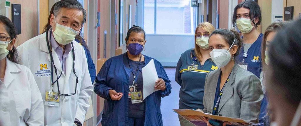
Vision technicians are available in two familiar, trusted community clinics–the Hope Clinic, a free clinic in Ypsilanti, and the Hamilton Clinic, a federally qualified health clinic in Flint. The technicians perform tests to assess patient vision and eye health.
Patient information and eye screening data collected during exams are transmitted through the electronic medical record to an ophthalmologist at Kellogg who reviews and interprets the chart and imaging and creates a plan for follow-up care, if needed. Results and physician-directed care plans are transmitted back to the technicians, who educate patients about their diagnoses, help them obtain recommended follow-up, and dispense and fit low-cost glasses as appropriate. When a condition like glaucoma is diagnosed, the clinic’s staff is there to help the patient navigate follow-up appointments, insurance options and access to low- or no-cost treatment options.
MI
“MI SIGHT is currently one of three CDC-funded programs delivering low-cost ophthalmic care in underserved communities around the country,” says Principal Investigator Paula Anne Newman-Casey, M.D., M.S., “We’re hoping it will serve as a roadmap for similar interventions in other areas.”

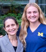
MI SIGHT also created an opportunity for undergraduate University of Michigan senior Megan Sabb , majoring in Molecular,


Cellular and Developmental Biology and Spanish, to gain hands-on experience in community outreach and research. She volunteered for MI SIGHT through a community outreach class and continued on with an independent study in Dr. Newman-Casey’s lab.
To gauge response to the program, Sabb interviewed a representative sample of 40 MI SIGHT participants, 20 from each clinic, and transcribed and coded their responses. “The overall feedback was so positive, confirming that participants from these communities value the program, particularly the combination of affordable glasses and free eye health exams,” she says.
Sabb, who plans to apply to medical school, received the Fight for Sight Summer Student Fellowship Award for her role in MI SIGHT.
SIGHT IS CURRENTLY ONE OF THREE CDCFUNDED PROGRAMS DELIVERING LOW-COST OPHTHALMIC CARE IN UNDERSERVED COMMUNITIES, WE’RE HOPING IT WILL SERVE AS A ROADMAP FOR SIMILAR INTERVENTIONS IN OTHER AREAS.— Paula Anne Newman-Casey, M.D., M.S.
The New Kellogg Vision Rehabilitation Section brings together cutting edge research and expanded clinical services to help patients with visual challenges maintain quality of life and independence. While traditional vision rehabilitation services focus on those with decreased acuity (sharpness of straight-ahead vision), the new Section at Kellogg expands our services for those with compromised peripheral vision and other conditions such as traumatic brain injury, according to inaugural Section Leader Sherry Day, O.D., FAAO.
Patients are referred to the section from vision specialties like Neuro-Ophthalmology, Retina, Glaucoma, and, increasingly, from community and UMHS non-ophthalmic services, including Physical Medicine & Rehabilitation, NeuroSport, the Stroke Center, the Concussion Center and Balance/Vestibular Testing & Rehabilitation. To help meet patient demand, Dr. Day and fellow optometrist
Donna Wicker, O.D., FAAO are joined by new Kellogg faculty members Erin Klukas, O.D., Jackie Nguyen, O.D., and Meagan Tucker, O.D. Dr. Tucker is a neuro-rehabilitation specialist who focuses on vision issues arising from vestibular disturbances, concussion, stroke and traumatic brain injury.

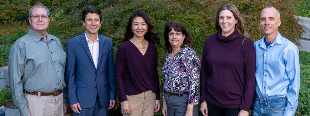
Occupational therapist Ashley Howson, M.S., OTR/L, and orientation and mobility specialist Russ Ellis, M.A., COMS provide additional support.
The new section is a natural incubator for innovation. “We share our clinical dilemmas with researchers, and they look to us for practical applications for their ideas,” explains Dr. Day. This translational approach has already yielded new insights and approaches.
James Weiland, Ph.D., Professor of Ophthalmology and Visual Sciences at Kellogg, Associate Chair for Research in Biomedical Engineering in the U-M Medical School, and Professor of Biomedical Engineering in the U-M School of Engineering, is working with his BioElectronic Vision Lab team to develop technological solutions for visual dysfunction like implantable bioelectronic retinal prostheses and wearable smart cameras. Ultimately it is hoped that the devices will provide assistance to patients of the vision rehabilitation section.
“ “
WE ARE A UNIQUE DESTINATION PROGRAM FOR CLINICIANS AND MULTIDISCIPLINARY RESEARCHERS TO COLLABORATE TO IMPROVE THE LIVES OF PATIENTS WITH VISION IMPAIRMENT.Sherry Day, O.D., FAAO Erin Klukas, O.D., K. Thiran Jayasundera, M.D., M.S., Megan Tucker, O.D., Jacqueline Nguyen, O.D. (not pictured)
Dr. Josh Ehrlich, M.D., M.P.H., a glaucoma specialist, has developed a patient-reported questionnaire that is translating the impact of peripheral vision loss into clinical information to help guide care.
Dr. Thiran Jayasundera, M.D., M.S., a Kellogg retina specialist, has partnered with the Department of Psychiatry to pair low vision rehabilitation with counseling and psychotherapy in order to address emotional stress in patients with vision loss due to inherited retinal diseases. The work should be applicable to other forms of vision loss as it is estimated that more than a quarter of those with significant vision loss develop depression or suffer other mental health consequences.
An annual survey that examines patient vision promises to help doctors improve their understanding of disease progression and the effectiveness of treatment at the U-M Kellogg Eye Center.

Jennifer S. Weizer, M.D., Professor of Ophthalmology and Visual Sciences and UMHS Service Chief of Ophthalmology, is leading an initiative to implement administration of the National Eye Institute’s Visual Function Questionnaire (VFQ-9) to all adult patients at the Kellogg Eye Center. Building on the innovative idea of Josh Stein, M.D., M.S., to incorporate the VFQ-9 into routine clinical care, the new initiative will see that all adult patients at Kellogg are included to capture their perceptions of how their vision impacts their functioning.
The nine-question test is easy to understand and uses a number scale to evaluate activities such as reading, driving, socializing and finding items in a kitchen cabinet in order to evaluate how eyesight affects patients’ lives.
Dr. Day stresses that vision rehabilitation should not be viewed as end-stage therapy for patients at the last phases of vision loss. “Doctors can and should refer patients to us any time they note a change in functional vision, rather than waiting until a patient is legally blind,” she says. “Early intervention is critical to help lead to the best outcomes. With appropriate devices, rehabilitation and resources, patients can more easily maintain independence, remain in school, pursue careers, work and care for their families. And like physical therapy, vision rehab can take place in parallel with ongoing surgical or medical treatments, sometimes with immediate positive impacts for the patient.”
“There’s a push in healthcare to become more patientcentered,” said Weizer, who specializes in glaucoma, patient safety and quality improvement. “Many are looking to physicians across the country to more directly incorporate patient feelings about their health, how it affects them and the way they function.”
While the VFQ has been used extensively for research, there was previously no widespread effort to ensure patients used the test to provide input as part of their clinical care. Peer-reviewed studies have shown that administering the VFQ is effective in assessing the natural history and effects of treatments for multiple eye diseases.
Eye clinics across U-M are administering the survey to their adult patients, either through their patient portals or as they check into a clinic appointment. The responses to the VFQ will help doctors improve their understanding of disease progression and the effectiveness of treatment at the U-M Kellogg Eye Center. “The answers to the survey will help us tailor our efforts and our treatments for more individualized care in the future,” said Weizer.
THE ANSWERS TO THE SURVEY WILL HELP US TAILOR OUR EFFORTS AND OUR TREATMENTS FOR MORE INDIVIDUALIZED CARE IN THE FUTURE.
Jennifer Weizer, M.D.
Kellogg continues to expand our ability to offer hope for infants and children and their families with acquired and inherited retinal diseases (IRDs). One of a handful of clinics in the country devoted to pediatric retinal disease, Kellogg offers the latest medical and surgical treatment options for children with IRD and other retinal conditions such as Retinopathy of Prematurity (ROP).
Kellogg is one of a few (14) U.S. treatment centers approved to offer Luxturna, the first F.D.A. approved gene therapy shown to improve vision in patients with IRDs linked to a mutation in the gene RPE65®. The treatment involves surgically injecting a working copy of the RPE65 gene under the retina of each eye. Because it is important for patients to have viable retinal cells, most patients undergo the surgery as children prior to the complete loss of retinal cells due to the disease. The procedure may also be an option for adults with RPE65-related IRDs that progress slowly or manifest later in life.
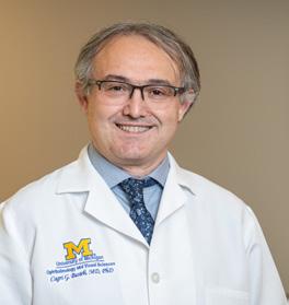
“We have offered this therapy since 2017, with impressive results,” says Cagri G. Besirli, M.D., Ph.D., who performs the procedure at Kellogg. “We see a dramatic improvement in low light vision, and as a result, parents tell us their kids are more active, engaged and confident.”
Dr. Besirli notes that in addition to Luxturna®, Kellogg offers clinical trials of several other gene therapies for pediatric retina patients, including X-linked retinitis pigmentosa and achromatopsia.
Other retina specialists who care for pediatric retina patients include K. Thiran Jayasundera, M.D., M.S., and Abigail Fahim,
M.D., Ph.D., who focus on retinal dystrophies; and pediatric ophthalmologists Pamela Williams, M.D., and Steven Archer, M.D., who specialize in the treatment of ROP, an abnormal growth of retinal blood vessels in premature babies.

These physicians and physician-scientists are supported by an attending rheumatologist, April Marquardt, M.D., and a dedicated electrophysiologist, Naheed Khan, Ph.D., along with a team of genetic counselors, researchers and technicians.
The team is expanding to meet the growing volume of complex pediatric patients and the growing array of treatment options. In addition to Drs. Besirli, Jayasundera and Fahim, Thomas Wubben, M.D., Ph.D. also is part of the team and Emily Eton, M.D., will come on board upon completing her fellowship in 2023. Dr. Eton’s research has already laid important groundwork for improving and expanding fellowship training opportunities in ROP and other pediatric retina disorders.
“By building the depth and cross-coverage of our team,” adds Dr. Besirli, “Kellogg is poised to host more translational research and clinical trials, and attract more trainees to the pediatric retina subspecialty.” Dr. Besirli and Dr. Jayasundera have already trained colleagues from Ohio State, Wayne State, University of Illinois-Chicago, Brazil and elsewhere around the world.
“
BY BUILDING THE DEPTH AND CROSS-COVERAGE OF OUR TEAM, KELLOGG IS POISED TO HOST MORE TRANSLATIONAL RESEARCH AND CLINICAL TRIALS, AND ATTRACT MORE TRAINEES TO THE PEDIATRIC RETINA SUBSPECIALTY.
Cagri Besirli, M.D., Ph.D.

Every year thousands of patients turn to Kellogg to restore, improve or preserve their eyesight. Through the Ocular Prosthetics Service, Kellogg also offers solutions and hope to people who lose eyes to illness or injury, and parents of babies born with undeveloped or underdeveloped eyes.
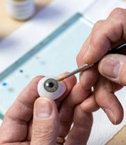
Ocularists are ophthalmic specialists trained to create and fit ocular prosthetics, or artificial eyes. Replicating a human eye requires an extraordinary combination of medical know-how, technical precision and artistry. Guiding patients and parents through the meticulous process requires tremendous patience and compassion.
With fewer than 140 board-certified ocularists in the U.S., few private practices and even fewer academic eye centers have one on staff. Kellogg has two, with more than 60 years of combined experience.
Greg Dootz, BCO, BADO, came to the discipline from an optical background, training and apprenticing at U-M before joining the Kellogg staff in 1980. He estimates that he has created prosthetic eyes for about 6,000 adults and 900 infants and children. “My patients have ranged from 2 days to 103 yrs old, and have come from as far as Italy and Russia.”
Eric Lindsey, BCO, BADO, was recruited by Kellogg in August 2022 after owning an ocular and facial prosthetics practice in California. His expertise is rooted in strong artistic fundamentals, having studied painting and sculpture in the U.S. and Italy before applying his talent to prosthetics. “It’s so rewarding to build relationships with patients, and to know that I’m making art that makes a difference,” he says.
Numerous steps are required to create and fit an ocular prosthesis. An impression of the patient’s eye socket is made, which forms the mold for a wax casting. Hours are spent in the lab sculpting and adjusting the casting. Multiple patient fittings are needed to position the iris and pupil and calibrate lid movement and motility. Alterations in one variable mean changes in them all, until finally a prosthetic can be cast in acrylic and hand painted.
The youngest patients, including babies born with anophthalmia or microphthalmia, require additional preparation in advance of their first prostheses. As early as possible—ideally within the first two months—a newborn’s eye socket is fitted with an expandable device called a conformer. Regular adjustments by the ocularist enlarge the conformer so the socket and surrounding structures develop properly as the child grows.
Eventually the child is fit with his or her first prosthetic eye.
“Especially for babies who have one functioning eye, our goal is to provide the first prosthesis by about age two,” explains Dootz. “It’s important that, from the very start, they see themselves as looking no different than anyone else.”
The ocularistry lab is located adjacent to surgical and medical clinics, making it easy to partner with patients’ care teams to address any issues or concerns. “That continuity of care gives patients and parents peace of mind,” says Lindsey, who has already collaborated on a number of complex cases. “It’s one of the biggest advantages of practicing at Kellogg.”
In addition to monitoring, troubleshooting, improving and replacing prostheses of existing patients, the team sees four to five new patients each week. “We follow these patients for life,” says Dootz, “so the bonds we build are strong.” So strong that an adult patient Dootz has worked with since infancy recently asked him to walk her down the aisle at her wedding.
“
IT’S SO REWARDING TO BUILD RELATIONSHIPS WITH PATIENTS, AND TO KNOW THAT I’M MAKING ART THAT MAKES A DIFFERENCE.
Eric Lindsey, BCO, BADO
Decades of teaching in the U.S. and around the world have made Jonathan Trobe, M.D., an ardent observer of how people— especially medical students—learn.

A professor of both Ophthalmology and Neurology, in 2014 Dr. Trobe led the multidisciplinary team that developed The Eyes Have It— a groundbreaking website and mobile app now widely used to impart ophthalmology basics to medical students and providers.
STUDENTS SUCCEED WHEN THE MATERIAL IS INTRIGUING, USEFUL AND ENJOYABLE, LEARNING SHOULD BE FUN, OR YOU WON’T DO IT.
Building on that success, Dr. Trobe has taken on an even more ambitious project: an online study tool devoted to his clinical specialty. Fifteen years in the making, he and a new team created Neuro-Ophthalmology at Your Fingertips. (https://michmed.org/fingertips)
Dozens of narrated videos, animations and anatomic illustrations bring to life 27 topics, each presented in a sequence of tabs:
• What is it?
• How does it appear?
• What else looks like it?
• What should you do?
• What will happen?
“This is the same approach used by the Audubon Society to help people differentiate between and retain information about birds,” explains Dr. Trobe. Users can expand and collapse each section, learning at their own pace without becoming overwhelmed by the depth of content. Common clinical observations – TIPS and TRAPS—are interspersed throughout. And a self-guided quiz section employs 100 clinical case studies to test the user’s knowledge. Since its launch three years ago, the site has continued to grow its user base within U-M and beyond, including students of ophthalmology, optometry, neurology, pediatrics, neurosurgery and emergency medicine. Embraced as a teaching tool by the North American Neuro-Ophthalmology Society (NANOS), it now resides within the Neuro-Ophthalmology Virtual Education Library (NOVEL) hosted by the University of Utah.
“Students succeed when the material is intriguing, useful and enjoyable,” says Dr. Trobe. “Learning should be fun, or you won’t do it.”
Dr. Trobe credits U-M and Kellogg leadership and a generous donation from former ophthalmology resident trainee Bithika Kheterpal, M.D., with making the project possible. Major contributions were made by David Murrel and Robert Prusak in art and design, Marc Stephens, Richard Hackel, and Tim Steffens in video editing, and Andrew Moses and Lisa Burkhart in programming.
Jonathan Trobe, M.D.
Part science, part art, ophthalmic imaging is an exacting discipline essential to documenting and diagnosing eye diseases. Each fall the American Academy of Ophthalmology (AAO) and the Ophthalmic Photographers’ Society (OPS) host a competition celebrating this best work of ophthalmic imaging specialists from around the world.
Images are entered in the print division (two-dimensional) and the stereo division (three-dimensional). There are multiple juried categories, including the various modalities for imaging the back of the eye, such as fundus photography, angiography, autofluorescence, and the front of the eye like slit lamp, gonio and external photography.


Facing tough competition from across the U.S. and as far away as Singapore, Australia, Thailand and Slovenia, Kellogg’s imaging team continued its impressive winning streak in the 2022 competition. Three team members combined to win 31 awards, and for the fifth straight year a Kellogg team member took the Best-of-Show award in the Stereo category.
Kellogg’s dominance in ophthalmic imaging dates back nearly 50 years. From 1975 to 2000, Csaba Martonyi, CRA, FOPS, served as Director of Ophthalmic Photography at Kellogg. A longtime active member and past president of the OPS, Martonyi was a technical and artistic pioneer and a revered teacher. Each year the Csaba L. Martonyi Award recognizes the image judged overall best-in show at the competition.
This year, an image produced by Tim Steffens, CRA, OCT-C, FOPS, the current Director of Ophthalmic Imaging & Information Systems, won the Martonyi Award—the third time in five years this honor has come home to Kellogg. Steffens also placed first in the “Eye as Art” category, which recognizes creativity and visual appeal.
“Our team is so proud of the work we do, and so honored to represent Kellogg at this prestigious event,” says Steffens.
“
“
OUR TEAM IS SO PROUD OF THE WORK WE DO, AND SO HONORED TO REPRESENT KELLOGG AT THIS PRESTIGIOUS EVENT.Tim Steffens, CRA, OCT-C, FOPS
You can’t learn surgery from a book. Hence the growing popularity of videotaped surgical procedures as a teaching tool for medical residents.
In ophthalmology, residents can go online to access countless high-quality videos of procedures like cataract or glaucoma surgery. These videos are produced by mounting a coaxial camera to the surgeon’s microscope, making it possible to capture exactly what the surgeon sees.
But as Nathan Liles, M.D., realized during his residency and pediatric ophthalmology fellowship at Kellogg, videotaping procedures that typically don’t use a microscope, like many forms of strabismus surgery, presents a challenge.

For non-microscope procedures, surgeons wears ‘loupes’, telescopic magnifying lenses mounted to eyeglasses to provide the necessary detail in a wide enough view. “Loupes don’t lend themselves to a camera mount,” he explains. “And it’s virtually impossible to capture an unobstructed surgeon’s point-of-view with a camera mounted to a wall or light source.”
For Dr. Liles, the solution was right under his nose—or rather, on the top of his head. “I thought about how head-mounted cameras are used to produce amazing point-of-view videos of sports like skiing and skydiving,” he says. “I figured making it work for extraocular surgery was just a matter of adding up-close definition.”
Cagri Besirli, M.D., Ph.D., recently found inspiration in metabolic reprogramming— research that earned him a Macula Society 2022 Young Investigator Award. “We looked at how metabolism can be reprogrammed to make cells more resistant to injury, boost their survival and slow vision loss,” said Besirli, a clinician-scientist and Associate Professor of Ophthalmology treating and researching retinal diseases. “The concept of aerobic glycolysis
After researching different commercial telescopic lenses, Dr. Liles found one that could be adapted to fit head-mounted cameras to provide 2.5x magnification, the same as that achieved with the surgical loupes.
Now a member of the Pediatric Ophthalmology faculty at Kellogg, Dr. Liles has used his device to record 4k-quality videos of dozens of surgeries, including many strabismus procedures. “In many cases I also filmed versions without magnification to broaden the field of view,” he explains, “to allow the viewer to see the hands of the surgeons and assistants.”
Kellogg surgical residents are currently using Dr. Liles’ videos to augment traditional forms of study. Work is underway to complete a website to house a full catalog of videos.
was discovered decades ago,” he said. “We are rediscovering the importance of the metabolic pathways partly because we understand them better, we have new tools to investigate them and there is broader interest, especially in cancer. We are leveraging what cancer researchers are discovering and applying those concepts to the eyes. We hope it will give us another tool to prolong vision.”
Ophthalmic technicians are an essential part of our care teams, facilitating high quality, efficient care for patients. Kellogg’s Technician-in-Training Program (KTITP) began in 2012 to address the urgent need for ophthalmic technicians. Since then, more than 100 people have been selected to take part in the rigorous program of classroom education and on-the-job training. Most graduates become technicians in ophthalmology and optometry practices. But for some, like 2017 graduate Audree Bass, the experience lights a fire that sends them down a different road.
“As an undergraduate, I knew I wanted a career in healthcare,” says Bass, who earned a B.S. in Biopsychology Cognition & Neuroscience from U-M in 2013. “But I wasn’t sure what direction to take.” She was considering medical school or a physician assistant program. Dr. Bass had learned about the KTITP program, but wasn’t sure.

KELLOGG STRESSED LISTENING SKILLS AND QUALITIES LIKE MATURITY, PATIENCE AND KINDNESS THAT ARE SO ESSENTIAL FOR BUILDING PATIENT RELATIONSHIPS.
— Audree Bass, O.D.
Her roommate, who was training in optometry, suggested Bass try working in an optometrist’s office while she considered her next move. She loved it. Not long after, a chance encounter with a Kellogg staff member in a restaurant again raised the possibility of working at Kellogg. Right after that conversation, Bass received another call from the supervisor for the Tech-inTraining Program. “After discussing it with my dad, we both felt that this door kept opening for a reason.”
Based on her experiences with the KTITP, Bass decided to become a doctor of optometry. She earned her degree from Ohio State University in 2021. Now a faculty member at Michigan and an attending optometrist, Bass practices at Kellogg facilities in Ann Arbor and Ypsilanti.
She credits the program with giving her a leg up over her fellow optometry classmates in both knowledge and confidence. “From day one I knew the fundamentals like refraction, slit lamp examination and retinal scanning, and I had a solid foundation in the pathophysiology of eye diseases.”
“Even more important,” she adds, “Kellogg stressed listening skills and qualities like maturity, patience and kindness that are so essential for building patient relationships.”
“Mastering this challenging curriculum sets our graduates up for success in whatever they pursue,” says Program Administrator Cathy Huebner.
Professional paths graduates have pursued include:
• M.D., Pediatrics
• M.D., Psychiatry
• Nursing
• Physician Assistant
• Orthoptics
• Clinic Management
• Clinical Research
• Public Health
• Global Health
Lesions on the eyelid or other tissue around the eye may develop in patients of all ages for a number of reasons. The vast majority of these lesions are benign, and do not require surgical removal. Examination by a specialist in eye plastics and orbital surgery can be key in assessing a lesion correctly. Unfortunately, most patients do not have ready access to these specialists.
Christine Nelson, M.D., FACS, is working on a way to remotely evaluate lesions around the eye using dermoscopy, one of dermatology’s go-to-diagnostic tools.

A dermoscope is a handheld device that provides a magnified view of skin lesions. Dermoscopy helps detect conditions like melanoma with sensitivity and specificity not achievable with the naked eye. Until now, its use has been largely limited to examining flat surfaces of the skin. “Most clinicians assume dermoscopy can’t be used to examine curved areas, like the areas under the brow or over the globe of the eye,” Dr. Nelson explains. “Our work challenges that assumption.”
Dr. Nelson, Section Leader of Eye Plastic, Orbital and Facial Cosmetic Surgery, with a dual appointment in Ophthalmology and Surgery, put together a cross disciplinary team of faculty and medical students to explore whether dermoscopy could be adapted to evaluate periorbital, eyelid and conjunctival lesions.
“EVENTUALLY, PROVIDERS IN REMOTE AREAS
COULD TRANSMIT DIGITAL IMAGES OF SUSPICIOUS LESIONS TO US, AND KNOW RIGHT AWAY WHETHER TO REFER A PATIENT FOR SPECIALTY CARE OR TREAT THE CONDITION LOCALLY..
Christine Nelson, M.D., FACSPracticing first on each other, the team began by developing techniques to capture images both near the eye and touching the eye, using a 10x dermoscope mounted to a standard cell phone camera. High-quality images of 91 lesions were then captured from 78 of 97 patients enrolled in the study. The images were assessed for diagnostic utility by an oculoplastic surgeon and two dermatologists. Based on their evaluations, 63 of the lesions were flagged for biopsy; one third of those were malignant.
The results of their IRB-approved proof-of-concept study were presented at the 2022 meeting of the American Academy of Ophthalmology.
“It’s a steep learning curve to become proficient at the techniques,” reports Dr. Nelson. “But we’ve demonstrated that it’s possible to create diagnostic-quality dermoscopic images safely and reliably on and around the eye.”
While still in its infancy, the virtual health approach has the potential to help put patients’ minds at ease without requiring them to travel long distances. “Eventually, providers in remote areas could transmit digital images of suspicious lesions to us,” she says, “and know right away whether to refer a patient for specialty care or treat the condition locally.”
The Oculoplastics Service at Kellogg uses a multidisciplinary team approach to provide comprehensive care for the full range of pediatric and adult orbital and eye plastic disorders, including thyroid eye disease (TED), inflammation, fractures, and vascular malformations, and conditions impacting the function and appearance of the eyelid, eye socket, and tear ducts. An expanded program and new leadership are building on Kellogg’s pioneering reputation in this subspecialty—marked by the game-changing development of the first FDA-approved treatment for TED.
Vinay Aakalu, M.D., M.P.H., has been named Section Leader and Medical Director of Oculoplastics, and Director of the new Ocular Surface Disease Research Program. He joins oculoplastics specialists
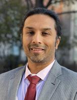
Hakan Demirci, M.D., Victor Elner, M.D., Ph.D., Denise Kim, M.D., and Christine Nelson M.D., FACS. A renowned clinician-scientist, Dr. Aakalu comes to Kellogg from the Department of Ophthalmology and Visual Sciences at the University of Illinois at Chicago College of Medicine (UIC).
Most diseases, including age-related macular degeneration (AMD), have a metabolic component. So manipulating metabolic pathways might be a possible treatment to preserve vision. Within the outer retina, a delicate balance must be maintained between the energy needs of the retinal pigment epithelium (RPE) and photoreceptors (PRs). A disturbance in this balance is associated with AMD.
While the link between RPE metabolism and AMD is well-documented, far less is known about PR metabolism in AMD. New research by Thomas Wubben, M.D., Ph.D., at the Kellogg Eye Center will address this critical knowledge gap. “We know that PR metabolism requires a lot of energy,” explains Dr. Wubben. “And we suspect that a disruption or uncoupling of the RPE/PR metabolic balance may be driven by PRs’ metabolic adaptations. Resolving this energy crisis in the outer retina has the potential to block the progression of AMD.”
In the lab, Dr. Aakalu was part of the team that first confirmed the presence of histatin peptides in tears and the cellular receptor for histatin peptides (TMEM97). Two of his current projects are supported by NIH R01 grants. One focuses on leveraging the wound healing properties of histatin peptides for the treatment of dry eye, corneal wounds and ocular surface inflammation. The second utilizes histatin peptides as a potential therapeutic approach to treat Niemann-Pick Type C disease, a rare, deadly neurogenerative disorder.
His focus in these translationally important areas led him to establish a drug development center for ophthalmic therapies. He plans to expand his focus on clinically meaningful orbital and ocular surface translational research at Kellogg. “The breadth of talent, world class facilities, and track record of breakthroughs in the treatment of orbital disorders brought me to Kellogg,” he says. “I am humbled by this opportunity to work alongside world leaders in oculoplastics and continue Kellogg’s legacy of transformative translational research.”
For this study, Dr. Wubben will fine tune an approach he previously developed in which PR metabolism is modulated by activating the target enzyme PKM2 in a model of AMD. The goal is to demonstrate that this approach can restore a healthy PR/ RPE metabolic balance and prevent PR loss. “We hope this research will point to new therapeutic targets to potentially transform how this blinding disease is treated,” he says. Dr. Wubben’s work is being supported by a Career Development Award from the Foundation Fighting Blindness, and a Bright Focus New Investigator Grant.
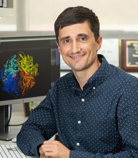
Charles Roginski was diagnosed with the dry form of age-related macular degeneration (AMD) decades ago. In recent years, the condition has robbed him of much of his sight.
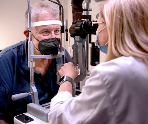
“I was told there are no treatments for dry AMD,” says Roginski, age 72. “But I wasn’t ready to give up. So I started researching clinical trials.” In 2019, he joined his first clinical study, a pharmaceutical trial conducted at the National Eye Institute (NEI) in Bethesda, Maryland.
That trial had no impact on Roginski’s vision, but he remained committed to participating in research. In February 2022, soon after learning he did not qualify for a different NEI cell therapy trial, Roginski received a call from Pam Campbell, a clinical research coordinator at Kellogg.
and tolerability of RPESC-derived RPE transplantation as therapy for dry AMD. He plans to enroll 18 patients in the trial, which will evaluate three different cell doses.
I WAS TOLD THERE ARE NO TREATMENTS FOR DRY AMD, BUT I WASN’T READY TO GIVE UP. SO I STARTED RESEARCHING CLINICAL TRIALS.
— Charles Roginski“I had spoken with Pam in the past,” he recalls, “and just when I was starting to feel discouraged, she phoned to ask if I was still interested in participating in a study.” Soon after, Roginski traveled from his home near Columbus, Ohio to Ann Arbor to make history.
In May 2022, he became the first patient in the first human trial of a transplant of retinal pigment epithelium (RPE) cells generated from human donor RPE stem cells (RPESCS).
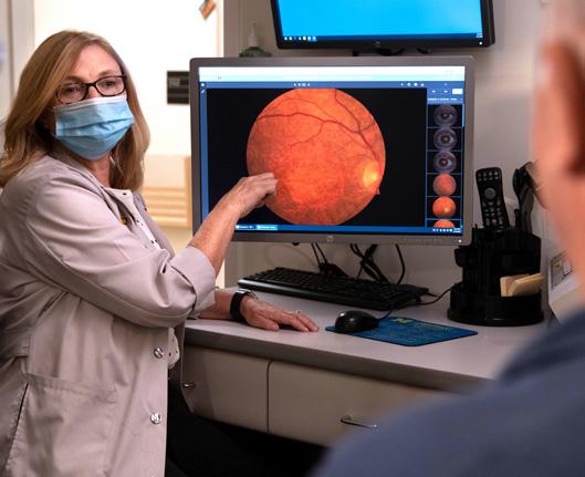
Rajesh C. Rao, M.D., is the Principal Investigator of a Phase 1/2a clinical trial intended solely to evaluate the safety
Dry AMD develops when cells lining the retina called the RPE degrade. The RPE is crucial to nourishing the photoreceptor cells that enable sight. Without a healthy RPE, photoreceptor cells die and vision is lost. One of the most compelling avenues of investigation in the quest for an AMD treatment is to use transplanted RPESCderived RPE to rebuild the layers of the diseased host RPE in the retina. Years of basic research in RPESCs by Sally Temple, Ph.D., and Jeff Stern, M.D., Ph.D., both of the Neural Stem Cell Institute in New York and adjunct faculty at Kellogg, yielded a novel approach to RPESC-derived RPE transplantation. The new method was shown to be effective at rescuing vision in pre-clinical animal models.
“In some trials evaluating therapeutic stem cell transplants, the stem cells are made from a patient’s own skin or blood cells, which are genetically induced to a stem cell that can be coaxed into almost any cell type in the human body—called induced pluripotent stem cells,” explains Dr. Rao. “But Drs. Temple and Stern achieved compelling results in the lab using allogeneic RPE stem cells, which are derived from human donor eye tissue obtained through an eye bank rather than from the patient.”
The stem cells chosen for this application have reached a specific point in their development; they are programmed to become RPE cells, but have not yet fully matured. Following isolation of RPESCs, they are differentiated (matured) to RPE, and these cells are transplanted into an area of the retina where host RPE cells are diseased or have died.
“We are grateful to Charles, and to all the patients who will partner with us in this study,” says Dr. Rao. “With their help,
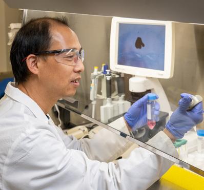
Animal models of disease, while extremely useful in basic science, do not always make the best models for the study of human disease. For example, it is difficult to study macular degeneration using a mouse model because a mouse retina does not have a macula. That’s why Drs. Rajesh Rao and Abby Fahim are spearheading the establishment of a new laboratory dedicated to directing human pluripotent stem cells into various retinal cell types involved in retinal diseases. “This new laboratory will allow us to build and study retinal organoids—three-dimensional ‘mini organs’ that more closely approximate the development, structure and function of the retina,” Dr. Rao explains. Building a human disease model is also far more complicated and time-consuming process, with more potential opportunity for contamination, than developing an animal model. A dedicated space reduces that risk.
Retinal organoid models will also be particularly useful in the study of a range of poorly understood pediatric eye diseases. For example, congenital malformations like microphthalmia, anophthalmia, and coloboma are very well suited to modeling by retinal organoid technology and may share some characteristics with congenital forms of macular degeneration. Dr. Fahim will lead an effort in the new facility to generate retinal pigment epithelial (RPE) cells. RPE cells nourish photoreceptors,
we eventually hope to demonstrate that transplanted stem cells can replace the lost RPE in the human retina, slowing or halting progressive vision loss from degenerative retinal diseases.”
Charles Roginski has very realistic expectations about the trial. “I participate in research because I know the psychological toll vision loss takes, and I want to be part of the solution,” he says. “Of course, I’d like to benefit myself. But if that doesn’t happen, it’s still worthwhile to help other patients down the line.”
the light-sensing cells responsible for one of the first steps of vision. Generating RPE cells may be helpful in modeling genetic forms of retinal dystrophies, and in better understanding RPE cell transplants in clinical trials for patients with dry AMD, such as the current Phase 1/2a trial (page 14) led by Dr. Rao.
“With this new lab, we’ll now have a central point for new collaborations among researchers from across the University. We hope it will be a catalyst for making regenerative medicine and stem cell-based disease modeling central research themes for Kellogg.” says Dr. Rao.
“THIS FACILITY WILL ALLOW US TO BUILD AND STUDY RETINAL ORGANOIDS — THREE-DIMENSIONAL ‘MINI ORGANS’ THAT MORE CLOSELY APPROXIMATE THE DEVELOPMENT, STRUCTURE AND FUNCTION OF THE RETINA.
Rajesh C. Rao, M.D.
Epigenetics is the process by which genes turn on or off. Epigenetic processes are instrumental in driving stem cells to become mature, specialized cells. Those on/off instructions may be altered in different phases and cellular compartments during cellular development, including when the instructions in DNA are copied into RNA (transcription), and when RNA is converted into proteins (translation).
Like DNA and histones (the proteins which DNA can wrap around), RNA can be modified by methyl groups, ultimately affecting the level at which the corresponding gene is expressed. The study of how RNA is methylated to affect gene expression is called “epitranscriptomics” or “RNA epigenetics”.
WE HOPE TO LEARN MORE ABOUT THIS AND POTENTIALLY OTHER ROLES METTL3 PLAYS IN IMPACTING GENE EXPRESSION DURING THIS CRITICAL WINDOW IN THE FORMATION OF THE RETINA..
Rajesh C. Rao, M.D.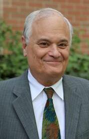
Scientists have only recently begun to investigate the epigenetics associated with RNA. Kellogg’s Rajesh Rao, M.D., an expert in DNA epigenetics, has received one of the first research grants ever awarded for RNA epigenetics in ophthalmology.
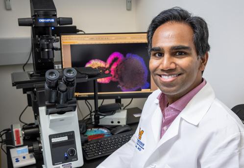
The Career Advancement Award from Research to Prevent Blindness (RPB) will support Dr. Rao’s foundational studies of the RNA epigenetics through which a stem cell becomes a retina cell.
Dr. Rao’s team is focusing on a protein called METTL3. “We know that METTL3 plays an important role in directing pluripotent stem cells (PSCs) to become retina cells,” Dr. Rao explains. A PSC is not yet fully developed, and still has the potential to become any type of cell in the body.
“Our hypothesis is that METTL3 encourages retina cell development by methylating RNA, which affects the stability of the transcript,” he continues. “We hope to learn more about this and potentially other roles METTL3 plays in impacting gene expression during this critical window in the formation of the retina.”
Dr. Rao plans a series of studies employing genetically engineered mouse models in which the METTL3 gene is disabled in the retina and doing the same in retinal organoid models—three-dimensional ‘mini organ’ structures derived from mouse and human PSCs.
“Translating our laboratory findings into clinical interventions is still far in the future,” cautions Dr. Rao. “But we hope these fundamental studies in RNA epigenetics will lay the groundwork to better understand how PSCs become retinal cells, with the goal of harnessing those principles to generate retinal tissues from stem cells more efficiently. Implications of this work could one day be applied to regenerative medicine such as stem cell-based transplants to treat retinal blindness.”
Terry J. Smith, M.D., the Frederick G.L. Huetwell Professor in Ophthalmology and Visual Sciences Emeritus and Professor of Internal Medicine, is one of 13 endocrinologists to earn the Endocrine Society’s 2022 Laureate Award. Dr. Smith received the Gerald D. Aurbach Award for Outstanding Translational Research, one of the society’s top honors.
Dr. Smith grew interested in Graves’ disease and thyroid-associated ophthalmopathy in medical school. More than three decades later, he and his team’s work led to the development of
teprotumumab—the first FDA-approved drug to treat thyroid eye disease. Prior to the discovery, steroids were the only medical—albeit nonspecific—treatment option for the condition. Othewise, surgery would be required. “Those earlier and available treatments were not disease-modifying, because they didn’t get at the heart of the disease,” he said. Smith’s lab continues to dig into the mechanisms of the disease and the actions of the medication to determine if teprotumumab or a similar drug may be applicable for other immune diseases.
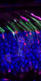
Photoceptors in the retina are among the body’s hardest working cells. They must detect light coming into the eye and stimulate a biochemical signal, which in turn triggers an electrical response that is transmitted to the brain, resulting in sight
Most of this happens in a highly specialized compartment of the photoreceptor called the outer segment.
The molecular components residing within the outer segment must be arranged just right for the signaling to succeed. The outer segment contains a number of light absorbing, connecting and structural components that work together to create the visual response to light. Because key components are made elsewhere in the photoreceptor cell, they must be properly transported to and organized within the outer segment.
Problems in delivering these molecules to the outer segment are associated with some of the most severe inherited retinal diseases, notably Retinitis Pigmentosa.
Cell and developmental biologist Jillian Pearring, Ph.D., has been awarded an R01 grant from the National Institutes of Health to find out more about the routes along which these components travel to the outer segment, and what can go wrong along the way.

“We know that once these molecules are produced, they are packaged into vesicles for their delivery to the outer segment,” Dr. Pearring explains. “And we’ve already identified two primary pathways along which they travel.”
WE HOPE THAT A BETTER UNDERSTANDING OF THE INTERCELLULAR TRANSPORTATION USED TO REACH THE OUTER SEGMENT WILL HELP GUIDE THE DEVELOPMENT OF NOVEL TREATMENTS TO CARE FOR PATIENTS WITH THESE BLINDING INHERITED DISEASES.
Jillian Pearring, Ph.D.The study will address key questions about the delivery pathways to the outer segment: Why do certain molecules choose one route over the other? If a route is altered or blocked, how will the cell respond?
“We hope that a better understanding of the intercellular transportation used to reach the outer segment will help guide the development of new treatments to care for patients with these blinding inherited diseases,” she says.
Many patients have their first exposure to opioids following surgery, when they are given prescriptions to manage postoperative pain. The risk that patients will develop issues with chronic opioid use has been documented in numerous surgical specialties. But until recently, the risk after eye surgery has not been described.
To document patterns of opioid prescription and usage as well as risk factors in ophthalmic surgery, a study team led by Kellogg surgeon and researcher Maria Woodward, M.D., M.S., conducted a retrospective cohort analysis based on private insurance claims. The results were published in the September 2021 issue of the journal Ophthalmology.
The dataset included information on 327,379 opioid-naïve patients who underwent ophthalmic surgery. The most common surgeries performed were anterior segment, oculoplastic and retinal procedures.
Of the 4.5 percent of patients who filled an initial perioperative opioid prescription, 3.4% developed new persistent opioid use (defined as use beyond the 90-day postoperative recovery period). Within that group, a prescription size of 150 morphine milligram equivalents or more was associated with an increased chance of refilling.
Among the 95.4% of patients who did not fill an initial prescription after surgery, the rate of persistent opioid use was 0.6 percent."
Our analysis establishes that exposure to opioids after incisional ophthalmic surgery is an independent risk factor for new chronic use in patients who were previously opioid naive ” explains Dr. Woodward.
The study also took into account additional patient-level risk factors, including a number of pre-existing health conditions and sociodemographic variables. “We observed that those who face a greater likelihood of persistent opioid use include women, people of Black race, residents of southern states, those with lower household incomes, and tobacco users,” she says.
“Like all surgeons, eye surgeons need to be mindful of the overall risks of prescribing opioids and the factors that elevate risk in certain patients,” notes Dr. Woodward. “We need to have frank conversations with our patients about pain, medications and expectations, and deemphasize opioid use whenever possible.”
Dr. Woodward, a member of the American Academy of Ophthalmology’s Opioid Task Force, collaborated with several U-M clinical and health services researchers on this study, including members of the Michigan Opioid Prescribing Engagement Network (M-OPEN), which is addressing the opioid crisis through novel research and evidence-based actions
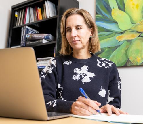
Kathy Whitney, a member of the Kellogg team for over 40 years, retired this fall. Kathy compassionately supported trainees in Ophthalmology, helping them navigate the challenges of clinical and surgical training as well as their lives outside of ophthalmology. A natural leader, she redefined the career path
of program coordinators and contributed to the success of multiple generations of Kellogg Ophthalmologists. Her legacy looms large, and the educators and mentees of Kellogg remain grateful for her many years of service to MIchigan.
WE NEED TO HAVE FRANK CONVERSATIONS WITH OUR PATIENTS ABOUT PAIN, MEDICATIONS AND EXPECTATIONS, AND DEEMPHASIZE OPIOID USE WHENEVER POSSIBLE.
Maria Woodward, M.D., M.S.
About 50 million people around the world today live with dementia—a number projected to triple by 2050.
Without effective dementia medications, leaders in the field are looking to prevent or slow its onset by addressing lifestyle and public health risk factors that we can change.
In the 2020 update to their landmark 2017 report, the Lancet Commission on Dementia Prevention, Intervention and Care listed a dozen modifiable risk factors for dementia: low education levels, hearing loss, hypertension, obesity, smoking, depression, social isolation, physical inactivity, diabetes, air pollution, alcohol use and traumatic brain injury. The commission estimates that overcoming these risk factors could prevent or delay four in ten dementia cases worldwide.
Compelling new research from a team led by Kellogg’s Joshua Ehrlich, M.D., M.P.H., indicates a critical risk factor is missing from that list: visual impairment. The study, published in JAMA Neurology in April, finds that an estimated 1.8 percent of U.S. dementia cases—more than 100,000 cases—may have been prevented by maintaining healthy vision.
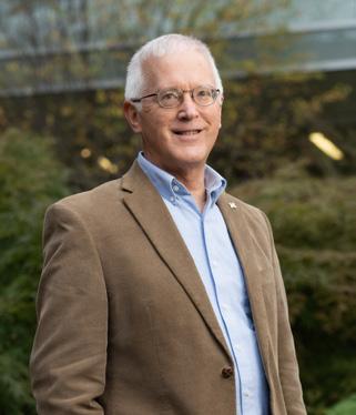
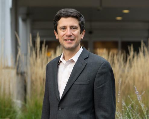
“Most people with dementia don’t have vision impairment, and most people with vision impairment won’t develop
dementia,” Dr. Ehrlich clarifies. “But this study indicates that, on average, those with vision impairment do seem to face higher dementia risk.”
The next step is to confirm the study’s findings with evidence from interventional studies. Dr. Ehrlich is part of a team developing a randomized clinical trial to evaluate the impact of vision correction on slowing cognitive decline. The CLEVER study (Cognitive Level Enhancement Through Vision Exams and Refraction) is expected to launch in India in the next year.
Dr. Ehrlich stresses that additional research is needed to determine whether a reduction in the incidence of dementia could be achieved through relatively economical, accessible interventions like eye exams, glasses and cataract surgeries.
David C. Musch, Ph.D., M.P.H., Professor of Ophthalmology and Visual Sciences in the U-M Medical School and Professor of Epidemiology in the School of Public Health, has been named a Fellow of the American Association for the Advancement of Science in recognition of his many contributions to clinical epidemiology, design principles and analytical techniques to provide evidence-based guidance for the prevention and treatment of ophthalmic disease. Musch’s work has led to improvements in research design, evident in his work leading a multi-center trial for glaucoma. “It led to years of NIH funding based on the knowledge we gained from that trial and the efficacy of treatments. We were also one of the first trials to include patient-reported outcomes,” he said. “It is now much more routine to ask patients how they are doing. It’s so important to obtain the patient’s perspective.” Dr. Musch also serves as Chair of the Data and Safety Monitoring Committees of many key clinical trials in vision research.
One promising front in the battle against age-related macular degeneration (AMD) is the development of genetic strategies to increase the resilience of the retinal pigment epithelium (RPE) and better defend against its deterioration.
In previous research, Kellogg physician-scientist Lev Prasov, M.D., Ph.D., described the role the gene myelin regulatory factor (MYRF) plays in the development of the RPE. Might that same transcription factor continue to play a protective role in the mature RPE?
Dr. Prasov’s latest MYRF research attempts to answer that question by better understanding the genes and pathways that support RPE function. In laboratory models of the mature RPE, he will conduct studies that both reduce and increase the function of MYRF and several of its target genes, measuring the impact of those changes on different aspects of RPE cell structure and physiology.
“In many tissues in the body, MYRF has been shown to play a supportive role in cellular structure and homeostasis,” he says. “We hypothesize that may be true in the cells of the adult RPE, where MYRF remains highly expressed. If so, this protective property of MYRF could represent a novel therapeutic target for AMD and other diseases of deteriorating RPE physiology.”
To establish the models used in these studies, Dr. Prasov is receiving support from colleagues with expertise in RPE culture and biology, including Jason Miller, M.D., Ph.D., at the Kellogg Eye Center, and Kapil Bharti, Ph.D., at the National Institutes of Health’s National Eye Institute (NIH/NEI).
The work is supported by a grant from the Bright Focus Foundation, which has named Dr. Prasov the inaugural recipient of the Dr. Joe G. Hollyfield New Investigator Award for Macular Degeneration Research
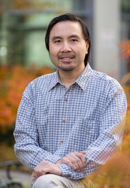
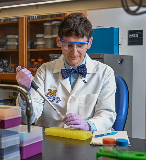
Typical indoor lights can disrupt our body’s internal clock and contribute to fatigue, depression, and sleep disorders. While researchers have spent decades fine-tuning the light spectrum of electric lighting to promote well-being, the results have been ineffective or visually unappealing. The University of Michigan recently received a U.S. patent for a multi-channel LED light fixture conceived by Kwoon Y. Wong, Ph.D., Associate Professor of Ophthalmology and Visual Sciences, that won’t interfere with our natural body clock. The retina contains special nerve cells that drive subconscious responses to light, particularly body clock activities such
as sleep timing and alertness. The new light fixture targets those specific cells rather than other retinal cells that we use to see objects. The result is that to our perception, the light appears constant while enabling the body’s natural clock to work with less interference. Professor Pei-Cheng Ku, Ph.D., of the department of Electrical and Computer Engineering, performed mathematical simulations with Wong’s team, including Ph.D. student Garen Vartanian and engineer Scott Almburg, to ensure there was no distortion of color perception. While prototypes cost upwards of $300, commercial production could drive the cost below $30.
Dr. Prasov has also received a Career Development Award from Research to Prevent Blindness (RPB) to continue his groundbreaking research linking a mutation in the gene DDX58 to a rare inherited syndrome typified by pediatric glaucoma.
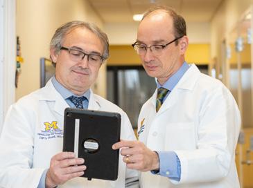
In 2020, an international team led by Dr. Prasov was the first to document a never-before-observed misspelling in the gene DDX58, associated with a rare syndromic form of glaucoma called Singleton-Merton syndrome type II. In this syndrome, the DDX58 variant triggers an inflammatory cascade throughout the body, leading to glaucoma, skin rash, calcium deposits in blood vessels, arthritis and other conditions.
“Because we saw quite a bit of variability in the symptoms of family members with the mutation, we suspect there may be non-genetic triggers at work as well,” says Dr. Prasov. “We plan to expose the mouse model to environmental factors such as viral infections, ultraviolet light, and dietary interventions to gauge their impact on the development of glaucoma.”
WE ARE CURIOUS ABOUT WHETHER THE INFLAMMATORY DDX58 PATHWAY IS ACTIVATED ONLY IN THE RAREST FORMS OF GLAUCOMA, OR WHETHER IT CAN BE IMPLICATED IN MORE COMMON FORMS OF THE DISEASE.
— Lev Prasov, M.D., Ph.D.“We currently know relatively little about the role of the immune system in the mechanism of glaucoma,” says Dr. Prasov. “This obscure mutation may actually have a lot to teach us about that connection.”
As part of the study, Dr. Prasov developed a mouse model with the same DDX58 variant found in two families with the syndrome. Preliminary data indicates that the model displays many of the same eye and skin features observed in humans with the mutation. Using the model, he will conduct a number of molecular studies to identify the cells and changes in gene expression responsible for driving the eye pressure elevations that ultimately lead to glaucoma in this syndrome.
“ “Studies will also be performed using the cell lines of patients with the DDX58 mutation, and those of glaucoma patients without the mutation. “We are curious about whether the inflammatory DDX58 pathway is activated only in the rarest forms of glaucoma, or whether it can be implicated in more common forms of the disease,” he explains. “If the latter, it could be a promising target with broader therapeutic value.”
Dr. Prasov emphasizes that interdisciplinary collaboration is vital to the success of this project. His colleagues include U-M Associate Chief of Basic and Translational Research in Rheumatology J. Michelle Kahlenberg, M.D., Ph.D.; U-M Professor of Dermatology and Skin Molecular Immunology
Johann Gudjonsson, M.D., Ph.D.; U-M Assistant Professor of Dermatology and Computational Medicine and Bioinformatics
Alex Tsoi, Ph.D.; Robert Hufnagel, M.D., Ph.D., of the NIH/ NEI; and a team of researchers at the University of Missouri.
Glaucoma and macular degeneration are leading causes of blindness in people over 60. Professor David Zacks, M.D., Ph.D., and Associate Professor Cagri Besirli, M.D., Ph.D., hope to change that. The pair received several international patents for molecular compounds developed and designed to treat these two diseases, as well as preserve vision in patients with retinal detachments. The treatments, licensed by ONL Therapeutics, inhibit activation of the Fas receptor, a key regulator of cell death in the retina. The compounds will be used either alone or in combination with other treatments (such as surgery), with the goal of keeping more retinal cells alive thus improving vision. To date, early studies have found the lead compound, ONL1204, safe and well-tolerated.
An ongoing collaboration between Kellogg and India’s Aravind Eye Care System (the world’s largest) took a big step forward in 2022.
Aravind and Michigan were awarded a D43 International Research Training Grant from the NIH Fogarty International Center. Led by Program Director/Principal Investigator David Musch, Ph.D., M.P.H., and Co-investigator Joshua Ehrlich, M.D., M.P.H., it is the latest joint effort between Kellogg and Aravind.
As part of the grant, Kellogg welcomed the first two Aravind visiting scholars in July. Cornea specialist Divya Manohar, M.D., and retina specialist Karthik Srinivasan, M.D., will spend a year immersed in clinical and epidemiological research fundamentals, from study design and ethics to statistical analysis.
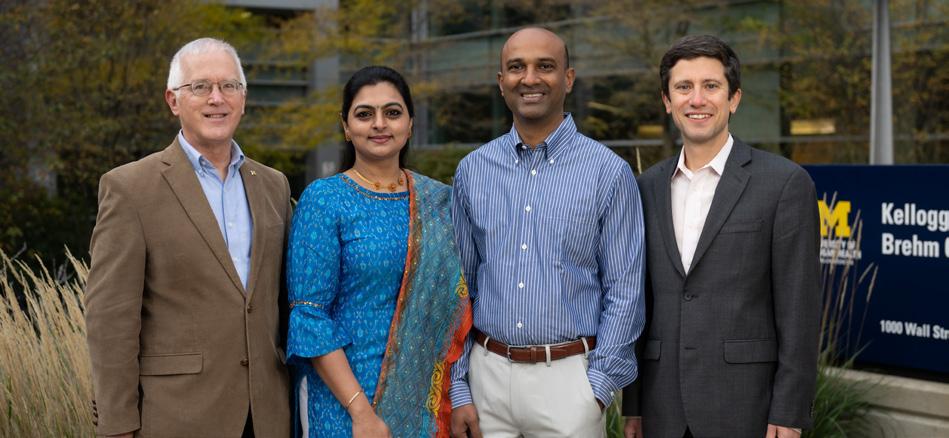
“Aravind is home to exceptional clinicians,” says Dr. Ehrlich. “This program provides an opportunity for the highly productive clinicians to take the time to further develop their skills to collect and analyze the clinical and population data needed to understand the many factors impacting the vision of Indians across the life-course.”
A total of ten visiting scholars will be part of the program at Michigan and Kellogg over the grant’s five-year duration. At the same time, Kellogg will connect with colleagues at Aravind through online workshops and journal clubs to enhance shared learning, especially about basic clinical and epidemiological research.
Joshua Ehrlich, M.D., M.P.H.
“The curriculum taps resources from within Kellogg and across the University,” explains Dr. Musch. “Divya and Karthik are taking courses at the School of Public Health and participating in educational opportunities offered by the Michigan Institute for Clinical and Health Research, while also working with their mentors on projects relevant to their clinical specialties.”
“We plan to transition all training activities to Aravind by the end of the grant period, adds Dr. Musch, “so another program deliverable is ‘training the trainers.’ Our goal is to have colleagues at Aravind who have the leadership, mentoring and teaching skills to prepare India’s next generation of epidemiological eye researchers and to share their expertise and insights with us as well.”
“
“
THIS PROGRAM WILL PREPARE A CORE OF TALENTED DOCTORS TO COLLECT AND ANALYZE THE CLINICAL AND POPULATION DATA NEEDED TO UNDERSTAND THE MANY FACTORS IMPACTING THE VISION OF INDIANS ACROSS THE LIFE-COURSE.
Divya Manohar, M.D., and Karthik Srinivasan, M.D., are the first of ten physicians from India’s Aravind Eye Care System joining U-M for a year under Kellogg’s D43 international research training grant. In addition to participating in a rigorous core curriculum in epidemiological research design and analysis, both will pair with mentors on clinical and epidemiological research projects tailored to their specific interests.
Dr. Manohar, a cornea specialist, is interested in conducting studies to understand how conditions like keratoconus and dry eye are impacting the eye health of Indians.
“Indians tend to develop keratoconus earlier in life than other ethnic groups,” explains Dr. Manohar. “Interestingly, that’s true whether they live in India or elsewhere. That leads us to suspect that there may be a genetic component at work, in addition to environmental factors.”
In India, as in the U.S., dry eye tends to get less clinical attention than other eye conditions. But this potentially debilitating disease seems to be on the rise in India, especially in younger people and people working in information technology. “I hope to conduct epidemiological research to test the hypothesis that heavy screen time is at least partially to blame,” says Dr. Manohar.
Dr. Manohar is collaborating with dry eye specialist Roni Shtein, M.D., M.S., cornea specialist Maria Woodward, M.D., M.S., and U-M pain researcher Daniel Clauw, M.D.
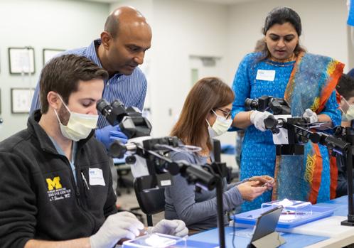
A retina specialist, Dr. Srinivasan is interested in how artificial intelligence (AI) tools like machine learning can be applied to ophthalmic diagnosis and treatment decision-making. He is working with Nambi Nallasamy, M.D., a faculty member at both Kellogg and the U-M Department of Computational Medicine
and Bioinformatics, and U-M gastroenterologist Akbar Waljee, M.D., M.Sc., in each of their respective clinical disciplines. Drs. Nallasamy and Waljee are developing models of precision care that use machine learning to tailor treatment to the specifics of a patient.
“This is a transformational approach to patient care,” says Dr. Srinivasan, who has already supplemented his coursework by participating in a U-M boot camp focused on AI coding. “Machine learning has the potential to deliver individualized treatment to large populations of patients, and to do so efficiently. It’s hard to imagine a better application than the treatment of patients with complex, costly conditions like retinal disorders, in low-resource areas like India.”
For Drs. Manohar and Srinivasan, devoting a year to study in the U.S. was not an easy choice. A married couple, they left their two children in India in the care of grandparents. “Although we miss them terribly, we could never have learned so much so quickly at home,” says Dr. Srinivasan. “Overall, the program has been a blessing for us both,” adds Dr. Manohar.
Thomas Gardner, M.D., M.S., has dedicated his career to understanding why diabetes damages the retina and how to help people with diabetes maintain their vision. The Professor of Ophthalmology and Visual Sciences, Molecular and Integrative Physiology and Internal Medicine at the University of Michigan, is also trying to alter the way we describe diseases from their appearance to the underlying biological mechanisms, the subject of his October 2022 Ian Constable Lecture at the Lions Eye Institute in Perth, Australia. The lecture honors Australian ophthalmologist and institute founder Ian Constable. Gardner proposes moving away from descriptive taxonomy in naming diseases and adopting a mechanistic approach. “How we name diseases influences how we think about, diagnose and treat them,” Gardner said. “In the age of precision medicine, we need specific names that lead to specific treatments therapeutics and outcomes.”
Glaucoma is the second leading cause of irreversible blindness in the U.S.—and the leading cause of blindness among African Americans. The standard treatment prescribed to nine in ten glaucoma patients is a daily regimen of eye drops to control their eye pressure.
With this proven, accessible, self-administered, relatively low-cost treatment, why are so many people still losing their sight to glaucoma? Studies report that more than 20 percent of patients do not instill drops properly, and at least 40 percent of the time patients do not keep to the prescribed schedule for using drops. “Patients who fail to use their drops, or use them incorrectly or inconsistently, are more likely to lose vision,” says glaucoma clinician and researcher Paula Anne Newman-Casey, M.D., M.S., Associate Chair for Clinical Research.

PATIENTS WHO FAIL TO USE THEIR DROPS, OR USE THEM INCORRECTLY OR INCONSISTENTLY, ARE MORE LIKELY TO LOSE VISION.
“Glaucoma primarily affects older adults, who are more likely to have sensory, fine motor skill and memory deficits, so the numbers should come as no surprise,” she says. “Typically, patients are diagnosed and sent home with a prescription for drops; only one in eight doctors makes any attempt to teach patients how to use them. Clearly, we need to do more to help patients get it right.” Even with coaching, many patients still have difficulty instilling drops.
That’s why Dr. Newman-Casey is a Multi-Principle Investigator of an ambitious project supported by an R01 grant from the NIH/National Institute of Biomedical Imaging and Bioengineering and a Physician-Scientist Award from Research to Prevent Blindness (RPB) to identify and measure each step in getting a drop into an eye by isolating every biomechanical, sensory, coordination and movement factor. “The study takes a page from the playbook of elite athletes and their trainers,” Dr. Newman-Casey explains. “They use sophisticated biomechanical tools to analyze every aspect of performance; the smallest tweak can mean a competitive edge.”
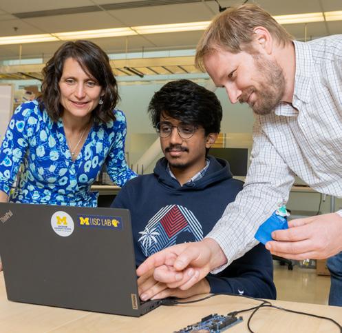
Similar microsensor technologies will be used in motion analyses of study participants who are reflective of the overall population of patients with glaucoma. Participants will be fitted with wearable sensors on the wrist, arm, head and thorax. They will use an eye drop bottle fitted with a special sleeve with additional sensors.
As a participant puts a saline drop in the eye, numerous data points will be captured. “An analysis of the data will give us a picture of whether certain movements predict whether a drop gets in the eye, and will help us determine the most efficient way to predict success,” she says. “A library of instillation profiles could be used to develop coaching strategies personalized to a patient’s particular biomechanical and sensorimotor challenges.”
The project will also evaluate the feasibility and acceptability of cues such as light or sound alarms (also built into the bottle sleeve) and/or telephonic or text message prompts to remind patients to use the drops. Finally, it will assess a method of communicating patients’ eye drop use information to the care team and return feedback and coaching tips to patients.
“We hope all of this learning will inform more effective, personalized strategies to improve patients’ success using all kinds of eye drops,” says Dr. Newman-Casey.
Dr. Newman-Casey credits her team of co-Principal Investigators and colleagues from across UM and halfway around the country, including:
• Stephen Cain, Ph.D., Assistant Professor of Chemical and Biomedical Engineering at West Virginia University and Director of the Advancing Wearable Systems for Out-of-the-lab Measurement and Evaluation (AWeSOME) Research Laboratory will lead the biomechanical analyses of movement as Multi-primary investigator.
• Neuro-physiologist Susan Brown, Ph.D., Associate Professor of Movement Science in the U-M School of Kinesiology and Director of the Motor Control Laboratory, is leading the design of the human movement studies.
• Alanson Sample, Ph.D., Associate Professor of Electrical Engineering & Computer Science and Director of the Interactive Sensing and Computing Lab, is refining the adherence monitor to maximize sensing functionality and energy efficiency.
• David Burke, Ph.D., Professor of Human Genetics in the U-M Medical School, brings critical experience in the development of low-cost health monitoring systems
Dolly Ann Padovani-Claudio, M.D., Ph.D., completed a residency and fellowship in pediatric ophthalmology and adult strabismus at Kellogg and is now a physician-scientist at Vanderbilt whose lab focuses on diabetic retinopathy. Padovani-Claudio is also dedicated to increasing diversity among clinicians and researchers in ophthalmology. “A diverse workforce representative of the patient population enhances trust and communication—and in turn, understanding of disease and management plans— enhancing compliance and improving outcomes,” she said. In 2021, 23 underrepresented in medicine participants of the National Medical Association Rabb-Venable Excellence in Ophthalmology Program, which she co-leads, matched to ophthalmology residency programs, three to four times more than in a typical year. “It is important to go beyond diversity—to inclusion and equity—providing resources that meet the career development needs of providers and sustains the impact of diversification efforts,” she said. Dr. Padovani-Claudio is an assistant professor and diversity liaison at Vanderbilt University.
Sunir Garg, M.D., who completed his undergraduate, medical, and ophthalmology education and residency training at the University of Michigan, received Kellogg’s 2022 Distinguished Alumnus Award. Garg is currently a Professor of Ophthalmology at MidAlantic Retina, the Retina Service of Wills Eye Hospital in Philadelphia, and is co-director of its Retina Research Department. His work has shed new light on the role of novel viruses and other potential pathogens as important factors in inflammation and infection in the eye. Dr. Garg also received the inaugural Champion Award from Women in Retina (WinR), a division of the American Society of Retina Specialists, for his efforts to help women gain prominence in their field during his time as Editor-in-Chief of Retina Times, the organization’s international publication.
Dr. Garg was inspired to choose a career in ophthalmology early in life as he watched his uncle, an ophthalmologist in Delhi, India, perform cataract surgery. “I’ve had amazing support from him and other mentors,” he said. “I’m fortunate to be able to help others succeed and grow personally and professionally.”
According to the JDRF (Juvenile Diabetes Research Foundation), nearly 80 percent of people with type 1 diabetes develop diabetic retinal disease. The Centers for Disease Control and Prevention lists diabetes as a leading cause of new cases of blindness in adults.
JDRF and the Mary Tyler Moore & S. Robert Levine, M.D. Charitable Foundation recently took a bold step to change this reality, launching the Mary Tyler Moore Vision Initiative, a multipronged scientific initiative to end diabetes-related vision loss.
A key step of the Mary Tyler Moore Vision Initiative is to establish a centralized human eye tissue biorepository in order to characterize the changes associated with diabetes. Because the retina is difficult to biopsy safely and without visual complications, understanding changes in human retinas has been a significant limiting factor for the development of novel therapies.
The Mary Tyler Moore Vision Initiative Ocular Biorepository and Tissue Sharing Network has been established by the U-M Kellogg Eye Center and the U-M Elizabeth Weiser Caswell Diabetes Institute, which hosts a JDRF Center of Excellence. It will make high quality human tissues and data accessible to the diabetic retinal disease research community worldwide.
The project is spearheaded by Kellogg’s Patrice Fort, Ph.D., M.S., a neuroscientist specializing in retinal neurodegenerations, particularly diabetic retinopathy. The specific protocols used to ensure the quality of ocular tissue samples and the associated data were developed in Dr. Fort’s lab over 10 years of collabora-
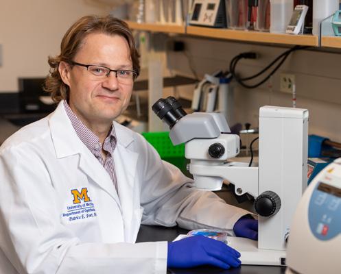
tion with national eye banks. Additional expertise and support are provided by the Central Biorepository of the U-M Medical School.
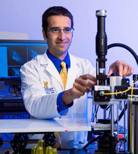
“Kellogg is fortunate to have longstanding partnerships with the nation’s top eye banks,” Dr. Fort says. “We are coordinating with them to procure high quality post-mortem tissue samples.”
While this is the first biorepository solely devoted to diabetes-related eye research, it builds on the success of another branch of diabetes research. “We are grateful for the guidance we are receiving from the Network for Pancreatic Organ Donors with Diabetes or nPOD, which has been providing pancreas tissue samples for research since 2007.”
The biorepository began operations in late 2022. The goal is to collect a total of 1,000 samples over the next four years, image, analyze, characterize and catalog each one and then coordinate their use by diabetes investigators around the world.
Yannis M. Paulus, M.D., FACS, Associate Professor of Ophthalmology and Visual Sciences and Associate Professor of Biomedical Engineering, was recently elevated to Senior Member of the Institute of Electrical and Electronics Engineers (IEEE), the largest engineering organization in the world, for his work advancing technologies to benefit humanity. Dr. Paulus fell in love with ophthalmology because of the impact it can have on the quality of life of those with vision loss. “I can’t think of a field that’s more rewarding,” said Paulus,
who works on novel retinal imaging systems, therapeutic techniques and technologies. The recognition is a nod to his work with the engineering community to meet clinical needs and improve lives. His lab aims to drive down costs of tools and equipment to help diagnose and treat those who might not be able to afford care. “Costs can be driven down when things are engineered at a scale that is more affordable.” Among his many contributions are pioneering the application of photoacoustic approaches for diagnosing and treating eye diseases.
“I didn’t start out wanting to become a doctor,” admits Timothy Soeken, M.D., Hailing from a small Texas town, Dr. Soeken was a first-generation college graduate, earning a degree in nuclear energy from Texas A&M. Next came the U.S. Air Force and flight training, after which he and his wife settled into military life and began raising a family.
His career path changed when the Soekens’ oldest child faced a health scare. Thankfully, their son recovered from his illness. Through that ordeal, Dr. Soeken experienced firsthand both the vulnerability felt by patients and parents, and the problem-solving skills and compassion exhibited by the very best physicians.
“I began to see myself in that role,” he recalls. “I thought my wife would balk at such a major course correction, but she was all for it.”
As it turned out, so was the Air Force. Nearly a decade older than his classmates, Dr. Soeken entered Baylor College of Medicine on a military scholarship. While completing an ophthalmology residency, his mentors raised the possibility of a fellowship in global ophthalmology.
Dr. Soeken chose to complete both a cornea fellowship and a global ophthalmology fellowship at U-M. The first Global Ophthalmology Fellow for both the Jerome Jacobson Program in International Ophthalmology and the U.S. military, Dr. Soeken’s U-M mentors included program co-directors H. Kaz Soong, M.D., and Joshua Ehrlich, M.D., M.P.H.
“Our goal was to help Tim gain as much experience in as many different parts of the world as possible,” says Dr. Ehrlich, “navigating complex situations with cultural sensitivity, working with multiple stakeholders, and mastering the skill set to perform eye surgery in low-resource settings.” Dr. Soeken traveled to the African nations of Kenya and Burundi, as well as to India, Mexico and Jamaica.
“Cataract surgery is so different in many developing countries,” explains Dr. Soeken, “both in the techniques used, and in the condition of the patients, who present with far more advanced disease.”
Dr. Soeken is now one of only a small number of U.S. doctors experienced in multiple methods of performing cataract surgery with limited resources, a necessary skill for medical aid missions.
Major Soeken is now back on base in San Antonio, caring for active and retired military and civilian patients. He has completed subsequent medical travel to Honduras and Guatemala and is planning more trips to Central America in 2023 in his role directing the humanitarian health work for the US armed forces.
Pamela Williams, M.D., Clinical Instructor of Ophthalmology and Visual Sciences, has been named Chair of the Advisory Board for the Children’s Eye Foundation of the American Association for Pediatric Ophthalmology and Strabismus (AAPOS) “Our mission is to end preventable blindness and improve the lives of visually impaired children,” she said. “The earlier we can detect and treat vision loss, the better.” The foundation’s programs in the U.S. and other
countries screen, treat and provide financial assistance to uninsured or underinsured children. One program, Stop Infant Blindness in Africa (SIBA), addresses Retinopathy of Prematurity, an eye disease in pre-term infants. With improvements in medical care, these babies are able to survive but may face issues with their vision due to their early birth. “By teaching physicians in Africa how to screen and treat patients, we can help improve vision outcomes for preterm infants.”
“CATARACT SURGERY IS SO DIFFERENT IN MANY DEVELOPING COUNTRIES, BOTH IN THE TECHNIQUES USED, AND IN THE CONDITION OF THE PATIENTS, WHO PRESENT WITH FAR MORE ADVANCED DISEASE.
Timothy Soeken, M.D.
How do we know whether today’s medical students are receiving enough education and training in ophthalmology? Currently, there are no objective, standardized clinical competencies for the specialty.
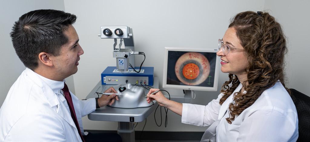
Ariane Kaplan, M.D., Associate Professor and Director of Medical Student Education at Kellogg, is working to fill that void. Using the standardized competencies established for residency education by the American College of Graduate Medical Education (ACGME) as a template, she has developed the first Ophthalmology Clinical Competencies (OCCs) for medical students.
“Like the ACGME competencies for residents, these OCCs codify the ophthalmology knowledge and skills a medical student should have attained by graduation,” explains Dr. Kaplan. “They give students concrete goals for developing their clinical skills, while giving educators guidelines for creating an ophthalmology curriculum.”
A pilot of this first generation of OCCs is currently underway at Kellogg, evaluating their usability for students and faculty raters/graders. “We want to make sure that we are measuring the right things,” Dr. Kaplan says, “and that the scoring structure yields reliable, consistent results from one student to another, and one faculty evaluator to another.”
Dr. Kaplan began the project when she was selected to participate in the U-M Medical School’s Medical Education
ArianeScholars Program (MESP). The MESP promotes educational scholarship and improves teaching and leadership skills of Medical School faculty leaders.
Preliminary feedback on the OCCs is very positive. “If shown to be useful for medical students and educators,” she says, “this could lead to the development of similar clinical competencies in other domains.”
“
“ LIKE THE ACGME COMPETENCIES FOR RESIDENTS, THESE OCCS CODIFY THE OPHTHALMOLOGY KNOWLEDGE AND SKILLS A MEDICAL STUDENT SHOULD HAVE ATTAINED BY GRADUATION.
Kaplan, M.D.Research Intern Keith Miller with Ariane Kaplan, M.D.
All ophthalmology residents must fulfill the competencies outlined by The Accreditation Council for Graduate Medical Education (ACGME). With the many advances in eye surgery, providing greater opportunities for residents from around the country to enhance their skills has been challenging.
A program to provide broader participation developed by the Program Directors Council (PDC) of the Association of University Professors of Ophthalmology (AUPO) was approved in 2018. Representatives from top academic ophthalmology programs across the U.S. began developing the AUPO Surgical Curriculum for Ophthalmology Residents (SCOR) program, which launched in 2021.
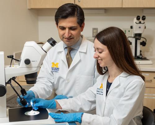
Shahzad Mian, M.D., past Program Director of the Kellogg residency and past Chair of the PDC was selected to direct the initiative. A corneal transplant surgeon and educator, Dr. Mian also serves as Vice Chair of Clinical Sciences and Learning at Kellogg.
AUPO/SCOR is a standardized system to train and assess the competency of residents in all disciplines of ophthalmology on a broad range of basic and advanced surgical skills. “The program is aimed at residents in their fourth year of postgraduate training (PGY4s),” explains Dr. Mian. “Traditionally, the first three years focus on clinical patient evaluation and medical management. Once those skills are mastered, the focus shifts to surgical skills in year four.”
The 2021 rollout began with the launch of the online platform and the first two in-person skills transfer workshops, attended by a total of 144 residents. Another 72 residents attended a third workshop held at the 2022 American Academy of Ophthalmology meeting.
“We are pleased with how the program is scaling,” says Dr. Mian. “To date we’ve trained 228 residents representing more than 80 programs, with help from 100-plus faculty members from more than 40 programs.” The plan is to double the capacity for in-person training in 2023.
“About 500 ophthalmology residents graduate each year in the U.S.,” Dr. Mian adds. “We’re making good progress toward our goal of giving them all the opportunity to participate.”
The curriculum is comprised of four components:
• On their own, students access an online learning platform for foundational medical knowledge.
• Students attend a one-day in-person skills transfer workshop to receive one-on-one training in approximately 25 surgical procedures.
• Assessment tools built into the workshop allow instructors to gauge how well residents are mastering the skills.
• Finally, train-the-trainer tools build the skills of both workshop instructors and residency program faculty at academic institutions.
Steven M. Archer, M.D., the Ida Lucy Iacobucci Collegiate Professor in Ophthalmology and Visual Sciences, was selected to present the 7th annual Kushner webinar, sponsored by the American Association for Pediatric Ophthalmology and Strabismus (AAPOS).
The annual presentation was established by the Department of Ophthalmology and Visual Sciences at the University of Wisconsin
—Madison to honor esteemed ophthalmologist and professor Burton Kushner, M.D.
Each year’s presenter is an outstanding pediatric ophthalmologist or strabismologist selected by a committee of members of the Pediatric Ophthalmology Service at the University of Wisconsin—Madison and a representative of the AAPOS Board of Directors.
In January 2022, Dr. Archer delivered his presentation, “Superior Oblique Apocrypha.”
Otana Jakpor, M.D., M.Sc., a third-year resident at Kellogg, has had the opportunity to combine her interests in pediatric ophthalmology and global and environmental health.
Dr. Jakpor worked on a project in Grenoble, France to explore the possible connection between air pollution and the incidence of refractive error in children. “When Covid-19 made it impossible to pursue a planned project in China, I was given the green light to pursue this opportunity with former medical school colleagues who shared my interest in environmental health.” Jakpor is raising attention to a growing understanding of the role of air pollution and a range of eye conditions, including glaucoma and retinal vascular diseases. Particulate matter of 2.5 microns in size or smaller (“P2.5”) has been associated with a number of health conditions such as hypertension and stroke as well as glaucoma. “I’m so grateful for the support I received from Kellogg leadership and my faculty
mentor, Dr. Joshua Ehrlich,” says Dr. Jakpor, who plans a clinical and research practice in pediatric ophthalmology that addresses global and environmental health issues.
Here in Ann Arbor, Dr. Jakpor is participating in a project launched through the U-M Medical School’s Health Equity and Quality Scholars Program. Her team is expanding the languages available for printed discharge instructions for patients following cataract surgery.
“Beyond standard post-operative instructions, there is often a need to provide patient-specific guidance about details like eye drops or follow-up appointments,” she explains. “In these instances, the care team uses a customizable template to generate post-op instructions. Working with Michigan Medicine’s outstanding Interpreter Services staff, our group is making this customizable template available in more languages.”
Patrice Hicks, Ph.D., M.P.H., has received an Institutional Research and Career Development Award (IRACDA) through the NIH’s National Institute of General Medical Sciences. The IRACDA program provides training, mentorship and assistance to students from groups underrepresented in biomedical science, combining traditional postdoctoral research and academic teaching opportunities.
Currently a post-doctoral research fellow mentored by Maria Woodward, M.D., M.S., and Paula Anne Newman-Casey, M.D., M.S., Dr. Hicks studies the role of neighborhood-level factors associated with outcomes for microbial keratitis and glaucoma. Going beyond traditional socioeconomic factors like age, income and education, her multi-factorial analyses also consider the impact
of neighborhood-specific barriers to opportunity and resources, such as safety, food insecurity and housing discrimination.
In recognition of her significant achievements in scholarship, leadership, character, service and advocacy, Dr. Hicks has also been inducted into the University of Michigan Rackham Graduate School’s chapter of the Edward Alexander Bouchet Graduate Honor Society. Named for the first African American doctoral recipient in the U.S., the Society promotes excellence and diversity in doctoral education.
“Kellogg has opened unlimited doors for me to pursue my interests in research, teaching and advocacy,” she says. “My goal is a career that combines all three.”

Stephen Armenti, M.D., Ph.D., completed a cornea fellowship at Kellogg in July 2022, and served as Chief Resident in 2019-2020 after his Kellogg ophthalmology residency. A cornea and cataract specialist, he is now Assistant Professor of Clinical Ophthalmology at the University of Pennsylvania Perelman School of Medicine.

At Kellogg, Dr. Armenti maintained a busy clinical practice and played a key role in the development of the Association of University of Ophthalmology Professors’ Surgical Curriculum for Ophthalmology Residents (AUPO-SCOR) program. Directed by Shahzad Mian, M.D., AUPO-SCOR is the first standardized system to train and assess the competency of ophthalmology residents on a broad range of surgical skills.
In recognition of his contributions in patient care, education, research and leadership, Dr. Armenti received the 2022 Resident Excellence Award from the American Society of Cataract and Refractive Surgery (ASCRS) Foundation.
Dr. Armenti continues to combine his passion for both patient care and surgical education at UPenn, where he trains residents in cornea and cataract procedures.
“My mentors on the comprehensive and cornea services were wonderful, especially Dr. Mian,” he says. “He helped me become a more compassionate doctor and a more confident surgeon. My time at Kellogg made me the physician I am today.”
Joah Aliancy, M.D., completed a glaucoma fellowship at Kellogg in July 2022. He is now a practicing ophthalmologist caring for glaucoma patients in Port Orange, Florida. While at Kellogg, Dr. Aliancy collaborated with Paula Ann Newman-Casey, M.D., M.S., Principal Investigator on the CDC-funded Michigan Screening & Intervention for Glaucoma & Eye Health Through Telemedicine (MI SIGHT) trial. He conducted a tandem analysis assessing the impact of healthrelated socioeconomic factors on a patient’s risk of screening positive for glaucoma and presented his findings at the March 2022 meeting of the American Glaucoma Society. For this work, he received a Glaucoma Foundation/Research to Prevent Blindness Glaucoma Fellowship.
In addition to a busy practice, Dr. Aliancy devotes time to educating and training ophthalmology residents in Haiti. He lectures at and is developing a glaucoma curriculum for the Hospital de l’Universite d’Etat d’Haiti, and looks forward to pursuing future outreach opportunities.
“The outstanding clinical training and research experience I gained at Kellogg reinforced my commitment to doing all I can to make the best possible glaucoma care available to patients everywhere, regardless of the socioeconomic challenges they face.”


The Kellogg Clinical Research Center (KCRC) serves as the hub for clinical research at Kellogg. The team includes clinical research coordinators, a full-time research technician, research photographer, research administrator and physicians. To the right are the types of active studies currently enrolling at the KCRC. The KCRC also performs eye exams for many cancer and other studies throughout the medical campus.
To find out more about studies in which you may be eligible to participate: https://umhealthresearch.org
Sally Temple, Ph.D., an adjunct faculty member at Kellogg, is among the 2022 inductees into the National Academy of Medicine. Co-Founder and Scientific Director of the Neural Stem Cell Institute, Dr. Temple’s pioneering work in stem cell biology is driving new therapies for neurodegenerative disorders of the brain and retina. As detailed in this year’s report, Kellogg is the first site of a Phase 1/2a clinical trial of the transplantation of allogeneic retinal pigment epithelial stem cells into the retina as a treatment for dry AMD, an approach based on Dr. Temple’s pre-clinical modeling work with her husband, retina specialist Jeffrey Stern M.D., Ph.D.
This year KEC hosted the 45th Annual Midwest Glaucoma Symposium which included local, regional, national and international guests. Over the 1.5 days of the conference, speakers shared their insights and thoughts on various topics pertaining to glaucoma research, and clinical and surgical glaucoma.


Kellogg's Annual Fall Reunion Weekend was back in person this year and Kellogg alums came together from across the country for a day filled with learning and reunions.
This year, Thomas Gardner, M.D., M.S., helped organize the first Mary Tyler Moore Vision Initiative’s Diabetic Retinal Disease Clinical Endpoints Workshop, in collaboration with JDRF and the Elizabeth Weiser Caswell Diabetes Institute. The conference brought together experts throughout the field, including academia,
industry, non-profit, government and lay representatives, to help determine innovative methods for measuring how diabetes affects the eye beyond the conventional methods we use today. Together, these global experts helped shape diabetic retinopathy research methods that will be used in studies throughout the world.


On September 16, the Department of Ophthalmology and Visual Sciences celebrated the inauguration of the Kenneth H. Musson, M.D. and Patricia M. Musson Research Professorship in Ophthalmology and Visual Sciences. Jonathan D. Trobe, M.D., professor of ophthalmology and visual sciences and of neurology, and section head of neuro-ophthalmology, was installed as its inaugural holder.
The Musson Professorship is intended to support a faculty member whose research focuses on neuroophthalmology—a specialty that encompasses nervous system disorders that affect vision, including those that cause double vision, pupillary abnormalities, ocular misalignment, or eye movement abnormalities.
Dr. Trobe’s interest in ophthalmology began when he was in medical school and suffered from keratitis, which required a corneal transplant. He is inspired when answering tough questions, saying that he appreciates “the opportunity to care for patients with medical problems that are challenging enough that community physicians or academic physicians in other specialties have sought my help.”
Dr. and Mrs. Musson established the professorship in gratitude for the residency Dr. Musson completed there so many years ago. After attending the University of Michigan Medical School, Dr. Musson spent two years on active duty in the medical corps of the U.S. Army Reserve—delaying the completion of his
education. To this day he remains grateful he was able to return to U-M after his service to begin his ophthalmology residency.
Dr. Musson began his career providing comprehensive ophthalmologic care at the Grand Traverse Ophthalmology Clinic and Munson Medical Center in Traverse City in 1969. He is retiring after 52 years of active practice. Dr. Musson is still actively engaged with his alma mater, and serves as a founding member of the Kellogg Eye Center Alumni Advisory Board. He has also been an active contributor to and member of professional societies including the Michigan State Medical Society, the Michigan Society of Eye Physicians and Surgeons, the American Academy of Ophthalmology, and the American College of Surgeons.
Patricia Myers Musson is equally committed to serving others. After enjoying a successful career as a teacher, Mrs. Musson was able to draw from her educational background in her volunteer work to help members of her own family as well as organizations in her community, including the Traverse City and Old Mission Peninsula School districts. Additionally, she and Dr. Musson are involved in many other charities and organizations across the area.
When asked about his philanthropy, Dr. Musson said, “We give back out of gratitude. It is important to support the quality of the Department of Ophthalmology and the Kellogg Eye Center, and to provide a future for the people who come after us.”
“
“
WE GIVE BACK OUT OF GRATITUDE. IT IS IMPORTANT TO SUPPORT THE QUALITY OF THE DEPARTMENT OF OPHTHALMOLOGY AND THE KELLOGG EYE CENTER, AND TO PROVIDE A FUTURE FOR THE PEOPLE WHO COME AFTER US.
Kenneth H. Musson, M.D.
Marilyn and Richard Witham are continuing their transformative support of the Department of Ophthalmology and Visual Sciences by funding a second professorship.
The couple’s recent gift of $2.5 million established the professorship in Ophthalmology and Visual Sciences to support a faculty member who directs a clinical research program. The professorship’s first recipient, Grant Comer, M.D., M.S., specializes in diseases and surgery of the retina and is known for his expertise across diabetic retinopathy, age-related macular degeneration, retinal vascular disease, infectious chorioretinitis, and retinal detachments. In pushing the boundaries of research to solve problems in new ways, he embodies the intent of this professorship, an approach that impressed the Withams when he was Mrs. Witham’s surgeon.
In fact, the care she received when she was first diagnosed with ocular cancer was the motivation behind the couple’s first professorship gift to the department in 2017, intended for a faculty member in ocular oncology. Its inaugural and current holder, Hakan Demirci, M.D., was another of Mrs. Witham’s physicians during her ocular cancer journey.
Mrs. Witham credits Dr. Demirci and Dr. Comer with saving her life and preserving her vision for as long as was possible.
The Withams are committed to ensuring that other patients can benefit from the leading-edge care that Mrs. Witham has experienced over the years. Mr. Witham has said, “We have met other patients from around the country and around the world at Kellogg, and we are not surprised that so many seek care here. We have learned a great deal about the faculty’s goals for the Kellogg Eye Center, and their success is widely recognized.”
Their most recent gift supports a professorship that will advance research into care and cures for ocular diseases of all kinds. The Withams look forward to hearing about the impact their gift will make for numerous projects. As medical director of the Kellogg Clinical Research Center, Dr. Comer is wellsituated to ensure that leading-edge research will move forward across several fronts.
The Withams reside in Muskegon, Michigan. Mr. Witham enjoyed a long entrepreneurial career in the plastic, electronic, and medical device industries, running many successful businesses over the years. He continues to consult for Motion Dynamics, AIM Corporation and several other companies. He has served on numerous boards, including the Kellogg Eye Center Leadership Campaign Board.
He also enjoys mentoring young business professionals. Mrs. Witham is an avid reader and actively engaged in her community through her service to the Greater Muskegon Service League and Women Who Care.
Mr. and Mrs. Witham are very close to and involved with their two sons, Chris and Kurt, and their families, including their six grandchildren and two great grandchildren.
“
“
WE HAVE MET OTHER PATIENTS FROM AROUND THE COUNTRY AND AROUND THE WORLD AT KELLOGG, AND WE ARE NOT SURPRISED THAT SO MANY SEEK CARE HERE.
Richard Witham
Jason Miller, M.D.,
named first
Age-related macular degeneration (AMD) is the leading cause of irreversible blindness in people over 50. It is even more prevalent than Alzheimer’s disease, with 11 million people affected in the United States and 170 million globally. Between 70% and 90% of cases are the dry form of the disease, which causes gradual blurring of central vision. While successful therapies are in place for wet AMD, there are no standard treatment options for dry AMD. An $11.5 million gift aims to change that.
James Grosfeld, an investor, philanthropist, and the former chairman and CEO of PulteGroup, Inc., has made the gift to launch a pioneering research initiative. The effort, which represents one of the largest investments of talent and resources in the country targeted at developing effective treatments for dry AMD, will increase discovery, collaboration, and leadership in the field.
Specifically, Mr. Grosfeld’s gift is supporting:
• The establishment of two endowed professorships focused on dry AMD research
• Increased laboratory staffing to increase the pace of research
• Collaborative grants that will link dry AMD investigators with other experts at the Kellogg Eye Center and across U-M to bring new techniques and approaches to bear on the disease
• A pluripotent stem cell facility to create cells for use in dry AMD-related research across disciplines
• Innovations in clinical research, including the collation of ophthalmic images to advance clinicians’ ability to track dry AMD progression
• Pilot funding for proof-of-concept experiments and research grants for trainees who seek to bring new ideas and perspectives to dry AMD research
“I am inspired by the passion, the commitment, and the ideas of the Kellogg Eye Center team,” Mr. Grosfeld says. “Increasing the speed and the breadth of discovery in dry AMD can make a significant difference in people’s lives.”
“It was a privilege to share with Mr. Grosfeld what a transformational investment in dry AMD research could mean, and we are very grateful for his enthusiasm, his generosity, and the belief he has placed in us,” says Paul P. Lee, M.D., J.D., the F. Bruce Fralick Professor and Chair of the U-M Department of Ophthalmology and Visual Sciences and director of the Kellogg Eye Center.
Jason M. Miller, M.D., Ph.D., a physician-scientist who specializes in retinal diseases and dry AMD research, will direct the initiative. He has been named the James Grosfeld Professor of Ophthalmology and Visual Sciences; the establishment of the professorship was celebrated at a ceremony on Aug. 23. Advancing understanding of dry AMD to identify pathways toward effective therapies has long been a goal for Dr. Miller, a third-generation ophthalmologist. “I came to the Kellogg Eye Center to build a laboratory program for dry AMD, which impacts so many people. We have made several discoveries that have given us a clear direction toward potential treatments. At the same time, we have been building partnerships that will both accelerate our work and enable us to translate our efforts into clinical applications.”
Ph.D.,
James Grosfeld Professor of Ophthalmology and Visual Sciences and will play key role in new initiative
INCREASING THE SPEED AND THE BREADTH OF DISCOVERY IN DRY AMD CAN MAKE A SIGNIFICANT DIFFERENCE IN PEOPLE’S LIVES.
James Grosfeld
AMD is marked by the accumulation of lipid-rich deposits in and around the retinal pigment epithelium (RPE), a part of the eye that keeps the retina’s light-sensitive photoreceptors alive. Dr. Miller’s research has led to a hypothesis that improving the RPE’s handling of lipid (fat) will slow the progression of dry AMD. His team is advancing understanding of how the RPE processes lipid. Their discoveries are already helping ophthalmologists make clinical determinations about care.
The new research initiative will create significant opportunities to collaborate with faculty across the Kellogg Eye Center, the University of Michigan, and with peers across the country, Dr. Miller says. “The Kellogg Eye Center and the University of Michigan have world-class faculty studying problems as diverse as diabetes and heart disease. The research tools and methods they use, when applied to dry AMD, will spark new discoveries about this devastating disease.” The initiative will also draw on U-M expertise in ophthalmic imaging, drug development, and clinical trials.
“This gift is designed to have a maximum impact on research progress,” says Mark W. Johnson, M.D., a professor of ophthalmology and visual sciences and head of the retina division at Kellogg, which is one of the largest in the country. Johnson introduced Grosfeld to the dry AMD research taking place at Kellogg and will serve on an advisory board overseeing the initiative.
“In catalyzing work across disciplines, we are enabling the creative application of a wide range of scientific techniques and approaches to the challenges of dry AMD,” says Marschall S. Runge, M.D., Ph.D., dean of the University of Michigan Medical School, executive vice president for medical affairs, and CEO of Michigan Medicine. “Mr. Grosfeld’s visionary support will enable us to make important advances toward saving sight today and will create a legacy of sight-saving achievement.”
MR. GROSFELD’S VISIONARY SUPPORT WILL ENABLE US TO MAKE IMPORTANT ADVANCES TOWARD SAVING SIGHT TODAY AND WILL CREATE A LEGACY OF SIGHT-SAVING ACHIEVEMENT.Marschall S. Runge, M.D., Ph.D.

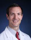
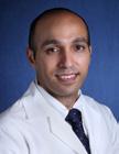
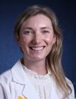
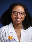
Sameerah Ali, M.D., earned her medical degree from Indiana University. She then went on to complete her ophthalmology residency at the University of Cincinnati. She joins as Associate Professor of Ophthalmology providing comprehensive ophthalmology care in Canton and at the VA hospital.
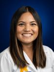
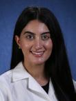
Meagan Tucker, O.D., attended optometry school at Indiana University. Following optometry school, she completed a residency at Gundersen Health System in ocular disease and neuro-optometric rehabilitation for patients with acquired brain injuries. Dr. Tucker will be providing primary care optometry in Brighton and neuro-optometric evaluations and rehabilitation in Ann Arbor.
Audree Bass, O.D., completed the University of Michigan ophthalmic technician training program prior to attending optometry school. She earned her doctorate from The Ohio State College of Optometry and completed the combined ocular diseaselow vision residency through the Ohio State College of Optometry and Columbus Veterans Affairs programs. Her service specialities include low vision, specialty contact lenses, and ocular disease management.
Sebastian Werneburg, Ph.D., earned his M.Sc. in Biomedicine from the Hannover Medical School, Germany, and his summa cum laude awarded Ph.D. in Neuroscience from the University of Veterinary Medicine Hannover, Germany. He completed his postdoctoral training at the University of Massachusetts Chan Medical School in Worcester, USA, before joining the Kellogg Eye Center as Assistant Professor in 2022. Dr. Werneburg’s research focuses on neuron-glia interactions, the mechanisms underlying de- and regeneration of neural circuits in the visual system, and developing novel therapeutic strategies for neurodegenerative diseases.

Julia Dalia, M.D., earned her medical degree from Saint Louis University School of Medicine. She then went to complete her ophthalmology residency at Rush Medical Center in Chicago, IL. She joins as Associate Professor of Ophthalmology providing comprehensive ophthalmology care at Huron River Dr. Office and the VA hospital.
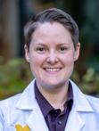
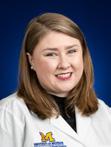
Erin M. Klukas, O.D., attended optometry school at the Illinois College of Optometry in Chicago. Following graduation, she pursued a residency specializing in ocular disease, low vision and acquired brain injury rehabilitation at the West Haven VA Medical Center, in Connecticut. She will be providing low vision rehabilitation in Ann Arbor, as well as providing primary care optometric services in Northville.
Jackie Nguyen, O.D., earned her doctorate in optometry from the University of California, Berkeley. Following optometry school, she completed her residency in Pediatric Optometry at Vanderbilt Eye Institute. Dr. Nguyen specializes in pediatric optometry and pediatric low vision care, and sees patients at the Grand Blanc, Northville, and Wall Street clinics
Vinay Kumar Aakalu, M.D. M.P.H., is a fellowship-trained Oculoplastic Surgeon and Clinician Scientist. He joins KEC as the Oculoplastic Section Leader and Medical Director after serving as an Associate Professor at the University of Illinois at Chicago. He completed medical school at the Mount Sinai School of Medicine in New York and completed ophthalmology residency and ASOPRS fellowship at the University of Illinois at Chicago. His NIH, DOD and VAfunded research focuses on ocular surface diseases and neurodegenerative disorders. He has a strong interest in translational research and clinical application of research innovations.
Steven F. Abcouwer, Ph.D.
Fellow, Silver, Class of 2023, Association for research in vision and ophthalmology (ARVO)
Chief Editor, Journal of Ophthalmology
Editorial Board Member, Investigative Ophthalmology and Vision Science
Editorial Board Member, Retina Section, Frontiers in Ophthalmology
Editorial Board Member, American Journal of Physiology: Endocrinology and Metabolism
Editorial Board Member, Frontiers in Immunology
Editorial Board Member, Frontiers in Neurology
Scientific Review Editor, Molecular Vision journal
Member, Department of Defense FY22 Peer Reviewed Medical Research Program (PRMRP) for the Congressionally Directed Medical Research Programs (CDMRP), Diabetes Treatment Clinical Trials Study Section
Member, National Institutes of Health (NIH) Study Section ZRG1
MGG-G (70) R RFA-RM-22-015: Investigational New Drug-enabling Studies of Somatic Genome Editing Therapeutic Leads (U19)
Reviewer, Stanford Diabetes Research Center (SDRC) 2021 Pilot and Feasibility Project Grant Program
Reviewer, University of Colorado Diabetes Research Center 2021 Pilot and Feasibility Grant Program
Reviewer (Ad Hoc), Czech Science Foundation (GACR, the main public funding agency in the Czech Republic)
Steven M. Archer, M.D.
Castle Connolly America’s Top Doctors
Presenter, 8th Constance E West Memorial Lecture, Cincinnati Society of Ophthalmology and Cincinnati Children’s Hospital
Lecturer, 7th Annual Kushner Webinar, American Academy for Pediatric Ophthalmology and Strabismus
Kari E. Branham, M.S., CGC
Chair, Genetics Review Committee, Foundation Fighting Blindness Clinical Consortium
Co-Chair, National Society of Genetic Counselors Ophthalmology and Hearing Loss Special Interest Group
Expert Review Panel Member, Early Onset Retinal Degeneration Variant Curation, ClinGen
Expert Review Panel Member, Retinal Disease Gene Curation, ClinGen
Member, Scientific Advisory Board, Foundation Fighting Blindness
Member, Evidence Based-Guideline for Retinal Dystrophies Workgroup, American College of Medical Genetics
Member, Editorial Board, Investigative Ophthalmology & Visual Science
Theresa Cooney, M.D.
Castle Connolly Top Doctors
Sherry H. Day, O.D., FAAO
Board Member, Vision Rehabilitation Committee, American Academy of Optometry Foundation
Board Member, Vision Rehabilitation Committee, American Optometric Association
Co-Chair, Vision Rehabilitation Committee, Michigan Optometric Association
Monte A Del Monte, M.D.
Secretariat Award, American Academy of Ophthalmology
22nd Marshall M. Parks Memorial Lecture, American Academy of Ophthalmology
8th Constance E West Memorial Lecture, Cincinnati Society of Ophthalmology and Cincinnati Children’s Hospital
Presenter, Keynote lecture, 2022 Midwest/West Joint Regional Meeting of the American Association for Certified Orthoptists
Invited Keynote International Speaker, 4th International Symposium of Pediatric Ophthalmology and Strabismus, Shanghai, China
Invited faculty, International Online Lecture Series, Aravind Eye Care System, Superior Oblique Palsy-Nuts and Bolts in Management
Hakan Demirci, M.D.
In-Service Exam Subcommittee, Education Committee, American Society of Ophthalmic Plastic and Reconstructive Surgery
Joshua Ehrlich, M.D., M.P.H.
Reviewer, Special Emphasis Panel, Fogarty International Center, National Institutes of Health training grant: “Building Non-Communicable Eye Disease Research Capacity in India”, National Eye Institute, National Institutes of Health
Co-Chair, International Education Sub-Committee, American Glaucoma Society
Editorial board, Ophthalmic Epidemiology
Associate Director, Innovative External Networks Core, P30 Michigan Center on the Demography of Aging
Glaucoma Visiting Professor and Scholar-in-Residence, Wills Eye Hospital
Angela Elam, M.D.
Mentoring Award, Women in Ophthalmology (WIO)
Closing Keynote Session, “Envisioning Equity in Eye Care”, Association for Research in Vision and Ophthalmology (ARVO)
Patrice Fort, Ph.D., M.S.
Invited Keynote Lecture, 31st Annual Meeting of the European Association, the Study of Diabetes Eye Complications Study Group (EASDec)
NIH study section: Pathophysiology of Eye Disease 2 (PED2)
Grant Reviewer, Retina France
Editorial Board, PlosOne
Thomas Gardner, M.D., M.S.
Invited Lecturer, Hinkle Lecture, Penn State College of Medicine
Invited Lecturer, Ian Constable Lecture, Lions Eye Institute, University of West Australia
Kanishka T. Jayasundera, M.D., M.S.
Board of Directors, Pan-American Ophthalmological Foundation
Chair, American Glaucoma Society Cares
Lancet Global Health Commission on Global Eye Health
Member, Scientific Advisory Board, Foundation Fighting Blindness
Mark Johnson, M.D.
Michael R. Petersen Leadership Lecture, University of Cincinnati
Castle Connelly Top Doctors
Associate Editor, The American Journal of Ophthalmology
Editorial Board, Retina
President, The Macula Society
Program Director, American Academy of Ophthalmology Retina Subspecialty Day
Member, Gass Club
Ariane Kaplan, M.D.
Council Member, Medical Student Education, Association of University Professors of Ophthalmology
Book 1 Committee, Basic and Clinical Science Course, American Academy of Ophthalmology
Paul Lee, M.D., J.D.
Board of Directors, Society of Heed Fellows
Past President, Board of Directors, Association of University Professors of Ophthalmology
Keynote Speaker, Stephen M. Podos Colloquium and ARI Awards Symposium, Alcon Research Institute
2nd Chairs Lecture, Bascom Palmer Eye Institute, University of Miami
Invited Speaker, American Academy of Ophthalmology Mid-Year Forum
32nd Wilmer Memorial Lecture, Wilmer Eye Institute, John Hopkins
Invited Lecturer, Monroe Hirsch Symposium, “Public Health/Health
Economics of Health Screening Programs and Access to Care,” American Academy of Optometry
Visiting Professor, Yale University
Moderator, “Envisioning Equity in Eye Care,” Association for Research in Vision and Ophthalmology (ARVO)
Paul R. Lichter, M.D.
Castle Connolly Top Doctors
US News and World Reports Top Doctors
Shahzad I. Mian, M.D.
Executive Committee, Board of Directors, Cornea Society
Board of Directors, Eversight Eye Bank
Board of Directors, Eye Bank Association of America
Castle Connelly Top Doctors
Editor in Chief, Surgical Curriculum for Ophthalmology Residents, Association of University Professors in Ophthalmology
Editorial Board, Cornea
Associate Editor, Cornea Open
Guest Editor, Cornea, Current Opinion in Ophthalmology
Examination Writing Committee, Examiner, American Board of Ophthalmology
Senior Medical Director, Eversight Eye Bank
Subspecialty Day Advisory Committee, American Academy of Ophthalmology
Preferred Practice Patterns, Cornea, American Academy of Ophthalmology
Vice Chair, Medical Advisory Board, Eye Bank Association of America Planning Committee, World Cornea Congress
Jason Miller, M.D., Ph.D.
Gragoudas Award, Macula Society
Invited Lecture, Macula Society
Megherio Award, Retina Society
Invited Lecture, Retina Society
Lecture, Ophthalmic Genetics and Visual Function Branch Seminar Series, National Eye Institute (NEI)
Lecture, Stanford Vision Science Seminar Series, Stanford University David Hinton Scholarship, Ryan Initiative for Macular Research
David Musch, Ph.D., M.P.H.
Scientific Reviewer: Health and Medical Research Fund, Government of the Hong Kong SAR
Member, Editorial Boards: JAMA Ophthalmology, Retina, and Eye and Vision
Member/Chair: numerous NEI/NIH and company-sponsored Data and Safety Monitoring Committees for ongoing, multicenter, randomized clinical trials
Paula Anne Newman-Casey, M.D., M.S.
Physician Scientist Award, Research to Prevent Blindness
Glaucoma Subcommittee Chair, Annual Meeting Program Committee, American Academy of Ophthalmology
AGS Cares Task Force, American Glaucoma Society
Susruta Lecture, NYU Research Day
Secretariat Award, American Academy of Ophthalmology
Student Fellowship Award for Mentoring, Goldman Sachs Philanthropy Fund Summer
Best Paper, American Academy of Ophthalmology Glaucoma Free Paper Session
Yannis Paulus, M.D.
Scientific Reviewer, Vision Research Program (VRP) for the Department of Defense Congressionally Directed Medical Research Programs (CDMRP)
Review Committee, Study Section, National Eye Institute R21 Secondary Data Analysis
Grant Reviewer, Moorfields Eye Hospital Eye Charity NHS Foundation Trust, United Kingdom
Senior Member, Institute of Electrical and Electronics Engineers (IEEE)
National Science Foundation SBIR/STTR Phase II Panel Review Committee Digital Health
Grant Reviewer, United Kingdom Research and Innovation Medical Research Council
Massey Foundation TBI Grand Challenge Award
Grant Reviewer, University of Maryland, Maryland Industrial Partnerships Program (MIPS)
Scientific Reviewer, NIH Study Section Brain Imaging, Vision, Bioengineering and Low Vision Technology Development (BIVT)
Grant Reviewer, The Retina Society Research and Education Grants
Grant Reviewer, Food and Health Bureau (FHB) of the Health and Medical Research Fund (HMRF) for The Government of the Hong Kong Special Administrative Region (HKSAR)
Grant Reviewer, Israeli Ministry of Innovation, Science and Technology
Scientific Reviewer, NIH Council ZEY1 VSN 04 NEI Mentored Clinician Scientist Applications
Jillian N. Pearring, Ph.D.
NIH Early Career Reviewer, BDE Study Section, National Eye Institute, National Institutes of Health
Young Investigator Travel Awardee, RD2021 Platform Session Moderator, Photoreceptors, RD2021
Lev Prasov, M.D., Ph.D.
Co-Chair, Variant Curation Expert Panel Anterior Segment Dysgenesis, ClinGen
Gene Curation Expert Panel, Glaucoma and Neuro phthalmology, ClinGen Guest editor, Genes
Research to Prevent Blindness Career Development Award
Dr. Joe G. Hollyfield New Investigator Award, Macular Degeneration Research
Rajesh Rao, M.D.
Career Advancement Award, Research to Prevent Blindness
Study Section Reviewer, Neurobiology-F (NURF) VA Merit Grant Review Program
Study Section Raeviewer, NEI RFA: Age-related Macular Degeneration (AMD) Integrative Biology Initiative: Discovery of AMD Pathobiology using Patient-Derived Induced Pluripotent Stem Cell-derived Retinal Pigment Epithelium special emphasis panel: ZEY1 VSN (10).
Study Section Member, the Aging and Development, Auditory, Vision and Low Vision Small Business Study Section (ETTN 12/SBIR/STTR), Center for Scientific Review, National Institutes of Health (NIH)
Anjali Shah, M.D. Blue Cross Blue Shield Foundation Physician Investigator Research Award
Alan Sugar, M.D., M.S.
Working Group, World Association of Eye Hospitals International Keratoconus
James Weiland, Ph.D.
Bioengineering of Neuroscience and Vision Technologies study section, National Institutes of Health
Vice President, Medicine and Biology Conferences, Institute of Electrical and Electronics Engineers (IEEE)
Kwoon Wong, Ph.D.
Associate Editor, Frontiers in Cellular Neuroscience
Maria A. Woodward, M.D., M.S.
Awards Committee, American Academy of Ophthalmology
Secretariat Award, American Academy of Ophthalmology
Deputy Section Lead, American Academy of Ophthalmology Council
Thomas Wubben, M.D., Ph.D.
Foundation Fighting Blindness Career Development Award
David Zacks, M.D., Ph.D.
Committee Member, Data and Safety Monitoring NEI-sponsored NAC Attack trial - a Phase 3 multicenter clinical trial of oral treatment with N-acetylcysteine (NAC) in patients with retinitis pigmentosa.
Amy Zhang, M.D.
Chair, Glaucoma Essentials Symposium, American Society of Cataract and Refractive Surgery (ASCRS)
Invited Section Editor, Advances in Ophthalmology and Optometry
Jason Zhang, M.D. Member, Multimedia Subcommittee, Education and Communication Committee, American Glaucoma Society
Each year, Kellogg offers an informative series of continuing medical education (CME) programs designed to share new approaches to the diagnosis and management of eye disease across subspecialties.
For more information, visit: www.umkelloggeye.org
For questions, contact Jennifer Burkheiser, CME Coordinator, at (734) 763-2357 or kelloggCME@umich.edu.


 V. Elner, MD, PhD
A. Kaplan, MD
C. Gappy, MD
J. Bixler, MD W. Cornblath, MD
D. John, MD, FRCSC
P. Newman-Casey, MD, MS
J. Weizer, MD
J. Stein, MD, MS
P. Grenier, OD
N. Khan, PhD
X. Liu, MD, Ph.D
J. Nguyen, OD
S. Hansen, MD
S. Khanna, MD
A. Maa, MD
S. Ono, PhD
P. Hitchcock, PhD
D. Kim, MD
M. McKee, MD
G. Oren, MILS
C. Hood, MD
E. Klukas, OD
J. Miller, MD, PhD
Y. Paulus, MD
J. Richards, PhD A. Robin, MD
J. Rosenthal, MD, MS
T. Sassalos, MD
T. Seng, OD, FAAO
A. Shah, MD
M. Shah, MD
T. Smith, MD K. Soong, MD
G. Wang, MD, PhD
S. Weidmayer, OD, FAAO
J. Weiland, PhD
S. Werneburg, PhD
A. West, MD
D. Wicker, OD, FAAO
P. Williams, MD
K. Wong, PhD
L. De Lott, MD, MS
M. Del Monte, MD
K. Deloss, OD, FAAO H. Demirci, MD
T. Deveney, MD
C. Dewey, OD
J. Ehrlich, MD, MPH S. Elner, MD
V. Aakalu, MD, MPH F. Abalem, MD
S. Abcouwer, PhD R. Ali, PhD, BSc
S. Ali, MD
D. Antonetti, PhD S. Archer, MD B. Ayres, MD A. Bass, OD
M. Huvard, MD
Z. Kresch, MD
S. Moroi, MD, PhD
J. Pearring, PhD


 P. Lee, MD, JD
K. Jayasundera, MD, MS
S. Day, OD, FAAO A. Elam, MD
S. Mian, MD
R. Shtein, MD, MS
A. Zhang, MD M. Woodward, MD, MS
D. Green, PhD
H. Kaur, MD
C. Lin, PhD
C. Nelson, MD
R. Rao, MD
W. Sray, MD A. Sugar, MD
J. Sundstrom, MD, PhD
B. Tannen, MD, JD
S. Temple, PhD
D. Thompson, PhD
J. Trobe, MD M. Tucker, OD
A. Verkade, MD
S. Wood, OD, MS, FAAO
A. Wu, MD
R. Wu, MD
T. Wubben, MD, PhD
G. Xu, PhD
D. Zacks, MD, PhD
J. Zhang, MD
J. Dalia, MD
A. Fahim, MD, PhD
C. Farkash, OD P. Fort, PhD
C. Foster, OD B. Furr, CO, PhD
P. Gage, PhD
T. Gardner, MD, MS
C. Besirli, MD, PhD A. Bicket, MD B. Boland, OD K. Branham, MS, CGC
R. Chen, PhD G. Comer, MD, MS
T. Cooney, MD J. Cropsey, MD
B. Hughes, PhD
A. Lagina, OD, FAAO
K. Mundy, MD
H. Petty, PhD
A. Jacobson, MD
H. Leung, OD, PhD
D. Musch, PhD, MPH
C. Podd, OD
V. Jeyaraj, MD
P. Lichter, MD, MS
M. Nagashima, PhD
L. Prasov, MD, PhD
M. Johnson, MD
N. Liles, MD, MPH
N. Nallasamy, MD
Vinay Kumar Aakalu, M.D., M.P.H.
Fernanda Abalem, M.D.
Steven Abcouwer, Ph.D.
Robin Ali, Ph.D., B.Sc.
Sameerah Ali, M.D.
David Antonetti, Ph.D.
Steven Archer, M.D.
Bernadete Ayres, M.D.
Audree Bass, O.D.
Cagri Besirli, M.D., Ph.D.
Amanda Bicket, M.D.
Jill Bixler, M.D.
Brittany Boland, O.D.
Kari Branham, M.S., CGC
Roland Chen, Ph.D.
Grant Comer, M.D., M.S.
Theresa Cooney, M.D.
Wayne Cornblath, M.D.
John Cropsey, M.D.
Julia Dalia, M.D.
Sherry Day, O.D., FAAO
Lindsey De Lott, M.D., M.S.
Monte Del Monte, M.D.
Karen Deloss, O.D., FAAO
Hakan Demirci, M.D.
Tatiana Deveney, M.D.
Courtney Dewey, O.D.
Joshua Ehrlich, M.D., M.P.H.
Angela Elam, M.D.
Susan Elner, M.D.
Victor Elner, M.D., Ph.D.
Abigail Fahim, M.D., Ph.D.
Cherie Farkash, O.D.
Patrice Fort, Ph.D.
Carlton Foster, O.D.
Bruce Furr, CO, Ph.D.
Philip Gage, Ph.D.
Samantha Gagnon, O.D., FAAO
Christopher Gappy, M.D.
Thomas Gardner, M.D., M.S.
Daniel Green, Ph.D.
Paul Grenier, O.D.
Sean Hansen, M.D.
Peter Hitchcock, Ph.D.
Christopher Hood, M.D.
Michael Huvard, M.D.
Bret Hughes, Ph.D.
Adam Jacobson, M.D.
K. Thiran Jayasundera, M.D., M.S.
Vanitha Jeyaraj, M.D.
Denise John, M.D., FRCSC
Mark Johnson, M.D.
Ariane Kaplan, M.D.
Harjeet Kaur, M.D.
Naheed Khan, Ph.D.
Sangeeta Khanna, M.D.
Denise Kim, M.D.
Erin Klukas, O.D.
Zvi Kresch, M.D.
Amy Lagina, O.D., FAAO
Paul Lee, M.D., J.D.
Helios Leung, O.D., Ph.D.
Paul Lichter, M.D., M.S.
Marschall S. Runge, M.D., Ph.D.
Executive Vice President for Medical Affairs, Dean, University of Michigan Medical School, C.E.O., Michigan Medicine
David C Miller, M.D., M.P.H
President, University of Michigan Health System, Executive Vice Dean for Clinical Affairs, Medical School
Debra F. Weinstein, M.D.
Executive Vice Dean for Academic Affairs, Medical School
Chief Academic Officer for Michigan Medicine
Steven L. Kunkel, Ph.D.
Executive Vice Dean for Research, Medical School
Chief Scientific Officer, Michigan Medicine
Patricia D. Hurn, Ph.D.
Dean, University of Michigan School of Nursing
The Regents of the University of Michigan
Jordan B. Acker, Michael J. Behm, Mark J. Bernstein, Paul W. Brown, Sarah Hubbard, Denise Ilitch, Ron Weiser, Katherine E. White, Santa J. Ono (ex officio)
© 2022 Regents of the University of Michigan
A Non-discriminatory, Affirmative Action Employer
Nathan Liles, M.D., M.P.H.
Cheng-Mao Lin, Ph.D.
Xuwen Liu, M.D., Ph.D.
April Maa, M.D.
Matthew McKee, M.D.
Shahzad Mian, M.D.
Jason Miller, M.D., Ph.D.
Sayoko Moroi, M.D., Ph.D.
Kevin Mundy, M.D.
David Musch, Ph.D., M.P.H.
Mikiko Nagashima, Ph.D.
Nambi Nallasamy, M.D.
Christine Nelson, M.D.
Paula Anne Newman-Casey, M.D., M.S.
Jackie Nguyen, O.D.
Santa Ono, Ph.D.
Gale Oren, M.I.L.S.
Yannis Paulus, M.D.
Jillian Pearring, Ph.D.
Howard Petty, Ph.D.
Colleen Podd, O.D.
Lev Prasov, M.D., Ph.D.
Donald Puro, M.D., Ph.D.
Rajesh Rao, M.D.
Julia Richards, Ph.D.
Alan Robin, M.D.
Julie Rosenthal, M.D., M.S.
Therese Sassalos, M.D.
Traci Seng, O.D., FAAO
Anjali Shah, M.D.
Manjool Shah, M.D.
Roni Shtein, M.D., M.S.
Terry Smith, M.D.
H. Kaz Soong, M.D.
William Sray, M.D.
Joshua Stein, M.D., M.S.
Alan Sugar, M.D.
Jeffrey Sundstrom, M.D., Ph.D.
Bradford Tannen, M.D., J.D.
Sally Temple, Ph.D.
Debra Thompson, Ph.D.
Jonathan Trobe, M.D.
Meagan Tucker, O.D.
Angela Verkade, M.D.
Grace Wang, M.D., Ph.D.
Sara Weidmayer, O.D., FAAO
James Weiland, Ph.D.
Jennifer Weizer, M.D.
Sebastian Werneburg, Ph.D.
Adrienne West, M.D.
Donna Wicker, O.D., FAAO
Pamela Williams, M.D.
Kwoon Wong, Ph.D.
Sarah Wood, O.D., M.S., FAAO
Maria Woodward, M.D., M.S.
Annie Wu, M.D.
Rebecca Wu, M.D.
Thomas Wubben, M.D., Ph.D.
Guan Xu, Ph.D.
David Zacks, M.D., Ph.D.
Amy Zhang, M.D.
Jason Zhang, M.D.
Editor: Julie Rosenthal, M.D., M.S.
Design and Art Direction: David Murrel
Head Writer: Shelley Zalewski
Contributing Writers: Margarita Bauza, MargaretAnn Cross, Kristy Demas, Melinda Mullen
Editorial Assistant: Jo Kristine Scott
Photographers: Michigan Photography: Eric Bronson, Roger Hart, Daryl Marshke, Scott Soderberg, Austin Thomason
Department of Communication: Chris Hedly, Hunter Mitchell
Kellogg Eye Center Photography: Tim Steffens
FOR PATIENT APPOINTMENTS, PLEASE CALL 734.763.8122
For additional copies, please contact:
University of Michigan
Department of Ophthalmology and Visual Sciences
W.K. Kellogg Eye Center
1000 Wall Street
Ann Arbor, Michigan 48105
www.umkelloggeye.org
Department of Ophthalmology and Visual Sciences
1000 Wall Street
Ann Arbor, MI 48105 #
The University of Michigan Kellogg Eye Center is proud to be ranked in the top 10 in the country by U.S. News & World Report—recognizing our outstanding care for patients with complex eye conditions.
This year we celebrated 150 years of excellence in ophthalmology. Kellogg has seen extraordinary growth in all aspects of patient care, research and education since the department was established in 1872. Every day, our clinicians, scientists, trainees and staff work together to shape the future of eye care and vision science. We are proud to be part of Michigan Medicine.
