RESEARCH SPOTLIGHT
Veterinary pathology residency at UKVDL
“You do what for a living?”
In casual conversation, it can be hard to explain what I do day to day as a second-year veterinary anatomic pathology resident at the University of Kentucky Veterinary Diagnostic Laboratory. When people ask, I tell them, “I am a veterinarian who does work behind the scenes at a veterinary diagnostic laboratory.”
That means I use my knowledge from both my exhaustive reading and collaboration with senior pathologists to determine how and why an animal dies. Necropsy (an animal autopsy) is an important tool which functions to give owners closure, and which can allow veterinarians and clients to make informed decisions regarding herd health.
Since coming to Kentucky a year and a half ago as a veterinarian fresh out of veterinary college, I have learned so much. I have studied pathology texts, journal articles, thousands of images of classic diseases, written hundreds of descriptions and gained a particularly indepth understanding of equine pathology.
Although training at the UKVDL is designed to educate us in all domestic animal species, pathology residents here can’t help but benefit from the equine-focused veterinary expertise and caseload that comes with being in such close proximity to the equine industry in Kentucky. At the end of residency, we will step into the field as fully-fledged anatomic pathologists, and at least two things are certain; our coworkers will come to us to consult on equine cases, and our time at the UKVDL will have prepared us to be excellent well-rounded diagnosticians.
CONTACT:
Madison Conway, DVM
Second year resident, Anatomic Pathology
University of Kentucky Veterinary Diagnostic Laboratory Lexington, KY Madison.Conway@uky.edu
The UKVDL is one of the leading diagnostic pathology institutions in the United States, especially for diseases related to horses.
As a first-year anatomic pathology resident who just graduated from veterinary college this past spring and started my veterinary pathology career in July, the UKVDL has already provided me with so many interesting and eye-opening cases. Our job as residents is to study animal diseases with our outstanding pathologists and apply that knowledge and training to real cases. We prepare both biopsy and postmortem examination reports for clients, which we review with pathologists.
I always feel so happy and honored to read the histology slides from these cases and figure out an answer for our clients. This also helps our veterinary clients to find the best treatment for their patients.
In addition to diagnostic services, residents are also responsible for teaching pathology to students currently in veterinary college, as well as participating in educational activities with outside institutions, doing research projects and presenting at national conferences.
A strength of the UKVDL is the much larger number of equine cases compared to any other diagnostic laboratory in the country. This allows us to conduct more research to benefit our equine veterinarians and horse owners.
I have enjoyed working with people in the different departments within the lab (like Virology, Bacteriology, Molecular Biology, Clinical Pathology and Toxicology) and learning about all the different diagnostic tools we have at our disposal. The challenge for me right now is how to absorb all the knowledge required to be a veterinary pathologist and write an accurate and clear report for our clients.
Being a veterinary pathologist has always been my dream; I will keep learning and honing my skills at the UKVDL, just like Plato said, “Success is the result of perseverance to the very end.”
CONTACT:
Danyue Kang, DVM
First year resident, Anatomic Pathology University of Kentucky Veterinary Diagnostic Laboratory Lexington, KY
Danyue.Kang@uky.edu
EDITORS
Lynne Cassone
Lutz Goehring
Allen Page
STAFF
Holly Wiemers
EDQ@uky.edu
NATIONAL
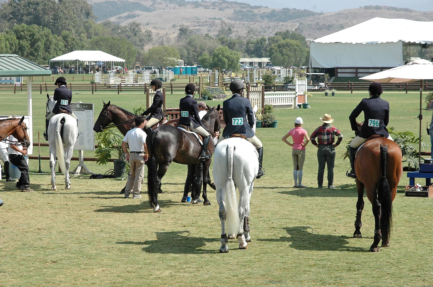
Biosecurity planning for equestrian events
“Show is Cancelled. You are Quarantined.”
These are phrases no equine event manager or exhibitor wants to hear. Unfortunately, the increased number of equestrian events, the increased international and interstate equid movement and the increased number of equids co-mingling at individual events have resulted in an increase in the potential for infectious disease agent incursion and spread.
Biosecurity, the measures taken to prevent pathogen entry and spread on a premises, is the key to protecting both equids and event venues from devastating disease outbreaks. There is no one-size-fits-all biosecurity plan for equine events, as each venue event is unique. Thus, identifying and assessing biosecurity risks for the population of equids at the event and on the event grounds are essential to developing the most effective biosecurity plan. Performing a biosecurity risk assessment to identify the disease risk factors for the venue, event and horse population is the first step.
Although the venue typically has a consistent footprint, weather can impact disease agent entry and transmission. For example, the Santa Ana winds in California, or the hurricanes that come in through the Gulf of Mexico, have the potential to carry pathogens or infected vectors (insects) onto or around a venue. Additionally, droughts almost certainly impact the availability of hay, resulting in the sourcing of hay for an event from further away. This sourcing of hay can potentially lead to the transmission of unfamiliar pathogens, toxins or insects, such as blister beetle, to a venue. Lastly, hot, humid days increase the likelihood of horses clustering under shade and/or utilizing communal water sources, which results in a higher risk
of respiratory pathogen spread. When looking at equestrian events, there is no cookie cutter event biosecurity plan for the venue. Each event is unique with regards to stabling and competition plans for the venue grounds. Events involving multiple disciplines may designate a day or consecutive days for each discipline, while others may have all disciplines on the venue grounds at the same time. Thus, a disease outbreak occurring in one discipline would more significantly impact a show that includes many disciplines on the grounds, especially if horses of various disciplines are housed together.
Another aspect to consider is that each discipline has different horse management and competition practices that could impact disease risk. For example, it is more common to see horses tied along the fence outside the competition arenas at western events. In contrast, one may find that it is more common to share a single nose rag to wipe several muzzles outside the show ring at a hunter competition. During an event, the amount of horse-to-horse contact on the grounds can also impact these outcomes.
Once a risk assessment has identified potential disease entry and transmission points, the appropriate biosecurity measures can be identified to mitigate the risk. The event manager, in consultation with the event veterinarian, should compile these biosecurity measures into a written biosecurity plan for the event. Some basic concepts to consider in a biosecurity plan include healthy equine entry requirements, limiting horse to horse contact, limiting human contact to essential personnel, avoiding the sharing of equipment unless it has been cleaned and disinfected between uses and monitoring the health of all equines on the premises. Entry to the venue should be restricted to horses accompanied by an owner-signed health
declaration that attests the horse has been monitored for a minimum of three days prior to arrival with twicedaily temperature readings below 101.5°F and no observable clinical signs of disease. Furthermore, the plan should require and verify proof of vaccination for equine influenza and equine herpesvirus-1 within the prior six months.
Unfortunately, even with these measures implemented, horses may still develop an infectious disease at an equine event. Therefore, prompt identification and isolation of the sick horse limits disease spread beyond the index (first) case. Identification of the sick horse requires protocols to be in place that require all horses on the grounds be monitored for any signs of illness or fever (temperature over 101.5°F), and, if present, be reported to a veterinarian. If the veterinarian suspects an infectious disease case, that horse must be immediately isolated in appropriate isolation stabling. Oftentimes, space is limited on event grounds and appropriate isolation is not possible at the venue. These event grounds must identify a place to isolate off-site in advance, as finding a place at the time of identification of a sick horse delays the isolation and increases exposure risks to all horses on the grounds. Some options for off-site isolation include empty fairgrounds, stables or fields with run-in sheds.
Once an infectious disease has been confirmed, identifying any exposed horses to monitor can be a significant challenge, especially if the facility or event have many potential avenues for horse-to-horse or human-to-horse contact or sharing of equipment. Horses that can touch over the tops of the stalls or through the bars, or horses that are loped in an arena for an extended period time very close to another horse have an increased potential for spread of respiratory pathogens. However, it is often difficult to identify all horses, people and equipment that have touched the affected horse. Thus, it becomes important to have protocols in place for all those handling multiple horses to prevent fomite (inanimate objects, such as buckets, rags, tack, etc.) transmission of pathogens.
Proper cleaning and disinfecting protocols are essential to decreasing the risk of infectious disease agent spread. Stalls and common areas should be regularly cleaned and disinfected with an appropriate disinfectant for the surface. For example, bleach is inactivated by organic material, thus it is ineffective in a stall that has manure stains covering the surface. Metallic surfaces, like bars in a stall, require a non-corrosive disinfectant to prevent degradation of the surface. Venue management should consult their veterinarian to determine the most effective disinfectant(s) for the various surfaces on their grounds.
Ultimately, identifying the disease risk factors for the specific event, developing a written biosecurity plan and implementing a few key biosecurity measures can protect the equids on the event grounds as well as protect the show from potential cancellation and quarantine due to an infectious disease agent.
CONTACT: Katie Flynn, DVM
Equine
Health and Biosecurity Veterinarian
United States Equestrian Federation Lexington, KY, USA kflynn@usef.org
Material published in the Quarterly is not subject to copyright. Permission is therefore granted to reproduce articles, although acknowledgment of the source and author is requested.
Maxwell H. Gluck
Equine Research Center Lexington, Kentucky USA, 40546-0099
Telephone (859) 257-4757
Fax (859) 257-8542 gluck.ca.uky.edu
RNA vaccines: Can they protect from equine herpesvirus infection and its complications?
RNA vaccines were tremendously successful during the Covid-19 pandemic due to excellent stimulation of immune responses combined with a fast and easy production process.
Traditional vaccines are typically based on subunit, inactivated or attenuated pathogens, which require long development times, making a rapid response to newly emerging pathogens difficult. In contrast, the RNA in RNA vaccines encodes the primary immunogenic protein(s) of the pathogen and can be developed quickly once the genetic sequence of a pathogen is known. This RNA is formulated or ‘packaged’ in e.g. lipid nanoparticles, cationic polymers or hyperbranched polyglycerol (hPG) amines to allow for efficient transfer into host cells where the RNA of interest is translated into an immunogenic protein.
This protein then triggers an immune response against the very same protein expressed by the pathogen. Currently, RNA vaccines are being explored for a wide range of human and animal pathogens. The main advantages of RNA vaccines are that the production of RNA vaccines is technically simple, fast, easy-to-adapt, cost-effective and free of animal material.
There are currently two types of RNA vaccines, the conventional mRNA vaccine and self-amplifying (sa)RNA vaccines. The conventional formula contains a fixed concentration of packaged mRNA while saRNA vaccines may contain less target RNA than the conventional formula, but in addition contain a sequence encoding for an enzyme called ‘replicase,’ which amplifies the RNA sequence of interest, i.e., itself. The advantage is a ‘smaller’ package with a longer duration of protein expression depending on the replicase half-life.
While vaccines for some pathogens are easy to design because there is only one, or few. immunologically important protein(s) for protection, other pathogens are more complex and require a multiple-target or cascade approach. This is one of the biggest limitations of RNA vaccines, as only a limited number of RNA sequences of interest – up to three – can be packaged into a single product or injection. Another disadvantage of RNA vaccines is storage, as most require storage at either -20°C or even at -80°C.
Equid alphaherpesvirus 1 (EHV-1) and 4 (EHV-4) are common equine viral pathogens. Horses in North America are routinely vaccinated against several viruses, including Western and Eastern Equine Encephalitis virus (WEEV, EEEV) and against West Nile virus, as well as equine influenza virus (EIV), with all of these vaccines performing reasonably well. In contrast, EHV-1 and EHV-4 outbreaks still occur in well-vaccinated populations. Thus, it is not surprising that the first efficacious nucleic acid vaccines have been developed against viruses for which protective immune responses can be induced with one (or few) immunogenic proteins, including coronavirus, flaviviruses and influenza viruses.
Unfortunately, EHV-1 and EHV-4 are not one of these viruses. Currently, challenges of developing effective mRNA or saRNA vaccines against equine herpes viral infections include the inherent antigenic complexity and immunosuppressive functions of EHV-1 and -4. With funding available through the Grayson Jockey Club Equine Research Foundation in Lexington, Kentucky, and in collaboration with Dr. Paul Lunn, University of Liverpool, UK, and Dr Juergen Richt, Kansas State University, we are currently testing the safety and efficacy of EHV-1 mRNA and saRNA vaccines.
Based on previous experience, our vaccine prospects include nucleic acid sequences encoding EHV-1 glycoprotein D and the Immediate Early (IE) gene. The rationale is based on i) available results from previous studies with EHV-1, and ii) on the Shingrix® vaccine success story of a subunit vaccine that only contains the glycoprotein (g)E (analogous to EHV-1 gD) preventing shingles in adults after childhood Varicella-Zoster infection (chickenpox).
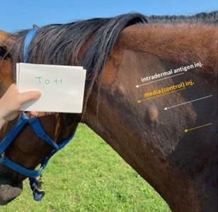
We expect these two components to be highly immunogenic, and to stimulate both cellular and humoral (antibodies) immunity. Figure 1 shows the neck of a horse four months after the second immunization with an EHV-1 mRNA vaccine containing gD and IE mRNA. Sizeable wheals (in duplicate) developed 24 hours after injection of deactivated EHV-1 as a sign of local inflammation caused by migration of mostly T-lymphocytes, while wheals were absent following intradermal injection of media alone.
This project is ongoing, and we will provide updates on the progress. We anticipate that testing the vaccine in a neurological model of EHV-1 will show at least some protection from EHM which will be an improvement over currently available vaccines. In the future, this strategy can be further refined and tested for duration of immunity or effectiveness as a booster following vaccination with conventional vaccines. Because of the high sequence homology of the selected viral proteins, it is likely that this vaccine would also offer cross protection to EHV-4. Finally, once we can show the applicability of the technology for EHV-1 in horses, it could readily be adapted to other viral pathogens of horses.
CONTACT:
Skin wheals 24 hours after intradermally injected deactivated EHV-1 injection (white arrow) and media (control) injection (yellow arrow). Injections were performed in duplicate.
Lutz S. Goehring, DVM, MS, PhD Wright – Markey Professor of Equine Infectious Diseases
Gluck Equine Research Center Department of Veterinary Science University of Kentucky Lexington, KY l.goehring@uky.edu
Gisela Soboll Hussey, DVM, PhD Professor College of Veterinary Medicine Michigan State University East Lansing, MI Husseygi@MSU.edu
Klaus Osterrieder, ECVM
Dean of the Jockey Club College of Veterinary Medicine and Life Sciences at City University; professor of virology and chair at Freie Universität Berlin, Germany; and adjunct professor of virology at Cornell University Tierärztliche Hochschule Hannover, Lower Saxony, Germany Klaus.osterrieder@tiho-hannover.de
Fourth Quarter 2024
International report on equine infectious diseases
This report collates information on equine infectious diseases provided by diagnostic laboratories in Lexington, Kentucky, the University of Kentucky Veterinary Diagnostic Laboratory (UKVDL) and Equine Diagnostic Solutions (EDS), Inc. We have further included information from the International Thoroughbred Breeders Federation, the International Collating Centre (ICC) in Newmarket/Cambridge, United Kingdom, and information from the American Association of Equine Practitioners’ Equine Disease Communication Center (EDCC). The retrieved information mostly covers North America and Europe with only incidental reports on disease outbreaks or single case from elsewhere.
As in previous quarters, there is regular reporting of Strep equi spp equi, both from outbreaks as well as from single cases, throughout North America and Europe. There are few notifications of Equine Influenza from various North American regions – some epidemiologically linked to horse transportations to and from events, and reports from Europe – mostly British Isles, but including one report from Sweden. The Lexington-based laboratory EDS reports several (Eq) Influenza positive nasal swabs from various regions throughout the USA, including cases from Kentucky. An outbreak investigation on Contagious Equine Metritis (CEM) caused by the bacterium Taylorella equigenitalis in the United States is ongoing. The south of Sweden and northern Germany report single cases of T. equigenitalis positive cases, all in Icelandic horses. EHV-3 infection, an alphaherpesvirus and cause of pustular vulvovaginitis, is rarely reported. For this quarter, there is one report from Kentucky.
So far, there have been several cases of respiratory EHV-1 and EHV4 infection with reports mostly originating from Europe. Two EHV-4 positive abortions were reported from France without any reports of EHV1 abortions so far. For the fourth quarter 2024, the UKVDL has received close to 140 aborted fetuses. There have not been EHV-1 abortions so far, and the most frequent diagnosis has been (suspect) umbilical cord torsion or unknown cause.
Other causes for infectious abortions: Both, Belgium and UKVDL reported two abortions caused by Leptospira spp. A few EHM single cases/outbreaks have been reported from North America, and a few from Northern Europe (two in France, Netherlands and Sweden). Mosquitoborne (ARBO) diseases Eastern Equine Encephalitis virus (EEE) and West Nile virus (WNV, in North America have been decreasing likely due to the expected decrease in mosquito activities, while WNV cases have also been reported from Europe for all endemic areas. The Caribbean island Guadeloupe reported a WNV case.
While there are sporadic cases of Equine Infectious Anemia reported from Europe, there are a number of reports on EIA cases across the United States that were epidemiologically linked to iatrogenic transmission and transportation to different venues in New Mexico, Texas and California.
Nigeria reported a case of African Horse Sickness (AHS).
The United Kingdom reported their annual numbers of horses succumbed to equine grass sickness (EGS) during through the ICC. The east coast of Scotland has always been the epicenter of EGS since its first descriptions of the disease and most of the research around this disease has been conducted there. With different theories on the ethiopathogenesis of this disease, there is encouraging news from Scotland. In an editorial in the Equine Veterinary Journal (Vol 57 (1)), Dr. Bruce McGorum and team from the Royal (Dick) School of Veterinary Studies, University of Edinburgh, United Kingdom, in collaboration with Newcastle University, Istituto Zooprofilattico Sperimentale delle Venezie and University of Padova, Italy, unveiled what sounds like a break-through in grass sickness etiopathogenesis. Through their ongoing studies it became likely that a neurotoxin in the form of a phospholipase A2 could be responsible for the clinical signs. The authors suggest that this toxin could be either ingested or in vivo produced by microbes in the horse gut.
EGS is not limited to the United Kingdom with cases also reported from countries along the North Atlantic and Baltic Sea, albeit in much lower numbers. In South America, EGS is known as ‘Mal Secco,’ while it is likely that other animal dysautonomias affecting cats, dogs, hares, rabbits, llamas, alpacas and sheep may be linked to the same neurotoxin, which hopefully will provide strategies for prophylaxis and intervention. Congratulations to the team of researchers!
CONTACT:
Edward Olajide, DVM
PhD Graduate Student
Gluck Equine Research Center
Department of Veterinary Science University of Kentucky Lexington, KY
Edward.olajide@uky.edu
Lutz S. Goehring, DVM, MS, PhD
Warren Wright, Sr. – Lucille Wright Markey Endowed Chair in Equine Infectious Diseases
Gluck Equine Research Center
Department of Veterinary Science University of Kentucky Lexington, KY l.goehring@uky.edu
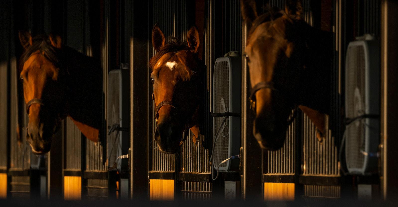
Photo Courtesy Mark Pearson Photography.
Whole genome sequencing of Thoroughbred horses: A tool for surveillance and management
The Welfare and Safety of the Racehorse Summit meetings began in 2006 with the objective of improving the safety and soundness of the Thoroughbred racehorse. One of the central questions resulting from these meetings was whether the genetic structure of the population had become compromised, such that the Thoroughbred had become less sound and durable during the last few decades. Powerful genomic tools are now available to address this question.
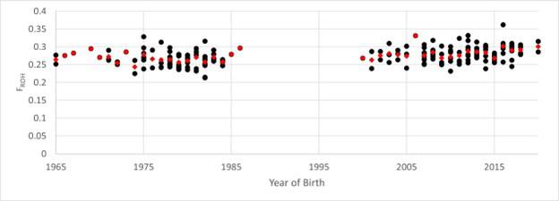
As a first step to do so, we used whole genome sequencing to identify every base of DNA in an animal’s genome. We studied the genomes of two groups of U.S. Thoroughbred horses: Group 1 (82 horses born between 1965 and 1986) and Group 2 (103 horses born between 2000 and 2020). Previous genetic studies used methods that measured, at most, 0.03% of the DNA to report statistically significant increases in inbreeding over time. “Statistically significant” is a scientific term meaning that there is a difference between measured groups but does not address the magnitude of the difference nor its consequence. Using the whole genome sequence of each horse, we calculated its inbreeding coefficient, which is illustrated in the Figure below, taken from our recent publication.
Each black dot represents the inbreeding estimate (FROH) for one of the 185 Thoroughbred horses, ordered according to its year of birth. The red dots represent the average for a year. The average inbreeding measured for Group 1 (0.266) was statistically different (P<0.001) from the average for Group 2 (0.283), however, the differences were small, such that one cannot identify whether a horse belonged to Group 1 or Group 2 based on its measure of inbreeding. Indeed, an increase in inbreeding is expected in a closed stud-book population under selection. The average increase from Group 1 to Group 2 suggests that breeders are successfully removing deleterious variants while selecting for desirable variants.
The two groups were also evaluated for maternal lineage using mitochondrial DNA, as well as for the prevalence of known genetic variants for disease, performance and color. Additionally, regions of inbreeding were assessed to determine whether they occurred recently (in the past five to 10 generations) or if they were more likely to date back closer to the foundation of the breed.
Only two of 19 known disease variants were found among the 185 Thoroughbreds, and those were present at low frequencies (Fragile Foal Syndrome: expect one in 62,500 affected Thoroughbreds; Hypoparathyroidism: expect one in 27,778 affected Thoroughbreds)
demonstrating that the most common Mendelian diseases of horses are uncommon in the breed. We also observed a statistically significant and relatively large increase in the frequency of the MSTN variant when comparing the two groups (Group 1: 0.427; Group 2: 0.539). This MSTN variant was previously reported to be more prevalent among champions of short races and less common among champions of longer races. This observation may reflect selection for performance at short distances during the last 50 years. In the future, we propose the use of whole genome sequencing to provide specific data that breeders can use in assessing and managing the genetic health of the population.
The cost of whole genome sequencing is rapidly dropping, meaning that it can be a valuable tool for surveillance of the genetic integrity of the Thoroughbred. Furthermore, when hereditary factors are thought to negatively affect complex traits such as soundness, fertility or fetal loss, whole genome sequencing may allow for the identification of genomic regions that contribute to the problem. In summary, these tools will prove to be valuable additions to the experience, understanding and intuitive grasp of genetics by Thoroughbred breeders.
The manuscript, with more details, is freely available at: https://www. nature.com/articles/s41598-024-73645-9
CONTACT:
Ernest Bailey, PhD Professor, Gluck Equine Research Center Department of Veterinary Science University of Kentucky Lexington, KY ebailey@uky.edu
Ted Kalbfleisch, PhD Professor, Gluck Equine Research Center Department of Veterinary Science University of Kentucky Lexington, KY ted.kalbfleisch@uky.edu
Jessica Petersen, PhD Associate Professor Department of Animal Science University of Nebraska - Lincoln Lincoln, Nebraska jessica.petersen@unl.edu