9 minute read
Super scanner There are

The brain on screen
The brain is our most complicated and least understood organ. This makes it almost surprising that it functions so well in most people. But there is a lot that can go wrong. Luckily, we now have MRI scanners with which we can examine the brain in detail.
By Joris Janssen
It is located in a special wing of UMC Utrecht: one of the most powerful MRI scanners in the world in a clinical environment – an environment in which it can help patients directly, as opposed to an environment focused solely on research. For two neurologists, this machine is a blessing for their research. Geert Jan Biessels uses it to study patients with dementia due to damage to small blood vessels in the brain. Kees Braun uses the scanner to treat children and adults with epilepsy that is difficult to control. Two different disciplines with something in common: technological progress promises improved treatment by visualizing the brain.
Anyone thinking of dementia, often thinks of Alzheimer’s disease. This disease causes pathogenic protein to damage the brain, leading to memory problems, among other things. However, this is often only part of the story. Nine out of ten people who develop Alzheimer’s in later life have another problem: damage to small capillaries in the brain. Sometimes, this type of capillary damage is even the main culprit behind the dementia-like symptoms. In those cases, patients are diagnosed with vascular dementia. Overall, capillary damage is responsible for roughly one third of all dementia symptoms. Half is due to Alzheimer-like processes, and the rest is caused by a catchall of other conditions. So, although it is not the most common form, many people with dementia are faced with the vascular variety, to a greater of lesser extent.
Tiny capillaries ‘The study of dementia due to capillary damage is lagging behind the study of dementia due to Alzheimer’s,’ says Geert Jan Biessels, head of general neurology and leader of the Vascular Cognitive Impairment research group at UMC Utrecht. ‘It’s a shame, because there is a very real opportunity to do something about it.’
This disadvantage is, in part, caused by lagging investments, but there is also a practical cause. Until recently, it was impossible to obtain a good visual of the culprits behind the condition: the small
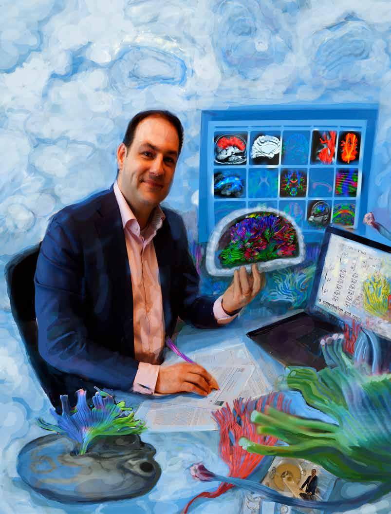
MRI scans are essential when uncovering whether Alzheimer’s, capillary damage or a combination of the two is behind dementia symptoms. BRAM BELLONI

Damage to small capillaries in the brain is the cause of roughly one third of all dementia symptoms. BRAM BELLONI

capillaries that supply the brain with blood. Damage to these capillaries affects the connections in the brain, causing the fiber-optic network of the brain to malfunction. ‘Compare it to streaming a series or a movie on Netflix with a slow connection,’ Biessels explains. ‘It is not that people with this type of dementia make lots of mistakes or really forget things, it just takes a lot more time to retrieve the information.’
Scans made by the average MRI scanner are not detailed enough to show the tiny capillaries in the brain, let alone abnormalities or damage to these capillaries. The only thing these scans show is the result of the damage: blotches in the fiber-optic network caused by small hemorrhages or cerebral infarctions. This imposes two major limitations. The blotches can be cause by something other than damage to the small capillaries. The presence of blotches alone does not have to mean that someone actually has vascular dementia. In addition, it means that you can’t identify the condition until its final stage, when it has already caused a great deal of damage. Visualizing the tiny capillaries in the brain can help to identify the problem sooner and can enable fundamental research into ways of dealing with the disease at an early stage.

UMC Utrecht is unique And that is where the powerful 7-Tesla MRI scanner comes into play. An MRI scanner creates images of structures in the body by setting hydrogen atoms in motion using strong magnetic fields and radiofrequency pulses. When the atoms settle down again, they give off a signal. This signal can then be picked up and turned into an image. The different types of tissue in the body contain different quantities of hydrogen atoms. This creates a contrast, allowing you to see the difference between, for example, blood, fat, and organ tissue on the image.
The stronger the magnet, the higher the contrast that can be achieved. The strength of a magnet is expressed in Tesla. Most MRI scanners in hospitals have a 1.5 or a 3-Tesla magnet. The magnet in the MRI scanner at UMC Utrecht is a bit stronger.
As the name indicates, it has a strength of 7 Tesla. When UMC Utrecht got the scanner in 2007, there were only twenty of them worldwide. This number has gone up a bit since then, but most of these scanners are at research institutes. The MRI scanner in Utrecht is unique because of its strength and because it is in a clinical environment, where many patients have now been examined using the scanner. With a magnet that is many times stronger, contrasts become visible that would otherwise have remained hidden. ‘We can
now see the circulation of blood through capillaries as thin as eyebrow hairs,’ says Biessels. ‘We can see how circulation varies during the cardiac cycle, which shows us how hard the capillaries are.’ Thanks to this increased contrast, for the first time, Biessels and his colleagues are able to make a connection between the damage seen on other scans and underlying abnormalities in the tiny capillaries. ‘For the first time, we are now able to describe disease of the capillaries in terms of those capillaries themselves.’
Examining the brain at this level of detail is an outcome for many more brain diseases than vascular dementia alone. For Professor of Pediatric Neurology Kees Braun, it is also a boon. He treats children with so-called refractory epilepsy, which is difficult to treat, and he researches how improved imaging of the brain can improve the efficiency of treatment.
Epilepsy is a condition in which electric

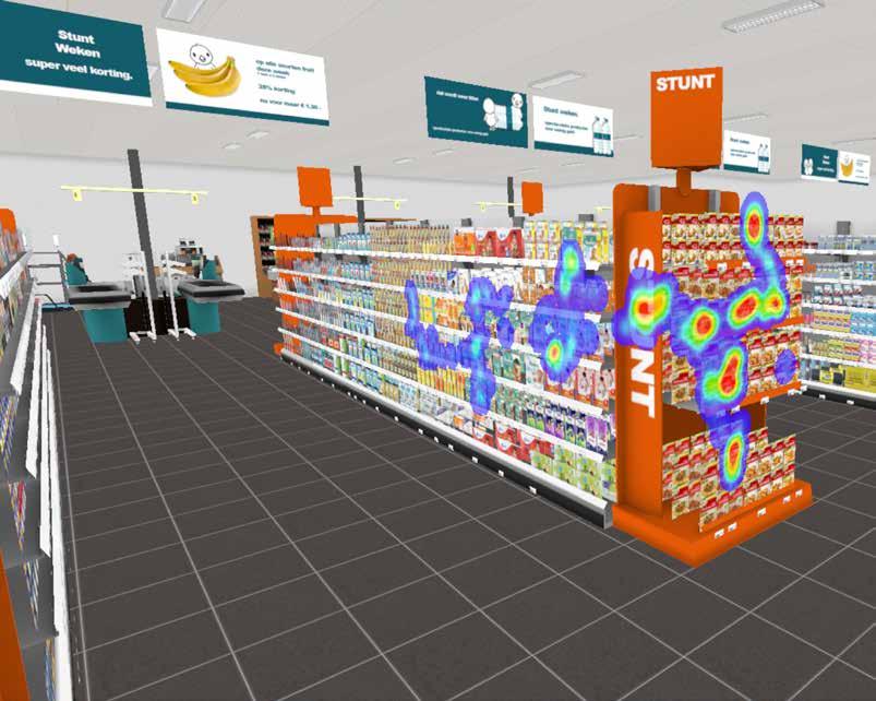
The human eye alone is no longer enough to interpret brain scans. Smart software helps doctors and researchers with this difficult task.
discharge occurs involuntarily and fitfully in the nerve cells of the brain. The result resembles a short circuit that can lead to twitching, decreased consciousness, and speech or vision difficulties, depending on where in the brain the short circuit is located. In roughly two-thirds of the cases, medication can eliminate nearly all symptoms. For the remainder of patients, medication does not offer a solution. These patients are diagnosed with refractory epilepsy, the variety that Braun and his colleagues study.
Vulnerability An operation may be a possible solution for patients with refractory epilepsy. If the source of the attacks is known, that source can be removed or disconnected from the rest of the brain, provided that it is not in an area of the brain that is so important that the neurosurgeon best leave it alone. In some severe cases, surgeons can even disconnect an entire side of the brain in young children. A condition for this type of operation is that the source of the epilepsy is local. A specific area of the brain has to be identified as the source of the problem. If involuntary electric discharge occurs throughout the entire brain, for example due to a genetic predisposition, an operation will be of no use.
A local source of electric discharge can have various causes. ‘It could be a tumor, an infarction, a scar, inflammation or an abnormal development of the cerebral cortex,’ says Braun. ‘In those cases, an operation could help. So being able to make a diagnosis is very important. Imaging using an MRI scanner is crucial in this process.’
The better the scans are at visualizing the problem area, the greater the chance of a successful operation. This is particularly true for abnormal developments of the cerebral cortex, a condition known in medical terms as focal cortical dysplasia. ‘The technology that visualizes these small, local abnormalities has been improved over the past decades,’ says Braun. ‘This has allowed us to improve the selection of people who qualify for an operation. We had already made good progress with the previous generation of scanners, but our research shows that the chance of detection using the 7-Tesla MRI scanner is increased by another 20 percent.’ However, more powerful scanners are only part of the equation, in both Kees Braun’s
epilepsy research and Geert Jan Biessels’ dementia research. A large part of the progress also lies in a smarter way of looking at the scans. For example, in Braun’s research, new statistical techniques ensure that computers are getting better at identifying abnormalities in the brain. Braun: ‘A computer can compare the scan of a patient to the scans of a large group of healthy people and identify the areas of the patient’s brain that are significantly differ-

ent from those of the control group. In this way, the computer can show you an abnormality that you would have missed with the naked eye.’
The field of vascular dementia also benefits greatly from statistically combining large amounts of data. Biessels hopes that an international cooperation project that he helped start will soon produce the first ‘vulnerability map’ of the brain. This combines the data of around one thousand patients from Asia and Europe to form a map that shows the extent of impact of damage to specific parts of the brain. ‘In the past, we only looked at the total amount of damage, but that’s too simple. The consequences of damage at a critical point are much greater. This map will show us in what areas damage has the most impact.’
Developing a platform As crazy as it may sound, statistical techniques created by electricity companies are also contributing to a better view of the condition of a person’s brain. Biessels: ‘These companies are very interested in what happens when, for example, a truck hits an electricity pylon. Will it take out the entire network, or not? Many measurements have been developed to indicate how robust and efficient a network is. We can also apply them here to see the condition of brain networks.’
More and more disciplines are becoming involved in research using brain scans and more and more international partnerships are being set up. ‘We are really pushing the scanner to its limits,’ says Biessels. ‘This results in so much data that you can no longer assess it with the naked eye. The limit of human perception is so easily reached that you need computer technolo