


The Anchorage & Valley Radiation Therapy Centers have been providing Alaskans with compassionate care and state-of-the-art cancer treatment since 1993. Home to Alaska’s only Gamma Knife radiosurgery system, our clinics provide cutting-edge treatment technologies for optimal outcomes.
We believe you shouldn’t have to travel far from home to receive top-of-the-line radiation treatment, and our compassionate, highly experienced team is there for you every step of the way So you can get back to living your life.





Publisher: Andy Pennington
Editor: Nina Wladkowski
Special Projects Director: Brandi Nelson
Sales: Ryan Estrada
Adam Garrigus
Victoria Hansen
Joleesa Stepetin
Justin Thompson
Josh Trouy
Advertising Operations: Lisa McGuire
Graphic Designer: Jian Bautista

Caroline Clune, M.D., Mayo Foundation for Medical Education and Research, Premium Health News Service
A mammogram showed a lump in my breast, and my doctor said it’s benign breast disease. I’m glad it’s not cancer, but I’m still worried. What does this mean? Does it increase my risk of getting breast cancer in the future?
Changes to your breasts can cause a lot of worry. This is understandable. As you discovered, though, not all breast changes are a result of breast cancer.
Any breast symptoms, such as a breast lump, nipple discharge or breast pain, should be evaluated by a medical professional. If the symptoms are diagnosed as benign, it means they are not cancer. Noncancerous breast symptoms are known as benign breast disease.
Some cases of benign breast disease are discovered during a screening mammogram. Some are felt at home. For any lump or symptoms felt at home, it’s recommended that you seek a thorough examination with a health care professional. If there are findings on a mammogram, your health care team will decide if additional imaging is required. This could include another mammogram to get more images of the
spot and an ultrasound of the breast. Often, we can determine whether the cause is benign or not through imaging alone. Sometimes a biopsy may be necessary.
The good news is that benign breast disease is not cancer. However, some benign breast disease needs treatment and can increase the risk of developing breast cancer in the future. Ask your health care professional which type of benign breast disease you have and if it increases your personal risk.
If you have a history of cancer or other concerns, consider seeking out a specialist at a breast care center.
Here are a few of the most common types of benign breast disease:
• Fibroadenoma: Fibroadenomas are the most common benign tumors in the breast. Most often, they occur in people between ages 15 and 35. They often present as a firm, round, smooth and rubbery breast lump on a breast exam. Many fibroadenomas are managed by following repeat ultrasounds over time. They do not increase your risk for breast cancer in the future.
• Breast cyst: Sometimes, cysts can develop in the breast. Cysts are fluid-filled masses. They can present as lumps noted in breast tissue or found on a mammogram. They don’t always cause symptoms, but breast cysts that grow can lead to breast pain and tenderness. They are common between the ages of 35 and 60, and can fluctuate with menstrual cycles. Breast cysts do not increase your risk of breast cancer.
• Mastitis: This is inflammation of the breast tissue caused by blocked milk ducts or bacteria in the breast. It commonly affects women who are breastfeeding, but mastitis can occur in women who aren’t breastfeeding. The inflammation leads to breast pain, swelling, warmth and redness. Mastitis is treated using antibiotics and pain relievers. It does not increase your risk of developing breast cancer.
• Papilloma: A papilloma is a growth in a milk duct and can present as nipple discharge. It also may present as a small lump behind or next to the nipple. A biopsy can help understand whether papillomas need to be treated, as they sometimes can contain atypical cells that can increase your risk for breast cancer. Treatment also depends on the size, if there are multiple lumps or if they are causing symptoms. Surgery may be recommended to remove the papillomas as well.
• Atypical hyperplasia: This type of benign breast disease is diagnosed by a breast biopsy of an abnormal finding on an exam or breast imaging. It is an accumulation of abnormal cells in the milk ducts or lobules of the breast. Atypical hyperplasia isn’t cancer, but it increases the risk of breast cancer. For this reason, the area sometimes is removed with surgery. Often, health care teams recommend intensive breast cancer screenings and medications to reduce breast cancer risk.
All breast changes should be discussed with your health care team. In addition, an annual physical exam is a good way to review your risk for breast cancer and discuss an appropriate screening schedule for you.Caroline Clune, M.D., Primary Care Internal Medicine, Mayo Clinic Health System, La Crosse, Wisconsin
Mayo Clinic Q & A is an educational resource and doesn’t replace regular medical care. This Mayo Clinic Q&A represents inquiries this healthcare expert has received from patients. For more information, visit www.mayoclinic.org.


If you are suffering from neck pain, migraines, headaches, radiating pain, muscle spasm, upper, mid or low back pain then you need to investigate this. The Atlas Orthogonal and Cox Flexion Distraction Procedures are gentle, specific and proven effective for thousands of patients. We are in-network with BC/BS, Aetna, Tri-west & Medicare. Call 907-677-6953 to schedule your Complimentar y 15 minute
Pre-Consultation with our doctors today!


Lauren Cornell, M.D. and Kristin Robinson, M.D., Mayo Foundation for Medical Education and Research, Premium Health News Service
DEAR MAYO CLINIC: I recently had my annual screening mammogram and was told everything was normal. However, I just read an article that said health care professionals must inform patients if they have dense breasts. My clinician did not share anything. Why is this information important? How do I know if I have dense breasts? What additional screening is required?
ANSWER: Breasts come in all shapes and sizes. Breast density is not determined by how a breast looks or feels. Breast density refers to the amount of fibroglandular tissue in a woman’s breast compared to fatty tissue.

Fibrous connective tissue gives the breast shape, and glandular tissue responds to hormonal influences, produces milk and is where breast cancer forms at the cellular level. Fat in the breast is, well, just fat. On a mammogram, the fibroglandular tissue looks white, while the fat looks black. The whiter the breast appears on a mammogram, the denser it is considered. Dense breast tissue by itself does not need to cause alarm. Approximately 40% to 50% of women have dense tissue. But women with dense breast tissue are at a higher risk for breast cancer compared to women without dense breasts. In addition, dense tissue can mask abnormalities on the mammogram, making it harder to detect small cancers.
Generally speaking, mammograms can detect approximately 80% of breast cancers in women without dense tissue, but that number decreases to below 60% in women with dense breast tissue. The more dense the breast tissue appears on a mammogram, the further the detection rate falls. That is one of the reasons for the most recent ruling by the Food and Drug Administration that aims to help further standardize information that health care organizations share. In 2009, Connecticut enacted the first breast density reporting law requiring facilities

performing screening mammograms to inform patients about breast density, and this more recent ruling will standardize reporting at the federal level. Mayo Clinic has been providing women with information about breast density for years.
Data show us that mammograms consistently reduce breast cancer mortality by detecting breast cancer at smaller sizes and earlier stages compared to women who do not have mammograms, regardless of breast density. Mammograms still detect microcalcifications and areas of abnormal tissue that might indicate cancer is hiding. But women who have dense breast tissue should discuss the option of supplemental screening in addition to undergoing a yearly mammogram.
Breast density is only one factor that contributes to someone’s overall risk of developing breast cancer. The decision to undergo additional imaging may depend on your age, exactly how dense your tissue is, your personal preferences and whether you have additional risk factors for breast cancer, including family history of breast cancer or ovarian cancer, prior breast biopsies or exposure to hormones.

Today, there are several options for breast imaging that may be used in conjunction with the mammogram for women with dense breast tissue, including:
• Tomosynthesis, or a 3D mammogram. This is performed at the same time as the traditional 2D mammogram. Tomosynthesis only increases cancer detection slightly, but it does reduce the likelihood of false positive findings. This translates to a lower recall rategetting the dreaded phone call to return for an additional imaging.
• Whole breast ultrasound. This technique has been used historically for women with dense tissue, although the added cancer detection rate is relatively low compared to using mammography alone. The major downside is that whole breast ultrasound has a fairly high false positive rate.
• Contrast-enhanced digital mammography. This can be done at the same time as the traditional screening mammogram, but uses an iodine-based contrast, delivered through an IV prior to the test. This is a newer imaging strategy, but initial research studies have been very promising.
• Molecular breast imaging (MBI). MBI is a test that uses a radioactive tracer and a special camera that looks at the energy level of cells. Cells that are growing quickly take up more of the tracer than do slowly growing cells. Cancer cells often grow quickly, so the idea is that they will take up more of the tracer. MBI is a great option for women with dense tissue who do not have a lot of other risk factors for breast cancer.
• MRI. This test has the highest cancer detection rate, finding significantly more cancers than mammograms alone. MRI is the costliest test to image breast tissue, and current guidelines recommend MRI only for women at the highest risk for breast cancer - women whose lifetime risk exceeds 20% to 25% based on several factors. MRI also is beneficial for women previously diagnosed with breast cancer and who have dense breast tissue, or if diagnosis occurred before age 50.

The decision to pursue any of these options should be made based on several factors, including which tests are accessible in your area, your insurance coverage, the pros and cons of each test and your individual cancer risk.
Your primary care clinician is a good person to have conversations with, but you also can ask for a referral to a breast health specialist who can help guide you further to ensure you have the most appropriate imaging based on your breast density and overall cancer risk. - Lauren Cornell, M.D., Hematology/Oncology, and Kristin Robinson, M.D., Radiology-Diagnostic, Mayo Clinic, Jacksonville, Florida
Mayo Clinic Q & A is an educational resource and doesn’t replace regular medical care. This Mayo Clinic Q&A represents inquiries this healthcare expert has received from patients. For more information, visit www.mayoclinic.org.


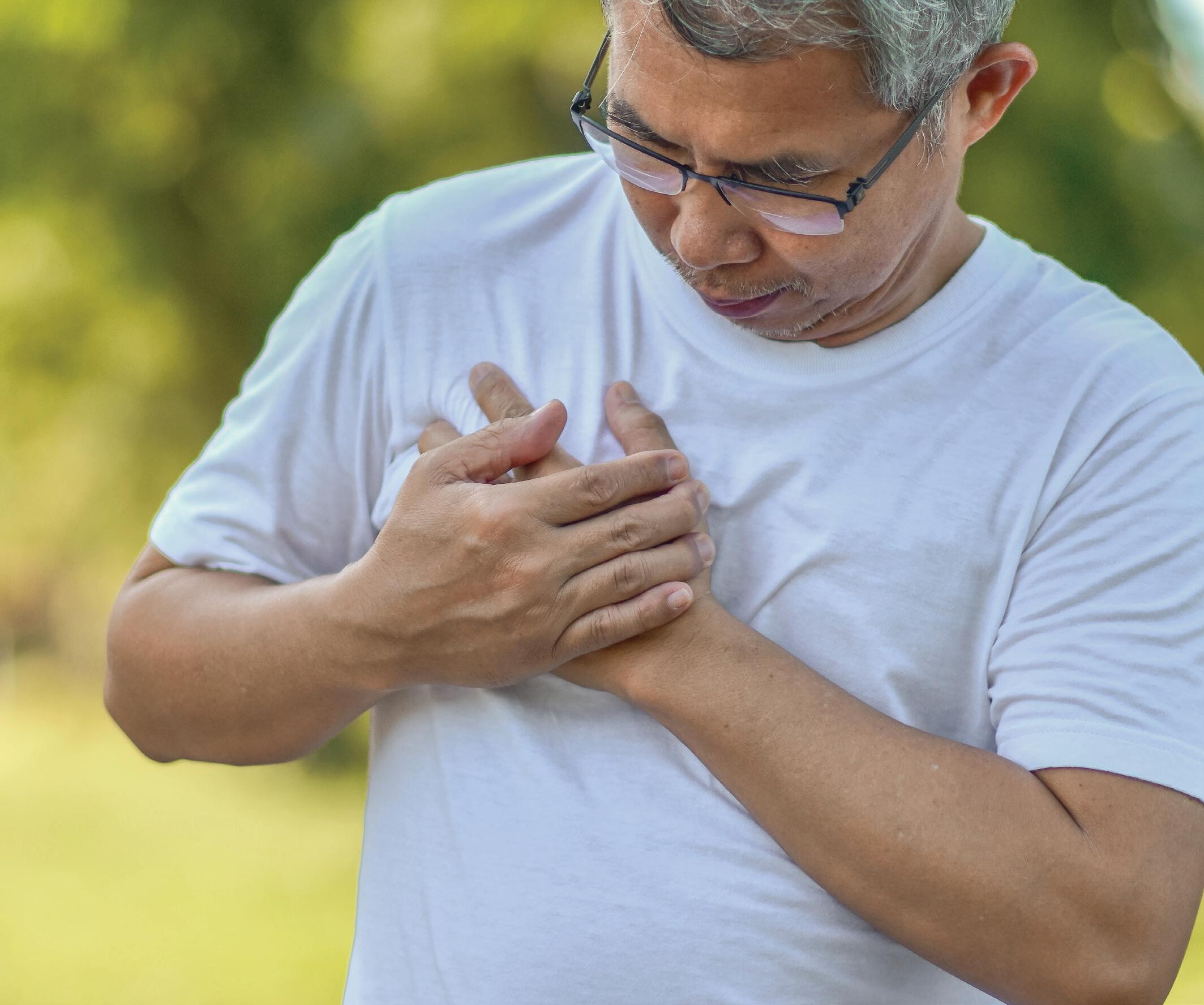
Q:I am a 53-year-old man. I think my breasts are larger than average but they usually don’t bother me. For the past two weeks or maybe longer, one spot in my left breast feels harder and is slightly tender. Should I be worried about breast cancer?
A:The most likely cause of this harder area is a non-cancerous lump related to a condition called gynecomastia. However, you definitely need to seek medical evaluation. Any new breast lump in a male - or female - must be evaluated by a health professional.
Getting back to the condition you most likely havegynecomastia. This is benign breast enlargement in a male. Most often both breasts are involved. When it is only one area of one breast, as you describe, that’s when it’s especially important to be sure that it is not breast cancer.
Some men have prominent breasts simply because they are overweight. But true gynecomastia is caused by an enlargement of the breast’s glandular tissues, not by an excess of fat. The glandular tissue
is concentrated under the nipple, but fat is spread around the whole breast area.
Gynecomastia is very common in teenage boys. About one-third of them will show some breast enlargement at the time of puberty. It’s less common in adulthood. Two-thirds of the cases involve both breasts. The breast enlargement is usually mild and painless. But about a third of men complain of tenderness.
When a reason for the gynecomastia can be identified, it is usually caused by a medication or liver disease. There is a long list of other much less common causes.
Of the many medications that sometimes cause gynecomastia, these are at the top of the list:
• Digoxin (Lanoxin) - used for heart disease
• Aldactone (Spironolactone) - a type of water pill
• Cimetidine (Tagamet) - a stomach acid blocker
• Ketoconazole (Nizoral) - used for fungal infections
• Finasteride (Proscar) and dutasteride (Avodart) - used for an enlarged prostate.
Other possibilities:
The tender spot could be an abscess. But I would expect it to be more painful than what you describe, and the overlying skin to be red.
You might have a fatty lump called a lipoma. This is a non-cancerous, localized growth of fat cells. But usually these are not tender to the touch.
Another consideration is an entity called runner’s nipple. Pain and tenderness of one or both nipples are quite common in people who do a lot of running due to the mechanical irritation of the runner’s shirt rubbing up and down against the nipples. But usually this won’t cause a lump.
Howard LeWine, M.D., is an internist at Brigham and Women’s Hospital in Boston and assistant professor at Harvard Medical School. For additional consumer health information, please visit www.health.harvard.edu.

Tucker Coston, M.D., Mayo Foundation for Medical Education and Research, Premium Health News Service
DEAR MAYO CLINIC: I am 48 and am being treated for metastatic breast cancer. Despite my diagnosis, I live a fairly normal life, am physically active and strive to optimize my health from a noncancer standpoint. An acquaintance who works in health care advised that I should watch closely for blood clots and be monitored. What is the risk of developing a blood clot, and how can I minimize my chances? How will I know if I have one?
ANSWER: Dealing with a cancer diagnosis is never easy, especially when you have so many potential things to worry about, including side effects from treatments. Discussion around the risk of blood clots often can be overshadowed in patients with cancer because many other issues take priority during office visits.
While blood clots can be dangerous, many strategies can minimize the risk. But it is important to discuss your individualized risk with your health care team.
Blood clotting is a complex, carefully regulated process in the body that prevents life-threatening bleeding when blood vessels are damaged. This becomes a problem when the clotting processes become imbalanced and form inappropriate clots within blood vessels. When this occurs in the vessels that carry blood back to the heart, it is referred to as a deep-vein thrombosis, or DVT. This type of clot is most common in the veins of the legs, but it can occur in any vein in the body.
Occasionally, a deep-vein thrombosis can break loose in the vein where it formed and travel to the vessels of the lungs. This is referred to as a pulmonary embolism, or PE. A pulmonary embolism carries the highest risk for severe illness or death.
Many processes can cause imbalance in the body’s blood clotting systems, increasing the risk for deepvein thrombosis and pulmonary embolism. Among the most common risks is prolonged immobilityoften days to weeks of immobility, such as during a hospital stay or after surgery. Additional risk
factors include damage to the walls of the blood vessels. This is sometimes caused by a process called atherosclerosis due to diabetes, high blood pressure or smoking, as well as increased production of blood-clotting proteins.
Though some people may have high blood clotting proteins due to a genetic condition, this can occur because of infection, serious illness or other diseases. Cancer is one such condition that causes imbalance in the blood clotting system by increasing the clotting proteins in the blood. Therefore, cancer patients are at increased risk for deep-vein thromboses and pulmonary embolisms, with the degree of risk depending on cancer type and degree of spread in the body.
When a deep-vein thrombosis occurs, it presents most often as swelling of the affected limb with accompanying pain or pressure, redness and prominence or tenderness of the veins. Occasionally, these symptoms are subtle. In some patients, they may be completely absent. When


a deep-vein thrombosis travels and becomes a pulmonary embolism, this can present as shortness of breath, chest pain with breathing, cough or rapid heartbeat.
If you begin to experience any symptoms out of the ordinary, especially emergent issues, it is critical to be evaluated by a health care professional as soon as possible.
Call 911 and use emergency services if necessary. It is important to be aware of the symptoms, but it's equally important to realize that these symptoms are not exclusive to blood clotting issues.
The mainstay of blood clot prevention involves maintaining your health, which it sounds like you are already doing. Keep up your activities, as staying active promotes healthy flow through your veins. Also, maintaining a healthy diet and a healthy weight, in addition to avoiding cigarette smoke, promotes the health of the walls of your blood vessels.


Comprehensive hearing evaluations
Hearing aid fittings & repair
Real-Ear-Measurements
Custom ear plugs & protection
Tinnitus management
Rehabilitative & preventative counseling

If necessary, blood thinning medications, referred to as anticoagulants, also are available by prescription from a health care professional. Careful consideration about the risks and benefits of these medications is necessary, but they are sometimes appropriate to add to the other measures to prevent blood clots. These same anticoagulants are used - sometimes at higher doses - to treat blood clots if they occur.
I always advise patients to talk with their cancer specialist and primary health team to determine the individual risk of developing blood clots and formulate an individualized plan for blood clot prevention. It is also valuable to talk about managing noncancerous medical issues that may contribute to risk. - Tucker Coston, M.D., Hematology/Oncology, Mayo Clinic, Jacksonville, Florida
Mayo Clinic Q & A is an educational resource and doesn’t replace regular medical care. E-mail a question to MayoClinicQ&A@mayo.edu. For more information, visit www.mayoclinic.org.


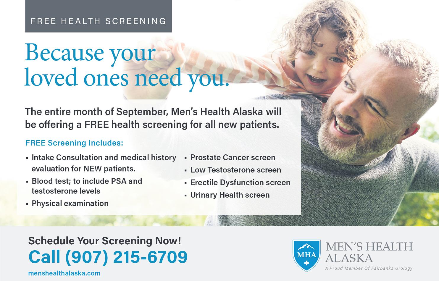
According to the American Cancer Society, the lifetime risk of cancer in the United States is one in three. But, many people may not realize that more than half of all cancer deaths may be preventable by making healthier food choices, maintaining a healthy weight and keeping physically active.
What can you do to help prevent cancer?
Load up on plant-based foods. No single food or food group can protect fully against cancer. But, plant-based foods contain vitamins, minerals, antioxidants and phytochemicals that may help lower the risk for many types of cancer. Their dietary fiber content also contributes to the healthy bacteria in our gut and helps keep us feeling fuller for longer. This can help us maintain a healthier body weight. Research suggests that it is the synergy of these dietary components working together that offers the greatest protection against cancer. So, fill your plate two-thirds full with a wide variety of colorful vegetables and fruits, whole grains and legumes.
Choose fish or poultry most often. The World Health Organization reviewed more

than 800 studies and found that eating processed meat or red meat every day increased the risk of colorectal, pancreatic and prostate cancer. So, limit your intake of red meats - beef, lamb, pork, and goat - and avoid frequent consumption of processed meats - bacon, sausage, ham, hot dogs and deli meats.
Snack on small portions of nuts and seeds. Nuts and seeds, preferably unsalted or low salt, contain numerous bioactive components that are both cancer-protective and cardioprotective. They also make a great snack compared with the empty calories found in traditional snack foods like ice cream, potato chips, pretzels, cookies and candy.
Limit your intake of salty foods and foods processed with salt. In addition to contributing to an increased risk of high blood pressure and stroke, diets high in salt have been linked to an increased risk of stomach cancer. Work on limiting your intake of the “Salty Six” - breads and rolls, cold cuts and cured meats, pizza, poultry injected with saline solutions - check the label - canned and restaurant soups and chili, and restaurant
entrees. Choose whole, unprocessed foods, fresh, frozen or low sodium canned vegetables, and cook more often with herbs and no-salt spice blends.
Choose water over wine. Many people assume that alcohol is good for us because it has some heart health benefits. But, worldwide studies have shown that regular consumption of alcohol can increase the risk of mouth and throat cancer, liver cancer, colorectal cancer and breast cancer. If consumed at all, limit daily intake to up to one drink for women and up to two drinks for men.
Use a food first approach over dietary supplements. Although it can be appealing to take dietary supplements with the hope that they will help prevent cancer, research has been inconclusive regarding their efficacy. In addition, dietary supplements are not consistently regulated and some have been found to contain harmful ingredients or little to none of the purported nutrients.
Putting your financial resources toward high-quality whole foods is a more sensible approach to help prevent cancer.

If you are still interested in taking supplements, you may want to discuss your concerns with a Registered Dietitian who can help evaluate your individual health needs.
Aim for a healthy weight. Having a body mass index of 30 or higher has been implicated in at least 13 different types of cancer. Use the 80/20 rule to help maintain a healthy weight and to help prevent cancer. So, 80% of the time, choose a wide variety of vegetables, fruits, nuts, seeds, whole grains, legumes, fish, nonfat or low-fat dairy, and plant-based oils. Then, the remaining 20% of the time, allow yourself room to enjoy other types of foods, such as desserts and goodies for special occasions and celebrations.
Reprinted with permission from Environmental Nutrition, a monthly publication of Belvoir Media Group, LLC. 800-829-5384. www.EnvironmentalNutrition.com.



Mended Hearts Inc. is the largest cardiovascular peer-to-peer support network in the world.We are a nonprofit organization offering the gift of hope to cardiovascular patients, their caregivers, and families for over 70 years! Mended Hearts has served millions by providing support and education, bringing awareness to issues that those living with heart disease face, and advocating to improve quality of life across their lifespan.
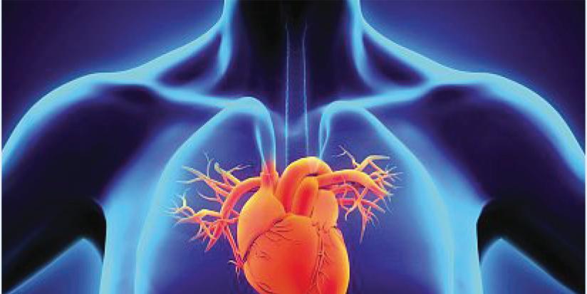
Thousands of Alaskans are living with cardiovascular disease. Many go undiagnosed until a medical emergency occurs. They and their caregivers and loved ones face many emotional and physical challenges during recover y. Our Mended Hearts volunteers provide encouragement, educational information, and peer-to-peer emotional, social, and practical support to them from diagnosis through recover y Because we are heart patients as well, we understand that adjusting to life after a heart event is challenging. Heart disease is complex, and few patients and their caregivers experience it the same way. There may be appointments to track, new medications to take, lifestyle changes to make, and many other issues to consider However, you are not alone on this journey Mended Hearts-Anchorage is. a dedicated group of volunteers whose mission is to provide support and encouragement to you, your caregiver and family. I invite you to learn more about our local chapter at www alaskamendedhearts.org and www.mendedhearts.org.

We invite you to join Mended Hearts-Anchorage and become part of a close-knit group that is focused on providing morale support for those with heart disease in our community.
Joining Mended Hearts connects you instantly to patients and caregivers just like you who understand the needs of cardiovascular disease patients. The Mended Hearts program provide counseling and emotional support at the hospital bedside, one-to-one support after discharge, plus a variety of helpful support group meetings, educational materials, online events, and social activities.

You can join the nation’s largest cardiovascular disease support network today and be part of our caring support network. Free memberships are available, and we welcome all. If you join at a donation level, you will get some gifts from Mended Hearts and Mended Little Hearts.Membership levels and gifts are shown here.
The Mended Little Hearts program has been supporting children and families since 2004. Our mission is to inspire hope and improve quality of life, so the littlest heart patients are not defined by their heart conditions but are stronger and more courageous because of them. Our chapter provides Bravery Bags to those children traveling out of state for medical procedures It’s not “just a bag” and can mean so much to those in the hospital. y or
Mended Hearts also offers a program for children with heart defects known as Mended Little Hearts


DEAR MAYO CLINIC: Since age 20, I have had annual visits with a gynecologist. I am now in my late 30s, and my physician recommended that I get an HPV test in addition to a Pap test to check for cervical cancer. What is the importance of both? And what is my risk of cervical cancer?
ANSWER: Screening for cervical cancer can cause a lot of confusion because guidelines and tests have evolved over the years.
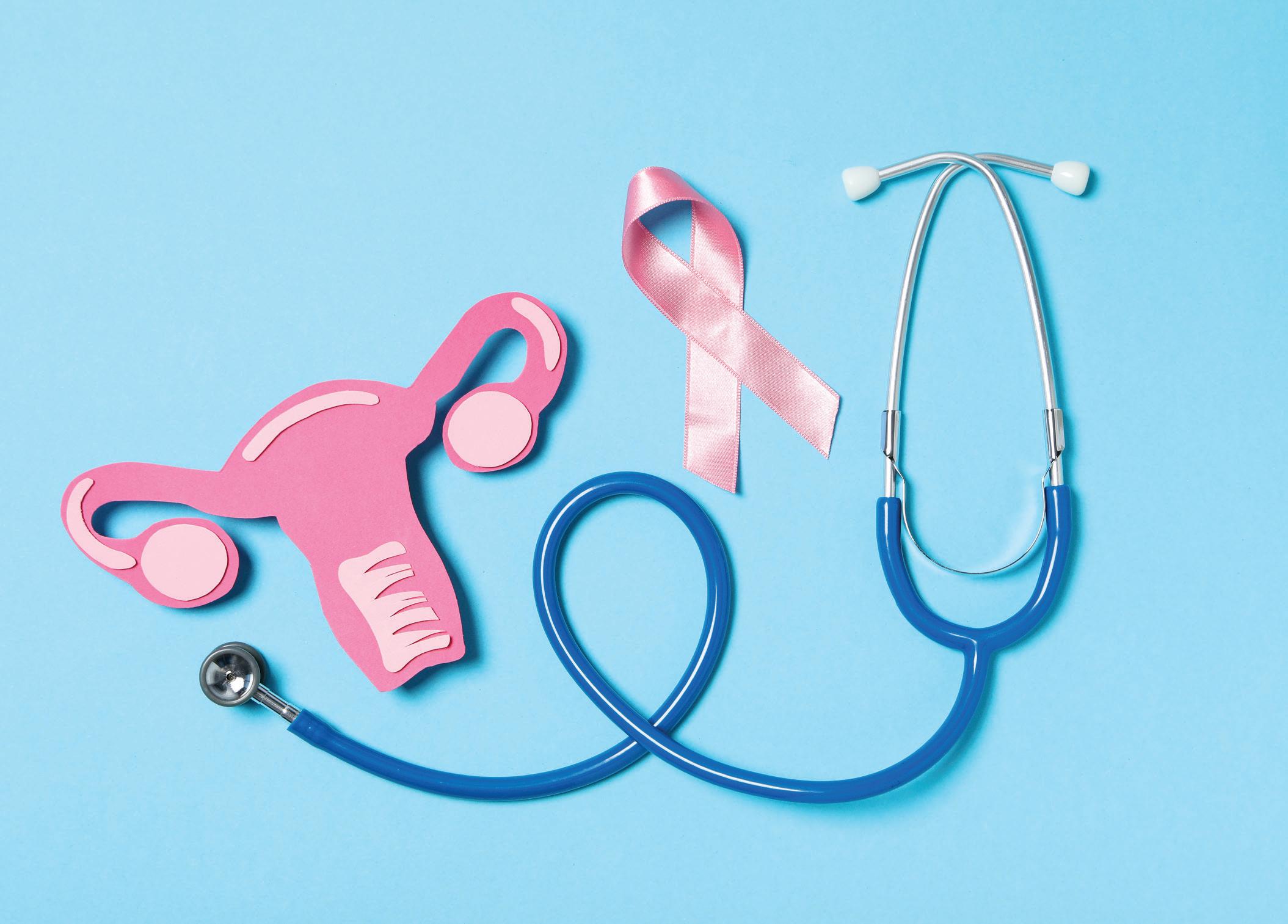
A Pap test is an important screening exam that’s used to check for abnormal cells on the cervix that could indicate early-stage cervical cancer or precancerous cells. The test for human papillomavirus, or HPV, also is important because if you have that virus, it raises your risk for developing cervical cancer. Knowing if you have HPV or not can inform and direct your health care in the future.
Cervical cancer occurs in the lower part of the uterus known as the cervix, which connects the uterus to the vagina. The most common types of cervical cancer are squamous cell carcinoma - occurring in squamous cells, which line the outer part of the cervix - and adenocarcinoma, which develops in glandular cells in the cervical canal.
Anyone with a cervix is at risk of developing cervical cancer. According to the Centers for Disease Control and Prevention, more than 13,000 new cases of cervical cancer are diagnosed each year in the U.S. Hispanic women have the highest rates of developing the disease, while African American women are at greatest risk of dying from this cancer.
Additional risk factors for cervical cancer include:
• MULTIPLE SEXUAL PARTNERS
• SEXUAL ACTIVITY AT A YOUNG AGE
• SEXUALLY TRANSMITTED INFECTIONS
• WEAKENED IMMUNE SYSTEM
• SMOKING
Numerous strains of HPV infection play a role in causing almost all cervical cancer. Many women’s immune systems combat HPV, preventing the virus from causing cancer. However, some women are more susceptible to cervical cancer as HPV lives in their bodies for years and aids in the emergence of cancer cells.
Historically, women often have visited with a gynecologist or other health specialist annually to have a Pap test, which involves collecting and examining cells from the cervix. If the test finds abnormal cells, additional tests to look further for precancerous or cancerous cells may be recommended.
More recently, HPV testing has been recommended for women - along with the Pap exam - as it can provide useful information about a person's risk for cervical cancer. This is particularly important since most women will not experience any signs of cervical cancer in early stages.
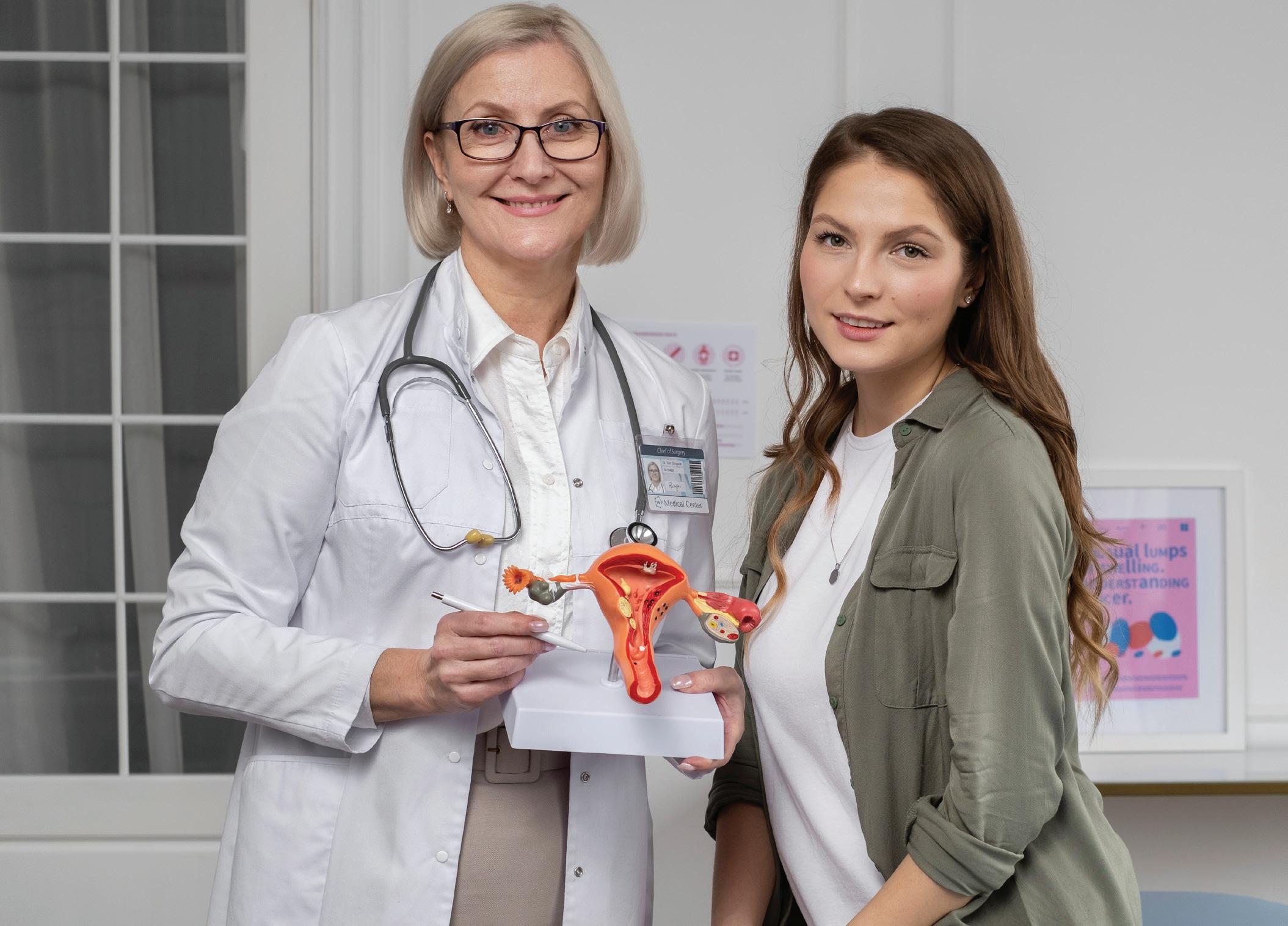
An HPV test can be done at the same time as a Pap test and uses the same cell sample collected for the Pap test. The HPV test doesn’t tell you whether you have cancer. Instead, it detects the presence of HPV in your body. If you have HPV - particularly the two common subtypes that are most closely linked to cervical cancer - then you are at higher risk for developing cervical cancer. Knowing that, you and your health care team can decide how often you need Pap tests and other tests to monitor your condition going forward. Not all types of HPV increase the risk of cervical cancer, and if you are found to have an HPV infection, it is important to note that it does not mean you will develop cervical cancer
The HPV vaccine protects against the types of HPV that are most likely to cause cervical cancer. The vaccine works better when given to girls starting at age 9, but it has now been approved for women up to 45. Depending upon your risk factors, if you have not previously received the vaccine, it may be valuable for you.
How often you need a Pap test and an HPV test depends on your individual situation. In general, the recommendation is to have both tests done every five years for women older than 30 who haven’t had an abnormal Pap test in the past. That may shift, however, depending on your medical history, as well as other health issues you may have. Talk with your health care team about the schedule that’s best for you. — Tri Dinh,
and Gynecology, Mayo Clinic, Jacksonville, Florida
M.D., Obstetrics
Mayo Clinic Q & A is an educational resource and doesn’t replace regular medical care. T his Mayo Clinic Q&A represents inquiries this healthcare expert has received from patients. For more information, visit www.mayoclinic.org.

DEAR MAYO CLINIC: I am an African American woman in her 40s. I recently had a well-woman checkup done and was told that I also should have a cardiac workup.
Although I don’t eat as healthy as I should and high blood pressure and cholesterol run in my family, I have not begun menopause yet. Why do I need to be concerned about heart disease now?
ANSWER: Heart disease is the No. 1 cause of death in women in the U.S. Many African American women are not aware of that fact, or that African American women have an even higher risk of dying from heart disease - and at a younger age - than white women, according to the National Heart, Lung and Blood Institute.
Each year, more African American women die from heart disease than breast cancer, lung cancer and strokes combined. But many do not realize the factors that increase their risk of developing heart disease or that they are at increased risk.
African American women face a high burden of negative social determinants of health. Although it may not apply to you specifically, it is valuable to help bring awareness of how issues such as chronic stress related to food insecurity, racism, the wealth gap and socioeconomically disenfranchised communities can prevent some people from living a healthy lifestyle and controlling many heart disease risk factors.

There is an increased awareness in trying to change the narrative on heart health and African American women. It is commendable that your health care professional proactively noted your risk, regardless of whether you are dealing with any of these specific health disparities.
I recommend that all women follow heart disease prevention strategies and consider the American Heart Association’s Life’s Essential 8 to achieve ideal heart health:
• MANAGE BLOOD PRESSURE. Statistics tell us that African Americans have the highest hypertension rates in the world. Untreated, high blood pressure increases the risk of heart attack, stroke and other serious health problems. Given your family history, it is important to have your blood pressure checked regularly.
• CONTROL CHOLESTEROL. While national guidelines for women, in general, recommend screenings for cholesterol at age 45, if you have a known risk for coronary artery disease, screening earlier is appropriate. This can guide any additional tests that may be needed to check for specific areas of concern related to the heart’s function.

• REDUCE BLOOD SUGAR. Diabetes is a significant heart disease risk factor for African Americans. At least annually, your blood glucose level should be checked. If you do have diabetes, be proactive with your management.
• GET ACTIVE. African American women are the least physically active group of women in the U.S. Embrace being physically active if you can. Guidelines recommend 150 minutes of vigorous activity a week, which can be challenging for many women. You can start small. Just keep moving.
• EAT BETTER. Familiarize yourself with the Mediterranean diet. It was noted that heart disease is not as common in Mediterranean countries as it is in the U.S. Numerous studies have confirmed that the Mediterranean diet helps prevent heart disease and stroke. Plant-based foods, such as whole grains, vegetables, legumes, fruits, nuts, seeds, herbs and spices, are the foundation of the diet. Olive oil is the main source of added fat. Fish, seafood, dairy and poultry are included in moderation. Red meat and sweets are eaten only occasionally.
• LOSE WEIGHT. Obesity can affect the heart’s ability to pump effectively. Take measures to lose and manage weight to reduce the risk for cardiac conditions.

• STOP SMOKING. One of the best things you can do for your heart is to stop smoking or using smokeless tobacco. Even if you’re not a smoker, be sure to avoid secondhand smoke. Chemicals in tobacco can damage the heart and blood vessels. Cigarette smoke reduces the oxygen in the blood, which increases blood pressure and heart rate because the heart has to work harder to supply enough oxygen to the body and brain.
• GET HEALTHY SLEEP. Insomnia and sleep apnea are linked to high blood pressure and heart disease, and also can increase the risk of stroke. Lack of sleep also can affect weight. For people with diabetes, good sleep habits can help improve blood sugar.
Personally, I always recommend that African American women be diligent in protecting their hearts, and that also includes taking time for themselves. Self-care really does matter, and that includes scheduling a heart health checkup at an earlier age. Make heart health a priority now. - LaPrincess Brewer, M.D., Cardiovascular Medicine, Mayo Clinic, Rochester, Minnesota
Mayo Clinic Q & A is an educational resource and doesn’t replace regular medical care. This Mayo Clinic Q&A represents inquiries this healthcare expert has received from patients. For more information, visit www.mayoclinic.org.

Winter’s relative lack of daylight and sunshine, especially in cloudy or more northerly climates, may make you feel less energetic and more blah than you do in the brighter months. For about five percent of the U.S. population, however, the “winter blues” have a more serious effect on mood and daily functioning - possibly because they have seasonal depression, also known as seasonal affective disorder, or SAD.
SAD is an all-too-appropriate acronym for this subtype of major depressive disorder. Symptoms can include oversleeping, overeating and social withdrawal, along with low mood, low energy and trouble concentrating, and they last nearly five months on average, typically beginning in late fall or early winter and progressively worsening until they begin to fade in spring or summer. Even though SAD’s seasonality sets it apart from other types of depression, it is considered
a major mental health disorder due to its duration, its annual recurrence and the effect of its symptoms on daily quality of life.
Unfortunately, there’s no definitive explanation for what causes SAD. Some research suggests that people with SAD may have disrupted levels of serotonin, a brain chemical that regulates mood, and melatonin, a hormone that helps maintain the normal sleep-wake cycle. These disruptions would make it hard to adjust to seasonal changes in day length, explaining why SAD is more common and more severe in states where the sun rises the latest and sets the earliest in winter. While there’s evidence that nutrition can play a role in the treatment of mental health conditions, is this also true for SAD?
Conventional wisdom from the early days of SAD-awareness in the 1990s was that people with SAD should eat a highcarbohydrate diet to increase levels of serotonin. However, a 2020 systematic




review found that while people with SAD experience stronger cravings for carbohydraterich foods, randomized controlled trials have found that actually eating more carbs doesn’t ease symptoms.
What about vitamin D, the “sunshine vitamin”?
While low vitamin D levels have been associated with increased risk of depression, this review cites a study that found that people randomly assigned to take a high dose of vitamin D for 12 weeks during the winter experienced no significant differences in depressive symptoms compared with participants assigned to take a placebo. A study looking at the effects of supplemental vitamin B12 also failed to find that it helped with SAD symptoms.
“While there absolutely is research showing correlations with nutrients and mental health, there are many limitations to this research, and people can often find themselves relying on expensive supplements without addressing the root cause,” says Anafer Barrera, a Coloradobased registered dietitian and Certified Intuitive Eating Counselor with experience in mental health. “When symptoms persist due to a lack

of effect from the nutrient, people can often end up feeling worse, like there must be something really wrong with them if X supplement didn’t cure their blues.”
Barrera says the priority for someone with SAD should be to make sure they’re eating enough. “When anxiety and depression rise, food can become intolerable or just a hassle. The best thing to do is to find out how you can navigate these barriers to make sure you’re getting enough calories, protein, carbs, fat and fiber to fuel your brain and body on a physical level.”
She recommends that people struggling with SAD meet themselves where they’re at in terms of how much time and energy they have to focus on nutrition. “Ideally, we’re getting a good variety of food, with a healthy dose of it made at home from fresh ingredients,” she says, with an important caveat: “Sometimes you desperately need convenience foods, simple recipes like cereal and milk, fruit and yogurt, quesadillas and avocado, or the classic boxed pasta and jarred pasta sauce to get your needs met and tackle the more important things in life.”
Barrera says while food can provide a sense of control, responsibility and thus empowerment in our lives, which can be good for mental health - that sense of control can also be a major factor in the development of an eating disorder.
That 2020 review found that people with SAD are more likely than people without SAD to eat just because food is there - even if they aren’t hungry or are already full - including eating significantly larger dinners and snacking more in the evening. More alarmingly, people with SAD are at increased risk of engaging in emotional eating, binge eating and other disordered eating behaviors, including those related to bulimia nervosa.
Because there’s a shortage of high-quality research on the role of nutrition in managing SAD symptomsin part because of lack of clarity on SAD’s underlying causes - don’t treat nutrition as an alternative to seeking mental health support. “Regardless of the cause, nutrition will likely only be a fraction of the solution,” Barrera says.
According to the National Institute of Mental Health, while psychotherapy and antidepressant medications are two treatment options, for those seeking a lifestyle-based approach, another primary treatment for SAD is light therapy - sitting in front of a special bright light box for 30-45 minutes a day - to try to make up for some of the sunlight missing in winter.
Reprinted with permission from Environmental Nutrition, a monthly publication of Belvoir Media Group, LLC. 800-8295384. www.EnvironmentalNutrition.com.

Q:
I struggle with thick skin on my heels all year long. But in the winter, they crack and get quite painful. Why does that happen and what can I do to help prevent it?
A:Calluses are especially common on the heels, because they take a lot of pressure as we walk. That pressure, combined with skin that’s often dry, makes us susceptible to a common and potentially debilitating condition called cracked heels.
Calluses form as a result of friction. Sometimes it’s caused by the way your foot strikes the floor, and sometimes it’s due to loose shoes, which cause your feet to move around and rub against the shoe. With all of that friction, the body protects itself by forming thicker skin on the heels.
When callused skin is very dry, it tends to crack. If you picture a piece of dry, brittle bread, you can imagine that it would crack open if you stepped on it.
Winter is a common time to develop cracked heels. Here are steps to take now to protect your heels:
• Keep your feet covered. Wear socks and cushioned shoes when you’re up and about.
• Moisturize feet regularly. Use thick moisturizers with emollients - a mixture of water and oil, the kind that’s so thick you have to scoop it out of a jar.

• Cover your feet after moisturizing, so you don’t slip and fall.
• Treat calluses immediately. Use a cream that contains the ingredient urea twice a day, and only on the calluses. Urea helps thin them and break them down.
• Use a pumice stone. If urea cream isn’t helping, soak your feet and afterward rub callused heels with a pumice stone to sand off the calluses.
• Check feet for calluses regularly. They pose higher risk if you have diabetes, which can cause nerve damage that keeps you from feeling an injury, or peripheral artery disease, which can impair the circulation in your feet.
Cracked heels can usually be treated at home, with similar measures as you do for prevention. But you need to be sure that the affected areas are calluses and not plantar warts or psoriasis, which would require different treatment.
If there is any pain, redness, pus or swelling, contact your doctor or podiatrist. It could indicate an infection - an urgent health matter for anyone, especially if you have diabetes or PAD. To treat the infection, your doctor will prescribe a topical or oral antibiotic. Then y ou have to return two weeks later, to make sure you’re healing correctly and the pain is going away.
The doctor may also remove the callus with special tools such as scalpels and power sanders made for the feet.
Howard LeWine, M.D., is an internist at Brigham and Women’s Hospital in Boston and assistant professor at Harvard Medical School. For additional consumer health information, please visit www.health.harvard.edu.


