9 minute read
On the Sunny Side of Science Noah Bussell
from Flux
On the Sunny Side of Science
BY NOAH BUSSELL
Advertisement
While the lamps along the Charles Bridge and the vibrant colors of the aurora borealis exemplify an ethereal yet romantic quality of light, to a scientist, light is also immensely practical. From photosynthesis to drug design, interactions between matter and light are responsible for the progression of many organic processes, our ability to elucidate the structures involved, and these structures’ significant contributions to the mechanisms of chemical action at work.
The relationship between light, structure, and energy is one of the most fundamental and widely utilized concepts in science—and science fiction for that matter. In Jean Luc Goddard’s dystopian film Alphaville, where science and logic are the law of the land, the “laws’’ are E=mc2 and E=hν. The latter serves as the scientific statement that quantifies light’s energy as well as the fundamental equation underlying spectroscopy. While current developments in light-matter research are highly unlikely to plunge us into the technocratic society of Goddard’s film, absent of emotion and free thought, they instead have immense potential to (and currently are working to) usher us into a healthier and more sustainable future.
Even historically, advances in our ability to harness and manipulate light have been linked to the advancement of science. Due to the strong relationship between the structure and function of organic systems, obtaining detailed information about the structure of biomolecules by manipulating and measuring light allows for advances in
fundamental research and health. In 19521953, the English biochemists James Watson and Francis Crick were attempting to uncover the structure of DNA via model building while similar efforts coupled with the chemical analysis of Nobel Laureate Linus Pauling—who inaccurately proposed a triple helix structure—did much of the same. Nonetheless, it was an image procured by Rosalind Franklin, dubbed Photograph 51, captured via the interaction of X-rays with DNA molecules that led to the fundamental breakthrough. Light provided the answer. Watson and Crick’s postulate in their seminal 1953 Nature paper that “the specific pairing we have postulated immediately suggests a possible copying mechanism for the genetic material” was precinct, and the discovery has revolutionized the field of biochemistry. Their specific contributions to the project were perhaps exaggerated, however. This was in accordance with the sexist environments of the Cavendish and King’s College laboratories where Watson and Crick and Rosalind Franklin were respectively conducting their research.2
As our knowledge of light-matter interactions, and how to manipulate them, continues to advance, scientists are progressing in their ability to make more precise measurements and study increasingly complex systems. Notably, in 2020, a scientific group spread across several European technical institutions published their research on the morphology and energetics of a protein that plays a critical role in energy transport within living systems. Their PNAS paper outlines the mechanism

Figure 1: Scene from Jean Luc Goddard’s Alphaville portraying the “laws” of the dystopian technocracy, Source: Boake1 .

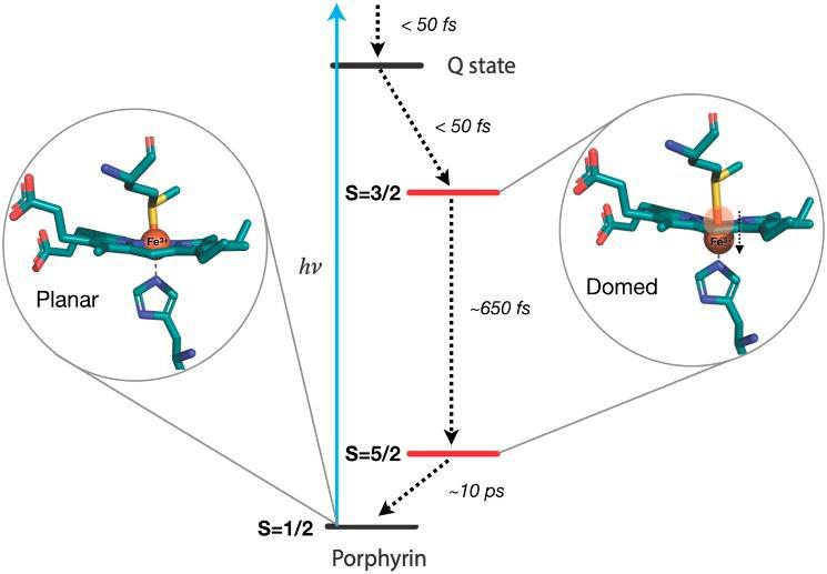
Figure 2: (a) Chemical structure of cytochrome c; (b) Structural changes of ferric cytochrome c via excitation and relaxation through spin states, Source: Bacellar, et al.3
“In the case of cytochrome c, the researchers discovered that the protein takes up a domed configuration very soon after excitation by light. As cytochrome c undergoes a change in energy it accordingly shifts to this new conformation.” “Ultrafast spectroscopy is an especially useful experimental approach as it can distinguish the various shapes and sizes of molecules on the femtosecond time scale (1 millionth of 1 billionth of a second).”
by which this protein, known as ferric cytochrome c, “relaxes” and simultaneously shifts in shape after light excitation.3 “Ferric” simply refers to the fact that the iron atom in the protein has +3 charge as opposed to the +2 charge of ferrous iron, a subtle yet important distinction in this study.
Cytochrome c is an integral component in a collection of proteins that compose the electron transport chain (ETC), a biochemical network essential to life. Cytochrome c specifically channels electrons and their energy to proteins downstream in the chain and towards the production of adenosine triphosphate (ATP), commonly referred to as an energetic “currency” in biochemistry.4
Alongside this role in the ETC, cytochrome c can also increase in energy by absorbing light and then subsequently relax in energy. This relaxation occurs through “spin states” in the cytochrome c system; electrons can be either in a spin-up or spindown state, and collectively, these spins can arrange parallel or antiparallel. This alignment in turn affects the energy of the cytochrome c molecule.
In certain spin states, molecules have high energy, affecting molecular bonds and geometry. To think of it one way, atoms are attracted to some particles in their environment and repulsed by others. Naturally, they try to move to locations where they are most attracted—when the atoms find a sweet spot where there are a lot of attractive interactions and few repulsive ones, the molecule is in a state of low energy. By exciting cytochrome c proteins with light, electrons are pushed to higher energy states. Then, to compensate for the simultaneous change in bond lengths as atoms move around, the molecule needs to change geometry.
In the case of cytochrome c, the researchers discovered that the protein takes up a domed configuration very soon after light excitation. As cytochrome c undergoes a change in energy, it accordingly shifts to this new conformation. While doming had been shown to play a key role in the respiratory function of ferrous iron proteins, this finding challenges a prior hypothesis that ferric cytochrome proteins (such as the cytochrome c protein in this study) did not undergo doming. Thus, this study provokes inquiries into the biochemical relevance of doming and future studies of ferric cytochrome proteins.
To probe the precise structure of these molecules, researchers had to use ultrafast spectroscopy techniques, physical techniques that measure the absorption and emission of light to garner information about chemical systems on miniscule time scales. Ultrafast spectroscopy is an especially useful experimental approach as it can distinguish the various shapes and siz-
es of molecules on the femtosecond time scale (1 millionth of 1 billionth of a second).
One type of ultrafast spectroscopy used in this study is known as X-ray emission spectroscopy (XES). By “pumping” cytochrome c systems with high energy X-ray light, electrons that are held very closely to atomic nuclei can be ripped away, leaving behind a desirable, low-energy vacancy for another electron to fill.5 This process outputs a spectroscopic signal known as a “Kβ-line,” which is a stream of light that marks this vacancy filling and details where the electrons that filled these gaps came from. While many of these signatures exist, Kβ-lines are specifically significant in the cytochrome c complex. These Kβ-lines are a signature of electrons (here, referred to as 3p electrons) coming from regions where there exists interaction with other electrons that are responsible for most of the molecule’s spin behavior (3d electrons in the cytochrome c system). As such, Kβlines are effectively a marker of both 3p and 3d electrons.
In the context of this study, in a low spin state (more antiparallel alignment), it is easier for 3p electrons to produce Kβlines. This results from the enhanced ability of 3d electrons in low spin states to push 3p electrons away towards the low-energy vacancies. Thus, the Kβ signals that chemists measure are stronger in low-spin states. Using this principle, the group discovered that ferric cytochrome c relaxes to low energy states after light excitation by progressing through these spin states, as opposed to other thermal mechanisms that were previously postulated.
To validate their findings, the group used an array of other techniques as well. In fact, there is currently an abundance of research into light-matter interactions, using a whole array of these spectroscopic techniques, with applications ranging from fundamental scientific inquiry to developments in health and sustainable energy.
For instance, Professor Naomi Ginsberg’s laboratory at the University of California, Berkeley in collaboration with scientists from Lawrence Berkeley National Laboratory and the Kavli Energy NanoSciences Institute have shown how proteins can serve as a scaffold for the arrangement of chromophore (color-related) complexes. This was accomplished in part through the use of ultrafast spectroscopic techniques. These protein-chromophore complexes mimic the photosynthetic process for light harvesting, an active area of research relevant to the reduction of atmospheric carbon dioxide concentrations. By modifying the lengths and configurations of organic
“linker” molecules, the proximity and interaction of chromophores, proteins, and their solvent environments are altered with the potential for the development of more efficient light harvesting systems.7 This means that just as plants harvest sunlight
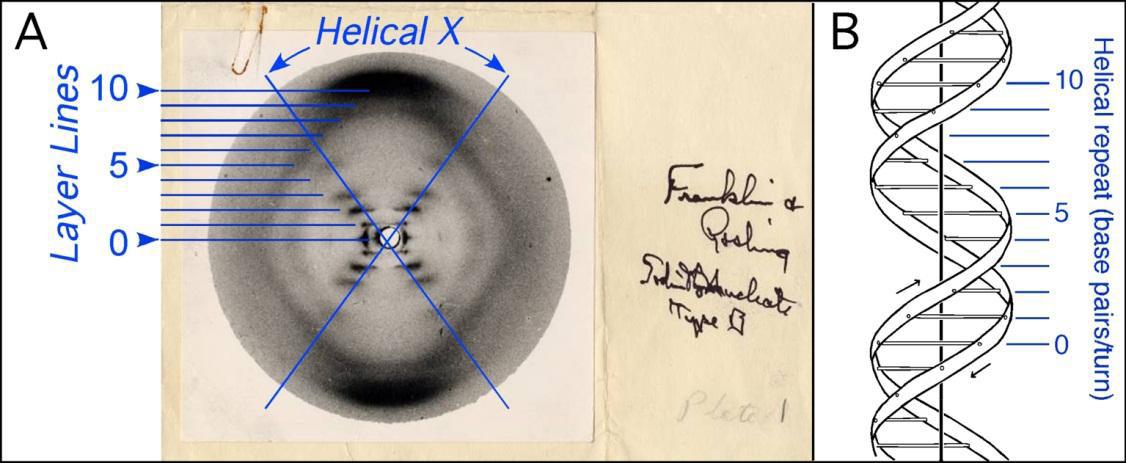
Figure 3: Photograph 51 with analysis, Source: Ho & Carter6 .
to synthesize chemicals that are useful for them, scientists are tweaking the process so that we can channel the sun’s energy to the production of renewable energy.
Today, spectroscopy techniques are rapidly advancing, and physical chemists are pushing the field further forward. Dynamic information can be collected on a range of systems on the femtosecond time scale, allowing researchers to not only visualize the structures of molecules, but also the ways in which charge, heat, and energy flow through. Light is even being used to activate drugs, enabling highly targeted, and consequently safer, cancer therapies.8
In addition to its plentiful practicality and the fundamental role it plays in nature, there is something more to light that equations and spectroscopy can’t quite capture. The physicist Erwin Schrödinger once wrote in his book, Mind and Matter, that “the sensation of colour cannot be accounted for by the physicist’s objective picture of light-waves.”9 Regardless, as scientists and philosophers excite organic systems with light to unearth their structure and artists excite our emotions with it, light’s practical and sentimental nature continues to advance and enrich the world around us.
REFERENCES
1. Boake, T. M. (2008). Alphaville discussion questions. Arch 443/646:
Architecture and Film Fall 2008. http://www.tboake.com/443_ alphaville_f08.html 2. Maddox, B. (2003). Rosalind
Franklin: The dark lady of DNA.
HarperCollins. 3. Bacellar, C., Kinschel, D., Mancini, G.
F., Ingle, R. A., Rouxel, J., Cannelli,
O., Cirelli, C., Knopp, G., Szlachetko,
J., Lima, F. A., Menzi, S., Pamfilidis,
G., Kubicek, K., Khakhulin, D.,
Gawelda, W., Rodriguez-Fernandez,
A., Biednov, M., Bressler, C., Arrell,
C. A., … Chergui, M. (2020). Spin cascade and doming in ferric hemes:
Femtosecond X-ray absorption and
X-ray emission studies. Proceedings of the National Academy of Sciences, 117(36), 21914–21920. https://doi. org/10.1073/pnas.2009490117 4. Rosamond, R., Keeler, A., & Diaz, J. (n.d.). The role of cytochrome c in the electron transport chain. Chemistry
Texas A&M University. https://www. chem.tamu.edu/rgroup/marcetta/ chem362/HW/2019%20Student%20
Posters/The%20Role%20of%20
Cytochrome%20c%20in%20the%20
Electron%20Transport%20Chain.pdf 5. Bergmann, U., & Glatzel, P. (2009).
X-ray emission spectroscopy.
Photosynthesis Research, 102(2), 255. https://doi.org/10.1007/s11120-0099483-6 6. Ho, P. S., & Carter, M. (2011). DNA structure: Alphabet soup for the cellular soul. In H. Seligmann (Ed.),
DNA replication. IntechOpen. https:// doi.org/10.5772/18536 7. Delor, M., Dai, J., Roberts, T. D.,
Rogers, J. R., Hamed, S. M., Neaton,
J. B., Geissler, P. L., Francis, M. B., &
Ginsberg, N. S. (2018). Exploiting chromophore–protein interactions through linker engineering to tune photoinduced dynamics in a biomimetic light-harvesting platform.
Journal of the American Chemical
Society, 140(20), 6278–6287. https:// doi.org/10.1021/jacs.7b13598 8. Liu, J., Chen, H., Ma, L., He, Z.,
Wang, D., Liu, Y., Lin, Q., Zhang,
T., Gray, N., Kaniskan, H. Ü., Jin,
J., & Wei, W. (n.d.). Light-induced control of protein destruction by opto-PROTAC. Science Advances, 6(8). https://doi.org/10.1126/sciadv. aay5154 9. Schrödinger, E. (1958). Mind and matter. University Press.


