15 minute read
Corrupted Clocks: How Parasites Utilize the Circadian Rhythm (Dr. Filipa Rijo-Ferreira
from Flux
Corrupted Clocks:
How Parasites Utilize the Circadian Rhythm
Advertisement
INTERVIEW WITH DR. FILIPA RIJO-FERREIRA
BY: CAROLINE KIM, ANJULI NIYOGI, MICHAEL XIONG, ESTHER LIM
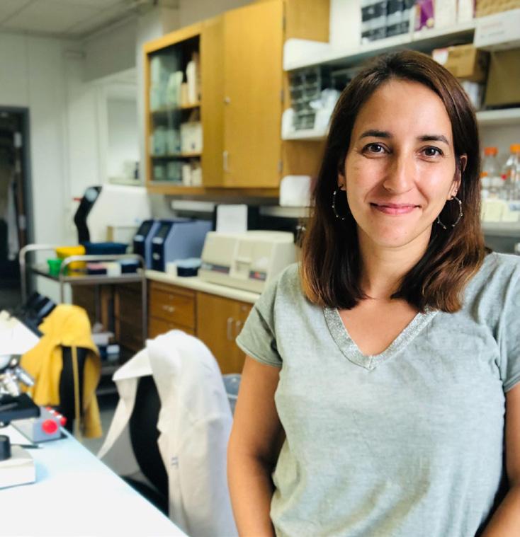

Dr. Filipa Rijo-Ferreira is an assistant professor of Infectious Diseases and Vaccinology at Berkeley Public Health. Much of her research focuses on the relationship between circadian rhythms—the cycles by which organisms keep track of time—and parasitic infections. Some of her groundbreaking work includes the discovery of circadian rhythms within parasites, such as those responsible for malaria and sleeping sickness, as well as the ability of parasites to influence the circadian rhythms of their hosts. She hopes that these innovations can help inspire ways for scientists to advance medication and treatment of infectious diseases.
BSJ: What led you to pursue research in infectious diseases, specifically regarding the circadian rhythms of parasites?
FRF: When I was in college, I started becoming very interested in the world of microbiology and how much the immune system of the host and pathogens, viruses, bacteria, or parasites have co-evolved and learned to be together. Parasites, for example, are able to maintain long chronic infections; they do not ‘want’ to kill the host, whilst the host is trying to get rid of the parasite. This really intrigued me. Then I started being interested in neuroscience. I worked in a lab studying a parasite-related condition called sleeping sickness, but there was so little known about the neuroscience side. Sleeping sickness parasites lead to the disruption of sleep, and in particular, patients are not sleeping more but rather, are sleeping at the wrong time of the day, causing increased daytime instead of nighttime sleep. When I went to graduate school, I could not let go of this question that I had been so intrigued about. I wanted to do a project combining both parasitology and circadian rhythms, and that is how it all started.
BSJ: What is sleeping sickness and what are the symptoms associated with it? Which areas of the world does it predominantly affect?
FRF: Sleeping sickness is caused by a parasite called Trypanosoma brucei. The fly that transmits the infection is endemic in Africa, especially sub-Saharan Africa. The disease itself has an early blood-stage and is then able to cross the blood-brain barrier and reach the brain. That is when, traditionally, most of the complicated sleep disruption occurs. But there are many different disruptions beyond sleep; there is also dysregulation of rhythmic temperature. Usually, temperature profiles get disrupted, hormone secretion gets disrupted, and some very complex neuropsychological disorders come with it. It creates inflammation in the brain and eventually leads to a coma. It is almost 100% lethal if left untreated and unfortunately, the treatments that we have available are also quite toxic. It is believed that about 5% of people that have taken the treatment die of the treatment itself. However, the treatments are still worth taking because the other route is very devastating. There is a lot of further research that needs to be done on the pharmaceutical side, but also on understanding how the disease happens.
BSJ: One of the most important observations made was a phase advance in the circadian rhythms of the mice. What is a phase advance, and what are its consequences?
FRF: A rhythm usually has a period, a phase, and an amplitude. It is an oscillation: the phase is the peak of that rhythm, and advance means that it is happening earlier. If we think about sleep having its phase in the night, if there is now a phase advance, that means that sleep is happening earlier. That is most likely what is causing the traditional disruption in humans, in which people experience daytime sleepiness and nighttime insomnia.
BSJ: It is known that body temperature and circadian rhythms go hand in hand. How did you measure altered body temperature regulation, and what did you discover?
FRF: When you are active, your temperature rises. Temperature is higher during the day for us and lowers at night when we sleep. It is the opposite for mice that are nocturnal. We can implant a telemetry device in the mouse abdomen that is recording the temperature fluctuation into a receiver in real-time. We were able to find that in the first few days upon infection, there was a high
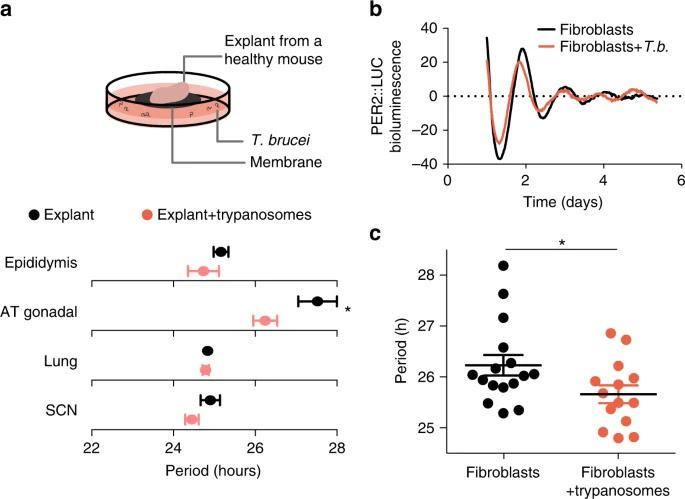
Figure 1: Trypanosoma brucei shortens the period of cells. Infection by T. brucei reduced the length of daily rhythms in mouse tissue samples from a number of different organs. A bioluminescent reporter of the circadian cycle (PERIOD2::LUCIFERASE) showed that circadian rhythms of cells in the presence of parasites drifted away from those of noninfected cells over the course of several days. On average, the period of infected fibroblast cells was shorter than that of healthy fibroblasts.
increase in temperature like a fever. Then, the body of a mouse that is infected is able to re-adjust the temperature rhythms, becoming normal again. As they get sick, they will have a hypothermia-like phenotype, so the temperature also drops. There is dysregulation in both the rhythmicity and even on the absolute levels of the temperature.
BSJ: You measured the locomotor activity of infected mice, which is driven by the suprachiasmatic nucleus (SCN). Could you describe the SCN and its relationship to circadian rhythms?
FRF: The SCN is a very small nucleus in the hypothalamus of mice and humans. You have direct connections with the eyes through the optic chiasm. That is how our circadian rhythms are linked to environmental changes from light to dark. We can get our rhythms synchronized with the environment directly because the eye is connected to the SCN. This nucleus is made of 20,000 neurons and they are all coupled to each other; they keep a rhythm because we have a molecular clock inside these neurons and virtually all cells of our body. We have some genes that are transcription factors and transcribe many other genes, among them their own repressors. This creates a loop: the activators transcribe the repressors, which then repress the activators from initiating transcription of them and other genes. This is what is going on in the SCN. It is keeping time, receiving information from light, and then sending out this information through the body to keep other organs and cells synchronized with one another. That is through direct connections like neuronal connections, and also humoral connections like hormones.
BSJ: What is the function of the Suramin drug? How did you use it in your study to analyze the mechanism of action of T. brucei?
FRF: Suramin is one drug that is used clinically to treat sleeping sickness. It acts only on the blood stage of the parasite, because this drug in particular is not able to cross the bloodbrain barrier. As I mentioned, there are two phases. If you are able to detect the infection early on, you can give this drug. It kills the parasite. It is believed to act on the metabolic pathways of the parasite. For this study, we took advantage of the drug not crossing the bloodbrain barrier but being able to save mice from dying early on, to better study and mimic human infection. In the mouse model, mice do not have a long lasting chronic infection, possibly because they
did not co-evolve with this parasite. Once we sub-cure the mouse by killing parasites and decreasing the inflammation in the periphery, the mouse survives for much longer. Only the few parasites that have already crossed to the brain are going to remain. So we are able to mimic low parasitemia (low levels of parasites in the blood) and the long-term behavior changes that we see later on in humans.
BSJ: Your study establishes that the T. brucei parasite causes disruptions in the host’s circadian rhythm and sleep patterns. How do the findings of this research change the way sleep sickness and sleep disorders are viewed? What do you think are the next steps for this research?
FRF: Before, we were aware of sleep disorders and circadian disorders, but most of them were commonly associated with genetic variations or mutations from the host itself. Now, this is a new perspective of a parasite or pathogen that is somehow able to modulate and disrupt the clock. This is new information that we did not have before because now it is not just about natural variation in the human population, but specific pathogens that are able to modulate the clock. In regards to what comes next, that would be trying to find out how the parasite is doing all of that, as it is important to understanding sleeping sickness. One of the things we found is that the parasite appears to be secreting a molecule, but we do not know what it is. If we are able to identify this molecule, it could be useful. If it is not pathogenic, we could use the molecule itself to modulate the clock of humans. Imagine you are going through jet lag, or if you have a phase delay disorder or delayed sleep pattern, and you want to bring your phase earlier, you may benefit from using that molecule.
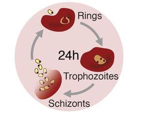
Figure 2: The life-cycle of the malaria parasite Plasmodium chabaudi.
Within the bloodstream, P. chabaudi reproduces by infecting red blood cells. The parasite begins as an immature trophozoite within the red blood cell, appearing as a ring. Next, the parasite matures into a schizont, which replicates and bursts from the cell, ready to begin another round of infection.
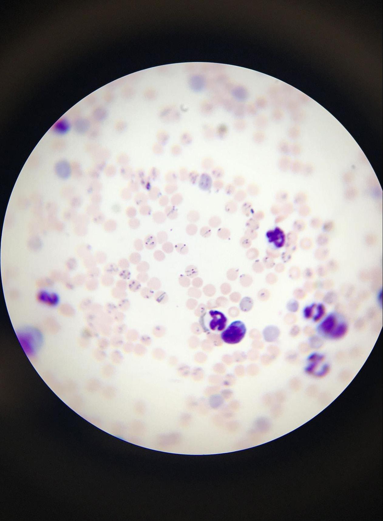
Figure 3: Image of red blood cells of mice infected with Plasmodium chabaudi, a parasite of the Plasmodium genus responsible for causing
malaria. Small purple-dyed dots within the red blood cells refer to the parasite; large dark purple circles refer to immune cells.
BSJ: In one of your studies, you infected mice with the parasite Plasmodium chabaudi, one of the many species in the Plasmodium genus that is responsible for causing malaria. What were the main goals going into this study, and how were they shaped by your previous research with T. brucei?
FRF: Once we became aware that parasites and circadian clocks can interact with one another, and learned that parasites themselves like T. brucei may have their own clock, we came to this question about malaria. We were very intrigued about the rhythmic fevers that malaria is known for. Even before we knew it was caused by a parasite, we already knew about these fevers that would come and go at certain times of the day. This periodicity actually has a multiple of 24 hours, which is a circadian characteristic, as you can imagine. It was very difficult to distinguish if the fevers in malaria were a consequence of the host circadian rhythm, or if it was more complex than that, and actually had a contribution from both the host and the parasite. And that was the main goal: Can we test if there is a contribution beyond the circadian rhythm of the host—is
BSJ: You discuss how the intrinsic clock of the malarial parasite is the underlying mechanism for malarial rhythmic fevers. What cellular processes occurred during these fevers?
FRF: There is this amazing phenomenon. These unicellular parasites invade red blood cells, but they do it coordinately. All the other parasites are also invading red blood cells of their own. They are multiplying and replicating inside the red blood cell, and then they burst the red blood cell. When they do this, they burst all millions of red blood cells at once, exposing newly formed parasites and all the metabolic waste that the parasite generated while living in these red blood cells. That triggers a massive immune response which causes fevers.
BSJ: How is the cell cycle and gene expression of a parasite linked to its intrinsic clock?
FRF: That is actually a very difficult question. We know from other organisms, like mammalian cells, that the circadian clock and the cell cycle are usually independent, but they do regulate one another. So they may also be linked to the parasite. But we also know from the mammalian system that cells that have grown enough to cover an entire petri dish, called confluent cells, do not divide anymore. There is no more space to create new ones, so they
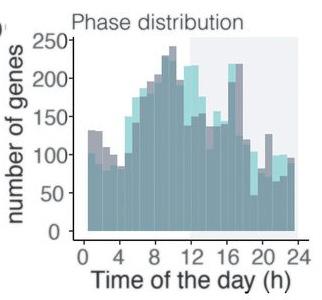
Figure 4: Graph depicts the distribution of gene expression of the Plas-
modium chabaudi parasite throughout a 24-hour cycle. The green distribution represents the parasites of mice that received food spread out during the day, whereas the blue distribution represents parasites that followed a regular feeding schedule (control). The distribution concludes how host cues, such as feeding patterns, did not significantly influence gene expression of the parasite. do not divide anymore, but they still have circadian rhythms. One does not drive the other, even though they might interact with one another. In the malaria research that we did, we were looking into blood, where there is definitely also replication of the parasite. We are now going to have to dissociate these two, and try to understand which genes are cell cycle-related, and which genes were circadian rhythm-related. Most likely, we need to go to a stage of the parasite that has no division.
BSJ: In the process of proving that the malarial parasite has rhythms of its own, you had to control for several confounding factors, like food and light exposure. How could these factors have influenced the circadian rhythms you observed?
FRF: The way to prove that there is an intrinsic clock is through removal of all of the possible confounders. For example, light is known to be an important biological cue that many organisms respond to, and our team was quite worried as to whether or not this was also important for malaria parasites. We tested this by eliminating any light in the parasite’s environment, and instead, having complete darkness to ensure that the parasite’s rhythms were generated on its own. After we established light was not a required cue, we also thought of considering food, because there have been studies showing that parasites sense when the food is being consumed and align the timing at which they burst red blood cells. If you gave the food at the wrong time of the day, then the parasites would burst at an abnormal time. What we did to solve this was to introduce automated feeders to the mice to abolish a normal feeding rhythm. We gave food evenly distributed throughout the day so that the mice were eating small portions of food every hour and a half, which is very different from what they normally do, which is eating a lot in 12 hours and not eating at all in the other 12 hours. Even after removing the food confounder, we still observed a consistent rhythm in the parasite, and so we were able to conclude that food is a potential factor that the parasites sense, but is not completely necessary for their synchronization.
BSJ: The models that you developed led you to the conclusion that malaria parasites use host activity to synchronize their internal clocks. Why might the malaria parasite benefit from having its own circadian rhythm rather than simply taking cues from its host?
FRF: This is really the big “why” question: Why would parasites go through the effort of having an intrinsic clock if they can just rely on the host? I think the most important aspect to consider in answering this question is understanding the difference between responding and anticipating. When we think of plants, for example, there is a very obvious need for sunlight to conduct photosynthesis. If plants had a circadian clock that allowed them to anticipate when light was coming, they could be more prepared to photosynthesize by making all of the relevant proteins earlier, even halfway through the night because, in a few hours, the sun will come out. If they had just waited for the sun, on the other hand, they wouldn’t be prepared to photosynthesize as effectively until much later in the day. We can think about this reasoning in the context
of parasites. By predicting when nutrients are going to be available, they could time when to replicate. Our immune system is also highly rhythmic—the number of lymphocytes in circulation is very different from night and day. Sometimes they are in the lymph nodes and other times, they are in the blood. If the parasites can predict these and other changes in their environment, they might be at an advantage.
BSJ: How do the results of your study give us more insight into how we can best treat these parasitic infections?
FRF: This new research on circadian rhythms and parasitic diseases may open doors for further research on when the best time to treat these infections is. When we need to take medication, we are often not informed of when it is the best time to take it because we have not studied it. Regarding the sleeping sickness parasite, our team found that the parasite is more susceptible to the suramin drug at specific times of the day. Just from this information, we can improve the outcome of the disease by administering the drug when the parasite is much more vulnerable. There is a window of opportunity to kill parasites more effectively, and this is something that is important to consider in treating parasitic infections.
BSJ: What is the most rewarding aspect of your work? What do you love most about being a scientist?
FRF: For me, the most rewarding part is the teamwork that goes into trying to discover what is unknown. And this kind of work cannot be done alone—being able to work with students, postdocs, and other faculty members has made my work very fun and rewarding. In this particular malaria paper that we have just discussed, I had a very strong collaboration with a mathematician who helped us model it. And that is just one example of how interdisciplinary science can be and how fun it is.
REFERENCES
1. Headshot: [Photograph of Filipa Rijo-Ferreira]. Image reprinted with permission. 2. Figure 1: Rijo-Ferreira, F., Carvalho, T., Afonso, C., Sanches-Vaz,
M., Costa, R. M., Figueiredo, L. M., & Takahashi, J. S. (2018).
Sleeping sickness is a circadian disorder. Nature Communications, 9(1). https://doi.org/10.1038/s41467-017-02484-2 3. Figures 2 and 4: Rijo-Ferreira, F., Acosta-Rodriguez, V. A.,
Abel, J. H., Kornblum, I., Bento, I., Kilaru, G., Klerman, E. B.,
Mota, M. M., & Takahashi, J. S. (2020). The malaria parasite has an intrinsic clock. Science, 368(6492), 746–753. https://doi. org/10.1126/science.aba2658 4. Figure 3: [Microscope image]. Rijo-Ferreira Lab. Image reprinted with permission.


