Radiography and Painting
I. TEXT
 Elisabeth Ravaud F
Elisabeth Ravaud

 Elisabeth Ravaud F
Elisabeth Ravaud
Ever since Louis Pasteur was entrusted with the first Chair of Physical Chemistry as Applied to the Arts at the Ecole des Beaux-Arts1, more than 150 years ago, an understanding of works of art has required a detailed analysis of their material makeup. Indeed, this involves—in a manner comparable to deciphering an archive—examining the techniques of the artist or artisan, the materials he or she has chosen and their provenance, and the evolution of these over time. Most of the methods used to decipher these works—beyond the emotions they provoke, their beauty, their profound meaning, and their occasional ability to surprise—derive from other fields, technological or scientific in nature. Thus, today, laboratories specializing in the analysis of cultural property routinely employ methods developed for industrial and medical applications or for use in fields far removed from art, such as astrophysics.
Radiography, the subject of the present publication, to a certain extent breaks this rule of technology transfer, since this method, as the author points out in her first chapter, was applied at quite an early stage within the field of cultural property, at the same moment that its well-known and highly successful medical applications were underway. Hence Wilhelm Conrad Roentgen’s use of this method in examining ancient coins bonded by corrosion and, from 1896, its use by his disciples to identify pigments in colour boxes or the presence of retouching on the surfaces of paintings. The relationship between the imaging techniques used in the fields of medicine and heritage has deepened over time, each discipline obeying the imperative to respect both the object of study and the diagnostic procedure to be performed. The author herself, in the trajectory of her career and in her own diagnostic approach, perfectly exemplifies the scientific link between medicine and the heritage sciences. With a doctorate in medicine and a specialism in radiology and medical imaging, she worked for several years in a hospital setting before joining, in 1993, the Laboratoire de Recherche des Musées de France, which in 19982 became the Centre de Recherche et de Restauration des Musées de France (C2RMF), where she is today responsible for the scientific study of easel paintings.
The C2RMF, a department granted national authority under the Ministry of Culture, is responsible for implementing the official policies of the Museums of France in regard to the research, preventive conservation, and restoration of objects in these institutions’ collections. Located at the crossroads of science and art, the C2RMF encompasses an examination and analysis laboratory, restoration studios, and two documentation centres open to the public in Paris (within the complex of the Louvre Palace) and Versailles (at the Petite Ecurie du Roi). A multidisciplinary team of 157 colleagues brings diverse expertise to bear upon the study and conservation of cultural property, uniting the humanities and social sciences with the experimental sciences: curators, restorers, archivists, librarians, physicists, chemists, geologists,
1. In 1860.
2. Upon its merger with the Service de Restauration des Musées de France.
photographers, radiologists, and IT specialists (or computer scientist?). Their research focuses on the materials, manufacturing processes, and dating of objects, as well as an analysis of the effects of environmental factors and an evaluation of conservation and restoration methods. The studios of the C2RMF receive a wide variety of objects, including easel paintings, sculptures, archaeological artefacts, furniture and craft objects, textiles, graphic works, and contemporary art. Some of the greatest masterpieces of French collections are analysed and restored here by in-house and freelance restorers. C2RMF professionals also provide museum directors with methodological and technical advice concerning the protection, conservation, and restoration of their collections.
It is this very context that enabled Elisabeth Ravaud to study an extensive number of easel paintings3, particularly masterpieces of the Italian Renaissance by Leonardo da Vinci and Titian but also works by seventeenth-century French masters such as Nicolas Poussin. In order to understand a painter’s technique, or to be able to authenticate, confirm, or deny an attribution or detect past interventions, Ravaud employs a wide range of techniques: of primary importance is the visual and critical analysis of the work itself, observed directly or through optical microscopy, and of the various types of radiographic images, from infrared to X-ray. Her joint expertise in radiology and art history, her experience of more than 27 years, and her continual exchanges with curators have allowed her to develop a method of interpreting radiography that she terms the radiological semiology of paintings. She teaches us that the X-ray of a painting—a two-dimensional projection of a three-dimensional object—cannot be interpreted on its own, as we often assume. To fully understand it we must simultaneously play with technical parameters, rely on the repertoire of radiological signs that the author has built up over time, and create a dialogue between different sources of information that include alternative types of images, material analysis, archival resources, and consultation with art historians, in a multidisciplinary approach characterized by moderation and prudence. We must applaud here the approach of an author who, unsatisfied with studies of the greatest masterpieces of painting, took the initiative to formulate and publish her own method first within the framework of a doctoral thesis and then in the present publication, assuredly an invaluable resource for researchers in the heritage sciences.
Devoting today a book to a scientific technique, the radiology of paintings, which has existed since 1895 and which has been in widespread use since the middle of the 20th century for the study of paintings, may seem surprising. My scientific and professional career explain this choice.
My initial training was in the medical field with a specialisation in medical imaging and more particularly in radiodiagnosis. Mastering various medical imaging techniques and the interpretation of radiographic images, I worked as a radiologist for over ten years in different Parisian university hospital radiology departments. In parallel with this medical activity, for pleasure, I studied Art History at university. Having to complete a practical training course as part of my Art History degree, my lecturers encouraged me to apply to the Laboratoire de Recherche des Musées de France (Musées de France research laboratory). This was the start of an adventure which was to lead me to undertake this work, first in form of a PhD thesis in Art History, at Paris Sorbonne University.
During the course, I was confronted for the first time with radiographies of paintings. At that time, I was able to realise that the origin of numerous images visible on these radiographies, was unknown. Of course, I had studied in detail the works of the pioneers in the field such as Madeleine Hours. I was able to understand the spectacular radiographic discoveries of hidden compositions and unexpected pentimenti. On the other hand, I very quickly noted that systematic scientific studies on the radiography of paintings remained very rare, in total contrast with the vast scientific literature which I had encountered on medical radiology. My immediate conviction was that the radiographies of paintings were being insufficiently exploited. This observation pushed me to join the Laboratoire de Recherche des Musées de France in 1993. As soon as I arrived, I began a more thorough study of the existing literature but numerous questions on radiographic signs remained unanswered and I could find no work on radiological semiology such as the hundreds or even thousands which are available in the field of medical radiology.
In the light of this, it seemed natural to me to produce a radiological semiology for artists’ paintings. This research work was perfectly consistent with my activities and my position at the Laboratoire de Recherche des Musées de France, which in 1999 became the Centre de Recherche et de Restauration des Musées de France (C2RMF). It is important to underline that my activities at the C2RMF give me access material of rare quality since the laboratory undertakes work for all the French museums and possesses records of over 10,000 radiographies of paintings. This work would have been impossible without access to these exceptional iconographic records.
Although the work of identification, inventory, description and comprehension of radiological signs had already been completed in the medical field, this work remained to be done in the field of radiography of paintings. In order to undertake this work, I had to breakdown the interpretation reflexes which I had acquired during my years of practice in medical radiology and analyse the interpretation process by adapting it to this new application field.
In this book, the first chapter provides a history of the radiography of paintings, attempting to understand the birth and development of this new discipline as well as the limits reached. This is followed by a description of the main aspects of the methodology used during this research. The following three chapters cover successively each of the constituent parts of a painting (pictural layer, preparatory phases, support) by describing the radiological images observed, and explaining the formation mechanism of these images and giving a brief context placing the item under study in its historic perspective. The following chapter describes the images of the main deteriorations and restorations observed on the paintings.
The final chapter illustrates the benefits of radiography in the history of art, the history of pictural practices and conservation restoration, incorporating all the data resulting from such examinations with the other information available on a painting, based on concrete examples encountered during 25 years spent at the Centre de Recherche et de Restauration des Musées de France.
It was decided to accept certain redundancies between chapters such that each chapter can be read without the need to refer to other parts of the book.
The end of the 19th and beginning of the 20th centuries saw the emergence and development of a new analytical tool for artists’ paintings : radiography, an imaging technique based on the action of X-photons on matter. In parallel with the development of radiography of the human body, radiography was deployed for objects and painted works. The first part of this chapter describes the historical particularities of its expansion from the very end of the 19th century to the present day. Based on this observation, the second part of the chapter, reveals the new methodology for the interpretation of X-rays based on image formation mechanisms.
1895-1918 : From the discovery of X-rays to the advent of radiography in museums.
George Johnstone Stoney (1826-1911), an Irish physicist, predicted in 1874 the existence of the electron. Based on calculations, this prediction led to numerous experimental studies using a device called a cathode ray tube. In 1897, an Englishman, Joseph John Thomson (1856-1940), using such equipment, was able to demonstrate the extraction from matter of negatively charged particles. By experimentation, he proved the existence of the electron and thus validated Stoney’s prediction. The atom ceased to be indivisible. This was a major turning point in physics. The “Plum Pudding”, stuffed with raisins which could now be extracted (the electrons), became the accepted model for the structure of matter.
In 1895, the German physicist Wilhem Conrad Röntgen conducted experiments at Würzburg university on the effects of electric charges on rare gases inside a Crookes tube, a sealed glass tube containing a partial vacuum and incorporating a cathode and an anode1. On the evening of 8th November 1895, he related to a journalist from Mac Clure Magazine, that he had observed that a barium platinium cyanide paper placed close to a powered Crookes tube covered with black cardboard, was marked with a black line2. No light came from the covered Crookes tube. Röntgen therefore concluded that the mark observed was the consequence of invisible radiation whose property is a capability for penetrating the glass of the tube and the cardboard. As its nature was as yet unknown to the physicist, the radiation was called “X” rays in line with the terminology used in algebra. He reported his discovery in a preliminary communication submitted on 28th December 1895 to the Würzburg3 Physics and Medicine Society. The first human preserved radiography was his wife’s hand. It revealed the bone skeleton4. Radiology was born.
Röntgen immediately undertook numerous experiments in order to try to understand the origin, the nature and the behaviour of these unknown rays. In the manuscript of his preliminary communication, Röntgen described for example that a square section wooden bar painted on one of its faces with lead-based paint behaves differently according to its orientation relative to the radiation. If the painted face was parallel to the rays, no consequence
1. The name of the physicist has been variously spelled Röntgen or Roentgen.
2. The date of 8th November 1895 was quoted by Röntgen himself in the only interview which he gave to Mac Clure Magazine on 6th April 1896. (a) Boudenot, 2001, p. 197. (b) Pallardy, 1987, p. 29-56.
3. Röntgen, 1895. A more complete publication appeared in the same journal on 9th January 1896.
4. Roentgen biography on internet site: nobelprize.org/prize/physics/1901/ roentgen/biographical.
5. Pallardy, Pallardy, Wackenheim, 1989, p 83. Röntgen sent off-prints to other colleagues throughout Europe including Henri Poincaré, the French mathematician.
6. Glasser, 1934.
7. Barthelemy, Oudin, 1896, p. 150.
8. Communication to the Congrès de Médecine in Nancy, 6th August 1896. Radioscopy is the instantaneous and mobile display of the image produced by X-rays in real time on a fluorescent screen. Radiography provides an image frozen and recorded on a film.
9. Beclere, 1900, p.4, note 9
10. Bridgman, 1964, note 2.
11. Glasser, 1934, p. 348.
12. Bridgman, 1964, note 5.
13. Bridgman, 1964, note 6.
14. Bridgman, 1964, note 7.
15. Wehlte, 1932, p 70-80. The first X-ray machine installed in a German museum in 1924 in Munich had thus to be withdrawn because of the patent. Vanpaemel, 2010.
16. (a) Nahum, 2009, p 458-460. (b) Boudenot, 2001, p 208. Marie Curie was behind the initiative of this mobile radiological unit.
17. Pallardy, Pallardy, Wackenheim, 1989, p. 227.
18. Doctor Rene Ledoux-Lebard became the first professor in radiology in France in 1923. See Paunier, 1995, 1518-20.
19. Goulinat, 1974.
20. Alexandre Dauvilliers, was to become Professor of Physics at the College de France and a member of the Académie des Sciences. As a pupil of Professor Maurice de Broglie, at the end of the war, he worked on the spectral series of X-rays. Boudenot, 2001, p.199.
21. Jean Cailleux was the founder of a highly reputed art gallery at Rue du Faubourg Saint Honoré. Hours, 1980, p. 9.
was observed. If the rays reached the painted surface perpendicularly, a dark shadow appeared on the fluorescent plate.
From his different experiments, Röntgen concluded that the X-rays are distinct from cathode rays. They are absorbed by matter and their absorption varies according to the mass of the atoms, they are diffused by the matter (fluorescent radiation), they affect a photographic plate and they discharge electrically charged bodies.
This discovery was revealed by the Viennese press on 5th January 1896, following a leak by Franz Exner, an Austrian research scientist, to whom Röntgen had sent an off-print of his communication. The news spread like wildfire reaching Germany and Great Britain on 7th January, and New-York on 8th. In France, “Le Petit Parisien” announced the discovery on 10th January 18965. It earned Röntgen the first Nobel prize for physics in 1901.
The development of this technique was exceptionally fast, particularly in the medical field. In the year following Röntgen’s discovery, 49 books and brochures as well as 1044 essays were written in France on the scientific aspects and potential applications of X-rays, of which the majority were medical applications6. Two French doctors, Toussaint Barthélémy and Paul Oudin produced the first X-rays in France. Their research was presented to the Academie des Sciences on 20th January 1896 by Professor Raymond Poincaré together with X-ray prints sent by Röntgen7. During the summer of 1896, they developed a radioscopy process8. One of their boarding school friends, Antoine Béclère was enthusiastic about this technique and its considerable potential in medicine. In 1897, at his own cost, he installed an X-ray machine at Tenon hospital in Paris even before the building had any electricity supply. He began the first course in the world on Medical Radiology as he considered that radiology had become an essential technique for any medical practice. The first book on medical radiology was published in 1897. In 1899, he published an article titled “Radioscopy and radiography in hospitals” which sparked off a fierce debate9. It stated that radioscopy should precede radiography and be practiced by a doctor. He wrote “It is up to the doctor or surgeon, treating a patient, to personally undertake the examination of the patient. Indeed, although it is easy to view the images which appear on the fluorescent screen, it is not so straightforward to interpret them”. At that time, however, everyone claimed to be able to interpret X-rays. The essential nature of a report with its interpretation, signed by a doctor and accompanying the radiographic print, gradually became accepted. It was also recognised that a knowledge of anatomy and pathology are also indispensible in order to achieve a good quality radiographic examination.
In the field of radiology of paintings, development was also very rapid. In 1896, Toepler, one of Röntgen’s friends, X-rayed a box of paints and observed that the metallic pigments behaved differently from the non-metallic pigments10. That same year, König, one of Röntgen’s pupils, performed the first radioscopy of a painting in order to detect retouching11. Subsequently, applications of radiology for the study of paintings were published in different technical journals. In 1897 an article describing the search for a signature on a painting by Dürer using radiography was published in The Electrical Review12 in London. The same article was published the same year in The Electrical Engineer13 in Australia and in the British Journal of Photography14. In 1964, Bridgman published an article “The Amazing Patent on the Radiography of Paintings” which recounts these first studies published in generalist scientific journals as opposed to journals dedicated to heritage. At that time, it was again the immense potential and applications of this physical discovery which thrilled the scientists. In that article, Bridgman tells the story of Alexandre Faber, a radiologist in Weimar. The latter conducted experiments on the absorption of X-rays by different pigments in 1913, then applied for a patent in 1914 for the detection of overpainting on paintings by radiography. This patent, which remained in effect in Germany until 1937, paradoxically had the effect of significantly holding back the development of radiography of paintings in that country15.
On the eve of the First World War, medical radiology was already accepted as a recognised discipline. The French Army Health Service then acquired eighteen military motorised ambulances nicknamed the “little Curies” operating on the front in order to X-ray the wounded without having to transport them16. The distribution of chief radiologist doctors in the eighteen military regions is set out in a document dated 19th February 191617
The X-ray ambulance for the 9th military region in Tours was the site of a fortuitous encounter between four art enthusiasts : Rene Ledoux-Lebard18, an eminent radiologist doctor, chief of the region, Jean-Gabriel Goulinat19, artist, art critic and painting restorer, Alex Dauvilliers20, physicist and Jean Cailleux, painting expert21. Nursing officer Goulinat was recruited by the radiological team to “summarise in drawings the X-rays which the surgeons

volume two • illustrations

Copyright: C2RMF
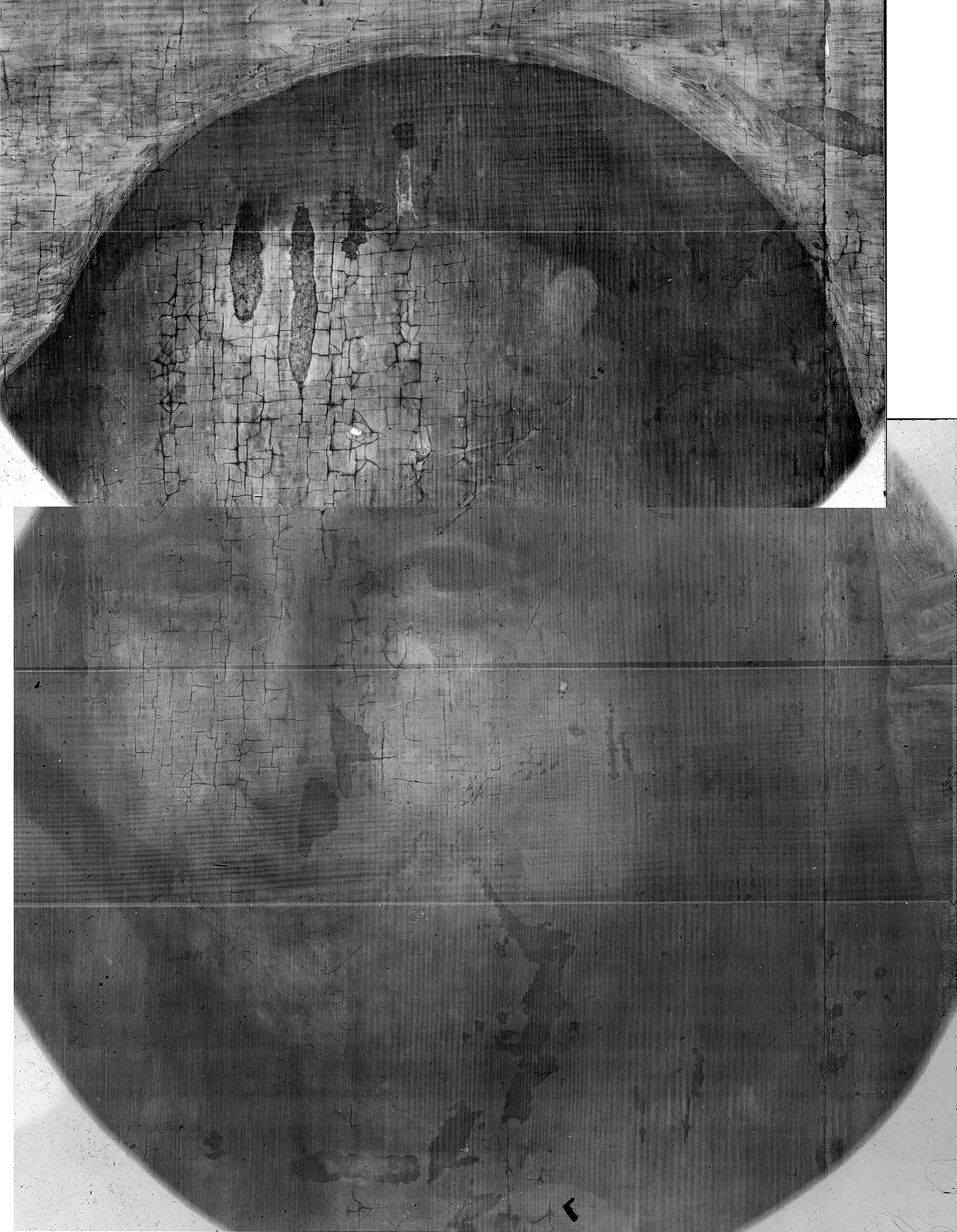
The radiographic contrast is here reversed from the original documents. It corresponds to the current X-rays. Note the differences between the radiograph of the Mona Lisa (Figure 1-3) and that of this copy: the wood is in oak, the flesh tones are rich in lead white and opaque, the face is altered by several paint losses.
Copyright: C2RMF
Copyright: C2RMF
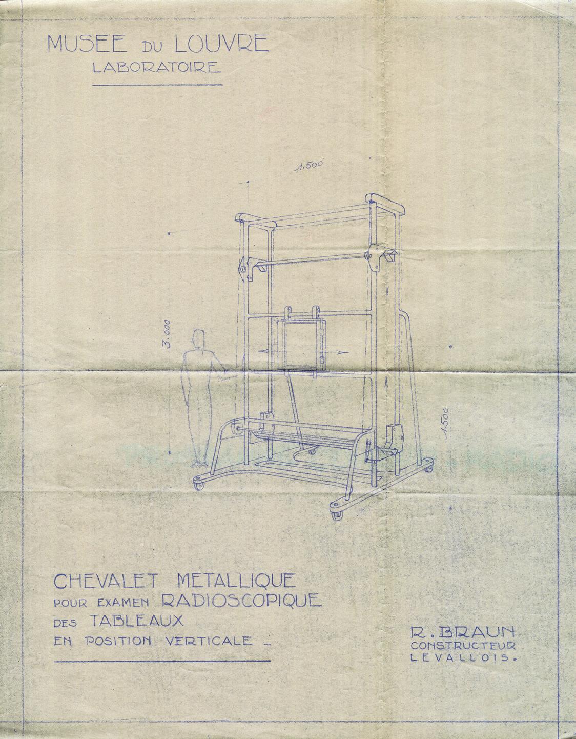

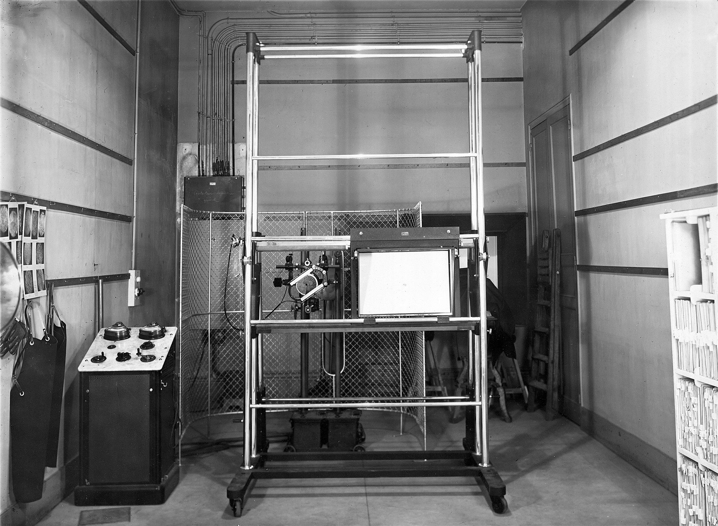
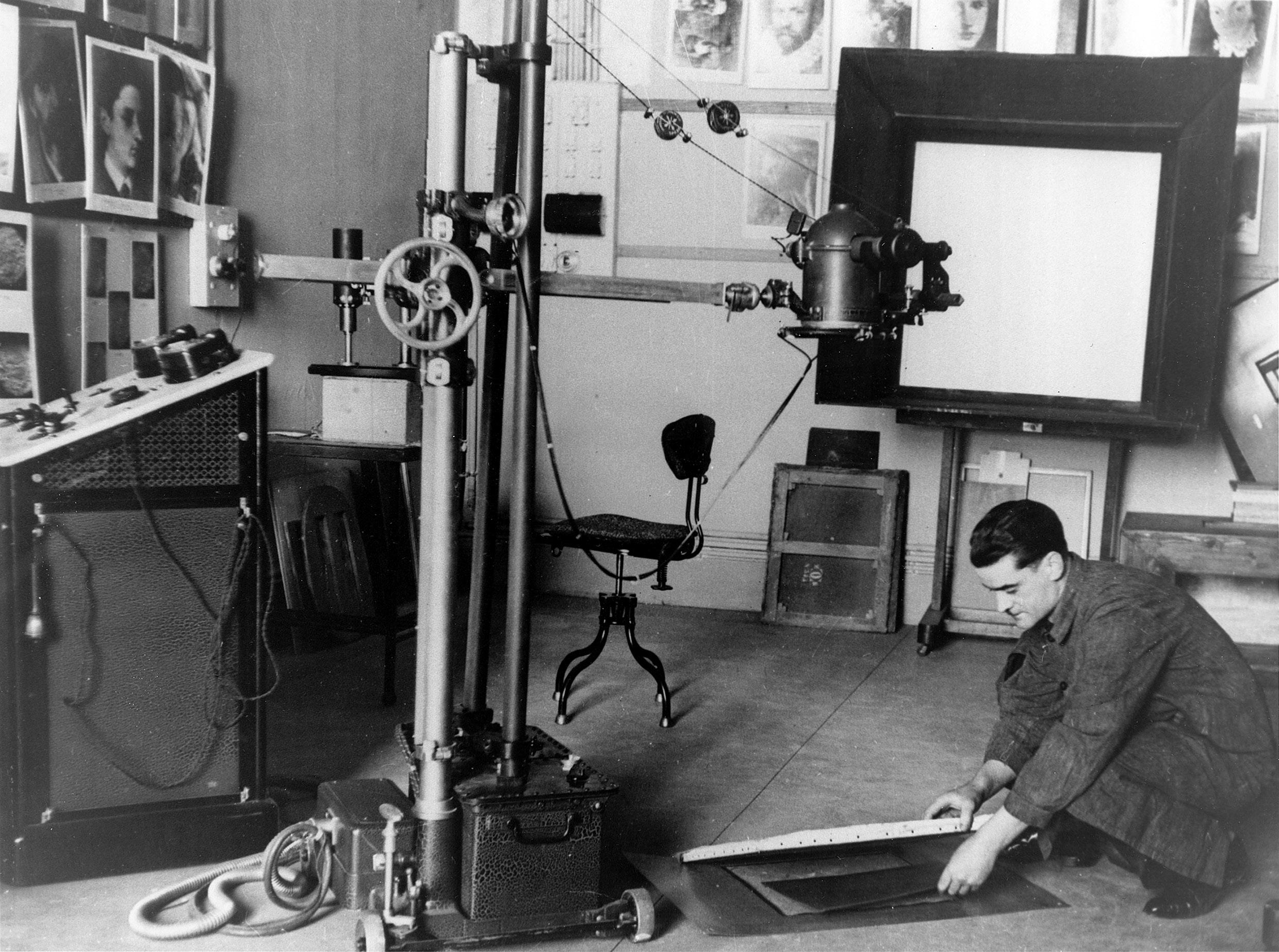
A new metallic armature was planned (a) and achieved (c) allowing a radioscopic examination of paintings in vertical mode. A new X-ray tube was bought shortly after 1952. It was placed on a multi levels support (b).
Copyright: a, b and c: C2RMF
Musée des Beaux-arts, Tours, 2007-2-2
50 cm x 35,5 cm
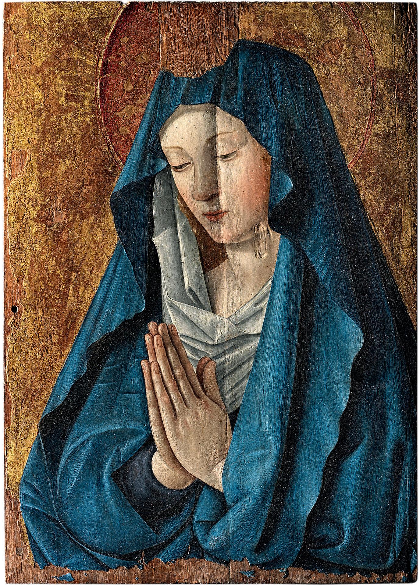
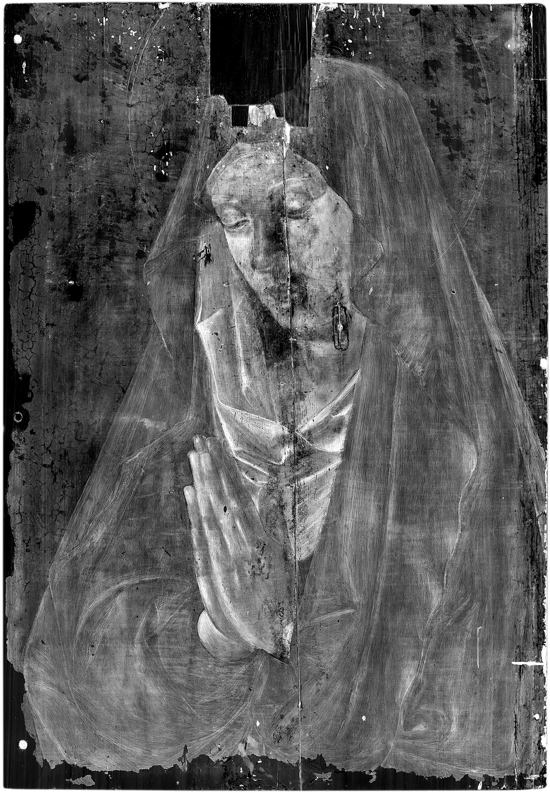
The Virgin at Prayer clearly exhibits a central vertical split enhanced by the raking light photograph (a). The radiograph (b) shows a linear vertical image, slightly sinuous crossing the height of the panel. The radiographic diagnosis of wood split is immediate since it refers to the well known image of a wood split that we can also observe directly on the panel. The top dark square image is a wood inlay.
Copyright: a and b: E. Lambert-C2RMF
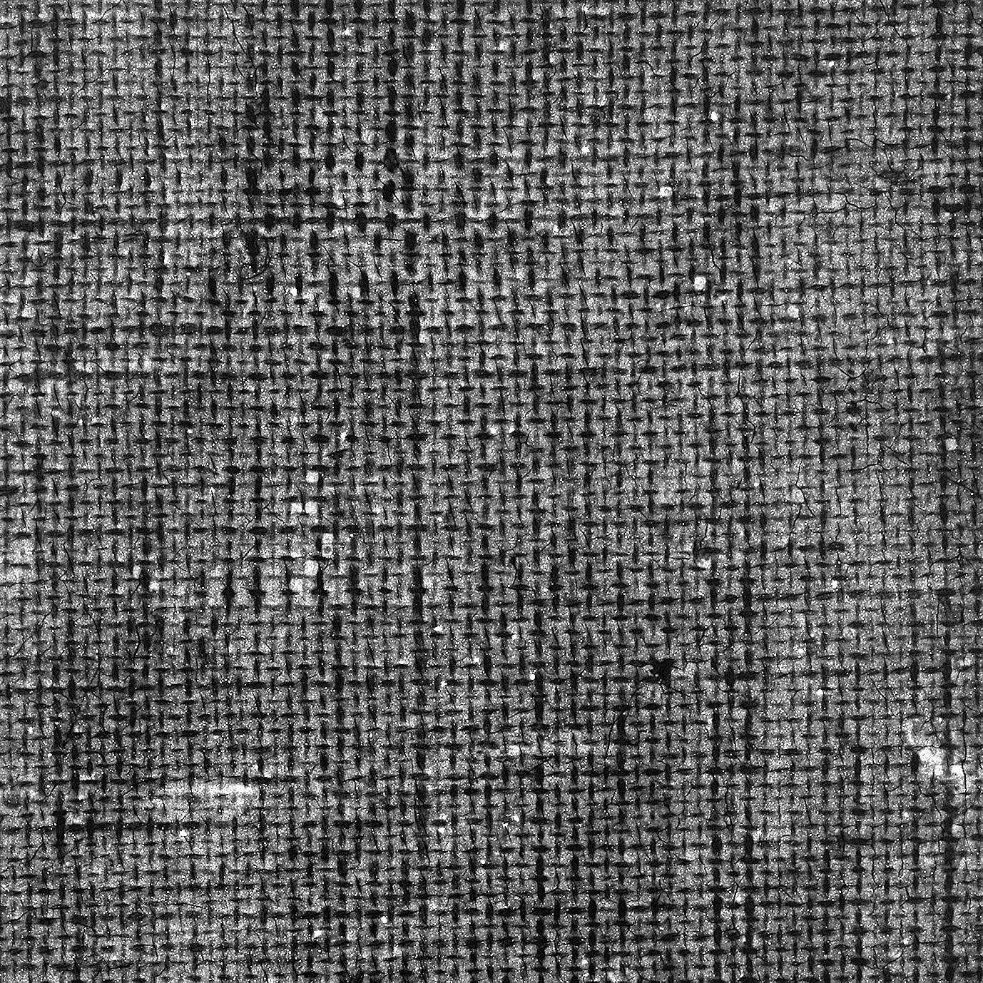
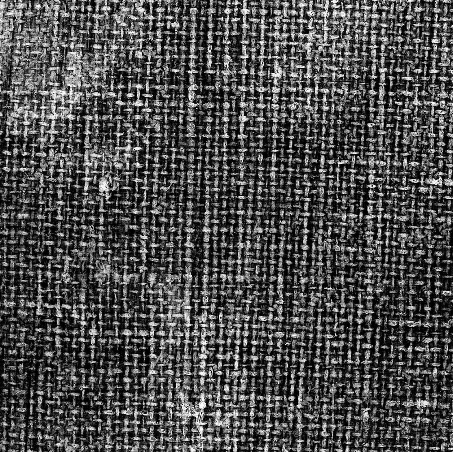
These radiographic details of two paintings by Nicolas Poussin The Triumph of Flora (top) and The Plague of Astod (bottom) in the Louvre, show in both cases an evocative image of canvas. The analysis of the image formation mechanism indicates that these two images correspond however to two different material realities. The image (a) corresponds to the actual existence of a canvas. The image (b) matches on contrar y to the lack of canvas, removed during a transfer.
Copyright: C2RMF
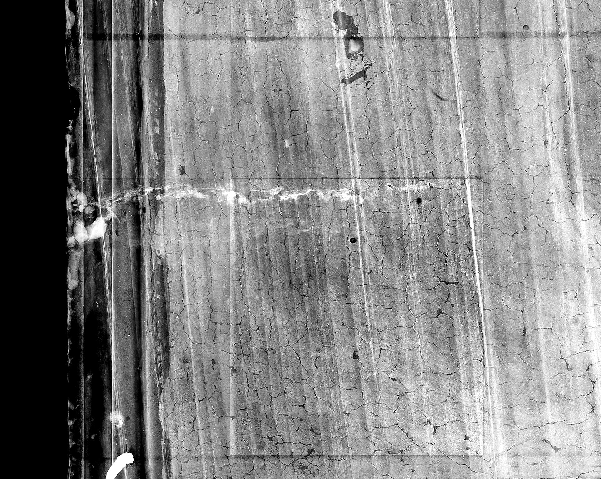
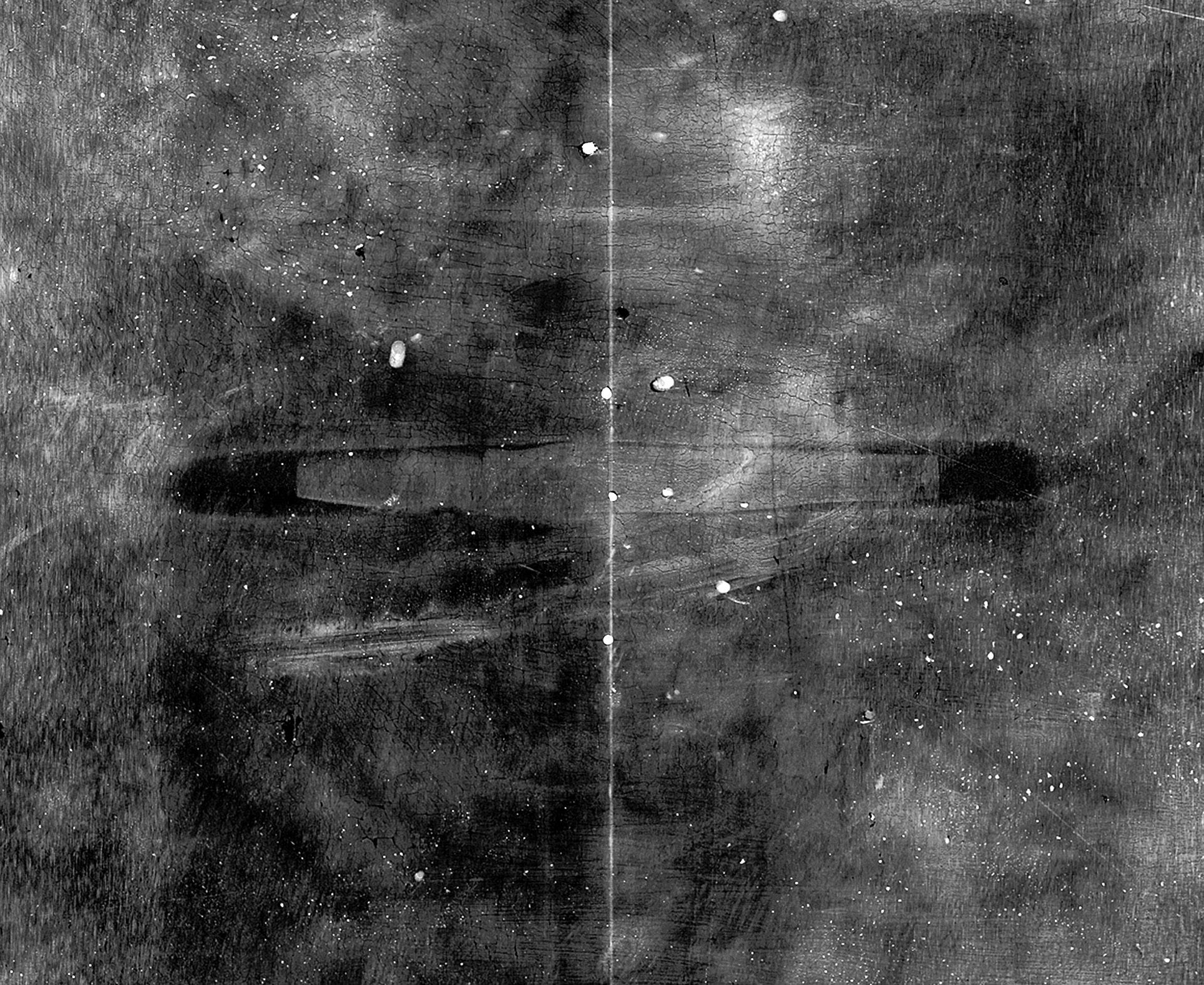
The radiographic detail (a) of the lower part of St. John by Sassetta (Louvre) shows a linear and irregular white image perpendicular to the edge of the panel. This is the image of half a dowel cavity in a thinned panel. By comparison the radiographic detail of The Virgin and Child with Saint Anne by Leonardo da Vinci (Louvre) (b) shows a dowelled joining between two boards in the preserved thickness of the panel.
Copyright: a and b: E. Ravaud-C2RMF

A painting is a flat laminate object in which the support (generally wood or canvas) is covered in succession by a sizing and a preparation or ground, intended to improve the flatness of the surface to be painted, the paint layer applied in one or more layers and a protective layer, usually a varnish.
Copyright: E. Ravaud
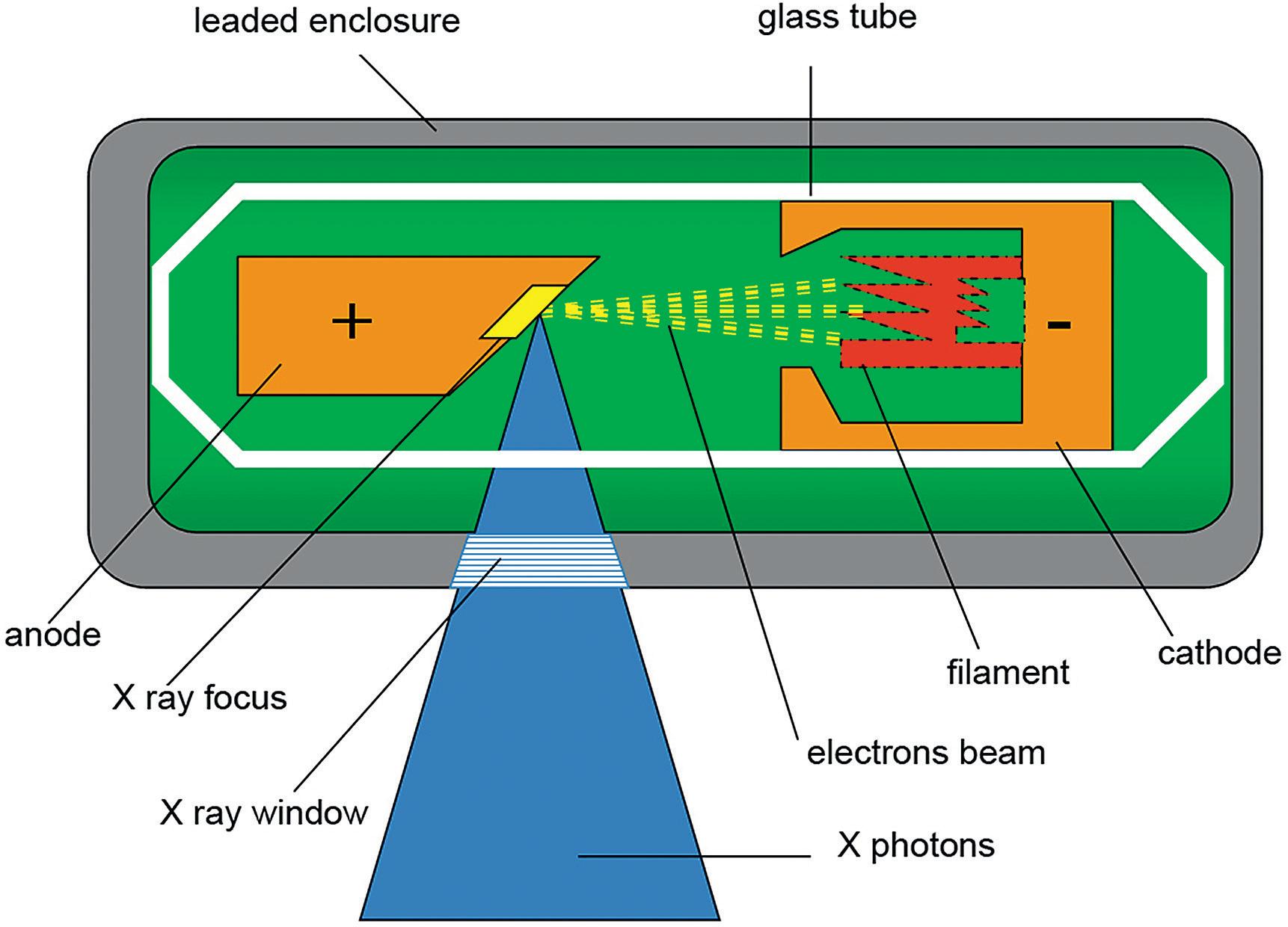
X-rays is produced by an electron beam from a cathode, impinging an anode, usually of metal. The generated X-rays are transmitted through a focusing window. Each X-photon propagates in a straight line but the overall beam forms a cone of increasing diameter with distance.
Copyright: T. Borel-C2RMF
Vinci
Portrait of a woman so called La Belle Ferronnière
Musée du Louvre, Paris, INV 778
63,5 cm x 44,5 cm
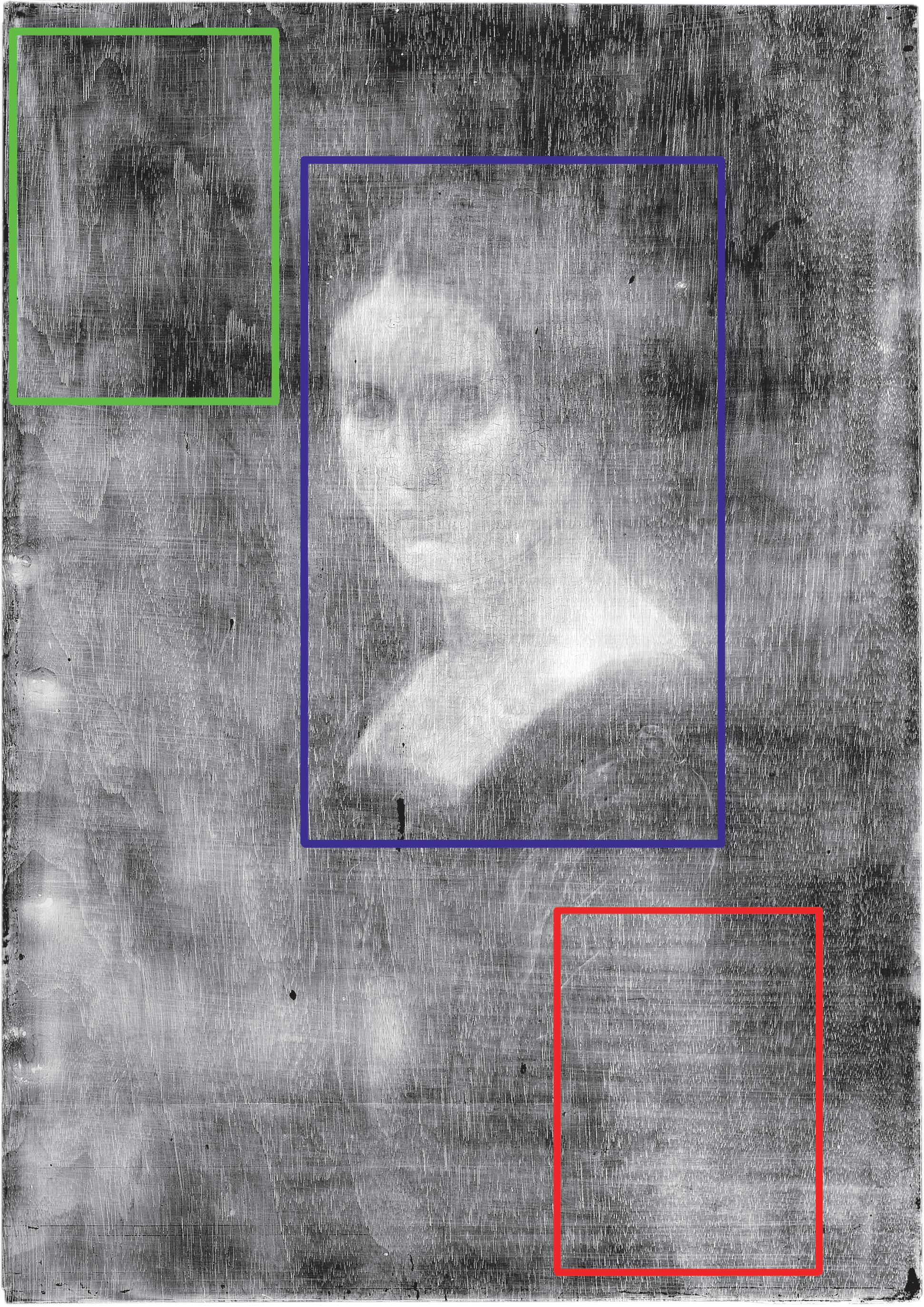
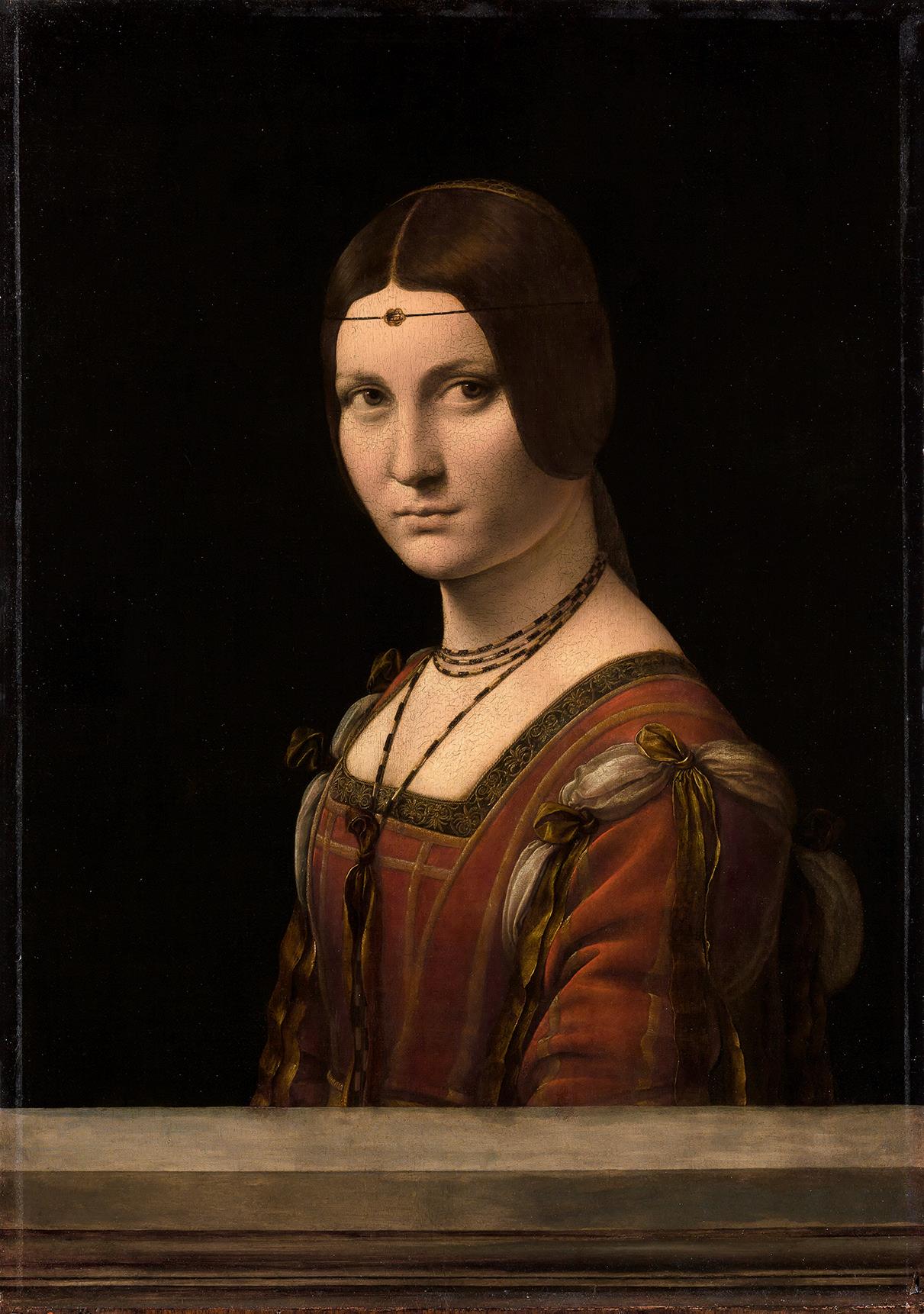
In the painting La Belle Ferronnière by Leonardo da Vinci (a), the radiograph superimposes the images generated by the wood panel (b-green rectangle: twisting images), the ground (b- red rectangle: horizontal striated image) and the paint layer (b-blue rectangle with the woman).
Copyright: a: T. Clot-C2RMF, b: E. Ravaud-C2RMF
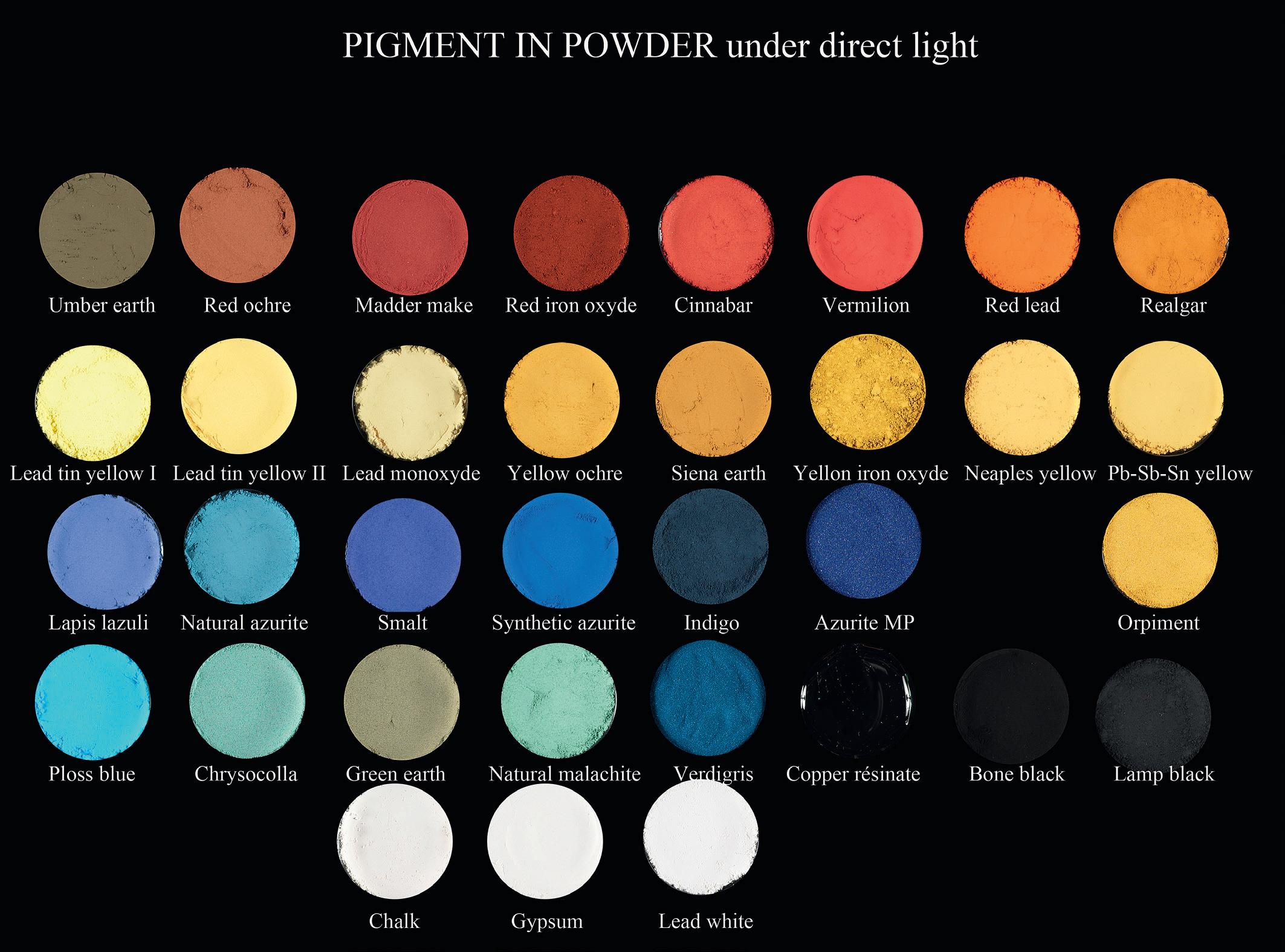
Copyright: E. Ravaud/ A. Paounov-C2RMF
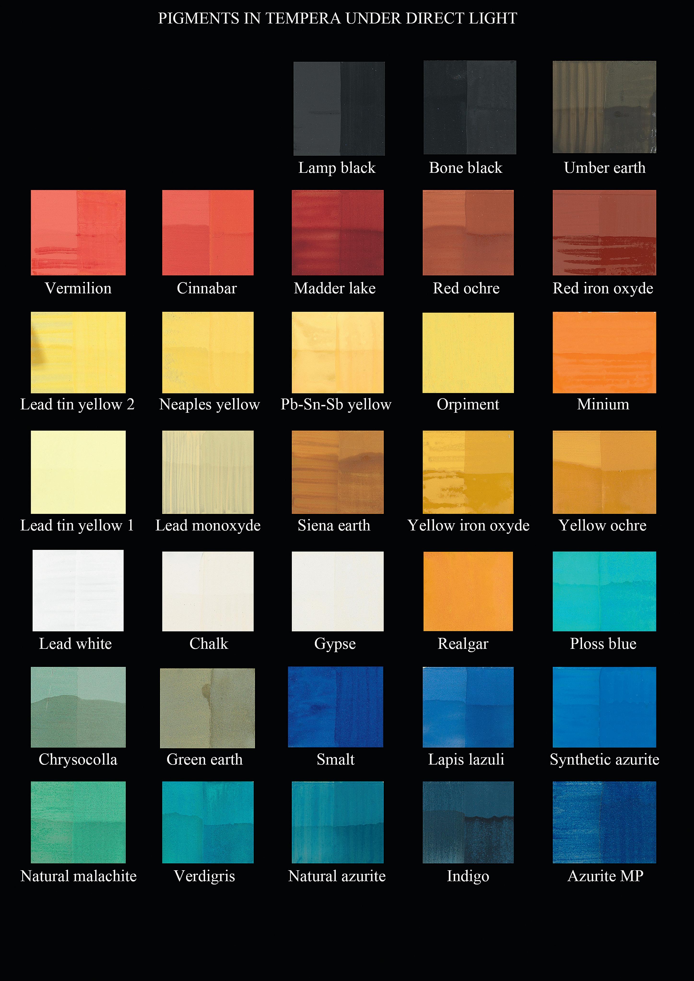
For each color mock up, it was applied a single layer on the left and two layers on the right. The bottom half is varnish, the top half is unvarnish.
Copyright: E. Ravaud/ A. Paounov – C2RMF
L’Arlésienne
Musée d’Orsay, Paris, RF 1953-6
92 cm x 73 cm
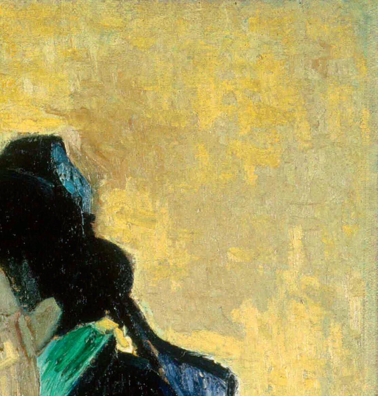


In Van Gogh’s L’Arlésienne in the Musée d’Orsay (a) (b), cross brushstrokes of the yellow background are particularly visible in radiography (c).
Copyright: a, b, c C2RMF
Radiography is a technique, which has been employed in the study of paintings for more than a century. The history of this method of analysis indicates that its development has been modest since the 1960s, as its use has been limited to reductive approaches that take into account no more than the immediately intelligible signs. By systematically considering the physical mechanisms involved in the creation of an image, this volume seeks to demonstrate that we can access new fields of radiological analysis by identifying two categories of ‘signs’: those that may be obvious, but whose meaning is misleading, and those which are not immediately comprehensible.
This study has been primarily based on a thorough and essential reviewing of current literature concerning the materials and processes used for the making of paintings. The semiological analysis is based on the understanding of the physical phenomena occurring in the formation of the image, and on correlations between the radiographic images of a painting and the information stemming from its observation, other scientific results and the restoration reports. Furthermore, a number of experiments were conducted in order to consolidate certain assumptions regarding image-formation mechanisms. Ultimately, this book hopes to show how data resulting from radiographic analysis can be seen and set in a broader context of information on a specific work, or a group of works, in order to enrich our knowledge of art history, history of technology, and conservation as well as restoration.