S.T.R.I.D.E. Professional Guide to Compression Garment Selection for the Lower Extremity
S.T.R.I.D.E.
Professional Guide to Compression Garment Selection for the Lower Extremity
An algorithm incorporating both textile characteristic and oedema presentation to optimize medical compression garment selection.
By Robyn Bjork and Suzie Ehmann
Declaration of interest: S.T.R.I.D.E. is a registered trademark of the International Lymphedema & Wound Training Institute (ILWTI). S.T.R.I.D.E. is a project developed by ILWTI, supported by an unrestricted educational grant by SIGVARIS GROUP. Inc. The publication and distribution of this document was supported by: 3M; Essity; L&R; SIGVARIS GROUP. Inc.
Suggest citation: Bjork R, Ehmann S. S.T.R.I.D.E. Professional guide to compression garment selection for the lower extremity. Journal of Wound Care 2019: 28:(6 suppl 1):1–44
Published by: MA Healthcare Ltd, St Jude’s Church, Dulwich Road, London, SE24 0PB, UK
All rights reserved. No reproduction, transmission or copying of this publication is allowed without written permission. No part of this publication may be reproduced, stored in a retrieval system, or transmitted in any form or by any means, mechanical, electronic, photocopying, recording, or otherwise, without the prior written permission of MA Healthcare Ltd for licensing, reprints and branded algorithms contact anthony.kerr@markallengroup.com
Although the editor and MA Healthcare Ltd and authors have taken great care to ensure accuracy, neither MA Healthcare Ltd nor the authors will be liable for any errors of omission or inaccuracies in this publication.
Editor: Rachel Webb
Managing director: Anthony Kerr
Designer: Sam Meaden
Foreword
The following supplement is a rare example of a paper that combines clinical experience and theoretical knowledge on textiles used in compression therapy. The authors’ intention is to propose a decision support system for choosing specific compression devices, which can be adjusted to counteract the individual signs and symptoms in an optimally adopted way.
The document concentrates on compression devices which can be self-applied by the patients —compression stockings and adjustable wraps. The acronym ‘S.T.R.I.D.E.’, incorporating both textile characteristics and clinical presentation, stands for: Shape, Texture, Refill, Issues, Dosage and Etiology.The intent of the mnemotechnical value is to highlight that successful compression includes more than dosage alone. In addition to dosage, etiology and patient presentation need to be incorporated, including a patient’s physical ability to use compression effectively as part of the daily routine, thereby promoting adherence.
The suggested algorithms provide a valuable guide to stride across the important, but still underestimated field of medical compression therapy and will help to put the prescription of a specific product on a more rational basis. Enjoy reading!
Hugo Partsch Emeritus Professor Medical University of Vienna, Austria
Dear Readers,
Thank you for joining us in a compression revolution! As two compression advocates and educators through the International Lymphedema & Wound Training Institute (ILWTI), we recognise that compression is a highly specialised skill that requires a keen understanding of individual patient assessment and knowledge of a wide array of compression products on the market. We developed S.T.R.I.D.E. as a means to simplify the process by which compression experts make garment selections every day, in order to ensure patient success. Poor compression choices too often leave patients feeling frustrated, hopeless, or despondent, and negatively impact their success with long-term oedema management. If you’ve ever encountered a patient saying ‘I tried it and compression doesn’t work for me’, this supplement is for you. We hope that it will demystify the science of compression and clinical pearls will help you successfully systematise your compression selection process. We want you to join us in changing the way that compression is represented, so that educated decisions can be made from actual compression profile data versus the long process of individual clinicians’ experiential and anecdotal results. We invite you to join us for continued education through ILWTI.com: on-line S.T.R.I.D.E. Certificate, and S.T.R.I.D.E. Certified Compression Specialist hybrid on-line/live certification training. Together, we can forge the future of compression through a compression revolution!
Sincerely,
Authors Robyn Bjork and Suzie Ehmann Contact: SuzieEhmann@ILWTI.com
S.T.R.I.D.E. professional guide to compression garment selection for the lower extremity
‘Taking compression garment prescription in S.T.R.I.D.E.’ — An algorithm incorporating both textile characteristics and oedema presentation to optimise medical compression garment selection
Objective: The effectiveness of a compression garment, such as a medical compression stocking (MCS) or adjustable wrap (AW), is dependent on dose (mmHg) as well as the physical and dynamic properties of the textile, including elasticity and stiffness.1–11
Transparency regarding the comprehensive compression profile is needed to make an appropriate MCS/AW selection. In addition, there are patient-specific characteristics (tissue texture, limb size, location of swelling, quality of oedema, patient functional level) that have clinical relevance on compression garment selection. Although compression therapy options for the management of chronic oedema states are readily available, including chronic venous disease (CVD) and lymphoedema, the practical application of current clinical guidelines for the selection of the appropriate compression garments is limited.
The purpose of this body of work is to provide an unbiased algorithm to assist with compression product selection, based on available medical literature, compression science, and clinical expertise. This algorithm, known as S.T.R.I.D.E., incorporates the comprehensive compression profile of MCS and AW currently
Cavailable. The S.T.R.I.D.E. algorithm incorporates compression dosage, dosage distribution, stiffness and other textile characteristics, and product categories/subcategories.
S.T.R.I.D.E. also includes a system for oedema assessment to best match the individual patient presentation with an appropriate compression product. Assessed patient characteristics include distribution of oedema and limb shape, tissue texture, oedema etiology, precautions/contraindications, and time to refill.
By defining compression prescription in terms of comprehensive compression product and individualised oedema profile, S.T.R.I.D.E. establishes a foundation for compression product selection to achieve optimal patient outcomes. S.T.R.I.D.E. also provides a platform to showcase the continuum of compression products on the market, and differentiate quality compression products from generic products produced by manufacture’s using low-quality materials and knitting technologies. In addition, S.T.R.I.D.E. will further define product profiles and efficacy through additional testing and research, in order to grow the body of clinical evidence to support compression use for chronic oedema conditions.
ompression is one of the cornerstones for the management of oedemas of various etiologies, as well as prevention and treatment of venous leg ulcers (VLUs).14,21
Application of compression has been shown to have a positive impact on both venous and lymphatic function. This impact includes improvement in trophic changes (lipodermatosclerosis, venous stasis, or eczema) and clinical symptoms (heaviness, itching, pain, or quality of life [QOL]) typically associated with chronic oedema.21–24
Pathophysiologic mechanisms for the positive impact of compression include the following:
1 International Lymphedema and Wound Training Institute, Alaska, US.
2 Atrium Health Stanly, North Carolina, US
Lymphatic:19,25–27
● Reduced formation of excess interstitial fluid by opposing fluid filtration from blood capillaries into the tissue, thereby decreasing the lymphatic load
● Shifting fluid into areas with functional lymphatics
● Increased lymphatic reabsorption and stimulation of lymphangion contractions
● Enhanced muscle pump resulting in increased frequency and amplitude of lymph collector contractions.28,29
Venous:15,18,30–32
● Reduced venous reflux and improved venous return
● Reduced venous hypertension
● Maximised calf muscle pump
● Elevated matrix metalloproteinase levels are reduced, promoting healing of VLUs.
Trophic changes: 2,19,20,25,33
● Reduced inflammatory response by release of antiinflammatory mediators
S.T.R.I.D.E. compression selection guide
● Resolution of fibrotic tissue producing a softening of skin.
Clinical symptoms:15,21
● Reduced pro-inflammatory cytokines resulting in an anti-inflammatory effect, reducing pain25,35 and promoting wound healing
● Reduced oedema allows for normal shoe wear and participation in normal Activities of Daily Living (AoDL), improving QoL.
Although the positive impact of compression use is evident throughout the literature, there are inconsistencies regarding the detailed reporting of compression dosage (in vitro and in vivo) and/or textile characteristic used to achieve stated results and insufficient documentation of descriptive comparative properties of textiles tested.14,15,17,18,21,34 A review of the literature on compression garments used for current clinical guidelines reveals variable descriptors of compression dosage (mmHg versus compression class versus descriptive terms including light, moderate, firm), infrequent in vivo assessment of compression pressure and generic mention of garment style of the product studied (knee high, ulcer care kit, AW).14,15,17,21,18,34 These inconsistencies and lack of information limit the practical application in daily clinical practice. As a result, the current guidelines available for the use of MCS and AW provide only recommendations in support of the use of compression garments, as summarised below, rather than actual clinical guidelines for prescribing compression garments:21
● MCS to provide improvement of CVD symptoms (oedema, skin changes, pain, QoL)
● MCS to reduce recurrence of VLUs
● MCS (‘ulcer kits’) to improve VLU healing and reduce pain
● MCS for acute venous disorders and in the initial phase after greater saphenous vein treatment
● MCS for treatment of symptomatic post thrombotic syndrome (PTS)
● MCS for lymphoedema management. Without more detailed information to specifically define the comprehensive compression profile of the compression products used, the clinician is led to believe that all compression garments matching a generic description cited in a reference, such as ‘moderate compression knee high’, ‘20 to 30mmHg knee high’ or ‘Velcro adjustable wrap applied at 20 to 30mmHg’, have similar compressive effectiveness. However, advancing study about the dynamic properties of compression textiles have produced research to refute this assumption.3,89,10,17,19,20,31,37–40
This research shows that compression dosage (mmHg) is only one of the variables that have an impact on oedema and the haemodynamic efficacy of a garment. In addition to dose, the elasticity and stiffness of the textile has been shown to have a great impact. Thus, effective compression garment prescription requires knowledge of both the dosage and dynamic compression profile of the textile.
COMPRESSION SCIENCE
MEDICAL COMPRESSION STOCKING DOSAGE
Compression is defined as the pressure applied to an area of the body by the recoil of an elastic garment, or the tension applied when donning an adjustable garment.1,41,42 Pressure is exerted on the limb because of the elasticity of the MCS and is related to the extension of the MCS.5 This compression pressure is transmitted to the underlying tissues and vessels and can be measured and expressed in mm of mercury of pressure (mmHg). The mmHg, measured at the ankle, is the compression ‘dosage’ and is also what is reflected on compression garment packaging.
The dosage of a compression garment is one of the factors that determines its haemodynamic efficacy and is engineered into the MCS by the type of yarns and knitting techniques used to produce the final garment textile.1 Inlay yarns and body yarns are knitted together to produce MCS textiles, illustrated in Fig 1.1.1 The body yarn delivers the thickness and stiffness of the textile, while the inlay yarn produces the compression pressure (Fig 2).1
CLINICAL PEARL Compression dosage (mmHg) is only one of the variables that have an impact on oedema and the haemodynamic efficacy of a garment. In addition to dose, the elasticity and stiffness of the textile has been shown to have a great impact.
In vitro dosage testing
The compression dosage for MCS can be measured in vitro by compression garment manufacturers using a dynamometer, such as a HOSY, HATRA, or Zwick machine.12 A section of the garment at the ankle region of the stocking, called the B point, is secured and stretched in a transverse direction, while a software programme records the amount of resistance of the garment to the applied force across the sizing range.38 This produces a hysteresis curve (Fig 3) of the measured dosages, reflective of the mmHg of inward force on a limb at each increment within the sizing range. For example, a 20–30mmHg garment means that, at the ankle, 20mmHg of inward force is exerted on the ankle
S.T.R.I.D.E. compression selection guide
if the girth matches the smallest circumference in the sizing range. And 30mmHg of inward force is exerted on the ankle if the girth matches the largest circumference in the sizing range. This type of laboratory testing is performed by reputable compression manufacturers and provides quality controls to verify the dosage specified on packaging.
In vivo dosage testing
Compression dosage is also measured in vivo by clinical researchers and by manufacturing companies of both MCS and AW. In vivo testing measures the interface pressure (IP), which is the pressure produced by a compression garment on the skin surface, using a pressure sensor such as a Kikuhime or Picopress (Fig 4).43 Recommendations regarding sensor types and placement can be found in the 2006 ICC Consensus document by Partsch et al.38
In vivo testing of compression IP is measured at B1, located at the medial myotendinous junction of the gastrocnemius muscle and Achilles tendon (Fig 5). B1 was chosen as it was found to be the area of greatest circumferential change with muscular activation, typically 10–15cm proximal to the medial malleolus.38 Although the measurement method and location for in vivo and in vitro testing are not the same, Partsch demonstrated that the correlation between the two was highly significant.44 It should be noted that IP is dynamic, changing with the stiffness of the textile, density of the tissues under the IP sensor, movement, change in position (supine versus sitting versus standing), as well as change in volume (circumference of the limb). The dynamic nature of compression is illustrated in Fig 6, which depicts IP fluctuations observed with change in position and/or movement.
Compression Dosage Gradient
Most current medical MCS are designed to provide gradient compression dosages with more compression at the ankle and progressively less moving toward the top of the garment. A graduated compression profile is engineered to oppose the anatomic hydrostatic pressure created when the body is in the upright position. This graduated compression profile is created by applying more stretch to the inlay yarn at the ankle during knitting and then gradually decreasing the tension toward the top of the garment.
It is very important to remember that the dosage reported on lower extremity compression garment packaging is a measure of the mmHg of pressure at the ankle only, across the sizing range. At the ankle, this point of measure is denoted as the B measurement (Fig 7). Many manufacturers follow the German RAL standard and construct their garments so that the percent of pressure relative to the ankle falls within a standard range. According to the RAL standard, calf dosages may vary between 50–80% of the ankle
Fig 1. Inlay yarns and body yarns are knitted together to produce medical compression stocking textiles
Fig 2. The body yarn delivers the thickness and stiffness of the textile, while the inlay yarn produces the compression pressure
Body yarn
Inlay yarn
Andrew Bezear and Cameron Law
Body yarn
Inlay yarn
S.T.R.I.D.E. compression selection guide
pressure, and thigh dosages between 20–60% of the ankle pressure. 45 The European Committee for Standardization (CEN) standards dictate that the pressure profile along the leg must be 70–80% at B1 and 50–80% at C and D compared with the pressure exerted at ankle level at B (Fig 7).43 This standard must be met for garment reimbursement in many countries. However, distribution of compression dosage as it relates to varying oedema presentations are not specified. Furthermore, this pressure gradient may vary between manufacturers and/or between individual products from a single manufacturer.
Variation in compression gradient and the impact on oedema management is an area of focused research and compression design. Bjork presented work demonstrating a new stiffer circular knit garment specifically engineered to provide graduated compression with more even distribution of compression from the metatarsal heads to the thigh.46 Table 1 compares the distribution of dosage and stiffness at the foot, ankle, calf and thigh of the stiffer circular knit garment compared with traditional circular knit and custom flat knit garments. The products were manufactured by the same company and tested in the same way, using a Zwick machine. Although they had the same prescribed dosage, distribution of that dosage was different between garments, as was the stiffness which we discuss later.47 In a preliminary case series, wear testing results were positive and indicated that the stiffer circular knit garment provided clinically efficacious levels of containment in the management of chronic oedemas where the oedema itself was more evenly distributed along the lower extremity.46
Haemodynamic response to altered compression gradient has also been studied. Mosti and Partsch48,49 observed that ambulatory patients’ using compressing stockings that exerted higher pressures on the calf than the ankle showed greater efficacy in increasing the venous ejection fraction from the leg. Therefore, they concluded that graduated pressure profile is not mandatory in the ambulatory patient.48,49 Limitations stated by the authors were that the study only looked at haemodynamic function with movement over a short time period, not over the course of a day, and only evaluated ambulatory patients.
Couzan et al.50 conducted a randomised double-blind multi-centre study of 401 patients with chronic venous insufficiency (CVI) to assess the efficacy of 30mmHg progressive compressive stockings (maximal pressure at calf), compared with ‘degressive’ compression (traditional graduated compression profile). This trial
demonstrated that progressive compression stockings are more effective than usual graduated compression stockings in the improvement of pain and lower leg symptoms in patients with CVI. Moreover, progressive compressive stockings were easier to apply, raising no safety concern at three months.
Fig 4. Example of a devices used to measure interface pressure in vivo, PicoPress
Fig 3. Example of hysteresis curve produced when testing medical compression stocking dosage and used to calculate textile stiffness in vitro
S.T.R.I.D.E. compression selection guide
There is a need for additional research to look at the impact of varying compression gradients and the corresponding haemodynamic function relative to varying oedema presentations. In particular, exploration is needed regarding which oedema presentations are best treated by different distributions of dosage. The distribution of compression dosage across the garment is important information that is currently missing from the compression profile descriptions on packaging, which further hinders matching appropriate compression selection to an individual patient presentation.
Compression dosage classes
It should be noted that there is variability with regard to the categorisation of compression dosages by mmHg or class, dependent on country of origin (Fig 8). These inconsistencies further impair the consumer’s ability to make an informed choice. For example, a Class III garment according to the French standard is 20–36mmHg, whereas Class III according to the German standard is 34–46mmHg. These inconsistencies further challenge the application of clinical research outcomes between countries, further muddying the water for researchers, clinicians and consumers alike.
Dosage diminished by garment fatigue
Ankle
Table 1. Comparison of three knitted compression garments from different compression categories. All garments are the same dosage and size, and all garments were manufactured by the same company and tested using a Zwick machine. A commercial software program was used to produce a hysteresis curve and calculate stiffness. (Data provided courtesy of Sigvaris Group, Inc., US) A Circular knit garment profile, stiffer circular knit garment profile and a flat knit garment profile
In addition to garment dosage, garment fatigue is yet another qualitative measure of compression efficacy that is rarely mentioned in compression studies. Compression garment manufacturers are able to implement quality controls that test and re-test garments’ ability to maintain the stated dosage from morning to evening, with repeated washing and drying, and with months of use. This is key in how the garment will perform in managing oedema. Reputable manufacturers will not only perform internal testing but also use outside laboratory testing to corroborate guaranteed compression dosages over the course of a day and months of use. This becomes the basis for guaranteeing their compression pressure, typically for six months. It is important for consumers to question how a garment has been tested and if dosages are guaranteed. If dosage is not guaranteed, the garment may not consistently deliver the therapeutic compression level needed to manage an individual’s oedema.
CLINICAL PEARL If a patient reports that their garment changed over time from more difficult to very easy to don, it could be because the garment is fatigued and no longer able to deliver therapeutic dosages.
S.T.R.I.D.E. compression selection guide
In contrast to rigorously tested garments, lesserquality garments may have the specified dosage right out of the box, but subsequently lose that dosage as the garment stretches and relaxes over the course of the day or with several weeks or months of use. This is called garment fatigue. In a 2009 dissertation, Van de Wegen-Franken40 tested garment fatigue in twelve different brands. The percentage drop in pressure over eight hours of use, morning to evening, varied from 7.8% up to 44.8%. These findings demonstrate that some garments lose their therapeutic value by the end of the day, precisely when many types of oedemas require robust support.
Knowledge of garment fatigue is necessary to ensure therapeutic efficacy of the stated compression dosage on garment packaging. Availability of garment fatigue testing ensures the consumer that the specified dosage will be provided throughout day as well as over the sixmonth warranty. If a patient reports that their garment changed over time from more difficult to very easy to don, it could be because the garment is fatigued and no longer able to deliver therapeutic dosages versus the patient becoming more proficient at donning techniques.
Adjustable wrap compression dosage
Like MCS, AW currently available on the market vary by textile composition as well as application process. Studies regarding the evaluation of dynamic performance of AW with regards to the dosage are limited and consist largely of case studies using a single type of AW.51 Published studies focus on investigation of a singular product line and consequently the results cannot be taken to represent an entire product line or AW as a compression category
CLINICAL PEARL In order to maintain medically efficacious dosages, it is very important to replace garments every six months
Similar to MCS, AW manufactures have developed light and firm, ready-to-wear (RTW) and custom products. We found no studies comparing the dynamic performance of different textiles used to construct the AW, such as light versions compared with classic styles. Lack of information regarding the textile properties of
Fig 5. In vivo testing of compression interface pressure is measured at B1, at the medial myotendinous junction of the gastrocnemius muscle and Achilles tendon
Fig 6. Graphic tracing of interface pressure measurements under inelastic and elastic compression with movement.
Lateral head of gastrocnemius
Medial head of gastrocnemius
Pressure transducer should be placed on the skin over this point
Achilles tendon
S.T.R.I.D.E. compression selection guide
Dosage prescription
Knowledge about the dosage of compression is important to address underlying haemodynamic response. It has been shown that low pressure is able to prevent or reduce oedema and that external pressure of 20mmHg is enough to narrow or occlude the veins in the supine position.36 However, in the upright position much higher pressures are required to achieve haemodynamic effects.
Top of Thigh
Mid-Thigh
Patella
Below Knee
Widest Calf
Below Calf
Smallest Ankle
Heel
Base of Toes
an individual AW limits consumers’ choice to evaluate its dynamic performance compared with other AW available on the market.
Similar to the dosage calculation for MCS, AW compression dosage is measured by the stretch of the textile when wrapped around the limb of a known circumference.52 Published works focus on the visual markings to indicate the IP upon donning the garment. These markings provide information on the resting pressure but not the dynamic change in compression pressure that would be created with a change in position or movement. In addition, there have been no studies looking at varying compression gradients with AW, such as greater compression at the calf versus the ankle, as they relate to haemodynamic performance or overall oedema management. One study did note that the application styles (interlacing versus overlapping) of the AW on the market may impact functional performance, but no objective measurements were reported.53
Compression science literature demonstrates that effective compression requires that the external pressure applied is greater than the intravenous pressure, thereby causing a narrowing of the leg veins and increased velocity of venous blood flow. Furthermore, a therapeutic dose of compression is dependent on position of the body because venous pressures change with a change in position. Compression pressures must increase to >50mmHg in sitting, and close to 70–80mmHg in standing, in order to counteract the intravenous pressure that occurs with change in position.36,54 However, it should be stressed that these very high levels of pressure noted in the literature represent the mmHg needed to cause a temporarily occlusion of the vein and are not a recommendation that they be statically maintained in compression garments.
Textile stiffness plays a big part in how high levels of pressure can be more safely and comfortably achieved. Very high pressures of elastic textiles are poorly tolerated and pose risks of traumatising soft tissues and compromising blood flow due to the inward and sustained, forceful recoil of the elastic material. Fortunately, compression garments can be constructed in a fashion to mitigate these deleterious effects by enhancing the textile stiffness, which produces high pressures intermittently to achieve the desired physiologic outcomes. It is important for clinicians and consumers alike to understand that the dosage listed on compression garment packaging refers to resting pressure only, the level of sustained pressure the garment exerts at the ankle when the person wearing it is not moving or contracting their muscles. In contrast, the optimised therapeutic benefit of a compression garment is achieved when patients exercise and move while wearing their garments.
STIFFNESS
Textile stiffness
Before discussing compression garment categories, it is important to explain stiffness, how it is measured and the gaps between engineering testing and data collection by compression garment manufacturers
Fig 7. Standard points of reference for circumferential measurements for custom garments, or compression pressure measurements
(T) Waist
S.T.R.I.D.E. compression selection guide
Class I 14–17mmHG 10–15mmHg 18–21mmHg
Class II 18–24mmHg 15-20mmHg 23–32mmHg
Class III 25–35mmHg 20–36mmHg 34-46mmHg
Class IV Not reported >36mmHg >49mmHg
versus stiffness testing by clinical researchers. The goal of this discussion and the principles of S.T.R.I.D.E., are to bridge that gap. As the authors of S.T.R.I.D.E. are able to acquire and integrate more data on compression product profiles in the future, clinicians will be enabled to make even more effective compression selection decisions.
Besides dosage, stiffness of garments is of crucial value to predict their effectiveness and tolerability for patients.9,11,20,38 Stiffness, which is also known as slope or elasticity coefficient, is defined according to the European Committee of Normalization (CEN) as the pressure increase produced by MCS per 1cm of increase in leg circumference at the B level (ankle), and is expressed in mmHg/cm or hectopascals per cm.43 In a practical sense, the stiffness is defined as the ability of the MCS/AW to resist expansion of the limb. Simply put, dosage describes the inward pressure that a garment exerts on the body as a result of its elastic recoil, while stiffness describes the resistance of the garment to the expansion, such as when an area of the body swells or tissue expands due to muscle contractions.
The importance of stiffness as a separate parameter in a complete compression profile is consistently evident in the published medical literature. It provides insight into the performance during posture changes and movement and impacts oedema prevention and venous haemodynamics.3,6,9–11,13,20,33,35,39 Multiple researchers have found variations of stiffness for MCS independent of dosage and type of knitting.3,6,9–11,13,20,33,35,39 In the studies by Van der Wegen et al.39,40,55 MCS with the same dosages had different stiffness and thus different physiologic impact on haemodynamics of the limb. In her doctoral thesis in 2009, Van de Wegen-Franken evaluated 18 different Class II, circular and flat knitted garments and found
US - no known standard
No standardized testing method documented
15–20 mmHg
20–30mmHg
30–40mmHg
40-50mmHg
>50mmHg
their static stiffness ranged from 1.7 to 10.32.40 Similarly, Hirai et al. found that the stiffness of elastic stockings during posture changes and exercise differs with brand, even in stockings belonging to the same compression class with the same dosage.9
In addition to the differences in stiffness observed among MCS of the same compression class, research has also demonstrated that the stiffness of an MCS has an impact on the haemodynamic effect, including decreased reflux, improved calf pump function, and decreased oedema formation.3,7,13 Van Geest et al.13 found there was a statistically significant difference in capillary filtration rate, and consequently in the development of oedema, between compression garments with the same dosage but different stiffness. Hafner and Junger3 observed similar findings in their randomised study demonstrating MCS of the same compression class or dosage, did not have the same acute effect on venous haemodynamics. Numerous compression researchers have demonstrated that the higher the stiffness of a compression garment, the larger was the improvement of haemodynamic parameters and the more effective was the reduction in oedema.3,7,13,20,39,57
CLINICAL PEARL Dosage describes the inward pressure that a garment exerts on the body as a result of its elastic recoil, while stiffness describes the resistance of the garment to the expansion, such as when an area of the body swells.
The research has shown that stiffness is a key descriptive characteristic of a compression textile (MCS /AW) impacting the haemodynamic effect of a compression garment. However, measurement of stiffness is not currently standardised, routinely tested, nor reported in compression garment labelling or packaging. One of the reasons for the gap between the
Fig 8. Comparisons of international classification systems for medical compression stockings
S.T.R.I.D.E. compression selection guide
availability of stiffness measurements from manufacturing for clinical practice is that calculation of stiffness varies, depending on the measuring device used to measure and whether it is measured in vitro versus in vivo
CLINICAL PEARL Numerous compression researchers have demonstrated that the higher the stiffness of a compression garment, the larger was the improvement of haemodynamic parameters and the more effective was the reduction in oedema.
In vitro stiffness measurement
In vitro assessment of stiffness of a compression textile can be precisely measured in a lab in a consistently reproducible manner. 12 Compression garment manufacturing companies can calculate garment stiffness of a MCS using the same information garnered during dosage testing. For example, using the Zwick CRE (constant rate of extension) machine, a stress/strain curve is produced during dosage testing. This curve, called a hysteresis curve, will look steeper in stiffer garments (Fig 3). The stiffness, or mmHg of resistance to each cm of stretch, is reflected in this hysteresis (stress/strain) curve and can then be mathematically calculated across the garment size.38
By deploying these testing methods at the foot, ankle, calf, thigh, or many more points in between, the dosage and stiffness, as well as the distribution of dosage and stiffness along the garment, could be precisely measured and documented as in Table 1. These compression profile data could become very valuable for compression garment selection and prescription. If consistently measured with the same type of equipment and testing methods, stiffness comparisons could also be made between products within one manufacturer’s product portfolio, as well as between manufacturers. Unfortunately, this type of testing and reporting to consumers is not yet routinely done by manufacturers, and standardisation of methods is not yet established. The authors of S.T.R.I.D.E. propose that compression garment manufacturers provide this type of complete compression profile to be integrated into the S.T.R.I.D.E. system, in order to develop more accurate, comparative algorithms that will aide consumers and clinicians in choosing compression to match their individual needs, based on compression science and engineering.
In vivo stiffness measurement
In vitro assessment of MCS provides information about how the textile performs in a semi-static state,
however, these laboratory measurements are far from the clinical use scenario. The stiffness of a garment as described above relates to the properties of the textile itself and is useful for garment manufacturing. In contrast, the IPs that are produced between the garment and the surface of the skin, at rest and with movement, are of interest to clinical researchers who desire to test the dynamic performance and effectiveness of garments on an individual patient or to compare different products on one patient. In vivo measurements of stiffness require measurement of IP, measured under static (resting) conditions, termed ‘resting pressure’, and dynamic (moving) conditions, termed ‘working pressure’.6
Measurement of stiffness of a medical compression device in vivo (static or dynamic) can be challenging. The measurement process is time and labour intensive and can vary depending on different tissue densities and location of the IP sensor or sensors.57,58 For example, IP between a garment and bone is much different than between a garment and fatty tissue, oedema of more watery consistency or fibrotic tissue. The leg position during measuring, the configuration and consistency at the measuring site, an individual’s muscle girth and strength, the presence of fat, and the tissue composition have all been shown to affect the measured interface pressure.6,7
In vivo static stiffness index (SSI)
A simple but practical measurement of in vivo assessment of a textile, as proposed by Partsch, involves the measurement of the SSI, which is calculated as the pressure difference between the standing and supine positions.59 To measure the SSI, IP is measured at B1 in two body positions, supine resting and standing, without change of the pressure sensor.6 The assessment is simple and can be performed even in those patients who may not have ambulatory ability. Research has shown that the SSI values correlate well with the values of stiffness measure in vitro 19,44,60
In vivo dynamic stiffness index (DSI)
Compression is most effective with movement. As such, several researchers have sought to further examine dynamic performance of garments by measuring DSI. DSI is defined as the difference between the maximum pressure peaks during walking and the resting pressure measured in the supine position. Dynamic stiffness measurements in vivo require sophisticated instrumentation that is not practical for daily use.40 However, various researches have presented adapted assessments of DSI that could be replicated in
S.T.R.I.D.E. compression selection guide
vitro, such as that described by Stolk et al. These researchers used an artificial leg segment model that could simulate walking, showing dynamic changes of circumferences and pressure.61 Additionally, Hirai and Partsch62 presented the results of a novel artificial leg model whose circumference could be mechanically extended in 1cm increments, enabling precise measuring of IPs with simulated limb expansion. The results of this study showed a significant correlation between the stiffness values measured with the artificial leg and those obtained from extensometer measurements and with data on the human leg in vivo using the SSI.62 These authors observed a positive correlation between the SSI and DSI at B1 concluding that a MCS with a high static stiffness would also have a high dynamic stiffness. Multiple researchers have found similar excellent correlation between SSI and DSI in vitro and in vivo testing.4,7,20,5,56,63
Although the research has demonstrated correlation between SSI and DSI, affording a standardised method for comparison of compression products, Hirai and Partsch acknowledged the inability of this method to replicate in vivo conditions, such as altered limb shape or altered tissue texture that are present in pathology. Thus, the applicability of these findings in clinical practice is limited. Additional research is necessary to objectively measure tissue density and the effect different compression textiles have on oedema management and soft tissue remodelling.
Resting and working pressures
The pressure created at the interface of the compression textile and the limb is known as the interface pressure (IP). In the supine position this is known as the resting pressure (RP) and corresponds to the labelled garment dosage. With movement or change in position, the IP will change. The amplitude of change of pressure will depend on the resistance to stretch, or stiffness of the MCS/AW. The less elastic or stiffer the material, the more resistance to stretch, and the greater the resultant increase in IP. The less stiff or more elastic the material, the more compliant with stretch and the smaller the resultant amplitude of change in IP. This pressure created with a change in position or muscular activation is known as the working pressure (WP) (Fig 6). WP reflects the resistance of the garment to expansion of the limb with muscle contractions. As a muscle contracts, the belly of the muscle shortens and increases in girth. A stiffer textile will resist that expansion, creating an inward, intermittent force that compresses the deep veins and increases the velocity of blood flow.
Calf muscle contraction, known as the calf muscle pump, is the principle mechanism by which blood is moved upwards in the venous system towards the heart against gravity in the upright position. Under normal conditions blood will be squeezed upward towards the heart with every muscle contraction and vein valves will inhibit a retrograde flow of the blood column due to gravity. This is augmented by the foot pump, which concurrently expels a column of blood of 100mmHg during weight bearing through a normal gait cycle. 64,65
When a needle is introduced into a dorsal foot vein and the intravenous pressure is measured, the pressure during quiet standing corresponds to the weight of the blood column and the right heart, which is between 80 and 100mm depending on the body height. When the subject starts walking, the intravenous pressure will drop to 20-30mmHg as a sign of a normal calf muscle pump. This normal pumping function is deficient, however, in patients with venous insufficiency. Due to incompetent venous valves or obstruction of venous outflow, the venous pump may press blood in a retrograde direction, resulting in ambulatory venous hypertension during walking. External application of stiffer compression garments can help restore optimised calf muscle pump function. In addition, foot compression can enhance the foot pump.65
Compression research has demonstrated that although compression devices may apply similar RP, textiles with no stretch or short-stretch characteristics produce higher peak pressures, WP, when standing or walking compared with the effects obtained with longstretch textiles.38 In short, the stiffer the compression product the greater the amplitude of peak pressures observed and the optimal capture of the calf muscle pump (Fig 6). Plethysmographic measurements of the ejection fraction of the calf pump showed significant improvement of the pumping function under inelastic bandages, but much less under elastic compression stockings.36 These findings can be explained by the fact that inelastic bandages with a resting pressure of 40mmHg will show pressure of around 80mmHg in standing, which is high enough to reduce the deep vein diameters. Thus, for patients with chronic oedema states due to venous hypertension, a compression garment that exerts a low comfortable pressure at rest, but a strong pressure during standing and walking to counteract ambulatory venous hypertension is ideal.2,7,66
The importance of oscillating forces for lymphatic drainage may explain why stiff compression material achieves a massaging effect in combination with muscular activity and is the preferred material for acute
S.T.R.I.D.E. compression selection guide
decongestion for individuals with lymphoedema. Stiffer compressive textiles produce a greater massaging effect by generating greater amplitude of change in IP with movement. The greater the amplitude of change, the greater the haemodynamic impact on the lymphatic vasculature. The massaging effect of inelastic compression created by the AW during upright active movement creates oscillating forces that transiently change the interstitial-intraluminal fluid pressure gradient, producing periodic movement of lymph into initial lymphatics.67
Despite the evidence regarding the physiological impact of textile types, neither garment stiffness nor distribution of stiffness is reported on the garment packaging, leaving clinicians to guess how a garment will actually perform. Clinical experts recognise that choosing a stiffer textile, rather than increasing the prescribed dosage, may have better outcomes for some patients. Though garment stiffness has been discussed for years internationally, no consensus to date has been reached as to a standardised method to test stiffness within and between compression manufacturing companies.
COMPRESSION GARMENT CATEGORISATION
S.T.R.I.D.E. organises compression garments by categories and subcategories. The main categories are circular knit, stiffer circular knit, flat knit, adjustable wraps, night garments and decongestive garments. Compression garments in these categories are available for many different body areas, such as the head and neck, trunk, arm and hand, pelvis, genitals, leg and toes. However, the current iteration of the S.T.R.I.D.E. algorithm guides specific product selection for the lower body only. Future iterations are intended for the upper body, trunk, bandaging products or compression pumps. Each of these has its own unique body of evidence in the research literature, and corresponding recommendations.
Compression garment selection is to be used when choosing compression for long-term maintenance, after the oedema is reduced (typically with compression bandages), with the exception of decongestive garments that are used in the acute, decongestive phase of treatment. However, some AW and night garments can also be used to augment acute decongestion or used for reduction of oedema back to a baseline level, if needed, during the long-term, maintenance phase of treatment. These options are reflected in the S.T.R.I.D.E. algorithm for lower extremity garments.
Categorisation of compression garments along a continuum, ranging from elastic to stiffer, affords the clinician insight into the expected dynamic performance
based on the textile construction. Subcategories further stratify the compression options in order to highlight different types of compression garments currently available within a compression category (Fig 9). In general, circular knit garments are the least stiff, progressively increasing in stiffness to stiffer circular knit and flat knit garments. AW and decongestive garments are the stiffest. Stiffness of night garments is dependent on the textile used in their construction. Those constructed from stretchier fabrics will have more elastic properties, whereas those constructed more like an adjustable wrap will display stiffer compression profiles. Within each category of compression, there is also variability. For example, some flat knit garments are ‘soft’ or ‘seamless’ that tend to be less stiff than the traditional or seamed versions. In addition, AW can be made of completely rigid, no-stretch textiles or a more flexible and breathable neoprene textile. It should be noted that there is no particular textile type that is superior to another, but rather each garment has its own, unique advantages and disadvantages because it relates to an individual oedema presentation.
Circular knit
Circular knit garments are knitted on a circular knit machine in a tubular fashion and are generally produced to a standard size called ready to wear (RTW). However, they can also be custom, ‘made to measure’. Circular knitting machines employ circular drums with rows of small needles that can knit very fine and premium yarns, producing garments that tend to be thinner and cosmetically more appealing (Fig 10). Elastic inlay yarns give the garment its dosage, while loop yarns contribute suppleness, wicking, sheerness versus opacity, and other properties.
Stiffer circular knit
As the construction and resulting dynamic performance of MCS has a bearing on garment selection, the authors have defined this as a unique category. Before this document, stiffer circular knit garments have not been formally recognised as a category for MCS. However, review of the literature does find reference to classification of circular knit stockings as ‘round knitted’ compression garments that were further divided into classic (long stretch), thick round knit, and firm (short stretch).8,9
These garments are knitted in the same way, and on the same machines, as other circular knit garments (Fig 10). However, the distribution of dosage along the garment is more even, sometimes deviating on purpose from the German RAL standard, to provide more
S.T.R.I.D.E. compression selection guide
Garment categories
containment of oedema along the limb. Generally, the stiffness of these garments ranges from 1.5 to 2 times as stiff as traditional circular knit garments.46 High modulus inlay yarns are used to more effectively resist garment fatigue and provide increased stiffness, and there is more dense knitting of interlocking loops in a pattern that resists expansion. These properties result in heightened containment of oedema compared to traditional, medical grade, circular knit garments. As such, this category is a hybrid option between circular knit and flat knit garments.
Flat knit
Flat knit garments are knitted on a flatbed machine comprised of rows of hundreds of needles in a linear configuration (Fig 11). The needles are larger gauge and able to knit thicker yarns than circular knit machines. Thus, in general, flat knit textiles are coarser than circular knit because of the thicker yarns.
Traditional flat knit garments are knitted in flat sheets that are easily customisable to any shape. These flat sheets are then knitted together, resulting in a seam. V-bed and glove machines are also flatbed machines but knit in a more circular fashion, producing a seamless, flat-knit garment with lighter containment.
Inlay yarns give flat knit garments their elastic properties and controlled dosage. Traditional flat knit garments are thicker and stiffer than circular knit garments, provide a higher level of containment, and are also better at bridging skin folds and less likely to
cut in or cause a tourniquet effect.1 Additionally, traditional flat knit garments have a more even distribution of dosage and stiffness than traditional medical circular knit garments. In the past, medical compression garments called ‘flat knit’ had similar engineering properties, manufactured according to RAL standards. However, from an engineering perspective, technically any garment knitted on a flatbed machine could be called ‘flat knit’. This includes loosely knit, winter sweaters. This has become problematic in recent years, particularly with lack of compression regulation and quality controls by the FDA in the US. New products are now emerging that are being called ‘flat knit’ but are unlike traditional flat knit garments. These garments are manufactured to have accurate dosages within medically accepted ranges, such as 20–30mmHg or 30–40mmHg. However, they do not provide enough containment for lymphoedema management because the stiffness properties are lacking. These products are deceptively marketed as flat knit garments that are easier to don and doff. In the US, due to lack of regulation, unsubstantiated claims can be made on packaging that are not true. Thus, clinicians are encouraged to procure compression garments made by reputable companies that have robust, voluntary internal quality controls.
Adjustable wraps
AW are versatile compression garments that are cut and sewn from textiles (Fig 12) and are generally stiffer than circular knit or flat knit textiles. AW use hook and
Fig 9. S.T.R.I.D.E. garment categories and subcategories
S.T.R.I.D.E. compression selection guide
loop (i.e. Velcro), elastic drawstrings or bra-type hook and hole closures, and the textile may interlace, overlap, or form a rigid sleeve. Common textiles used for construction include neoprene, Breath-o-Prene, or spacer fabric textiles. AW can be completely inelastic or short stretch, depending on the chosen textile. They are stiff and have high working pressures.
Many AW have built in or augmentative marking systems to determine the applied resting pressure. The packaging is often labelled according to the resting dosages within ranges that are consistent with MCS products, such as 20–30mmHg, 30–40mmHg, or 40–50mmHg. Dosages refer only to the pressure applied by the garment at rest, upon application, without movement or muscle contractions. Users should be instructed to apply AW with the prescribed resting pressure and re-adjusted when it drops due to oedema reduction.41,68
There are no formal studies measuring the elasticity versus stiffness of AW currently available on the
market. There are significant differences in the types of textiles used to manufacture AW, as well as the general construction and functional application, such as interlacing versus overlapping. These properties have an impact on the overall dynamic capacity of the compression garments, therefore, the consumer should not assume that all AW are equal in their function.
S.T.R.I.D.E. currently categorises AW into light, regular, and firm subcategories. However, further testing and data collection is needed to quantify the elasticity versus stiffness properties of each subcategory and to make comparison with products in other categories.
Night garments
As the name implies, these garments are primarily designed for nighttime use but are safe for day use as well (Fig 13). Typically, these items are ordered after decongestion of the limb and the limb volume has stabilised. The purpose of the night garment is to prevent rebound swelling, maintain volume reduction and address soft tissue changes associated with chronic oedema states.
As with other compression categories, there is a variety of options within this category to address mild, moderate and severe oedematous presentations. A unique feature of night garments is the incorporation of textured materials, including waved or chipped foam, to warm and soften fibrotic tissue in conjunction with circumferential compression. Various modifications are available to enhance the dynamic compression effect, including varying profile of thickness, and outer elastic or inelastic sleeves.
Stiffness and garment fatigue of night garments varies according to the type of textile and construction. Dosages
Fig 10. Circular knitting machine (a), examples of circular knit garments (b), example of stiffer circular knit garment (c)
specified relate to resting pressures and are indicated by the manufacturer. Application styles also vary between night garments. Some slip on and have an elastic oversleeve, while others are designed like an adjustable wrap.
Decongestive garments
Decongestive garments are AW that can be used in the acute decongestive phase of oedema reduction (Fig 14).
Custom application of multi-layered, short stretch lymphoedema bandages with integration of incongruent foam is the gold standard of lymphoedema treatment. However, decongestive garments can be used in place of multi-layered compression bandaging, or used to augment bandaging and the complete decongestive therapy programme.
Decongestive garments are specially designed wraps that can be custom cut to fit various limb shapes, and
re-sized and trimmed to accommodate for oedema reduction over time. These garments are often constructed like AW, from similar textiles. Because of the customisation, labelling with accurate resting pressures is difficult. However, like AW, these garments’ have high WP and low RP. In addition to custom cut and trim garments, some RTW AW garments can also be used for oedema reduction depending on the amount of volume that needs to be reduced.
S.T.R.I.D.E. ALGORITHM
The S.T.R.I.D.E. algorithm systematises the necessary characteristics vital to successful compression prescription. Each letter of ‘S.T.R.I.D.E.’ stands for a key group of considerations when selecting compression garments. Those elements are:
Fig 12. Examples of adjustable wraps
Fig 13. Examples of night garments
S.T.R.I.D.E. compression selection guide
S = SHAPE
Q: Where is the swelling located?
Q: Does the dimension of the limb match with standardised sizing charts, or is custom compression needed?
Q: What is the shape of the limb compared to the shape of the garment?
A golden rule of compression garment selection is that a limb should be measured and fitted for a garment when is in a decongested state (minimal to no oedema state). A limb that is still oedematous should be reduced with appropriate compression therapy for optimal fit of the final garment selection for long-term maintenance.
S = SHAPE
Shape of the limb and distribution of the oedema corresponding to garment types, shapes and sizing
T = TEXTURE
Texture of the oedema and soft tissues and corresponding types of textiles used to treat it
R = REFILL
Dynamics of the oedema, such as refill during the day or night, and time to refill
I = ISSUES
Issues including precautions, contraindications and challenges to successful compression usage
D = DOSAGE
The labelled dosage/ankle pressure (mmHg) versus resting and working pressure, distribution of pressure, and garment fatigue
E = ETIOLOGY
Consideration of underlying diagnoses, or etiologies, contributing to the oedema presentation and corresponding characteristics regarding tissue texture, shape/distribution and compression therapy needs.
All letters of the S.T.R.I.D.E. acronym are to be considered as a whole when selecting compression garments. However, for the purpose of a standardised process, each component can be addressed in sequence. As each letter is addressed, the information gained leads one closer to compression recommendations. Each step helps the clinician or consumer achieve greater compression success by ‘Taking compression garment prescription in S.T.R.I.D.E.’
The distribution or location of the oedema should guide garment selection. A complete portfolio of compression garments is available for various body areas. A careful history and clinical exam can help the clinician identify localised regions most affected by oedema. Lower body areas may include the pelvis and genitals, thigh, knee, lower leg, ankle, foot or toes, depending on the underlying oedema etiology or etiologies. Compression should encompass all areas of oedema. For example, if oedema presents from the toes up to and including the knee, thigh-high garments would be needed in lieu of a below knee garment. It is especially important for the clinician to ask about oedema presenting in the pelvis and genitals, and to perform a clinical exam. Many patients do not associate their leg oedema with genital oedema and may not offer that information independently.
CLINICAL PEARL The distribution or location of the oedema should guide garment selection. Compression should encompass all areas of oedema.
Limb shape and distribution of oedema, reflected in circumferential measurements, are an important consideration when choosing a compression garment. These garments are constructed to have controlled dosages across the sizing range, therefore, it is important to follow the manufacturer’s sizing chart when choosing one. A garment dosage of 20–30mmHg, for example, means that it exerts 30mmHg of pressure at the ankle when the ankle circumference matches the upper end of the sizing range, and 20mmHg of pressure at the ankle when the ankle circumference matches the lower end of the sizing range. All circumferential measurements reflected at various measuring points, such as the ankle, calf or thigh must fall into the RTW
Fig 14. Examples of decongestive garments
S.T.R.I.D.E. compression selection guide
sizing chart because dosage is carefully measured and standardised according to the sizing charts by medical compression garment manufacturers.
Sizing charts vary between products made by the same company, as well as between manufacturers. If a patient does not fit into the sizing range for one RTW product, they may fit into another. Thus, it is important to be familiar with quality compression garments from a variety of reputable manufacturing companies. More than one compression option is often available for an individual patient presentation, and the patient should be afforded the choice in which type they prefer to wear. If the patient’s circumferential measurements do not align with any RTW sizing chart, custom compression should be pursued.
In addition to sizing, a further refinement of garment selection pertains to the shape of the compression garment itself. Different body types naturally have different curves. One person’s legs may be curvy, another shaped more like a column, and others more like an inverted triangle slowly tapering toward the ankle. Besides the sizing, RTW garments often have an overall, built-in shape. An easy way to assess this is to lay a garment flat and observe its shape as it pertains to the limb to be covered. RTW AW garments, for example, may present with a sewn in curvature or more columnar shape. Matching the garment shape to the body shape produces the best fit. For example, applying an AW with a sewn in calf contour to a lower leg that has calf atrophy and a thin, columnar shape may result in pocketing. In contrast, applying a columnar shaped AW to a leg with calf contour may result in gapping. RTW MCS garments may present with slight natural curves, columnar or inverted triangle shaping. Choosing a shape that best matches the natural curves of the wearer may help a garment fit more ‘like a glove’.
T = TEXTURE
Q: What is the texture of the tissue?
Q: Does the tissue easily pit or does it have a more putty-like consistency?
Q: What is the best textile type to match the tissue texture?
Within the S.T.R.I.D.E. algorithm, ‘T’ relates to the texture of the skin and subcutaneous tissues in the oedematous area(s), as well as the type of garment textiles. Despite the fact that most compression prescription is currently written to include diagnoses and dosage only, the dynamic properties of textiles
may actually be more important in compression selection because they profoundly impact the physiological effects that the compression has on the body. The key is to match the tissue texture and oedema presentation to the corresponding textiles. With thousands of compression products on the market, representing a variety of knitting patterns and textiles, determining their function on an individual product basis can be confusing and overwhelming to clinicians. Based on the scientific and engineering principles of compression textiles, S.T.R.I.D.E. captures the key functions of various types of textiles and organises products into corresponding compression categories and subcategories. This is a starting point for discussing functionalities of those categories for compression garment selection. The system represents products manufactured according to quality standards, and each category of product is optimised to meet certain medical needs relating to oedema. It is the pairing of the two that will enable greater degrees of compression success and satisfaction in the marketplace.
Tissue texture
In S.T.R.I.D.E., tissue texture is described as watery, fatty, putty or woody. In addition, the skin is described as healthy or fragile. Descriptors that are easily associated with common words were chosen for tissue textures so that the clinician and consumer alike could relate to the terminology. Oedematous areas can present with different tissue textures, so a complete exam must include an assessment of all areas involved. Textile types are described as elastic, stiffer, textured, adjustable and layered. These characteristics relate to functionality of the textile and are used as the basis for compression garment categorisation within S.T.R.I.D.E.
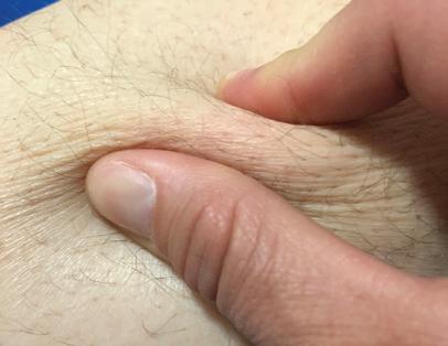
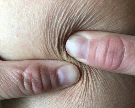
To assess tissue texture, gently encircle the limb with your hands and palpate the tissues from the toes to the hips. Note the consistency of the tissue, which can vary in different locations. Other simple tests that can be performed to assess tissue texture are the Pitting Test, Stemmer’s Test22 and Bjork Bow Tie Test. 69,70 To assess for pitting, apply sustained pressure to the tissue with the pad of the tip of the thumb, allowing it to sink into the oedema. Once indented, count how long it takes for the indentation to rebound (fill back in). Perform the Stemmer’s test by lifting and pinching the skin at the base of the second toe. Skin that cannot be lifted and pinched is positive and indicative of dermal and soft tissue thickening and fibrosis. The Bjork Bow Tie Test is an expansion of the Stemmer’s Test and can be
S.T.R.I.D.E. compression selection guide
performed anywhere on the body. Fig 15 explains how to perform this test, and its interpretation.
CLINICAL PEARL Simple tests that can be performed to asses tissue texture are the Pitting Test, Stemmer’s Test, and Bjork Bow Tie Test.
Watery tissue texture
Watery tissue texture is soft and pliable, easily pits and rebounds quickly, can be deeply pitting, and does not present with fibrotic tissue changes and therefore results in a negative Stemmer’s and Bjork Bow Tie Test (described in E=etiology). Watery oedemas are easy to reduce and manage with compression and typically require compression garments only during the day. Watery oedema worsens with dependency of the oedematous area, and reduces with elevation. It reduces quickly with adjustable wraps or multi-layered bandages and can be typically managed long term with circular knit garments at lower dosages. However, deeply pitting, watery oedemas may require stiffer circular knit or flat knit garments to prevent creasing into joint lines.
To perform the Bjork Bow Tie Test, in one maneuver, gently pinch, roll and twist the skin between the thumb and pointer finger, noting quality of tissue texture and thickness. Healthy skin can be lifted and pinched, should feel slippery between the layers when rolled and produce a ‘Bow Tie’ of wrinkles when twisted. Skin that tests positive for tissue texture changes will be thickened, less pliable, unable to be pinched and lifted, difficult to twist, and produce limited ‘Bow Tie’ of wrinkles. Negative test results include tissue that can be lifted, wrinkles when twisted and is of normal skin thickness for the tested body part.
Fatty tissue texture
Fatty tissue can be healthy fat with normal connective tissue architecture and healthy, elastic skin or it can be abnormal fat associated with lipoedema, with poor connective tissue support. Healthy fat can be supported with thinner, elastic garments, while these same garments tend to roll into creases and sink between folds in abnormal fatty tissue, causing uncomfortable binding and increased risk of linear skin damage. In contrast, stiffer and thicker textiles like flat knit garments can bridge across creases and help shape and support the abnormal fatty tissue.
Putty tissue texture
Putty tissue texture represents early fibrotic tissue changes resulting in a putty consistency. The tissue pits with deep and firm prolonged pressure, has a >30 second rebound, and a positive Stemmer’s and Bjork Bow Tie Test. Areas with putty consistency typically require day and night compression. Stiffer garments are required to provide adequate containment and textured compression is optimal to soften fibrotic tissue and facilitate lymphatic drainage.
up illustrating ‘Bow Tie’ of
Fig 15. Bjork Bow Tie Test
Negative Bjork Bow Tie Test
Positive Bjork Bow Tie Test
Alternate method Bjork Bow Tie Test
Close
wrinkles
S.T.R.I.D.E. compression selection guide
Woody tissue texture
Woody tissue texture represents more advanced fibrotic tissue changes with firm, woody consistency. It does not pit to deep pressure, and the Stemmer’s and Bjork Bow Tie Tests are positive. Areas that possess woody tissue texture require day and night compression, stiffer textiles and higher compression pressures, and textured compression options to warm and soften the fibrotic tissue in order to help facilitate soft tissue remodelling and lymphatic capillary regeneration. Although best treated before developing woody tissue texture, there are specialised, custom compression products with integrated textured foam that can help to reduce even the most advanced Stage 3 lymphoedema.
Fragile Tissue Texture
Thin, fragile skin has lost elasticity and tears or bruises easily. This is a common problem in the aged population and should be considered in compression garment selection. Selecting circular knit garments with double covered inlay yarns to decrease friction when donning the garment, layering an under-liner and secondary garment, use of silicone lotion on the skin to decrease friction, using special donning and doffing devices or choosing adjustable wraps are some options to help protect fragile skin.
CLINICAL PEARL To assess a garment for double covered yarns, turn it inside out. Stretch the textile and observe for flecks of bare elastic showing through, or a sheen from a higher concentration of uncovered inlay yarns.
Textile type
Elastic textiles
Elastic textiles have long stretch properties with higher resting and lower working pressures, and more elastic recoil contributing to sustained pressures. The dynamic performance of elastic textiles produces a lower static stiffness and thus a reduced containment effect. Some advantages of this type of textile include wider size ranges, and a variety of thinner, cosmetically appealing RTW options in a multitude of colours and prints. These garments are also generally lower cost. Disadvantages of elastic textiles include limited ability to bridge skin folds, limited ability to shape fatty tissue and diminished ability to contain more robust oedemas. Examples of more elastic textiles are circular knit garments. Sheer circular knit garments are the most elastic, followed by opaque garments, and then those made from natural yarns such as cotton. Some night garments are made of elastic textiles because they
are made by cutting and sewing stretchy fabrics. Elastic textiles are most useful for watery oedemas where there is normal connective tissue support and skin elasticity. As previously described, stiffer circular knit garments represent a category of circular knit garments that have unique engineering properties and are roughly double the stiffness, or half the elasticity, of traditional circular knit garments. Stiffer circular knit garments are considered, a more elastic textile compared with flat knit but stiffer when compared to traditional circular knit garments. Stiffer circular knit garments are useful for watery oedemas and fatty tissue, especially where the oedema includes the foot and thigh. In addition, using a stiffer circular knit garment during the day, combined with a textured AW night garment, may prove successful for management of putty tissue texture.
Stiffer textiles
Stiffer textiles have short stretch properties with lower resting and higher working pressures, and less elastic recoil. The dynamic performance of stiffer textiles produces intermittently high therapeutic IP while providing lower resting pressures. This dynamic compression profile allows for optimised outcomes at lower compression class prescription compared to elastic textiles. Some advantages of this type of textile are the ability to bridge across fat folds and lobules, ability to shape soft tissue, and containment of robust oedemas with more rapid refill times. Some disadvantages may include a thicker textile, need for custom sizing and associated higher costs, less recoil to accommodate to oedema reduction without readjustment, and in AW product lines reduced options for colours, patterns and prints.
CLINICAL PEARL To manually assess the stiffness of a circular or flat knit compression garment, insert two hands down into the garment, palms facing each other, and pull them apart to assess resistance to stretch. Stiffer textiles will offer increased resistance. Next stretch the garment vertically to assess vertical stiffness. Stiffer textiles will have less vertical elongation.
Because stiffness data is not currently provided by compression manufacturing companies, manual assessment of the textiles is often used. To manually assess the stiffness of a circular or flat knit compression garment, insert two hands down into the garment, palms facing each other, and pull them apart to assess resistance to stretch. Stiffer textiles will offer increased resistance. Next stretch the garment vertically to assess
S.T.R.I.D.E. compression selection guide
vertical stiffness. Stiffer textiles will have less vertical elongation. To manually assess the stiffness of AW, night garments, or decongestive garments, firmly grip a section of the textile between two hands and then pull them apart. Stiffer textiles will have minimal stretch and provide higher levels of resistance.
Textured textiles
Textured textiles can be found integrated into a variety of garment categories. These can provide a multidimensional effect on both oedema management and tissue texture, including not only more effective oedema reduction but also resolution of trophic changes. Options for textured textiles range from the course knit textiles found in flat knit garments to dense open or closed cell foam chips fabricated into vertical or diagonal channels of night garments. Most textured textiles are integrated into night garments as foam layers, which provide an added benefit of warming and softening fibrotic soft tissue. Disadvantages of textured textiles are they can be thicker and more bulky, making them more feasible to use at night versus day.
The use of textured textiles is an area of emerging research and study. The International Lymphedema and Wound Training Institute (ILWTI) coined the term Lymphatic Alternating Pressure Profiles (LAPP) to describe textured compression profiles with areas of alternating high and low pressure zones.71 Intermittent mechanical deformation of the skin and subcutaneous tissue under high/low pressure areas is thought to stretch anchoring filaments of the lymphatic capillaries, opening swinging tips and allowing fluid movement from the interstitial tissue into the functioning lymphatic capillaries. This fluid movement, further enhanced by movement within compression, stimulates the formation of new lymphatic capillaries.
These hypotheses are based on emerging evidence, although in vivo measurements have not yet been established as they relate to various compression garments. According to early research by Gerli and Alessandrini in 1995,72 the lymph capillary, anchoring filaments and elastic fibres of the connective tissue form one continuous functional unit. In 2002, Swartz et al.73 stated that lymphatics must act as a mechanical extension of the interstitium to be functional. They found that the microfibrils of anchoring filaments are expressed by lymph endothelial cells in culture and mechanical stimulation of the anchoring filaments may result in a biochemical response in the cell that mediates lymphangiogenesis.73 Further, their more recent published findings demonstrate that interstitial
fluid flow is necessary for lymphatic reorganisation, and interstitial fluid channelling precedes and may even direct lymphangiogenesis.74,75
CLINICAL PEARL To manually assess a garment for texture, inspect the garment for coarse yarns, built-in ridges, or chipped foam layers. Lymphatic Alternating Pressure Profiles (LAPP) may be enhanced by ridges, chipped foams, denser materials and greater differences between high and low interface pressures.
Adjustable textiles
All AW and decongestive garments are considered to be adjustable textiles that allow for alteration of the circumferential dimensions to address changing limb volumes. These garments allow the clinician, end user or caregiver to adjust the fit independently as the oedema reduces or to adjust for other volume changes or comfort level. Depending on the type of closure system and grip strength of the user, they can be more easily donned and doffed by some users. There are also options on the market that allow for one-handed, sidebending closure for individuals who are obese and cannot bend forward to adjust straps. Garments designed as adjustable textiles are made of stiffer textiles. Many night garments are also constructed as adjustable textiles.
Layered textiles
Layered textiles can be purchased together as a kit or as individual garments. Layering is an application option that changes the overall dynamic performance of the compression garment system, compared with the individual pieces alone. Clinical experts layer MCS garments to achieve desired functional outcomes with compression therapy. Layering is a useful method to increase stiffness, improve ease of donning, increase options for day-to-day compression wear for different activities, protect fragile skin, or enable donning over wound dressings.
Layering of MCS has been shown to increase stiffness. Ulcer care kits are an example of layering, consisting of two circular knit stockings — a light compression liner (8–10mmHg) and moderate compression (20–30mmHg) outer stocking. Layering of MCS increases not only dosage, but also stiffness.8,28,30,44,76,77 Hiari et al observed the IP was nearly double when the same two stockings were applied.8 However, the stiffness was dependent on the type of MCS layered. For example, higher stiffness was observed with layering flat knit MCS compared with layering circular knit MCS. 8 Overall performance of layering MCS should be
S.T.R.I.D.E. compression selection guide
considered on an individual basis, because each individual textile will have an impact on the dynamic performance of the layered system as a whole.8
A commonly used example of layering includes the application of two similar compression textiles one on top of the other, such as the case with ulcer care kits. For patients with more complex presentations, unique combinations of compression textiles can be paired. An example would be layering a class II flat knit knee-high garment under a class I flat knit footless thigh high for a patient who presents with full leg swelling with localised fibrotic changes limited to primarily the calf region. The double coverage over the calf would afford the benefits of textured compression and increased stiffness over the most affected area, while minimising the impact on patient comfort and mobility of the limb as whole. Layering of compression is also a way to optimise patient compliance.
CLINICAL PEARL Layering MCS is a useful method to increase stiffness, improve ease of donning, increase options for day-to-day compression wear for different activities, protect fragile skin, or enable donning over wound dressings.
R = REFILL
Q: Does the oedema increase during the day only, or day and night?
Q: How fast does the limb increase in size when compression is removed?
Oedema that refills during the day only, resolving with elevation or overnight, only requires day compression. In contrast, oedema that refills during both day and night requires day and night compression. Circular knit, stiffer circular knit and flat knit garments are designed for wearing during the day. AW and decongestive garments are designed for use both day and night. Night garments are designed for soft tissue remodelling and oedema reduction while sleeping, however, they can also be worn during waking hours.
In addition to day and night refill, the time to refill is an important factor. ‘slow refill’, as defined by S.T.R.I.D.E., means that, when compression is removed, the oedema slowly increases over the course of 24 hours or more. In contrast, ‘rapid refill’ means that, when compression is removed, the oedema starts increasing almost immediately and swells beyond the garment size within 8 hours. Refill time relates clinically to the level of containment needed to manage the oedema adequately. ‘Containment’ is the ability of a garment to prevent the
expansion of oedema and to maintain the ideal shape of a limb after therapeutic decongestion. Oedemas with very rapid refill require more robust containment and strict adherence to a compression regime, and a plan for independent oedema reduction when needed.
I = ISSUES
Q: Are there medical concerns that would limit compression use/application?
Q: What are the barriers to successful oedema management?
Q: What modifications can be made to overcome identified barriers?
Adherence with compression therapy regiments is dependent on a thorough assessment of the patient as a whole. Comorbidities, such as peripheral artery disease (PAD), chronic kidney disease (CKD), congestive heart failure (CHF), obesity and many other medical conditions must be considered. In addition, the patient’s physical and mental ability or support network, financial means, and his or her goals for compression therapy contribute to success or failure. The ideal compression product may be available, however, if the patient is unable to don and doff, or if the patient does not find the compression garment cosmetically acceptable, successful oedema management is not possible.
Medical compression is indicated for oedema caused by a variety of underlying etiologies. A compiled list of precautions and contraindications for the use of medical compression therapy are listed in Box 1, gathered from available consensus documents, scientific evidence and expert opinion. To the consumer, it should be noted that precautions and contraindications listed are guidelines only and do not replace medical advice.
Before application of compression bandaging or compression garments, a complete medical assessment of the individual should be performed, including evaluation of the arterial, venous and lymphatic vasculature. At a minimum, a venous duplex ultrasound to assess for blood clots in deep veins, and an Ankle Brachial Index (ABI) to assess for PAD should be conducted. Applying compression to a limb with severe PAD, or applying the wrong type of textile to a limb with PAD can result in tissue necrosis, amputation, or even death secondary to complications. Appendix 1 includes an ABI guidance document that can help the clinician interpret the ABI, as it relates to both wound healing and medical compression.
S.T.R.I.D.E. compression selection guide
BOX 1
Precautions
• Mild to moderate peripheral arterial disease (PAD)
• Peripheral neuropathy
• Chronic, compensated congestive heart failure
• Chronic renal failure
• Difficulty donning & doffing garment—consider patient’s dexterity, finger and grip strength, hip flexion, caregiver support etc.
• Dermatitis, eczema, and other skin conditions
• Lymphorrhoea
• Minor skin infections
• Fabric sensitivities
• Malignancy.
Contraindications
• Acute-untreated cellulitis or other major skin infections
• Acute-untreated deep vein thrombosis or thrombophlebitis
• Severe peripheral arterial disease (ABI <0.5 or absolute systolic ankle pressure 60mmHg or less), and s/p Arterial bypass grafting.
BOX 2
15–20mmHg
• Light compression for those with leg oedema 21
• CEAP* C0-1 and varicose veins in pregnancy, prevent leg swelling with prolonged sitting or standing prevent deep vein thrombosis with bedridden patient 21,77
20–30mmHg
• VLU prevention and treatment in those with no history of DVT14
• Mild oedema, varicose veins and venous ulcers18,32,77
• Use after sclerotherapy and for those with small telangiectsiases (C1), those with subjective symptoms associated with varicose veins and in acute phase of DVT 75
• CEAP C3-415
30–40mmHg
• VLU prevention & treatment in those with history of DVT14,15
• Following sclerotherapy large veins (C2), those with lipodermatosclerosis/tissue changes (C4b), promote healing VLU (C6), prevent VLU recurrence, lymphoedema77
40–50mmHg
• Severe oedema; severe venous disease;venous ulcers; lymphoedema1,18,32,68
• Deep vein thrombosis102
50–60mmHg
• Lymphoedema 32
* see Box 4 for the definition of CEAP classification
D = DOSAGE
Q: What is the appropriate dosage based on medical diagnosis, precautions, contraindications, and underlying oedema etiologies?
Q: Do certain areas of the limb require greater compression dosage due to size or texture?
Guidelines and/or recommendations for compression dosage (resting interface pressure measurements) prescription are found throughout the literature and are briefly summarised in Box 2.
It should be noted that these recommended compression dosages are based solely on a medical diagnosis and do not encompass individual patient characteristics such as limb size, tissue texture, distribution of oedema or functional ability of the patient. There is a tendency to prescribe higher dosages for more severe oedemas, which is often unrealistic for the patient to don or doff. Options to mitigate this challenge include use of donning and doffing devices, and referring the patient to a rehabilitation specialist to provide training using these devices. Further, the dosage recommendations do not consider other compression options that integrate the dynamic properties of individual textiles. For example, layering two garments of lower dosages, or choosing stiffer textiles at lower dosages, can be effectively used in lieu of simply prescribing higher dosages of compression The S.T.R.I.D.E. algorithm provides a systemised approach to compression garment prescription that incorporates both the etiology of the swelling and the individualised presentation.
As a practical example, note that the dosage recommendations in Box 2 for C4 venous disease is 30–40mmHg. In contrast, the S.T.R.I.D.E. algorithm not only includes 30–40mmHg compression options, but also lower dosage options such as layering two 15–20 circular knit garments, choosing a stiffer circular knit garment with 20–30mmHg dosage, a Flat knit garment class 1 or 2 (18–21mmHg or 23–32mmHg), or an AW with a resting pressure of 20-30mmHg. As the garment stiffness increases, dosage prescription can decrease.
CLINICAL PEARL Effective compression prescription requires matching the compression selection to the patient presentation, not to the diagnosis alone.
S.T.R.I.D.E. compression selection guide
Additionally, there are guidelines that have been published with regards to the use of compression for those with peripheral arterial disease (PAD) or mixed disease, summarised in Box 3.21,78–81
CLINICAL PEARL Layering two garments of lower dosages, or choosing stiffer textiles at lower dosages, can be effectively used in lieu of simply prescribing higher dosages of compression.
E = ETIOLOGY
Q: What comorbidities are contributing to the oedema/lymphoedema?
The new paradigm of the microcirculation positions the vascular endothelial glycocalyx layer (EGL) as the gatekeeper of fluid filtration from blood capillaries.69,70 Contrary to previous concepts of Starling’s Law, we now know there is no reabsorption back into the venous end of the blood capillaries and only diminishing fluid filtration across the capillary bed.82 All interstitial fluid (100%) is reabsorbed by the lymphatic capillaries alone. Consequently, all oedemas are on a lymphoedema continuum69,70 and represent relative lymphatic failure or insufficiency. 83
The lymphoedema continuum is shown in Fig 16. The bottom arrow pointing left to right illustrates that each individual falls somewhere between a fully optimised lymphatic system, where their lymphatic vasculature is perfect in form and function, versus a poorly optimised lymphatic system where there is some type of underlying congenital dysfunction, mechanical insufficiency or failure. The arrow to the left pointing bottom to top illustrates an increasing number of demands on the lymphatic system due to comorbidities and risk factors. The more demands on the lymphatic system and the less optimised the system is, the more likely it is to progress in the severity of chronic inflammation and tissue texture changes. This continuum demonstrates the need for diagnostic imaging such as indocyanine green (ICG) fluoroscopy, which shows lymph flow in real time, to assess an individuals’ lymphatic vasculature. This would help steer what steps need to be taken to manage their risks of lymphoedema, including a customised programme with more specific recommendations regarding their compression therapy regime.
All oedema is technically lymphoedema, albeit stemming from different underlying etiologies or diagnoses. Past definitions of ‘lymphoedema’ need to be replaced by the new evidence and broader definitions. Clinically, all comorbidities contributing
BOX 3
Ankle Brachial Index (ABI) Guidance—see ABI Guidance doc
• >1.40: falsely elevated
• 1 to 1.40: normal—safe to use all levels of compression (15–20, 20–30, 30–40, 40–50mmHg)
• 0.91 to 0.99: borderline PAD—safe to use all levels compression (15–20, 20-30, 30-40, 40–50mmHg)
• 0.8 to 0.90: mild PAD—apply compression with caution (15–20, 20–30, 30-40mmHg)
• 0.51 to 0.79: moderate PAD - Use modified compression with caution. Absolute systolic ankle pressure should be >60mmHg.103 Short stretch or inelastic garments safer than elastic compression for lower ABI. Use reduced compression pressure (15-20, 20-30mmHg) for elastic garments.
• ≤0.5: severe PAD, critical limb ischemia - unsafe to use compression. DISCLAIMER: This is a GUIDE ONLY and cannot replace clinical judgment. Each clinician is responsible for comprehensive evaluation and a plan of care appropriate for individual patient needs.
to the oedema should be captured on the diagnoses list, in addition to the diagnosis and appropriate staging of lymphoedema. S.T.R.I.D.E. bridges the gap between old and new by not only including traditional diagnoses and classifications, but also an evidencebased algorithm for compression garment selection that is based on the patient presentation, not just a diagnosis. Therefore, lymphoedema presenting as chronic swelling after an orthopaedic injury, a total knee replacement, or an infection, etc is just as applicable to using the S.T.R.I.D.E. system, even though these etiologies are not specifically discussed below. Matching the tissue texture characteristics to the appropriate textile characteristic provides guidelines for compression selection across the full continuum of oedema presentations.
Number of oedema etiologies (diagnoses)
Lymphoedemacontinuum
Fully optimised lymphatic system (Diagnostic ICG fluoroscopy)
Poorly optimised lymphatic system (diagnostic ICG fluoroscopy)
Fig 16. Lymphoedema continuum
S.T.R.I.D.E. compression selection guide
CLINICAL PEARL Matching the tissue texture characteristics to the appropriate textile characteristic provides guidelines for compression selection across the full continuum of oedema presentations.
As already stated, all chronic oedema is lymphoedema and can alter the surrounding tissue physiology resulting in a progression of fibrotic tissue texture changes in response to localised chronic inflammation.69,70,83 This lymphatic failure produces a cutaneous region susceptible to infection, inflammation and carcinogenesis and a disruption of adaptive immunity, called lymphatic dermopathy, which is failure of the skin as an immune organ.85 Simple, manual tests to identify early soft tissue changes indicative of this pathophysiology are the Stemmer’s Test22 and the Bjork Bow Tie Test,69,70 which is an expanded version of Stemmer’s Test, previously described, and can be performed anywhere on the body (Fig 15). When these tests are positive, they are indicative of chronic inflammation, tissue thickening and fibrotic soft tissue changes. It is important that all clinicians in every branch of clinical practice become proficient in these tests and use them to examine all areas of the body that are oedematous. Early identification of these deleterious soft tissue changes are key to early interventions, more effective compression choices, and reduction of potential complications such as pain, diminished quality of life, disfigurement, decreased mobility, emotional distress, chronic wounds, and recurrent cellulitis.
Incorporated into S.T.R.I.D.E. are common diagnoses leading to the need for compression therapy for the lower extremities. These are chronic venous disease (CVD), lymphoedema, lipoedema, dependent oedema, CHF, and CKD. Also included are other major contributing factors, such as obesity and the side effects of medications. Each oedema diagnosis has its own unique characteristics, however, most clinical presentations are a combination of comorbidities as illustrated in Fig 17. The multi-factorial nature of chronic oedema states is highlighted in 2016, study by Wang and Keast86 in which it was observed that patients with chronic oedema had 7.3 (±3.3) comorbidities. Thus, the following discussion of disease pathology resulting in chronic lymphoedema states is not meant to be all-inclusive, as most clinical presentations of lymphoedema result from a combination of comorbidities.
Chronic venous disease
CVD refers to chronic conditions related to or caused by veins that have become diseased or abnormal. These problems include varicose veins, leg oedema, CVI, venous leg ulcers (VLU), phlebitis and vascular malformations.87 Globally, CVD is expected to increase, due to advancing age and increasing obesity. The global population aged 60 years or over numbered 962 million in 2017, more than twice as large as in 1980, and the number of older persons is expected to double again by 2050, when it is projected to reach nearly 2.1 billion.88 Concurrently, worldwide obesity has nearly
Fig 17. Examples of multi-factorial lymphoedema
Phlebolymphoedema: chronic venous insufficiency with hemosiderin staining, lipodermatosclerosis, and secondary lymphoedema with positive Bjork Bow Tie Test, and papillomatosis.
Lipolymphoedema: classic lipoedema with loose, lobular, fatty lobes and relatively spared feet, coupled with secondary lymphoedema of the lower legs and feet, chronic inflammation and positive Bjork Bow Tie Test.
Softly pitting oedema from congestive heart failure with chronic inflammation, progressed to secondary lymphoedema with positive Bjork Bow Tie Test. Underlying chronic venous insufficiency as noted by light hemosiderin staining of left leg.
S.T.R.I.D.E. compression selection guide
tripled since 1975.89 These factors will contribute to the rise in CVI as well as painful, problematic VLU.
As of 2015, about 1% of the western population will suffer from a VLU during their lifetime.31 The annual US payer burden alone for VLU is estimated at $14.9 billion. 90 Compression therapy is widely recognised as key to the management of VLUs, with quicker healing times and reduced recurrence rates when compared with no compression.31 Unfortunately, the value of compression therapy has been underused, or perhaps more accurately misused due to the lack of clear descriptive explanation of compression textiles available on the market. Up until this point, prescription of compression has been based on dosage alone, despite a growing body of compression science that demonstrates compression dosage is only one part of the dynamic performance properties of a compression textile. It is a goal of S.T.R.I.D.E. to provide clear direction for practitioners and consumers towards effective compression product selection to reduce the economic burden of VLU, as well as improve quality of life for those patients living with CVI.
Relative to venous disease, the S.T.R.I.D.E. algorithm is organised by CEAP Classification (Box 4).91–93 C0–C2 includes ‘spider veins’ or varicose veins and corresponding prophylactic compression recommendations are reflected on the S.T.R.I.D.E. algorithm. Clinical compression recommendations corresponding to C3–C6, CVI, are also individually described on the S.T.R.I.D.E. algorithm. C3 is indicative of oedema, C4 skin changes, and C5 and C6 healed or active VLU, respectively. A distinguishing clinical characteristic of CVI is hemosiderin staining, a reddishbrown discolouration visible in the skin of the gaiter area. Other clinical signs of CVI include lipodermatosclerosis, varicosities, atrophie blanche and stasis dermatitis.
Oedema secondary to CVI slowly worsens through the day with prolonged sitting or standing and resolves overnight or with the legs elevated at the level of or slightly above the heart. Initially, the tissue texture is pliable and watery, but can progress to the development of fibrotic tissue with a putty-like texture as the concurrent lymphatic failure progresses. If concurrent with other oedema etiologies, foot and toe swelling may be present and must be considered when making a garment selection. For example, many traditional circular knit garments have a slight flare across the metatarsal heads and only 50% of the compression pressure in the foot as compared to the ankle. When foot swelling is present, a stiffer circular knit or a flat knit garment, a more columnar shaped foot, and
• C0 = no visible or palpable signs of venous disease
• C1 = telangiectasies or reticular veins
• C2 = varicose veins (diameter 3mm or more)
Chronic venous insufficiency (CVI)
C3 –C6 = Phlebolymphoedema = lymphoedema secondary to venous disease
• C3 = oedema
• C4a = pigmentation and eczema
• C4b = lipodermatosclerosis and atrophie blanche
• C5 = healed venous ulcer
• C6 = active venous ulcer
higher dosages distribution in the foot relative to the ankle should be considered. Further, as fibrotic soft tissue develops it is likely the lymphoedema secondary to CVI will no longer resolve completely overnight, and a textured night garment may be needed to facilitate soft tissue remodelling as well as evacuation of lymph from the interstitial space overnight.
Lymphoedema
According to the International Lymphoedema Framework 2019 Lymphoedema Impact and Prevalence International (LIMPRINT) studies, lymphoedema has always been a neglected global health-care problem.94LIMPRINT is an international epidemiology study to identify the prevalence and impact of chronic oedema/lymphoedema throughout the health and social care systems in participating countries. Chronic oedema/lymphoedema is defined as oedema present for more than three months. The lymphatic component is recognised in this study.
Data from 68 enrolled subjects in the Canadian LIMPRINT study showed that the most common underlying condition for lymphoedema was venous disease, 72% of patients had a history of cellulitis, and ~40% had an open wound.94 In 2008, it was estimated that 10 million people were suffering from lymphoedema in the US alone, more than multiple sclerosis, muscular dystrophy, ALS, Parkinson’s disease, and AIDS combined.96,97 Furthermore, according to WHO’s 2019 Morbidity Management and Disability Prevention (MMDP) information, an estimated 40 million people worldwide suffer from clinically significant manifestations of the lymphatic filariasis (LF) alone, predominately lymphoedema, elephantiasis and hydrocele.
As profound as these statistics are, we are only on the cusp of grasping the global impact of chronic oedema/lymphoedema, the continuum of oedema and how to use the continuum of compression
S.T.R.I.D.E. compression selection guide
BOX 5
Lymphoedema
• Stage 0: A latent or subclinical condition where swelling is not yet evident despite impaired lymph transport, subtle alterations in tissue fluid/ composition, and changes in subjective symptoms. It may exist months or years before overt oedema occurs
• Stage 1: Early accumulation of fluid relatively high in protein content (eg in comparison with ‘venous’ oedema) which subsides with limb elevation. Pitting may occur. An increase in various types of proliferating cells may also be seen
• Stage 2: Limb elevation alone rarely reduces the tissue swelling and pitting is manifest.
• Late stage 2: Limb may not pit as excess subcutaneous fat and fibrosis develop
• Stage 3: Lymphostatic elephantiasis, where pitting can be absent and trophic skin changes such as acanthosis, alterations in skin character and thickness, further deposition of fat and fibrosis, and warty overgrowths have developed.
It should be noted that a limb may exhibit more than one stage, which may reflect alterations in different lymphatic territories
therapies available to provide state of the art treatment. S.T.R.I.D.E. helps mitigate this gap by addressing the clinical presentations of the lymphoedema as they relate to compression selection. Underlying oedema etiologies are represented by their diagnoses and classifications in the S.T.R.I.D.E. algorithm, as well as tissue texture changes. Accordingly, if fibrotic tissue texture changes are present in CVI, for example, the compression selection defaults to the recommendations in the corresponding stage of lymphoedema. International Society of Lymphology (ISL) Staging for lymphoedema is summarised in Box 5 below.98
The duration (# hours /day) of compression wear for those with lymphoedema is dependent on the how the volume of the limb changes throughout the day. Lymphoedema may display dynamic volume changes with removal of compression. For example lymphoedema presentations of congenital malformation/primary lymphoedema or secondary lymphoedema resulting from extensive, surgical excision of lymph structures can lead to lymphoedema with very rapid refill. Rapid refill significantly impacts the compression garment selection. Higher levels of containment are needed, diligence is required on the part of the user to wear the compression without interruption, differing compression may be needed for different activities and risks, and night compression needs to be considered as well.
Chronic oedema resulting from lymphatic insufficiency that is not addressed leads to progressive fibrotic tissue changes such that the swelling no longer reduces overnight, or with elevation. The skin and subcutaneous tissue thickens and hardens, leading to initially subtle, and later obvious, positive findings
with the Bjork Bow Tie Test. Putty or woody fibrotic tissue changes consistent with late stage 2 and stage 3 lymphoedema require textured compression to help facilitate soft tissue remodelling. Even in cases of advanced lymphoedema presentations, such as ‘elephantiasis’ with ‘massive localised lobules’, appropriately chosen compression can still prove effective.
Lipoedema
There are four Stages of lipoedema and five Types99,100 summarised in Box 6. The stages depict the soft tissue characteristics and the types depict where the lipoedema is distributed.
Lipoedema is also known as the painful fat syndrome, but is differentiated from obesity. It is a chronic disease primarily affecting females, resulting in abnormal fat metabolism and accumulation and hyperplasia of subcutaneous fat cells. Disproportional fat distribution on the legs, without a similarly proportional buildup in the upper body/trunk region, is a key distinguishing characteristic of lipoedema. Typical features of lipoedema also include symmetrical swelling, bulging around the ankle (described as an inverted shelf or ‘Turks trousers’) and relatively spared feet. Unlike the smooth feel of obesity fat, reports indicate that lipoedema fat can present with palpable nodules –described as feeling like frozen peas in a bag or pearls.98
The overall consistency of the soft, fatty tissue is loose, with poor connective tissue support leading to skin that is easily bruised, hypermobility, venous disease and oedema. When oedema is present, the tissue feels heavier like a water balloon rather than light and fluffy. Palpation and gently lifting and dropping the tissue enables the clinician to manual assess for oedema. Garments are needed in all stages to support the tissue and prevent progression to Stage 4, lipo-lymphoedema. The garment textile selected must be able to help shape the fat, and bridge across fat folds.
Dependent oedema
Immobility, decreased mobility, ineffective mobility, and dependent positioning of limbs or other body parts can lead to ‘dependent’oedema. Dependent oedema is often associated with geriatric patients that spend much of their day in a wheelchair. Dependent oedema may be overlooked in other situations, such as hemiplegia following a stroke, a pannus that hangs over the edge of a bed, or a patient that sleeps in their recliner. Like any other etiology of lymphoedema, chronic dependent oedema can also present with tissue texture changes that then change the type of
S.T.R.I.D.E. compression selection guide
compression that may be most effective. In general, dependent oedema of the lower extremities is usually more watery and able to be managed with less containing garments worn during the day only, provided the limbs are elevated at night.
Congestive heart failure and chronic kidney disease
CHF and CKD are common, underlying etiologies of chronic oedema/lymphoedema. Because they are systemic causes, the oedema presents symmetrically in the lower extremities. Oedema from CHF is soft, doughy and deeply pitting, with swelling observed in the dorsum of the foot and toes as well as the limb. If the lymphoedema secondary to CHF is chronic or in combination with comorbidities, it can progress to fibrotic soft tissue changes, typically presenting symmetrically across the toes and dorsum of the foot and behind the malleoli. As noted in “I = Issues”, compression is contraindicated for acute-decompensated CHF. However, as long as the CHF is medically managed, compression therapy helps manage the edema at a level that is tolerated by the heart and preserves the integrity and health of the skin and soft tissues.
CKD also presents with unique compression needs. As CKD progresses, the increasing amount of fluid building up in the interstitium each day, and the increased outward pressure on the skin, will require higher containing garments. As dialysis begins and the oedema fluctuates pre and post treatment, adjustable garments are recommended to accommodate to the variable limb size. Like other oedema etiologies, using the S.T.R.I.D.E. algorithm to assess oedema characteristics, versus just the diagnosis, will help direct the textile choices that ultimately lead to compression success.
Obesity and medications
While outside the scope of this document, obesity plays a very important role in contributing to chronic oedema. Worldwide obesity has nearly tripled since 1975.89 In 2016, more than 1.9 billion adults (39%), 18 years and older, were overweight and over 650 million (13%) were obese.89 The obesity epidemic is evident in those patients with chronic oedema as was evidence in a 2019 study by Keast. It was observed that 47% of patients identified as having lymphoedema were morbidly obese with body mass index of >40.94
In addition to the impact of comorbidities, considering potential side effects of medications needs to be a part of every comprehensive medical assessment of oedema. It is important to look at all the contributing factors for each individual patient. As previously discussed, in most cases there are multiple underlying lymphoedema
BOX 6 LIPOEDEMA
• Stage 1: Normal skin surface with enlarged hypodermis.
• Stage 2: Uneven skin texture with indentations in the fat, larger mounds of tissue growing as unencapsulated masses.
• Stage 3: Thickening and hardening of the subcutis with large nodules and protruding fat pads, especially on the thighs and around the knees.
• Type II: Buttocks to knees, with folds of fat around the inner side of the knee
• Type III: Buttocks to ankles
• Type IV: Arms
• Type V: Lower leg
etiologies that need to be diagnosed and medically managed. However, by selecting garments on the basis of the clinical presentation of the lymphoedema, success can still be achieved with compression for even the most complex patients with multiple comorbidities.
S.T.R.I.D.E SUMMARY
Expert clinicians and compression fitters consider all components of S.T.R.I.D.E. when helping to select or recommend the best compression garment(s) for their individual patient. The components of S.T.R.I.D.E. — Shape, Texture, Refill, Issues, Dosage, and Etiology all work synergistically for optimal compression garment selection and improve the opportunity for patient success with compression recommendations. The S.T.R.I.D.E. Worksheet (appendix 2) is designed to help cue the clinician in the assessment process and serves as a tool for note taking. The S.T.R.I.D.E. Quick Reference Guide (appendix 3) is designed as a clinical tool that expands on the components on the S.T.R.I.D.E. Worksheet. The S.T.R.I.D.E. algorithm for lower extremity garments (appendix 4) points to specific textiles and dosage recommendations for diagnostic classifications of common lower extremity oedema etiologies, based on best available evidence and expert opinion.
Future iterations of S.T.R.I.D.E. will include algorithms for upper body garments, compression bandages, and other compression options. Future S.T.R.I.D.E.branded Product Guides will help categorise specific products made by reputable manufacturing companies who deliver standardised medical compression. Complete compression profiles and engineering data will be integrated. By creating parameters for compression categorisation, S.T.R.I.D.E. serves as a starting point for discussion and data collection, more cohesive research, clarity among clinicians and end-users of compression, and optimised success with compression therapies. JWC
All letters of the S.T.R.I.D.E. acronym are to be considered as a whole when selecting compression garments. However, for the purpose of a standardised process, each component can be addressed in sequence. As each letter is addressed, the information gained leads one closer to compression recommendations. Each step helps the clinician or consumer achieve greater compression success by ‘Taking compression garment prescription in S.T.R.I.D.E.’
CASE EXAMPLE I SHAPE
The location/distribution of the swelling is assessed to determine the area of compression coverage needed. As seen in the photo, the right leg requires compression below the knee and the left leg requires compression including the thigh as evident by swelling above the knee. In addition to the location of the swelling, the overall shape of the limb and circumferential measurements are assessed to see if they fit into RTW garment sizing options. The limb has an average shape and the circumferential measurements fall within standard categories of RTW garment options for both legs.
TEXTURE
Assessment of tissue texture provides clinical direction with regards to compression textile selection. The tissue of the right leg below the knee is a soft putty which pits with deep and firm prolonged pressure, with >30 second rebound, and has a positive Stemmer’s and Bjork Bow Tie Test. On the left leg, the distal 2/3 of the limb has a woody consistency. It does not pit to deep pressure, the Stemmer’s and Bjork Bow Tie Tests are positive. Stiffer textiles with higher compression pressures are best matched to these tissue textures. In addition, textured textiles can be chosen to soften the fibrotic tissues. Adjustable or layered garments are also an option as they provide a stiff compression profile and may afford the wearer greater ease with application.
REFILL
Assessment of the dynamic behaviour of the oedema throughout the day provides the clinician with
valuable information regarding the necessary duration of compression garment wear. During the initial assessment the patient reported that the limb swells progressively throughout the day when the limb is in dependent positions; In addition, he reports there is minimal volume reduction over night even with the limb elevated. As the limb has a rapid refill nature and swells both during the day and night, both day and night garments are needed. A garment with a firm containment is needed to prevent swelling during the daytime hours when the patient is in the upright position. A night garment with a textured profile could be used to address the fibrotic tissue changes as well as promote oedema reduction during night hours.
ISSUES
Compliance and concordance with compression therapy is dependent on a thorough assessment of the
CASE EXAMPLE 1
patient as a whole including associated comorbidities and physical abilities. Information gathered during the initial assessment for the patient pictured revealed no medical precautions/contra-indications for compression therapy. However, it was noted that due to his body habitus he had difficulty reaching his feet. In addition, he has moderate left side weakness and impaired dexterity of the left arm and leg secondary to a stroke. He is ambulatory, walking with a single point cane short community distances. A final concern voiced by the patient is lack of prescriptive coverage for compression therapy. Considering the limited reach and impaired dexterity of his left hand, an AW is no longer a feasible option as he is unable to reach and /or manipulate the straps to allow for adequate application. In addition, as he has no insurance coverage for compression products, layering of compression garments may not be economically feasible.
DOSAGE and ETIOLOGY
Recommendation for compression dosage based on underlying etiology of oedema presentation are summarised in the body of the S.T.R.D.E. manuscript and incorporated into spreadsheet as depicted below. The patient has CVD/CVI – C4 (yellow box) with a history of a deep vein thrombosis, post thrombotic syndrome. However, the presence of the woody, fibrotic tissue texture changes (see yellow circle) directs the prescriber to the row with compression options for late stage 2 lymphoedema. (see green box). From the other components of the S.T.R.I.D.E. algorithm it was identified that a stiffer compression application with texture was most appropriate. The compression choice that would best match to the patient’s current presentation would be a stiffer circular knit garment with a 20–30mmHg or 30–40mmHg dosage, or a class II or a class III flat knit garment. A knee-high garment could be used for the right leg, a thigh high is needed for the left.
CASE STUDY
Take it in S.T.R.I.D.E.
S hape
● Limb shape — normal
● Distribution — right leg below knee; left leg with some distal thigh swelling
● Garment — RTW OR custom –Knee high right, thigh high left
Texture
● Tissue – putty on right, woody of left distally
● Textile — stiffer textile to contain and bridge –Adjustable or layering an option
Refill — Rapid refill, with minimal reduction over night
I ssues — CVI, PTS, obesity
● h/o CVI with left sided weakness
● Cost - patient is paying out of pocket
D osage - 20–30mmHg or 30–40mmHg stiffer circular knit, layering an option
● Class II or class III flat knit
E tiology - Venous, lymphoedema
GARMENT CHOICE: flat knit class II applied with donning aide as pt not able to manipulate the AW/unable to reach feet - flat knit garment most economical option for patient as it offers stiff, textured textile to address underlying tissue texture changes in a single garment application.
Photos courtesy of Suzie Ehmann, used with permission
1 Clark M, Krimmel G. Lymphoedema and the construction and classification of compression hosiery. Lymphoedema Framework. Template for Practice: compression hosiery in lymphoedema. London:MEP Ltd, 2006.
2 Muldoon J, Charles H. Clinical effectiveness and patient considerations in oedema management. J Lymphoedema 2013;8(1):51–55.
3 Häfner HM, Jünger M. The Haemodynamic efficacy of six different compression stock-ings from compression class 2 in patients suffering from chronic venous insufficiency. Phlebology: The Journal of Venous Disease 2000; 15(3-4):126–130. doi:10.1177/026835550001500308
4 Neumann, MH. Elasticity, hysteresis and stiffness: the magic triangle. Veins Lymphatics 2013; 2(e6): 17–18
5 Van der Wegen- Franken. Muldert P, Tank B, Neumann HAM. Variation in the dynamic stiff-ness index of different types of medical elastic compression stockings. Phlebology 2008;23(2):77–84. doi:10.1258/phleb.2007.006018
6 Partsch H, Schuren J, Mosti G, Benigni JP. The Static Stiffness Index: an important parameter to characterise compression therapy in vivo. J Wound Care 2016 Sep;25(Sup9 Suppl 9):S4–S10. doi:10.12968/jowc.2016.25.Sup9. S4
7 Mosti G. Stiffness of compression devices. Veins and Lymphatics 2013; 2(1):1–2. doi:10.4081/vl.2013.e1
8 Hirai M, Koyama A, Miyazaki K et al. Interface pressure and stiffness in different combinations of compression material. Phlebology: The Journal of Venous Disease 2012; 27(2):82–89. doi:10.1258/phleb.2011.011010
9 Hirai M, Iwata H, Ishibashi H et al. Interface pressure and stiffness of various elastic stockings during posture changes and exercise. Vascular 2008; 16(2):95–100. doi:10.2310/6670.2008.00020
10 Hirai M, Niimi K, Iwata H et al. A comparison of interface pressure and stiffness between elastic stockings and bandages. Phlebology: The Journal of Venous Disease 2009; 24(3):120–124. doi:10.1258/phleb.2008.008057
11 Hirai M, Niimi K, Iwata H et al. Comparison of stiffness and interface pressure during rest and exercise among various arm sleeves. Phlebology: The Journal of Venous Disease 2010; 25(4):196–200. doi:10.1258/ phleb.2009.009064
12 Cornu-Thenard A, Benigni JP, Uhl JF. Terminolgy: resistance or stiffness for medical compression stockings? Veins and Lymphatics. 2013;2(4):11–12.
13 van Geest AJ, Veraart JC, Nelemans P, Neumann HA. The effect of medical elastic compression stockings with different slope values on edema. Measurements underneath three different types of stockings. Dermatol Surg 2000; 26(3):244–247. doi:10.1046/j.1524-4725.2000.09200.x
14 Ratliff CR, Yates S, McNichol L, Gray M. Compression for the primary prevention, treatment and prevention of recurrence of venous leg ulcers. J Wound Ostomy Continence Nurs 2016; 43(4):347–364. doi:10.1097/ WON.0000000000000242
15 O’Donnell TF Jr, Passman MA, Marston WA et al. Society for Vascular Surgery; American Venous Forum. Management of venous leg ulcers: Clinical practice guidelines of the Society for Vascular Surgery® and the American Venous Forum. J Vasc Surg 2014; 60(2 Suppl):3S–59S. doi:10.1016/j.jvs.2014.04.049
16 Lymphoedema Framework. Best Practice for the management of lymphoedema. International consensus. London: MEP Ltd, 2006.
17 Stout N, Partsch H, Szolnoky G et al. Chronic edema of the lower extremities: international consensus recommendations for compression therapy clinical research trials. Int Angiol 2012; 31(4):316–329
18 O’Meara S, Cullum N, Nelson EA, Dumville JC. Compression for venous leg ulcers. Cochrane Database Syst Rev 2012; 11:CD000265. doi: 10.1002/14651858.CD000265.pub3
19 Partsch H, Junger M. Evidence for the use of compression hosiery in lymphoedema. Lymphoedema Framework. Template for Practice: compression hosiery in lymphoedema. London: MEP Ltd, 2006.
20 van der Wegen-Franken CP, Mulder P, Tank B, Neumann H A M. Variation in the dynamic stiffness index of different types of medical elastic compression stockings. Phlebology: The Journal of Venous Disease 2008; 23(2):77–84. doi:10.1258/phleb.2007.006018
21 Rabe E, Partsch H, Hafner J et al. Indications for medical compression stockings in venous and lymphatic disorders: An evidence-based consensus statement. Phlebology: The Journal of Venous Disease 2018; 33(3):163–184. doi:10.1177/0268355516689631
22 Framework L. Best practice for the management of lymphoedema. International consensus. London: MEP Ltd. 2006:3-52.
23 EWMA Position Document - Understanding Compression Therapy 2003
24 Partsch H, Flour M, Smith PC; International Compression Club. Indications for compression therapy in venous and lymphatic disease consensus based on experimental data and scientific evidence. Under the auspices of the IUP. Int Angiol 2008; 27(3):193–219
25 Partsch H, Mortimer P. Compression for leg wounds. Br J Dermatol 2015; 173(2):359–369. doi:10.1111/bjd.13851
26 Partsch H, Moffatt C. An overview of the science behind compression bandaging for lymphoedema and chronic oedema. In: Compression therapy: A position document on com-pression bandaging. International Lymphedema Framework in association with the World Alliance for Wound
and Lymphoedema Care. 2012:12-22.
28 European Wound Management Association (EWMA). Focus Document: Lymphoedema bandage in practice. London: MEP Ltd, 2005.
29 Olszewski WL, Engeset A. Intrinsic contractility of prenodal lymph vessels and lymph flow in human leg. Am J Physiol 1980; 239(6):H775–H783
30 Mosti G, Mattaliano V, Partsch H. Influence of different materials in multicomponent bandages on pressure and stiffness of the final bandage. Dermatol Surg 2008; 34(5):631–639
31 Harding K et al. Simplifying venous leg ulcer management. Consensus recommendations. Wounds International 2015.
32 Alavi A, Sibbald RG, Phillips TJ et al. What’s new: Management of venous leg ulcers. J Am Acad Dermatol 2016; 74(4):643–664. doi:10.1016/j. jaad.2015.03.059
33 Casey S, St George J. An Introduction to Compression Therapy. Northeast Florida Medicine 2012;63(2):39–41.
34 Harding K et al. Simplifying venous leg ulcer management. Consensus recommendations. Wounds International. 2015.
35 Hirai M, Niimi K, Miyazaki K et al. Development of a device to determine the stiffness of elastic garments and bandages. Phlebology: The Journal of Venous Disease 2011; 26(7):285–291. doi:10.1258/phleb.2010.010041
36 Mosti G. Compression therapy in mixed ulcers. JMV-Journal de Médecine Vasculaire 2018; 43(4):223–224. doi:10.1016/j.jdmv.2018.06.004
37 van der Wegen-Franken K, Tank B, Neumann M. Correlation between the static and dynamic stiffness indices of medical elastic compression stockings. Dermatol Surg 2008; 34(11):1477–1485
38 Partsch H, Clark M, Bassez S et al. Measurement of lower leg compression in vivo: recommendations for the performance of measurements of interface pressure and stiffness: consensus statement. Dermatol Surg 2006; 32(2):224–232. doi:10.1097/00042728-200602000-00010
39 Van Der Wegen-Franken K, Roest W, Tank B, Neumann M. Calculating the pressure and the stiffness in three different categories of class II medical elastic compression stockings. Dermatol Surg 2006; 32(2):216–223. doi:10.1097/00042728-200602000-00009
40 Van der Wegen-Franken CP. (2009, October 22). Medical Elastic Compression Stockings. Erasmus University Rotterdam. Retrieved from http://hdl.handle.net/1765/51570
41 Damstra RJ, Partsch H. Prospective, randomized, controlled trial comparing the effectiveness of adjustable compression Velcro wraps versus inelastic multicomponent compression bandages in the initial treatment of leg lymphedema. J Vasc Surg Venous Lymphat Disord 2013; 1(1):13–19. doi:10.1016/j.jvsv.2012.05.001
42 Mosti G, Cavezzi A, Partsch H et al. Adjustable VELCRO® compression devices are more effective than inelastic bandages in reducing venous edema in the initial treatment phase: randomized controlled trial. Eur J Vasc Endovasc Surg 2015; 50(3):368–374. doi:10.1016/j.ejvs.2015.05.014
43 European Committee for standardization (CEN). Non-active medical devices. Working Group 2 ENV 12718: European pre-standard ‘Medical Compression Hosiery’. CEN TC 205. Brus-sels: CEN; 2001.
44 Partsch H, Partsch B, Braun W. Interface pressure and stiffness of ready-made compression stockings: Comparison of in vivo and in vitro measurements. J Vasc Surg 2006; 44(4):809–814. doi:10.1016/j. jvs.2006.06.024
45 Medical Compression Hosiery Quality Assurance RAL-GZ 387/1.
46 Bjork R, Leonard M, Parker N, Pour EB. Novel, High Containment, Circular Knit Garment Provides Improved Edema Management for Patients with Chronic Edema to Include Lymphedema. Poster session presented at the American College of Phlebology Conference, Nashville, TN. 2018 Nov.
47 Bjork R, Leonard M, Reyes M, Pour EB. Comparative Mechanical Testing of Knitted Compression Garments’ Dosage Distribution and Stiffness. Poster session presented at the Symposium on Advanced Wound Care, San Antonio, TX. 2019 May
48 Mosti G, Partsch H. Compression stockings with a negative pressure gradient have a more pronounced effect on venous pumping function than graduated elastic compression stockings. Eur J Vasc Endovasc Surg 2011; 42(2):261–266. doi:10.1016/j.ejvs.2011.04.023
49 Mosti G, Partsch H. Improvement of venous pumping function by double progressive compression stockings: higher pressure over the calf is more important than a graduated pressure profile. Eur J Vasc Endovasc Surg 2014; 47(5):545–549. doi:10.1016/j.ejvs.2014.01.006
50 Couzan S, Leizorovicz A, Laporte S et al. A randomized double-blind trial of upward progressive versus degressive compressive stockings in patients with moderate to severe chronic venous insufficiency. J Vasc Surg 2012; 56(5):1344–1350.e1. doi:10.1016/j.jvs.2012.02.060
51 Williams A. A review of the evidence for adjustable compression wrap devices. J Wound Care 2016; 25(5):242–247. doi:10.12968/ jowc.2016.25.5.242
52 Lawrence G. Juxta CURES: when is it appropriate? Wounds Essentials 2014;9(2):30–36.
53 Ehmann S, Bock K. Case report to demonstrate the need for selection
54 Partsch H. Venous narrowing by compression of the lower extremities: A prerequisite for improving venous hemodynamics. Vasa 2014; 43(4):235–237. doi:10.1024/0301-1526/a000356
55 van der Wegen-Franken CP, Tank B, Nijsten T, Neumann H A M. Changes in the pressure and the dynamic stiffness index of medical elastic compression stockings after having been worn for eight hours: a pilot study. Phlebology: The Journal of Venous Disease 2009; 24(1):31–37. doi:10.1258/phleb.2008.008037
56 Mosti G, Mattaliano V, Partsch H. Inelastic compression increases venous ejection fraction more than elastic bandages in patients with superficial venous reflux. Phlebology: The Journal of Venous Disease 2008; 23(6):287–294. doi:10.1258/phleb.2008.008009
57 Melhuish JM, Clark M, Williams R, Harding KD. The physics of sub-bandage pressure measurement. J Wound Care 2000; 9(7):308–310. doi:10.12968/jowc.2000.9.7.26001
58 Thomas S. Measuring sub-bandage pressure. J Wound Care 2000 Nov;9(10):491–492.
58 Partsch H. The static stiffness index: a simple method to assess the elastic property of compression material in vivo. Dermatol Surg 2005; 31(6):625–630. doi:10.1097/00042728-200506000-00002
60 Uhl JF, Benign JP, Cornu-Thernard A. Where should stiffness be measured in vivo. Veins and Lymphatics 2013;2(5):13–16.
61 Stolk R, Wegen van der-Franken CP, Neumann HA. A method for measuring the dynamic behavior of medical compression hosiery during walking. Dermatol Surg 2004; 30(5):729–736
62 Hirai M, Partsch H. The Mannequin-leg: a new instrument to assess stiffness of compression materials. Veins and Lymphatics 2013; 2(1):3. doi:10.4081/vl.2013.e3
63 Mosti GB, Mattaliano V. Simultaneous changes of leg circumference and interface pressure under different compression bandages. Eur J Vasc Endovasc Surg 2007; 33(4):476–482. doi:10.1016/j.ejvs.2006.11.035
64 Corley GJ, Broderick BJ, Nestor SM, Breen PP, Grace PA, Quondamatteo F et al. The anatomy and physiology of the venous foot pump. Anat Rec (Hoboken) 2010; 293(3):370–378. doi:10.1002/ar.21085
65 Broderick BJ, Corley GJ, Quondamatteo F et al. Venous emptying from the foot: influences of weight bearing, toe curls, electrical stimulation, passive compression, and posture. J Appl Physiol 2010; 109(4):1045–1052. doi:10.1152/japplphysiol.00231.2010
66 Partsch B, Partsch H. Calf compression pressure required to achieve venous closure from supine to standing positions. J Vasc Surg 2005 Oct;42(4):734–738. doi:10.1016/j.jvs.2005.06.030 Medline
67 Breslin JW. Mechanical forces and lymphatic transport. Microvasc Res 2014; 96:46–54. doi:10.1016/j.mvr.2014.07.013
68 Partsch H. Reliable self-application of short stretch leg compression: Pressure measurements under self-applied, adjustable compression wraps. Phlebology 2018
69 Bjork R, Hettrick H. Endothelial glycocalyx layer and interdependence of lymphatic and integumentary systems. Wounds International. 2018;9(2):50–55.
70 Bjork R, Hettrick H. Emerging Paradigms Integrating the Lymphatic and Integumentary Systems: Clinical Implications. Wound Care and Hyberbaric Medicine. 2018;9(2):15–21
71 International Lymphemda & wound Training Instutute. https://www.ilwti. com/ (accessed 22 May 2019)
72 Gerli R, Alessandrini C. Initial lymph vessels of the skin and elastic fibres form an integral morphofunctional structure. Italian journal of anatomy and embryology Archivio italiano di anatomia ed embriologia. 1995;100:579-87.
73 Swartz MA, Boardman KC Jr. The role of interstitial stress in lymphatic function and lymphangiogenesis. Ann N Y Acad Sci 2002; 979(1):197–210. doi:10.1111/j.1749-6632.2002.tb04880.x
74 Boardman KC, Swartz MA. Interstitial flow as a guide for lymphangiogenesis. Circ Res 2003; 92(7):801–808. doi:10.1161/01. RES.0000065621.69843.49
75 Rutkowski JM, Boardman KC, Swartz MA. Characterization of lymphangiogenesis in a model of adult skin regeneration. Am J Physiol Heart Circ Physiol 2006; 291(3):H1402–H1410. doi:10.1152/ ajpheart.00038.2006
76 Suehiro K, Morikage N, Murakami M et al. Putting a class I stocking over a class I stocking does not make a class II stocking. Ann Vasc Dis 2012;5(4):435–438. doi:10.3400/avd.oa.12.00037
77 Partsch H. Compression therapy: clinical and experimental evidence. Ann Vasc Dis 2012;5(4):416–422. doi:10.3400/avd.ra.12.00068
78 Aboyans V, Ho E, Denenberg JO et al. The association between elevated ankle systolic pressures and peripheral occlusive arterial disease in diabetic and nondiabetic subjects. J Vasc Surg 2008; 48(5):1197–1203. doi:10.1016/j.jvs.2008.06.005
79 Andriessen A, Apelqvist J, Mosti G et al. Compression therapy for
venous leg ulcers: risk factors for adverse events and complications, contraindications - a review of present guidelines. J Eur Acad Dermatol Venereol 2017; 31(9):1562–1568. doi:10.1111/jdv.14390
80 2011 ACCF/AHA Focused Update of the Guideline for the Management of Patients with Peripheral Artery Disease (Updating the 2005 Guideline) A Report of the American College of Cardiology Foundation/American Heart Association Task Force on Practice Guidelines.
81 de Carvalho MR, de Andrade IS, de Abreu AM et al. All about compression: A literature review. J Vasc Nurs 2016; 34(2):47–53. doi:10.1016/j.jvn.2015.12.005
82 Levick JR, Michel CC. Microvascular fluid exchange and the revised Starling principle. Cardiovasc Res 2010; 87(2):198–210. doi:10.1093/cvr/ cvq062
83 Mortimer PS, Rockson SG. New developments in clinical aspects of lymphatic disease. J Clin Invest 2014;124(3):915–921. doi:10.1172/ JCI71608
84 Földi M, Földi E, Strößenreuther R, Kubik S, editors. Földi’s textbook of lymphology: for physicians and lymphedema therapists. Elsevier Health Sciences; 2012 Feb 21.
85 Carlson JA. Lymphedema and subclinical lymphostasis (microlymphedema) facilitate cutaneous infection, inflammatory dermatoses, and neoplasia: A locus minoris resistentiae. Clin Dermatol 2014; 32(5):599–615. doi:10.1016/j.clindermatol.2014.04.007
86 American Venous Forum (AVF). https://www.veinforum.org/patients/ what-is-vein-disease/what-is-chronic-venous-disease/), accessed 042219
87 United Nations, Department of Economic and Social Affairs, Population Division (2017). World Population Ageing 2017 - Highlights (ST/ESA/ SER.A/397).
88 World Health Organization. https://www.who.int/news-room/ fact-sheets/detail/obesity-and-overweight, accessed 041319
89 Rice JB, Desai U, Cummings AK et al. Burden of venous leg ulcers in the United States. J Med Econ 2014; 17(5):347–356. doi:10.3111/1369699 8.2014.903258
90 American Vein & Lymphatic Society. http://www.phlebology.org/ patient-information/online-vein-self-assessment-vein/what-is-ceap, accessed 041319
91 American Venous Forum (AVF). https://www.veinforum.org/medicalallied-health-professionals/avf-initiatives/ceap-venous-severity-scoring/, accessed 041319
92 Eklöf B, Rutherford RB, Bergan JJ et al.; American Venous Forum International Ad Hoc Committee for Revision of the CEAP Classification. Revision of the CEAP classification for chronic venous disorders: Consensus statement. J Vasc Surg 2004; 40(6):1248–1252. doi:10.1016/j. jvs.2004.09.027
93 Moffatt C, Keeley V, Quéré I. The concept of chronic oedema-a neglected public health issue and an international response–the LIMPRINT study. Lymphat Res Biol 2019; 17(2):121–126. doi:10.1089/lrb.2018.0085
94 Keast DH, Moffatt C, Janmohammad A. Lymphedema Impact and Prevalence International Study: The Canadian Data. Lymphat Res Biol 2019; 17(2):178–186. doi:10.1089/lrb.2019.0014
95 Lymphatic Education & Research Network. https://lymphaticnetwork. org/living-with-lymphedema/lymphedema-and-lymphatic-diseases-affectmillions-and-concern-us-all, accessed 041319
96 Rockson SG, Rivera KK. Estimating the population burden of lymphedema. Ann N Y Acad Sci 2008; 1131(1):147–154. doi:10.1196/ annals.1413.014
97 Executive Committee. The diagnosis and treatment of peripheral lymphedema: 2016 consensus document of the International Society of Lymphology. Lymphology 2016; 49(4):170–184
98 Lontok E, Briggs L, Donlan M et al. Lipedema: A giving smarter guide. A publication of the Milken Institute Center for Strategic Philanthropy. 2017:1-40.
99 Dayan E, Kim JN, Smith ML et al. Lipedema—The disease they call FAT: An overview for clinicians. 2017.
100 Partsch H, Flour M, Smith PC; International Compression Club. Indications for compression therapy in venous and lymphatic disease consensus based on experimental data and scientific evidence. Under the auspices of the IUP. Int Angiol 2008; 27(3):193–219
101 Partsch H. Pathophysiology of veno-lymphatic interaction in leg ulcers and oedema 2nd Jobst Scientific symposium 2012.
102 Partsch H. The Role of Leg compression in the treatment of deep vein thrombosis. Phlebology: The Journal of Venous Disease 2014; 29(1_suppl suppl):66–70. doi:10.1177/0268355514528128
103 Lurie F, Lal BK, Antignani PL, Blebea J et al. Compression therapy after invasive treatment of superficial veins of the lower extremities: Clinical practice guidelines of the American Venous Forum, Society for Vascular Surgery, American College of Phlebology, Society for Vascular Medicine, and International Union of Phlebology. Journal of Vascular Surgery: Venous and Lymphatic Disorders, 2019; 7(1):17–28
S.T.R.I.D.E. Ankle Brachial Index (ABI) Guidance Document
DISCLAIMER: This is a GUIDE ONLY and cannot replace clinical judgment. Each clinician is responsible for comprehensive evaluation and a plan of care appropriate for individual patient needs.
‘In patients with critical limb ischemia, with systolic ankle pressure below 70mmHg or following arterial bypass grafting, compression stockings are contraindicated as there is a risk of ischemia or local skin necrosis, especially if the limb is elevated.’21
‘The reliance on a single value as a cut-off point for treatment has been debated as it (does not take)… into account differences in perfusion pressure between the three vessels at the ankle – a pressure difference of 15 mmHg or greater indicates a proximal stenosis or occlusion in the vessel with the lower pressure.’ (Andriessen et al, 2017)
ABI >1.4 (ACCF/AHA, 2011)
• Falsely elevated
• Common in diabetic population
• Due to calcification of vessel wall
• Calcification of wall does not automatically mean lumen has plaque or peripheral arterial disease (PAD)
• In 84.2% of cases, diabetic limbs with ABI ≥1.40 had abnormal results in at least one of the two noninvasive vascular indicators, suggestive of concomitant occlusive disease (Aboyans, 2008).
• Use TBI to confirm PAD and assess blood flow
• Refer to vascular/diabetic specialist
ABI 1.0–1.4
• Normal
• Safe to use all levels of compression (15–20, 20–30, 30–40,40–50mmHg)
• Safe to debride wound
• Good healing potential
ABI 0.91–0.99
• Borderline Peripheral Arterial Disease (PAD)
• Safe to use all levels of compression (15–20, 20–30, 30–40, 40-50mmHg)
• Safe to debride wound
• Good healing potential
ABI 0.8–0.90
• Mild PAD, or Lower Extremity Arterial Disease (LEAD)
• Apply compression with caution (15–20, 20–30, 30–40mmHg)
• Safe to debride wound
• Healing potential
• Manage risk factors
ABI 0.51–0.79
• Moderate PAD or LEAD
• Use modified compression with caution. Absolute systolic ankle pressure should be >60mmHg (Lurie et al 2019). Short stretch or inelastic garments safer than elastic compression for lower ABI. Use reduced compression pressure (15-20, 20–30mmHg) for elastic garments.
• Debride with caution
• Routine vascular specialist referral
• Limited healing potential
ABI ≤0.5
• Severe PAD or LEAD, critical limb ischemia
• Unsafe to use compression
• Maintain dry, stable eschar - debride only if unstable
• Poor healing potential
• Urgent vascular surgeon referral
• Rest pain, increased pain with limb elevation
Appendix 1. ABI guidance document
References:
Aboyans V, Ho E, Denenberg JO, Ho. et al. The association between elevated ankle systolic pressures and peripheral occlusive arterial disease in diabetic and nondiabetic subjects. J Vasc Surg 2008: 48(5):1197–1203
Andriessen A, Apelqvist J, Mosti, G et al. Compression therapy for venous leg ulcers: risk factors for adverse events and complications, contraindications–a review of present guidelines. J Eur Acad Dermatol Venereol 2017: 31(9):1562–1568
Rooke TW, Hirsch AT, Misra S et al. 2011 ACCF/ AHA focused update of the guideline for the management of patients with peripheral artery disease (updating the 2005 guideline) a report of the american college of cardiology foundation/american heart association task force on practice guidelines. J Am Coll Cardiol 2011; 58(19):2020-45
de Carvalho MR, de Andrade IS, de Abreu AM et al. All about compression: A literature review. J Vasc Nurs 2016; 34(2):47–53
Lurie F, Lal BK, Antignani PL, Blebea J et al.
Compression therapy after invasive treatment of superficial veins of the lower extremities: Clinical practice guidelines of the American Venous Forum, Society for Vascular Surgery, American College of Phlebology, Society for Vascular Medicine, and International Union of Phlebology. Journal of Vascular Surgery: Venous and Lymphatic Disorders, 2019; 7(1):17–28
Rabe E, Partsch H, Hafner J et al. Indications for medical compression stockings in venous and lymphatic disorders: An evidence-based consensus statement. Phlebology 2018; 33(3):163–184
Toe Brachial Index (TBI) Interpretation
• TBI above 0.7 is normal
• TBI below 0.64 indicates PAD
• TBI of 0.64 to 0.7 is borderline PAD
PATIENT NAME: DOB: DATE: COMPLETED BY:
S.T.R.I.D.E. Professional Guide to Compression Garment Selection WORKSHEET
SHAPE Assess distribution of oedema. Mark oedematous areas on body map & check corresponding compression garment types.
Compare measurements with sizing charts and choose ready to wear or custom.
TEXTURE Assess tissue texture(s) and choose applicable textile(s)
RTW
CUSTOM
REFILL Determine need for day or night compression, and containment level.
Refills during the day
Refills overnight
Choose day garments
Add night garments
ISSUES Consider precautions, contraindications, mobility, strength, financial, caregiver support, past experience, etc.
DOSAGE Prescribe appropriate dosage based on medical diagnoses, precautions, contraindications, and underlying oedema etiologies. (See ABI Guidance doc)
• 15–20mmHg, or German Class I = 18–21mmHg for legs and 15–21mmHg for arms
• 20–30mmHg, or German Class II = 23–32mmHg
• 30–40mmHg, or German Class III = 34–46mmHg
• 40–50mmHg, or German IV = >49mmHg
ETIOLOGY Document diagnoses and cofactors contributing to the oedema. etiologies. (See ABI Guidance doc)
CVI/Phlebolymphoedema
• C3
• C4a
• C4b
• C5
• C6 Lymphoedema
• Stage 1
• Stage 2
• Late Stage 2
• Stage 3
Appendix 2. S.T.R.I.D.E. Worksheet
• Stage 1
• Stage 2
• Stage 3
• Stage 4
Precautions
• Mild or moderate peripheral arterial disease
• Peripheral neuropathy
• Chronic, compensated congestive heart failure
• Chronic renal failure
• Difficulty donning & doffing garment - consider patient’s dexterity, finger and grip strength, hip flexion, caregiver support, etc.
• Dermatitis, eczema, and other skin conditions
• Lymphorrhoea
• Minor skin infections
• Fabric sensitivities
• Malignancy
Contraindications:
• Acute-untreated cellulitis or other major skin infections
• Acute-untreated deep vein thrombosis or thrombophlebitis
• Severe peripheral arterial disease (ABI <0.5 or absolute systolic ankle pressure 60mmHg or less), and s/p arterial bypass grafting
S.T.R.I.D.E. professional guide to compression garment selection
QUICK REFERENCE GUIDE
S = SHAPE Assess distribution of oedema. Mark oedematous areas on body map & check corresponding compression garment types. Compare measurements with sizing charts and choose Ready to Wear (RTW) or Custom.
RTW: Pre-made compression garments that can be ordered according to manufacturer sizing charts.
CUSTOM: Custom compression garments made to order according to precise individual measurements.
Foot & Toes
T= TEXTURE Assess the tissue texture and choose applicable textile.
Tissue Texture
Watery
• Soft, pliable, easily pits, <30sec rebound
• Negative Stemmer’s Sign & Bjork Bow Tie Test
• Contained with elastic or stiffer textiles
• Reduces with adjustable garments or multilayered compression bandaging
Fatty
• Spongy, redundant tissue
• Loss of anatomical architecture
• Negative Stemmer’s Sign & Bjork Bow Tie Test
• Supported and shaped by stiffer garments that bridge folds
Putty
• Early fibrotic changes with putty consistency
• Pits with firm, prolonged pressure, >30sec rebound
• Positive Stemmer’s Sign & Bjork Bow Tie Test
• Contained with stiffer textiles
• Softens with textured textiles
• Reduces with adjustable garments or multilayered compression bandaging
Woody
• Fibrotic changes with firm, woody consistency
• Does not pit to deep pressure
• Positive Stemmer’s Sign & Bjork Bow Tie Test
• Contained with stiffer textiles
• Softens with textured textiles
• Reduces with multi-layered compression bandaging
Fragile
• Thin, fragile skin prone to skin tears
• Skin protected with layering lighter compression garments, AW or double covered inlay yarns
Appendix 3. S.T.R.I.D.E. Quick Ref Guide
Textile Type
Elastic
• Textile with greater extensibility
• Sustained pressure
• Broad sizing ranges
• Lower containment
• i.e. Circular knit or cut and sew garments Stiffer
• Stiffer textile with less extensibility
• Offers higher containment
• Supports and shapes tissue
• i.e. stiffer circular knit, flat knit, or AW Textured
• Textured textile with Lymphatic Alternating Pressure Profiles (LAPP)
• Warms and softens fibrotic tissue
• i.e. Night Garments Adjustable
• Adjustable compression that can accommodate for changes in limb girth
• Oedema reducing capabilities
• Improved ease of donning & doffing
• i.e. AW and decongestive garments Layered
• Layering one compression product over another
• Useful over wound dressing, fragile skin
• Can improve ease of donning and doffing
• Increases stiffness
• i.e. ulcer care liner, 2 lighter compression garments
Head Upper Arm
Forearm
Torso
Pelvis Genitals
Chest Hand Fingers
Thigh
Knee Calf
Ankle
R = Refill Determine need for day or night compression, and containment level.
Refills during the day Choose day garments
Refills overnight Add night garments
SL = Slow refill: When compression is removed, oedema slowly increases over the course of 24 hours or more. Choose elastic or stiffer garments with light or moderate containment.
RR = Rapid refill: When compression is removed, oedema starts increasing almost immediately and swells beyond the garment size within 8 hours. Requires more robust containment. Choose stiffer or adjustable garments with firm containment. Consider layering garments during the day.
L = Light garment subcategory = Light stiffness/containment within the category
R = Regular garment subcategory = Moderate stiffness/containment within the category
F = Firm garment subcategory = High stiffness/containment within the category
I = ISSUES Consider precautions, contraindications, mobility, strength, financial, caregiver support, past experience, etc.
PAD = Peripheral arterial disease
ABI = Ankle brachial index
Indications: oedema/lymphoedema
Precautions: Contraindications:
• Mild or moderate peripheral arterial disease
• Peripheral neuropathy
• Chronic, compensated congestive heart failure
• Chronic renal failure
• Difficulty donning and doffing garment - consider patient’s dexterity, finger and grip strength, hip flexion, caregiver support, etc.
• Dermatitis, eczema, and other skin conditions
• Lymphorrhea
• Minor skin infections
• Fabric sensitivities
• Malignancy References
• Acute-untreated cellulitis or other major skin infections
• Acute-untreated deep vein thrombosis or thrombophlebitis
• Severe peripheral arterial disease (ABI <0.5 or absolute systolic ankle pressure 60mmHg or less), and s/p arterial bypass grafting
Disclaimer: Precautions and contraindications listed are guidelines only and do not replace medical advice.
• Andriessen A, Apelqvist J, Mosti, G et al. Compression therapy for venous leg ulcers: risk factors for adverse events and complications, contraindications–a review of present guidelines. J Eur Acad Dermatol Venereol 2017: 31(9):1562–1568
• 2011 ACCF/AHA Focused update of the guideline for the management of patients with peripheral artery disease (updating the 2005 guideline) a report of the American College of Cardiology Foundation/American Heart Association Task Force on Practice Guidelines 2001
• Lurie F, Lal, BK, Antignani PL et al. (2019). Compression therapy after invasive treatment of superficial veins of the lower extremities: Clinical practice guidelines of the American Venous Forum, Society for Vascular Surgery, American College of Phlebology, Society for Vascular Medicine, and International Union of Phlebology. Journal of Vascular Surgery: Venous and Lymphatic Disorders, 7(1), 17-–28
• Rabe E, Partsch H, Hafner J et al. Indications for medical compression stockings in venous and lymphatic disorders: An evidence-based consensus statement. Phlebology 2018; 33(3):163–184stockings in venous and lymphatic disorders: An evidence-based consensus statement. Phlebology 2018; 33(3):163-184
D = DOSAGE Prescribe appropriate dosage based on medical diagnoses, precautions, contraindications, and underlying oedema etiologies (see S.T.R.I.D.E. Spreadsheet and ABI Guidance Document).
DOSAGE = mmHg: Compression pressure measured in mmHg.
• 15–20mmHg, or German Class I = 18–21mmHg for legs and 15–21mmHg for arms
• 20–30mmHg, or German Class II = 23–32mmHg
• 30–40mmHg, or German Class III = 34–46mmHg
• 40–50mmHg, or German IV = >49mmHg
Note: Dosage is dependent on garment fatigue. Check with the manufacturer to verify that dosage does not drop over the course of the day, or with several months of use. Compression dosage should be guaranteed for 6 months.
Appendix 3. S.T.R.I.D.E. Quick Ref Guide
E = ETIOLOGY Document diagnoses and co-factors contributing to the oedema.
• C0 = no visible or palpable signs of venous disease
• C1 = telangiectasies or reticular veins
• C2 = varicose veins (diameter 3mm or more)
CHRONIC VENOUS INSUFFICIENCY (CVI)
C3 –C6 = Phlebolymphoedema = Lymphoedema secondary to venous disease. Include lymphoedema staging, in addition to CEAP classification.
• C3 = oedema
• C4a = pigmentation and eczema
• C4b = lipodermatosclerosis and atrophie blanche
• C5 = healed venous ulcer
• C6 = active venous ulcer
Eklöf B, Rutherford RB, Bergan JJ et al. Revision of the CEAP classification for chronic venous disorders: consensus statement. J Vasc Surg 2004; 40(6),:1248–1252
American Vein & Lymphatic Society: http://www.phlebology.org/patient-information/online-vein-self-assessment-vein/ what-is-ceap, accessed 041319
American Venous Forum: https://www.veinforum.org/medical-allied-health-professionals/avf-initiatives/ceap-venousseverity-scoring/ accessed 20 May 2019
LYMPHOEDEMA
• Stage 0: A latent or sub-clinical condition where swelling is not yet evident despite impaired lymph transport, subtle alterations in tissue fluid/composition, and changes in subjective symptoms. It may exist months or years before overt oedema occurs.
• Stage 1: Early accumulation of fluid relatively high in protein content (e.g., in comparison with “venous” oedema) which subsides with limb elevation. Pitting may occur. An increase in various types of proliferating cells may also be seen.
• Stage 2: Limb elevation alone rarely reduces the tissue swelling and pitting is manifest.
• Late Stage 2: Limb may not pit as excess subcutaneous fat and fibrosis develop.
• Stage 3: Lymphostatic elephantiasis where pitting can be absent and trophic skin changes such as acanthosis, alterations in skin character and thickness, further deposition of fat and fibrosis, and warty overgrowths have developed.
*It should be noted that a limb may exhibit more than one stage, which may reflect alterations in different lymphatic territories
Executive Committee. The diagnosis and treatment of peripheral lymphoedema: 2016 consensus document of the International Society of Lymphology. Lymphology, 2016; 49(4):170–184
LIPOEDEMA
• Stage 1: Normal skin surface with enlarged hypodermis.
• Stage 2: Uneven skin texture with indentations in the fat, larger mounds of tissue growing as unencapsulated masses.
• Stage 3: Thickening and hardening of the subcutis with large nodules and protruding fat pads, especially on the thighs and around the knees.
• Type II: Buttocks to knees, with folds of fat around the inner side of the knee
• Type III: Buttocks to ankles
• Type IV: Arms
• Type V: Lower leg
Dayan E, Kim JN, Smith ML et al. Lipoedema - The disease they call FAT: An overview for clinicians. Lipoedema
Simplified Publications, The Friedman Center for Lymphoedema Research and Treatment at The Center for Advanced Medicine at Northwell Health in collaboration with Lymphatic Education & Research Network (LE&RN) 2017
Lontok E, Briggs L, Donlan M. Lipoedema: A giving smarter guide. The Milken Institute, 2017
• DEPENDENT = Oedema resulting from dependency of body part and lack of movement.
• CONGESTIVE HEART FAILURE (CHF) = Oedema resulting from inability of the heart to effectively pump, resulting in fluid retention in the body.
• CHRONIC KIDNEY DISEASE (CKD) = Oedema resulting from insufficient or failure of kidney function, resulting in fluid retention in the body.
• OBESITY = Comorbidity contributing to the oedema
• MEDICATIONS = Oedema caused by medications, as a side effect.
Appendix 3. S.T.R.I.D.E. Quick Ref Guide
Professional Guide to Compression Garment Selection
S.T.R.I.D.E.™ LOWER BODY ALGORITHM
(Acknowledgements: Development of S.T.R.I.D.E.™ was supported through an unrestricted educational grant from Sigvaris Group, Inc. S.T.R.I.D.E. is a registered trademark of the International Lymphedema & Wound Training Institute (ILWTI).
Biker shorts, capri, genital pads, pantyhose, thigh-high, thigh or knee wrap, calf or below-knee, foot wrap, toe cap
Biker shorts, capri, genital pads, pantyhose, thigh-high, thigh or knee wrap, calf or below-knee, foot wrap, toe cap
TYPE I = Pelvis, buttocks and hips; TYPE II = Buttocks to knees, with folds of fat around the inner side of the knee; TYPE III = Buttocks to ankles; TYPE V = Lower leg
TYPE I = Pelvis, buttocks and hips; TYPE II = Buttocks to knees, with folds of fat around the inner side of the knee; TYPE III = Buttocks to ankles; TYPE V = Lower leg
TYPE I = Pelvis, buttocks and hips; TYPE II = Buttocks to knees, with folds of fat around the inner side of the knee; TYPE III = Buttocks to ankles; TYPE V = Lower leg
TYPE I = Pelvis, buttocks and hips; TYPE II = Buttocks to knees, with folds of fat around the inner side of the knee; TYPE III = Buttocks to ankles; TYPE V = Lower leg
TYPE I = Pelvis, buttocks and hips; TYPE II = Buttocks to knees, with folds of fat around the inner side of the knee; TYPE III = Buttocks to ankles; TYPE V = Lower leg
TYPE I = Pelvis, buttocks and hips; TYPE II = Buttocks to knees, with folds of fat around the inner side of the knee; TYPE III = Buttocks to ankles; TYPE V = Lower leg
TYPE I = Pelvis, buttocks and hips; TYPE II = Buttocks to knees, with folds of fat around the inner side of the knee; TYPE III = Buttocks to ankles; TYPE V = Lower leg
TYPE I = Pelvis, buttocks and hips; TYPE II = Buttocks to knees, with folds of fat around the inner side of the knee; TYPE III = Buttocks to ankles; TYPE V = Lower leg
RECOMMENDED TEXTILES RECOMMENDED DAY OR NIGHT COMPRESSION SLOW REFILL (SR) or RAPID REFILL (RR) ABI GUIDANCE (see ABI Guidance doc) - Applies to all oedema etiologies
N/A q >1.40: Falsely elevated
N/A
SR
N/A 1 to 1.40: Normal - safe to use all levels of compression (15-20, 20-30, 30-40, 40-50mmHg)
SR
SR
SR 0.91 to 0.99: Borderline PAD - safe to use all levels compression (15–20, 20–30, 30–40, 40–50mmHg)
20–30mmHg or 30–40mmHg, consider layering (R or F)
30–40mmHg or higher, consider layering (R or F)
30–40mmHg or higher, consider layering (R or F)
30–40mmHg or higher, consider layering (R or F)
20–30mmHg or 30–40mmHg, consider layering (L or R)
20–30mmHg, 30–40mmHg or higher, consider layering (R)
20-30mmHg, 30–40mmHg or higher, consider layering (R)
20–30mmHg, 30–40mmHg or higher, consider layering (R)
Class I (18–21mmHg) - Class II (23-32mmHg) (L or R)
Class I (18–21mmHg) - Class II (23–32mmHg) (R)
Class I (18–21mmHg) - Class II (23–32mmHg) (R)
Class I (18-21mmHg) - Class II (23–32mmHg) (R)
SR 0.8 to 0.90: Mild PADapply compression with caution (15–20, 20–30, 30–40mmHg)
20–30mmHg (R or F)
SR or RR
SR or RR
SR or RR
SR or RR
N/A 0.51 to 0.79: Moderate PAD. Use modified compression with caution. Absolute systolic ankle pressure should be >60mmHg (Lurie et al, 2019). Short stretch or inelastic garments safer than elastic compression for lower ABI, or use reduced compression pressure (15–20, 20-30mmHg) for elastic garments.
SR or RR ≤0.5: Severe PAD, critical limb ischemiaunsafe to use compression
SR or RR
15–20mmHg or 20 to 30mmHg (R or F)
20–30, 30–40mmHg or higher, consider layering (R or F)
15–20mmHg (L)
20–30mmHg (L or R)
20–30mmHg (L or R)
20–30, 30–40mmHg or higher, consider layering (R or F)
20–30mmHg (R or F) 20–30mmHg (L or R)
Class I (18–21mmHg) (L or R)
Class I (18–21mmHg) (L or R)
Class I (18–21mmHg) - Class II (23–32mmHg) (R or F)
30–40mmHg or higher, consider layering (F)
20–30 or 30–40mmHg, consider layering (L or R)
20–30, 30–40mmHg or higher, consider layering (R or F)
SR 20–30mmHg (R or F) 20–30mmHg (R)
Class I (18–21mmHg)Class II (23–32mmHg) (L or R)
Class II (23–32mmHg) - Class III (34–46mmHg) (L or R)
Class II (23-32mmHg) - Class III (34-46mmHg) (R or F)
Class III (34–46mmHg)Class IV (>49mmHg), layered class II (23–32mmHg) garment for easier donning (F)
Class I (18-21mmHg)Class II (23-32mmHg) (L or R)
SR DISCLAIMER: This is a GUIDE ONLY and cannot replace clinical judgment. Each clinician is responsible for comprehensive evaluation and a plan of care appropriate for individual patient needs.
20–30mmHg (R or F) 20–30mmHg (R or F)
SR 20-30mmHg or 30–40mmHg, consider layering (R or F)
SR or RR 30–40mHg or higher, consider layering (R or F)
Class I (18–21mmHg)Class II (23–32mmHg) (L or R)
Class II (23–32mmHg) - Class III (34–46mmHg) (R or F)
Class II (23–32mmHg),
Class III (34-46mmHg), Class IV (>49mmHg) (R or F)
Dependent oedema Resulting from dependency of body part and lack of movement
Congestive heart failure Heart is unable to pump blood effectively, resulting in fluid retention in the body
Chronic kidney disease Insufficient or failure of kidney function resulting in fluid retention in the body
Stage 0 lymphoedema A latent or sub-clinical condition where swelling is not yet evident despite impaired lymph transport, subtle alterations in tissue fluid/composition, and changes in subjective symptoms. It may exist months or years before overt oedema occurs
20–30 or 30–40 Resting mmHg (L or R)
30–40 resting mmHg (R or F)
30–40 or higher resting mmHg (F)
20–30 or 30–40 resting mmHg (L or R)
30–40 resting mmHg (R or F)
30–40 or higher resting mmHg (F)
Customisable wraps: 20–30 resting mmHg (L or R)
Customisable wraps: 20–30 or 30–40 resting mmHg (L or R)
Customisable wraps: 30–40 resting mmHg (R or F)
Customisable wraps: 30–40 or higher resting mmHg (F)
Stage 1 lymphoedema
Early accumulation of fluid relatively high in protein content (e.g., in comparison with venous oedema) which subsides with limb elevation. Pitting may occur. An increase in various types of proliferating cells may also be seen.
Stage 2 lymphoedema
Late Stage 2 lymphoedema
Stage 3 lymphoedema
Limb elevation alone rarely reduces the tissue swelling and pitting is manifest.
Limb may not pit as excess subcutaneous fat and fibrosis develop
Lymphostatic elephantiasis where pitting can be absent and trophic skin changes such as acanthosis, alterations in skin character and thickness, further deposition of fat and fibrosis, and warty overgrowths have developed
Stage 1 lipoedema
Stage 2 lipoedema
Stage 3 lipoedema
20–30 or 30-40 resting mmHg (R or F)
20–30 or 30–40 resting mmHg (R or F)
Defer to stage of lymphoedema
Stage 4 lipoedema
Normal skin surface with enlarged hypodermis
Uneven skin texture with indentations in the fat, larger mounds of tissue growing as unencapsulated masses
Thickening and hardening of the subcutis with large nodules and protruding fat pads especially on the thighs and around the knees
Lipoedema with lymphoedema (lipo-lymphoedema)
would like to acknowledge the support of our industry partners, who have provided unrestricted educational grants to enable the distribution of this supplement