Skin characteristics of lymphedema Clinical detection of breast cancer related lymphedema
By Carol ArmstrongWhile many cancer survivors do not develop lymphedema, for those who do, lymphedema can have a profoundly negative impact on quality of life. Early identification and intervention can improve lymphatic function1, slow, reverse, or halt the progression of lymphedema2, and reduce its physical, mental and economic burdens to both individuals and society3
Careful assessment of skin characteristics can identify changes associated with edema formation, and prompt patients to take appropriate action. These early skin signs may present along isolated areas within the limb or trunk, not necessarily the whole quadrant at risk. They precede pitting edema, skin thickening or subcutaneous fatty deposition associated with progression of lymphedema.
Skin polyhedral pattern
Skin lustre
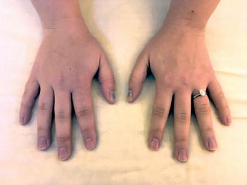
At rest, edema-free skin has a “matte” finish. Edematous skin sometimes develops a sheen, which becomes increasingly shiny as swelling progresses and especially when the skin is maximally stretched by fluid. These subtle changes to the lustre are usually in combination with increased pallor, stretching of the polyhedral texture lines and decreased vein visibility.
Wrinkles and fine lines indicate edema-free skin.
Skin colour
Edema-free skin in Caucasians has a translucent marbled palette of pink, blue and white tones, which give individual skin colour. In other ethnicities the individual tones may not be as clearly demarcated, but the veins are usually still visually identifiable.
As edema forms, skin takes on a uniform pallor in Caucasians and paler skin tone in non-Caucasians, so that the translucency


of marbling, decreased tendon definition.
and marbling effects are lost and veins gradually become obscured.
The top layer of the skin (epidermis) is composed of polyhedral shapes between pores, accomodating multidimensional skin stretch without tearing. In edema-free skin, these shapes are slack and may give the appearance of fine wrinkles, especially with age (Figure 1a). As fluid starts to accumulate in the skin, these changes may be detected: polyhedral shapes are stretched further apart, and may be more difficult to identify; the skin has less or no slack, and there are fewer or no wrinkles.


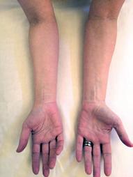
Pallor presentation medial pathway, veins obscured.
Carol Armstrong BA, Registered Massage Therapist, is trained in MLD/ CDT (1993), and Advanced Garment Fitting (2004) and practises comprehensive lymphedema care and compression garment fitting in Victoria, BC. She offers free public education sessions on lymphedema awareness on a monthly basis, and can be reached at carmstrongrmt@telus.net.
Based on common nodal excisions, either alone or in combination with radiotherapy, early edema-related arm pallor occurs most frequently along the medial lymphatic pathway from the mid-bicep to the midforearm (Fig 2a and 2b). When related to certain chemotherapy regimes, edema presents as generalized turgid arm swelling related to capillary permeability rather than one specific lymphatic pathway. For some patients, early pallor may first present in the hand, back of the elbow, or mid-forearm to wrist on the side at risk.
Skin-fold thickness
A normal edema-free skin fold is 1-2 mm wide. Thickness varies slightly based on body fat. The lightest of pinch pressure is used to pick up the thinnest skin fold possible (not the underlying tissue) and compared to the other limb. The greater the skin swelling, the thicker the skin fold will be. In later stages of


3a: Arms show more uniform pallor and loss of vein visibility towards the elbow in a very early stage of lymphedema.
3b: A slightly more advanced lymphedema, showing the more pronounced flexion ceases at the wrist, complete obscuration of the veins at the wrist, decreasing vein visibility along the forearm, and uniform pallor.
lymphedema, it is not possible to pick up the skin at all (positive Stemmer sign). Skin folds can be assessed over the knuckles, back of the hand, elbow and inner arm”.
Skin indentations
Indentations in edema-free skin are usually very narrow, approximately 0.5 mm wide, and disappear within 15-20 minutes of removal of the indenting item (e.g., clothing, jewellery). When edema exists, the indentations are broader (1-3 mm wide), appearing as paler “welts” with a more noticeable raised white border, and reddish indentations where the item is cutting into the skin (Fig 4a). The greater the swelling, the deeper and more enduring the indentation
Limb contour
If right and left limb contours are not generally symmetrical and there is no obvious explanation of difference based on medical history and limb dominance, swelling may be a factor. Compare arm contours when the elbows are bent and pointed towards the practitioner, or

knuckles when the hands are made into fists. Patients can self-check by looking at their reflection. Tendons and bony prominences have less definition as edema increases.
Patient-reported symptoms
Combining skin assessment with patient reported symptoms can validate the patient’s experience. In the very early stages of swelling, patients may report vague, subjective symptoms that frequently get dismissed until more serious physical discomfort ensues4. We have learned that these subjective feelings are valid: breast cancer patients who experienced such

5a: Hand on right shows altered knuckle contour, loss of polyhedral pattern and loss of tendon definition. Hand on left - polyhedral pattern and tendons are still visible.
5b: Loss of tendon definition (r).

symptoms in combination with other limiting factors, such as reduced range of motion, stiffness of joints or impaired sensation, were at higher risk for developing lymphedema5
Initially the limb may not have any visible swelling, but to the patient may feel “heavy”, “achy”, “uncomfortable”, “prickly” or simply “different from the other limb”. When there is actual visible swelling, the skin may feel “full”, “tight” or “restricted”. Other symptoms may include reduced sensation, pain, burning,

cording, or reduced range of movement on the affected side. Clothing and/or jewellery may feel more snug/tighter than usual.
Persistent swelling
Swelling or areas of “puffiness” that persist for more than two months after surgery/ radiation in the affected quadrant should be evaluated. The longer that fluid remains in the region, the more viscous it becomes, with concomitant inflammation and greater risk for secondary skin and tissue changes2
Areas at risk for swelling
If lymph nodes have been excised or radiated, the areas that would normally be drained by those nodes need to be monitored closely. For example, if the right axillary lymph nodes are affected by surgery and/or radiation, the region at risk includes the right trunk front and back from the breastbone to the spine and from the navel to the collarbone, any remaining breast tissue on the right side, the right arm and hand.
Breast and truncal edema
Breast edema may be very local or it may involve the whole breast, often with reported tenderness. The pores are often stretched apart (peau d’orange), the skin may have a uniform pallor or appear “pink” if radiated, and there may be marked indentations from bra seams. Truncal edema may involve the area just below the armpit, and/or extend to the back, the front chest wall and lower rib cage, and the upper one-third of the back of the arm.
Seroma is a fluid filled space that can sometimes form following breast cancer surgery. It frequently resolves on it’s own. If not, residual seroma located on the chest wall or remaining breast tissue is usually replaced by fatty tissue, and if the seroma is in the armpit, it can take on a harder encapsulated texture. Seroma is considered a higher risk factor for development of lymphedema7,8
Apparent delayed onset of swelling
Swelling may appear immediately following cancer treatment or many years later. When patients report the latter, close inspection can determine whether development is new, or if in fact there are significant secondary changes including thickening and subcutaneous fatty

Breast on right shows pores stretched apart—peau d’orange, larger contour. Lack of stretch mark striations present in unaffected breast on left of photo.
deposition present, indicating that the swelling has been present for some time.
Another consideration is that with ageing, many people gain body fat in the inner upper arms. Skin inspection can reveal whether increased limb girth is predominant on the side at risk for lymphedema, or present in both arms.
Summary
Close evaluation of the skin reveals much qualitative information and in combination with patient-reported symptoms, can facilitate

Polyhedral pattern
Fine lines and wrinkles
Pores
Lustre
Colour/tone
Veins
Skin-fold thickness
Indentations
Limb contour
m present m absent
m present m absent
m normal m stretched apart
m matte m sheen m shine
m marbled m uniform pallor
m visible m obscured
m thin m thickened
m marked m enduring “white welts”
m similar m different
discussion about interventions for managing early swelling. For some patient populations, taking early action decreases swelling, restores normal skin characteristics, and reduces progression. In others, such action mitigates the degree of chronic progression. These patients may still have isolated areas of swelling and secondary changes within the quadrant at risk rather than the whole quadrant, but these are largely manageable through exercise, compression, self-massage and management of body weight. I have seen such effects of good management repeatedly
Therapeutic Compression Garments by Wear Ease® Re c ov e ry f ro m

in my clinical practice; good results have been maintained for at least three years as long as patients remain diligent about self-care.
Studies such as “The Optimal Lymph Flow” (TOLF)6 will hopefully encourage a change in focus from treatment oriented to proactive risk reduction. With better education practices concerning lymphedema detection and management, fewer cancer survivors are likely to experience this condition. LP
A full set of references can be found online at www.lymphedemapathways.ca


