www.digitalsurf.com
CORRELATIVE MICROSCOPY ENABLES COMPREHENSIVE ANALYSIS OF GEOLOGICAL SAMPLES ISSUE IN THIS

NEW MOUNTAINS® 9.3
APPLICATION
Correlative microscopy : pyrite mineral spatial characterization
Accelerating surface analysis automation... and much more
APPLICATION
PRODUCT NEWS
New: MountainsImage®, bringing your microscopy analysis to light

Force spectroscopy:
Strengthening polylactic acid polymers by calcification
CUSTOMER SPOTLIGHT
FEATURE SPOTLIGHT
Using microwear analysis to understand what T-rex ate
Welcome to the fascinating world of fiber analysis
SURFACE METROLOGY
SURFACE METROLOGY Q&A
Q&A
How stable is my surface texture evaluation?
What cut-off value should I use?
Application experts at Horiba Scientific and JEOL Europe recently highlighted a technique for precisely relocalizing specific areas of a sample for analysis using various techniques including Raman microscopy, scanning electron microscopy (SEM) and energy dispersive spectroscopy (EDS).
NEWS & SOCIAL
EVENTS & SOCIAL
Control trade show
Events highlights
What’s hot online
What’s hot online
A unique combination of scientific equipment and analysis software enabled the comprehensive analysis of a pyrite sample and its surrounding materials.
… Turn to page 2 …
Did you know there is a whole catalogue of free webinars waiting just for you on our website?

Head on over to our Webinar Library & become a Mountains® software expert in no time!
Check it out: www.digitalsurf.com/learning/webinars/
CORRELATIVE MICROSCOPY PYRITE MINERAL SPATIAL CHARACTERIZATION
Full microscopic characterization of geological samples can be very complex, particularly in studies related to spatial distribution of mineral phases and composition of inclusions. Elemental, chemical and structural variations at the micro- or nano-scale are hard to observe using a single instrument. Evaluation by multiple techniques is key to understanding material properties. Application experts at Horiba Scientific and JEOL Europe recently highlighted a technique for precisely relocalizing specific areas between one instrument measurement and another, in order to perform comprehensive analysis of a pyrite sample with surrounding materials.
INTRODUCTION
Pyrite is a brass-yellow mineral with a bright metallic luster. It has a chemical composition of iron sulfide (FeS2) and is the most common sulfide mineral. The selected sample occurs in a chrysotile vein intersecting a serpentinite metaarkose. This type of metamorphic rock is the result of the interaction between sea water and peridotites, which are the rocks that make up the earth’s mantle.

IMAGING TECHNIQUES
Raman microscopy and scanning electron microscopy (SEM) including energy dispersive spectroscopy (EDS) were used to study the sample provided by the BRGM laboratory, France’s public institution for Earth Science applications. Thanks to their own specificities combined with a good spatial resolution, each of these techniques brings crucial information for mineral investigations.
Raman Microscopy is capable of characterizing the chemical composition and atomic structure of materials, as well as various mineral phases.

In SEM, secondary electrons (SE) reflect the fine topographic structure of the sample, backscattered electrons (BSE) reflect the compositional distribution on the sample surface and EDS is used to determine the elements contained in the sample.
A critical point in this correlative microscopy study was achieving measurement of the same specific area on the sample using both the SEM and the Raman microscope. This challenge was overcome thanks to the Horiba nanoGPS SuiteTM which provides accurate relocalization between different instruments (navYXTM), as well as software for positioning multimodal maps in the same coordinate system (graphYXTM based on Mountains® technology).

RESULTS
BSE imaging offers an easy and quick visualization of the specimen chemical variation, however, no chemical identification and characterization can be performed with this method.
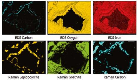
Therefore the first complementary technique applied to the sample was EDS mapping, which can bring information about the elemental composition, distribution and concentration. It is possible to observe the spatial distribution and the associated density of each chemical element detected by EDS.

To perform Raman analysis, the sample was then moved from the SEM chamber to the motorized stage of the Raman microscope. navYXTM technology allowed precise relocalization of the point of interest, enabling the Raman measurement at the same localization. With both EDS and Raman analysis, material phase identification was
READ MORE
achieved for this sample, the EDS analysis helping to sort between matching results in the Raman database. Cathodoluminescence was also used to identify an Al inclusion.
CONCLUSIONS
Raman measurement results associated with EDS mapping allowed the identification of the different minerals in the rings around the pyrite. Co-localization of measurements revealed the exact elemental and mineralogical make-up of the studied area. In particular, Lepidocrocite and Goethite, two minerals with the same chemical composition could not be identified using EDS mapping alone, but were successfully revealed thanks to Raman microscopy. The co-localization of different techniques and the complementary information leads to a complete knowledge of the specimen in question.
Colocalized microscopy techniques for pyrite mineral spatial characterization


João-Lucas Rangel1, Raghda Makarem2, Yusuke Uetake3, Guillaume Lathus3, Didier Hocrelle1, Thibault Brulé1, Jérémy Brites1
1HORIBA FRANCE SAS, 91120 Palaiseau, France 2HORIBA FRANCE SAS, 59120 Loos, France
3JEOL Europe (SAS), 78290 Croissy-sur-Seine, France
NEW: MOUNTAINSIMAGE® BRINGING YOUR MICROSCOPY ANALYSIS TO LIGHT
Mountains® software has been an ally for researchers analyzing surface topography since 1996. Over the years, new solutions have been added to the platform to make the study of other types of data like spectra or SEM and SPM images possible. Now comes the addition of MountainsImage® for the analysis of light microscopy images. Mathieu Cognard, Product Manager for MountainsImage® analysis software at Digital Surf, unveils to Surface Newsletter the new tools available and applications served with this recently released product line.
WHY MOUNTAINSIMAGE®?
“We realized microscopy users could benefit from a product line specifically designed for light microscopy image analysis” explains Mathieu. “Mountains® software already contained quite a few useful tools for light microscopy image processing, so we had a good base to work with. As with every product, it was extremely important for us to create a toolkit that would adequately answer the needs of its users. Because of this, new tools were developed with light microscopy users in mind. Adding this latest product to the Mountains® family was the next step towards completing our offer.”
The brand-new MountainsImage® dedicated package comes with three product levels for pre-processing and analyzing black & white or color images without topography. Users can expect some of the central features they have come to know in Mountains® software, as well as new dedicated ones for light microscopy analysis.
MOUNTAINSIMAGE® KEY FEATURES

MountainsImage® features include:
▶ loading and visualizing light microscope images
▶ basic correction and measurement tools
▶ image scaling
▶ viewing images in true or pseudo-color
▶ applying enhancements & filters to images
▶ extracting areas for analysis etc.
Other features are available such as image stitching for microscopists looking to combine multiple measurements into one set for analysis. The texture isotropy study and slices analysis study will be of great interest to those looking to segment images based on luminance.

ADVANCED FEATURES
Users who need more advanced tools when processing light microscopy images can access:


Top right. Fiber analysis study of a nerve bundle: segmentation image, diameter map, histogram of fiber diameters and parameters table. Middle right. Planar contour analysis with tolerance limits and image displayed beneath the contour. Bottom right. Color segmentation. On the left, true color image. On the right, color segmentation by selection of the components to detect (in this case gold and silver).


▶ Fiber analysis and particle analysis for the detection, quantification and classification of fibers and particles.

▶ Contour analysis including display of images beneath contour profiles, when analyzing horizontal object contours.
▶ Color segmentation (via particle analysis) which allows users to isolate zones based on color (for example to detect & visualize component coverage in wear applications).
“A wide range of formats are compatible with the software such as: BMP, EMF, GIF, JPG, PNG, TIFF, WMF” adds Mathieu.
“Numerous types of applications can be dealt with using MountainsImage®: studying the speed of hair growth, quantification and measurement of cells or studying corrosion on instruments, to name but a few.”
CONCLUSION
“We’re very proud of this new addition to the Mountains® software family. Our team has been working hard to bring to life a comprehensive solution for light microscopy users that renders the powerful and reliable results Mountains® users have come to expect from us” states Mathieu. “We are looking forward for users to try it out and share their findings with us.”
TRY IT OUT FOR YOURSELF
Get a MountainsImage® 30-day Free Trial and take advantage of all the features today: www.digitalsurf.com/free-trial
SEE IT IN ACTION
We’ll be presenting the Fiber analysis tool for microscopy images in our upcoming webinar on July 20 at 4:00 pm CEST, register here: bit.ly/fiber-analysis
USING MICROWEAR ANALYSIS TO UNDERSTAND WHAT T-REX ATE
Dr. Mugino O. Kubo Ph.D is Lecturer at the Department of Natural Environmental Studies at the University of Tokyo, Japan. A long-time user of MountainsMap® software, Dr. Kubo spoke to Surface Newsletter about her most recent research and how her team are implementing new, ground-breaking methods for studying the paleodiet of carnivorous dinosaurs, including the iconic Tyrannosaurus rex.

PLEASE CAN YOU INTRODUCE YOURSELF
I’m a vertebrate morphologist with a strong interest in the evolutionary relationship between ecology and morphology. I started my career with a comparative osteological study using sika deer living in various environments in Japan. I have been focusing on how different feeding habits result in changes in dental and skeletal morphology. I collected and analyzed several hundreds of deer skulls. Based on such abundant skeletal specimens, I clarified the relationship between ecology and dental morphology, which can be used to infer the paleodiet of extinct animals. Since I married a dinosaur paleontologist, Dr. Tai Kubo currently at Okinawa Institute of Science and Technology, I got further involved in paleobiology. We have started a collaborative project on the paleodietary reconstruction of dinosaurs using dental wear traces.
WHAT ARE YOUR CURRENT PROJECTS?
I introduced a confocal laser microscope in my lab in 2016. This instrument can obtain 3D surface data at the sub-micron level. We used it to evaluate the roughness of tooth surfaces. When animals eat, tiny marks at the microscopic level (microwear) are left on the surface of their teeth due to contact with the food. As the microwear reflects the physical characteristics of the diet, investigating it allows us to infer what the animals might have eaten while they were alive.
Since 2017, we have been working on research to accumulate microwear data

for diverse wild animals with known diets, including sika deer. Using these data as a comparison, we can reconstruct the ecologies of extinct species. We began our efforts with mammals and are now expanding to dinosaurs. Thanks to Dr. Daniela E. Winkler, who was a member of my lab from 2020 to 2022, we accelerated dental microwear research of dinosaurs. We published two papers on dinosaurs in 2022: on a Japanese sauropod, and on theropods, including Tyrannosaurus rex and its relatives (Fig. 2). We are now investigating hadrosaurs from various geological ages and extinct crocodiles from Cretaceous fossil localities.
WHAT IS THE INNOVATION OF YOUR RESEARCH?
Three-dimensional microwear analysis was proposed in 2004 by physical anthropologists and succeeded by mammalian morphologists and paleontologists. Currently, less than ten labs worldwide are actively conducting it. Each lab has its uniqueness in the scientific approach, and I would say our originality and novelty are in our focus on dinosaurs.
To understand the diet-microwear relationship in carnivorous dinosaurs, we conducted controlled feeding experiments using live alligators, through an international collaboration with Professor Richard Blob at Clemson University in the USA. Feeding experiments are usually only done with mammals, which are kept widely in laboratory environments: e.g., rats, guinea pigs, sheep, and pigs. But keeping and feeding alligators as a different challenge. Dr. Masaya Iijima, at that time Postdoc at Prof. Blob’s lab, fed individual alligators with different diets (crawfish, rats, quails, fish and reptile pellets) and collected a lot of shed teeth. The teeth were sent to Japan and scanned by the confocal microscope to observe microwear differences. We were excited to see clear differences in wear marks, with higher surface roughness in more physically demanding diets (crawfish and rats).
LEARN MORE
WHAT ROLE DOES MOUNTAINSMAP® SOFTWARE PLAY IN YOUR RESEARCH?
MountainsMap® plays an important role in our research projects. We use it from the initial stage of surface preparation through the calculation of surface roughness parameters defined by international standards (ISO 25178-2 and others). Our normal routine for 3D surface data includes leveling, removal of gross tooth curvature, removal of measurement noise using spatial filters and calculation of surface roughness parameters. This procedure is quite similar to that used in Dr. Ellen Schulz-Kornas’s lab (www.digitalsurf.com/ stories/revealing-diet-from-tooth-wear-surfaces-in-wild-chimpanzees), but we use different spatial filters that more effectively remove measurement noise which is specific to our machine.

Dr Mugino’s lab: https://sites.google.com/edu.k.u-tokyo.ac.jp/mugino-kubo-lab/home
Winkler, D.E., Kubo, T., Kubo, M.O., Kaiser, T.M. and Tütken, T. (2022), First application of dental microwear texture analysis to infer theropod feeding ecology. Palaeontology, 65: e12632. https://doi.org/10.1111/pala.12632

HOW STABLE IS MY SURFACE TEXTURE EVALUATION?


Most of us tend to take the measurements around us, such as the temperature indicated by our cars or poll percentages before an election, for granted. This is also the case in metrology where it can be tempting not to question results. However, measuring one 8 mm profile on a 40 cm metal part and calculating one Ra parameter does not entitle us to draw conclusions about the roughness of the whole part. That said, observing a change in value of 0.1% between two versions of your analysis software does not necessarily mean that there is a bug. In this article François Blateyron, senior surface metrology expert at Digital Surf, discusses why results may vary and good practices to ensure they remain stable.
VARIATIONS ON THE SAME PART
Surface texture specifications usually apply on a particular portion of a part, for example the upper face. If you measure a profile or a surface at different locations on the same toleranced portion, you will observe a certain dispersion in the parameter values. The smaller the measured area, the larger the dispersion. This relates to the scale of observation. Seen from a distance, the surface of a table is flat and relatively smooth. When looking at it with a microscope, you can see hills and dales, grooves and canyons. If the dispersion between several measures is high, it might mean that the measured area is too small for that texture, and/or that the surface is not homogeneous.
The example below shows 20 surfaces measured on the same sample of a brushed surface. We can see a large dispersion in values, even on parameters that are calculated as averages (such

as Rq and Ra). Each surface was measured as a square of 0.8 mm x 0.8 mm using an optical profiler. On such a texture, a larger field of measurement would have probably produced more stable results.
VARIATIONS WITHIN THE SAME MEASURED SURFACE
A similar exercise can be performed by extracting a sub-surface from an actual measured surface.
In the following example, sub-surfaces of 40% of the size of a surface were extracted with a horizontal and vertical shift of 10%, producing 49 surfaces (see opposite page). Here also, we observed dispersions, smaller than in the previous example (except for Ssk and Sku that are notably sensitive parameters). Similar dispersions could also be seen by changing the orientation of the measurement, by introducing slope or outliers or by changing the instrument technology (confocal, interferometer, focus variation).
VARIATIONS BETWEEN SAME MEASUREMENTS
You might think that measuring a surface several times at the same position without any changes would produce the exact same results every time. However, instruments contain electronic components that can produce noise, the measurement environment also emits small vibrations and the refractive index of air around the instrument may even vary slightly due to the body temperature of the operator.
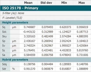
In the example above right, 10 measurements were made successively at the same position, in a short time period, yet we can still see small dispersions, especially on less stable parameters.
Above. 10 measurements made at the same position, yet we still see slight differences in values!
VARIATIONS IN CALCULATION ALGORITHMS
In a continuous effort to maintain and improve Mountains® software, we sometimes modify a particular algorithm in order to make it faster and/ or more accurate. As a result, a new version may show a small difference compared to an older version. Usually, that means that the new value is more accurate. In most cases, the difference is much smaller than the one observed when taking repeated measurements at different places of the same sample.
TAKEAWAYS
▶ Don’t take the result of a single measurement or a single parameter for granted
▶ Take measurements at several places on the same sample and average results


▶ Discard measurements if you see dirt or obtain too many outliers
▶ Carefully select the field of view (objective) or the type of instrument
▶ Make sure you have a good knowledge of the complexity of the surface texture to be able to adapt analysis tools accordingly
ADDITIONAL RESOURCES
▶ guide.digitalsurf.com/en/guide-stochastic-vs-deterministic.html
▶ guide.digitalsurf.com/en/guide-software-verification.html
NB: surfaces shown in this article come from the work of GDR 2077 SurfTopo (https://surftopo.cnrs.fr/)


CONTROL TRADE SHOW AND MOUNTAINS® 10 RELEASE
CONTROL 2023
Digital Surf was thrilled to be exhibiting at the 35th Control international trade fair which took place in Stuttgart, Germany from May 9-12.
589 exhibitors from 32 countries showcased their latest innovations and demonstrated the ever-increasing importance of quality assurance in production environments. More than 21.300 visitors attended this new edition spread out over four exhibition halls.
On a freshly redesigned booth with new corporate colors, Digital Surf was proud to present Mountains® 10, the new major and also the most comprehensive version release of its analysis software. The Digital Surf team was pleased to provide live demonstrations of Mountains® 10 new features dedicated to surface imaging and analysis.
It was great to see that Mountains® software was also on show throughout the exhibit floor on our partners’ stands, often fully integrated with different surface measuring instruments for various applications.


This year again, Control Messe was a success and a great opportunity to meet up with our clients and partners.
MOUNTAINS® 10 IS OUT!
On May 31, Digital Surf announced the release of the tenth major version of the Mountains® software analysis platform for surface metrology & microscopy.

With the arrival of the new MountainsImage® branch for light microscopy as well as several other new modules and features, Mountains® software is now more than ever the most comprehensive software solution for all your instruments. Check out what’s included in this new release: bit.ly/3ML7q7w
How to update
Access to this latest release is included in the Mountains® Software Maintenance Plan (SMP). Please visit our Software Updates page: bit.ly/Mountains-updates
MOUNTAINS® FIBER ANALYSIS WEBINAR
Good news, fiber analysis is now available with all Mountains® software product families!

Join us on Thursday July 20 @4PM CET for a webinar dedicated to this new feature.
In this webinar presented by Mathieu Cognard, Product Manager for SPM & Light Microscopy Applications at Digital Surf, you will learn more about how to analyze fibers in microscopy images (detection methods, visualization options, results generation & exports, etc.)
Register here: bit.ly/fiber-analysis
WHAT’S HOT ONLINE
Have you been to our YouTube channel ?
SPOTTED ON LINKEDIN
Digital Surf Sales Director, Arnaud Viot, payed a visit to our partners & friends over at JEOL Europe in Paris, to chat about their data analysis needs, check it out: bit.ly/3pkXpGM

We have lots of quick helpful videos, like the tutorials on Mountains® software basic and advanced features, check them out: bit.ly/2U2I2za

LIKED ON FACEBOOK
Lucky visitor to the Digital Stand at Control, Dr. Gerhard Moeller of Zeiss Microscopy, won a hamper full of delicious French goods: bit.ly/3NeoCTm

Surface Newsletter

Know a friend or colleague who would be interested in receiving the Surface Newsletter?
Let us know : contact@digitalsurf.com
The newsletter is available for download on our website www.digitalsurf.com
Useful LINKS
HQ, R&D Center
16 rue Lavoisier
25000 Besançon - France
Tel: +33 38150 4800
contact@digitalsurf.com
www.digitalsurf.com
TRY MOUNTAINS® SOFTWARE
Take Mountains® for a test drive
Visit www.digitalsurf.com/free-trial
CONTACT US FOR AN UPDATE
Contact sales@digitalsurf.com for information about updating from previous versions to the latest Mountains® version

WATCH A MOUNTAINS® TUTORIAL
Get the most out of Mountains® software by watching one of our video tutorials www.digitalsurf.com/tutorials
LEARN SURFACE METROLOGY
Dive into our free online surface metrology guide and learn about characterizing surface texture in 2D and 3D www.digitalsurf.com/guide
CATCH UP WITH US
Microscopy & Microanalysis (M&M) meeting
July 23-27, 2023 | Minneapolis, Minnesota, USA
JASIS | September 6-8, 2023 | Tokyo, Japan
SciX | October 8-13, 2023 | Sparks, Nevada, USA
Surface Newsletter, July 2023
Editor : Christophe Mignot
Content editor : Clare Jamet
Contributors : Laure Aubry, François Blateyron, Eugenia Capitaine, Mathieu Cognard, Mugino Kubo, Renata Lewandowska.
Copyright © 1996-2023 Digital Surf, all rights reserved
