8 minute read
M.Tani Terahertz Spectroscopy of Biological Molecules and Tissues

M.Tani1, F. Zhang2, T. Kawasaki3, M. Mizuno4, M. Hayashi5, K. Tominaga2, Y. Izumi6, G. Ohori3 , H.Kitahara1, T. Furuya1, K. Yamamoto1, K. Matsuo6, K. Tsukiyama3, K. Sasaki4, M. Kojima7 , Y.Suzuki8, T. Tasaki7, Y. Tatematsu1, M. Fukunari1 1 Res. Center for Development of FIR Region, University of Fukui, Fukui 910-8507, Japan 2 Molecular Photoscience Research Center, Kobe University, Kobe 657-8501, Japan 3 IR-FEL Research Center, Tokyo University of Science, Chiba 278-8510, Japan 4 National Institute of Information and Communications Technology, Tokyo 184-8795, Japan 5 Center for Condensed Matter Sciences, National Taiwan University, Taipei 10617, Taiwan 6 Hiroshima Synchrotron Radiation Center, Hiroshima University, Hiroshima 739-0046, Japan 7 Medical Research Institute, Kanazawa Medical University, Ishikawa 920-0293, Japan 8 Graduate School of Systems Design, Tokyo Metropolitan University, Tokyo 192-0397, Japan
Advertisement
Abstract— The authors report spectroscopic studies for (i) alanine crystal [1-2], (ii) hen egg-white lysozyme irradiated with mid-IR laser [3], and (iii) corneal tissues of rabbit eyes [4]. From these examples, it is elucidated that THz spectroscopy with theoretical analysis is a powerful tool for the studies of biological molecules and biological systems.
I. INTRODUCTION
IN recent years, with the progress of the terahertz (THz) wave light source, THz wave applications have attracted much attention. In future wireless communications, the carrier frequency will be extended to the THz frequency region (>0.1 THz), and there is also an increasing concern with the influence of THz waves to biological systems. When we study the influence or interaction of THz electromagnetic waves with biomolecules and biological systems, the absorbed power or energy is the key parameter. The influence or reaction induced in the biological system for the case of high-power and low-power irradiation should be much different. For the case of high power irradiation, the thermal effect may play an important role. To simulate the thermal effects, it is essential to know precise dielectric properties of the biological system under investigation in the THz frequency region. In this paper, the authors illustrate the usefulness of THz time-domain spectroscopy (THz-TDS) for evaluation of complex dielectric properties of biological macro molecules and tissues in the THz frequency region by THz-TDS studies and theoretical works carried out for (i) L-alanine (enantiomer) and DL-alanine (racemic compound) (ii) hen egg-white lysozyme after a high-power mid-IR irradiation and (iii) corneal tissues of rabbit eyes with high-power THz wave irradiation.
II. THZ SPECTROSCOPIC STUDY OF L-ALANINE (ENANTIOMER) AND DL-ALANINE (RACEMIC COMPOUND) Biomolecular crystal show relatively sharp absorption bands in the THz frequency region originating from inter-molecular vibrations, whose frequencies and intensities are governed by the crystal structure and its symmetry. For example, poly-crystalline L-alanine (enantiomer) and DL-alanine (racemic compound) show strikingly different absorption bands even though the constituent molecules are same except the handedness. In a recent theoretical study [2], we have revealed that such a difference arises from the IR-activities determined by the crystal space-group symmetry. In addition, it was also found that the intra-molecular motions play crucial roles in determining the IR intensities [1].
III. THZ SPECTROSCOPIC STUDY OF HEN EGG-WHITE LYSOZYME WITH HIGH-POWER MID-IR IRRADIATION Irradiation of mid-IR laser resonant to the amide I absorption band (6 m) can refold the amyloid fibrils of hen egg-white lysozyme (HEWL) (aggregate of lysozyme) to the native-like form and also recover the enzymatic activity of HEWL [5]. THz time-domain spectroscopy was carried out for native lysozyme, aggregate of lysozyme, aggregate with resonant irradiation at amide I, and aggregate with non-resonant irradiation by the authors of [3]. The native lysozyme exhibited a higher THz absorption against the aggregate, and the mid-IR resonant irradiation increased the aggregate absorption close to the native one by the conversion of fibrous aggregate to the non-aggregate state of HEWL [3]. This indicates that THz spectrum is indicative for the conformational changes and thus the activities of the enzyme.
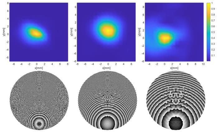
IV. THZ TIME-DOMAIN SPECTROSCOPY OF CORNEAL TISSUES OF RABBIT EYES The dielectric properties of the normal corneal tissue of rabbit eyes were measured in vitro in a range from approximately 0.1 to 1 THz by THz-TDS measurement by the authors of [4]. The difference in reflectance from the rabbit eye surface between normal tissues (control) and tissues exposed to high-power terahertz waves (@162 GHz) was also investigated [4]. It was verified that reflectance data calculated from the dielectric constant values and those measured in vivo agreed at frequencies under approximately 0.4 THz. When the rabbit eye was exposed to the terahertz waves, the average reflectance decreased significantly owing to the drying process, while the tears caused the reflectance increment. The results suggest that the complicated reflectance changes of the eye surface should be considered for the precise computational dosimetry under the high-level irradiation condition [4].
REFERENCES [1] M. Yamaguchi, et al, Appl. Phys. Lett. 86, 053903 (2005). [2] F. Zhang, M. Hayashi, M. Tani, K. Tominaga, “Interpretation of THz intensities of molecular crystals: the role of mixing between intermolecular and intramolecular vibrations” (in preparation). [3] T. Kawasaki, et al, J. IR, MMW & THz Waves 40, 998–1009 (2019). [4] M. Mizuno, et al, submitted to Biomedical Optics Express. [5] T. Kawasaki, et al, J. Synchrotron Rad. 23 pp.152-157 (2016).
Genome-wide mRNA-seq analysis in Human Fibroblasts exposed to 25 GHz
E. Regalbuto1 , V. Franchini1 , A. Anselmo1 , A. Fortunato1 , G. Alfano1 , S. De Sanctis1 , A. Sgura2 , S. Ceccuzzi3 , A. Doria3, E. Giovenale3, G.P. Gallerano3 , G.L. Ravera3 , L. Masuelli4 , R. Bei5 , P.M. D A gel 6, V. Cusimano6 and F. Lista1 .
1Scientific Department of Army Medical Center of Rome - Rome, Italy
2Department of Science, University of Rome R ma T e - Rome, Italy
3ENEA Research Center, Radiation Sources, Antennas and Diagnostics Laboratory - Frascati, Italy
4Department of Experimental Medicine U i e i f R me Sa ie a - Rome, Italy
5Department of Clinical Sciences and Translational Medicine Uni e i f R me T Ve ga a - Rome, Italy
6National Research Council of Italy-I i e f S em A al i a d C m e Scie ce A i R be i IASI, Bi Ma Lab, R me, I al
Abstract—This study investigated gene expression modulation in human fibroblasts in vitro exposed to 25 GHz. For this purpose a new high-throughput RNA sequencing approach, by Next generation sequencing platform, was used.
I. INTRODUCTION
THE increasing use of Microwave (MW) and Radiofrequency (RF) technology together with the future perspective of the fifth generation (5G) wireless communication introduction, raised questions about their possible biological and adverse health effects. Despite several in vitro and in vivo studies have been carried out to evaluate the biological effects potentially induced by this radiation, the results are rather controversial [1, 2]. In addition to the evaluation of classical biological markers (e.g. genetic damage, cell cycle/proliferation, apoptosis), in the last decades there is a growing interest in the identification of genes that could change their expression profile following exposure to radiofrequency electromagnetic field (RF-EMF). In this research field, the most common approach to identify RF-EMF sensitive genes consist in whole genome screening methods using microarray based technology. However the few large-scale studies performed until now reported unclear results [3,4] making difficult to establish a list of common RFEMF sensitive genes. In this context the aim of this study was to evaluate whole gene expression modulation in human fibroblasts in vitro exposed to 25 GHz CW radiation [5] using the highthroughput sequencing approach through the RNA sequencing (RNA-seq) technology.
II. RESULTS Gene expression analysis was performed by mRNAsequencing on Illumina NextSeq 500 platform on both exposed and sham RNA samples isolated 2 and 24 hours after 20 minutes of exposure to 25 GHz. In order to increase the statistical power 4 experimental replicates were performed. To identify genes differentially expressed Cuffdiff and EdgeR analysis tools were applied on data obtained from sequencing. This analysis showed in exposed cells, some genes with differential expression profiles. After Gene Ontology terms analysis these genes resulted involved in different biological processes. However, the genes identified in this study have to
be confirmed by RT-PCR, gold standard for the detection and quantification of gene expression profiling.
Fig.1. RNA-seq workflow including four basic steps: library preparation (1), cluster generation (2) and sequencing (3) performed on Next-Seq 500 system, and data analysis (4).
III. SUMMARY
The innovative high throughput RNA-seq technology, used in this study, overcomes the limitation of a preselected set of genes allowing the identification of known and unknown transcripts. This promising approach will contribute in the identification of sensitive genes and in understanding the underlying mechanism of possible biological responses and effects induced by RF-EMF.
REFERENCES
[1] L. Vershaeve, J. Juutilainen, I. Lagroye, J. Miyakoshi, R. Saunders, R. de Se e, T. Te f de, E. a R ge , B. Ve e a d Z. X , I i a d i i genotoxicity of adi f e e c field , Mutat Res, 705, 3, 252-268, 2010. [2] D. Manna and R. Ghosh Effect of radiofrequency radiation in cultured mammalian cells: A Review . Electromagn Biol Med. 35(3): 265-30, (2016). [3] J.P. McNamee and V. Chauhan Radiofrequency Radiation and Gene/Protein Expression: A Review. Radiation Research 172: 265-287, (2009). [4] D. Habauzit, C. Le Quement, M. Zhadobov, C.Martin, M. Aubry, R. Sauleau, Y. Le Drean Transcriptome Analysis Reveals the Contribution of Thermal and the Specific Effects in Cellular Response to Millimeter Wave Exposure. PLoS ONE 9(10): e109435, (2014). [5] V. Franchini, E. Regalbuto, A. De Amicis, S. De Sanctis, S. Di Cristofaro, E.Coluzzi, J. Marinaccio, A. Sgura, S. Ceccuzzi, A. Doria, G.P. Gallerano, E. Giovenale, G.L. Ravera, R. Bei, M. Benvenuto, A. Modesti, L. Masuelli, F. Li a Ge ic Effec I H ma Fib bla E ed T Mic a e Radia i Health Physics 115 (1) 126-139 (2018).