

DENTAL SOLUTIONS
Aoralscan Elite
The Aoralscan Elite is the world’s first device to integrate both intraoral scanning and photogrammetry into a single unit
Two-in-One System features
• Standard intraoral scanner (IOS) for capturing images of dentulous cases, allowing for detailed scans of teeth and oral structures.
• Intraoral photogrammetry (IPG) to accurately record the position of dental implants in edentulous cases.
This dual functionality makes it versatile for both traditional dental work and advanced implant procedures.
NEW NEW
Tooth Mousse Chocolate
GC Tooth Mousse™ comes in a delicious BRAND-NEW Chocolate that your patients will love. Topical creme with calcium, phosphate


Henry Schein
Endoflex
CFiles
Stainless steel files designed with enhanced tensile strength to ensure easy location and penetration of complex and calcified canals.
Improved cutting efficiency due to the spiral design. 21 & 25mm, available in various sizes


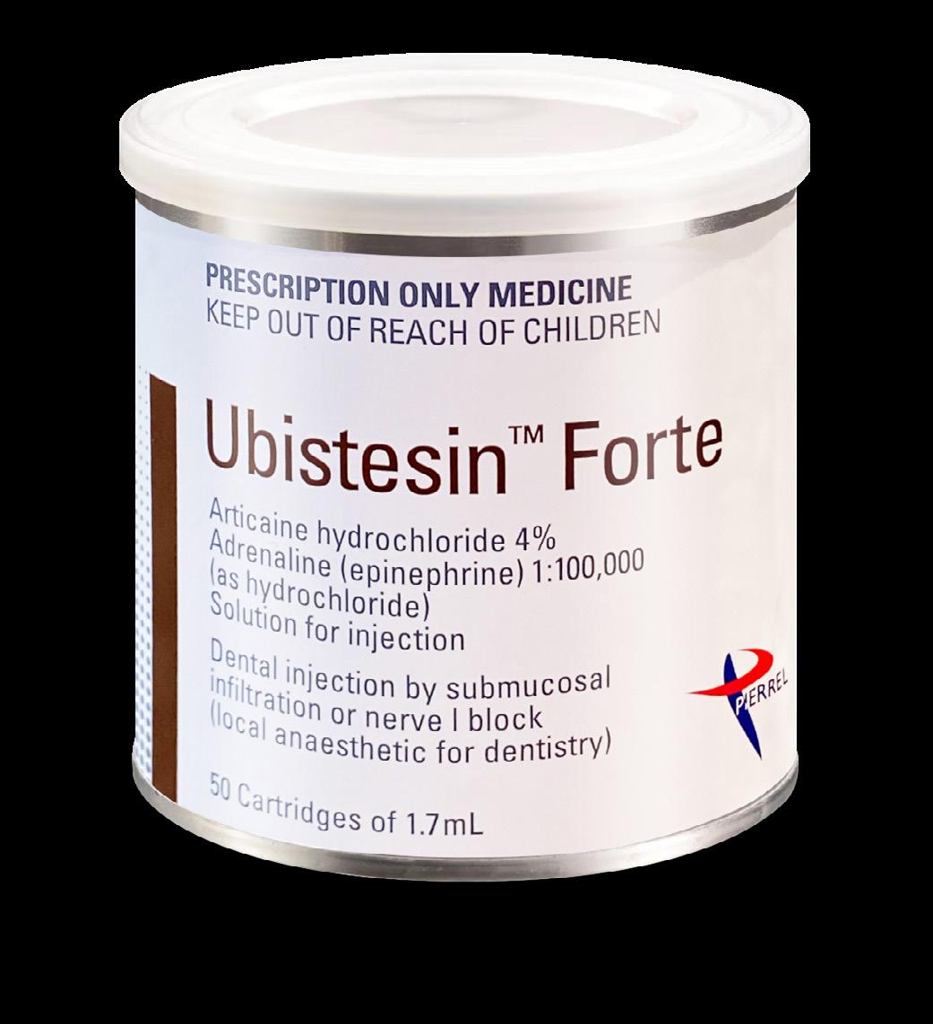
Pierrel S.p.A: Leading the Charge in Dental Anaesthetics and Innovation

Introduction
Pierrel S.p.A, a pharmaceutical company established in 1948, is a recognized leader in injectable drugs and dental products. Based in Capua (CE), Italy, Pierrel has built an impressive reputation over 70+ years of experience. The company is known for its contributions to pharmaceuticals and dental sectors and its commitment to quality, innovation, and sustainability. Pierrel is authorized by major global drug regulatory agencies, including the European Medicines Agency (EMA) and the U.S. Food and Drug Administration (FDA), allowing it to market products worldwide.
A Legacy of Excellence
From its roots in pharmaceuticals, Pierrel has become one of the leading manufacturers of dental anaesthetics. The company’s extensive portfolio includes notable products
like Ubisestin™, Xylestesin™, Mepivastesin™, and Orabloc. This success is a direct result of its ongoing dedication to product excellence, safety, and a comprehensive approach to research and development.
Since its plant was authorized by the EMA in 1989 and the FDA in 2009, Pierrel has remained at the forefront of the injectable drug market, particularly in the field of dental anaesthesia. The company’s production site spans 40,000 square meters, housing manufacturing, laboratories, and warehouses to store products at varying temperatures.
Pierrel’s
Acquisition of 3M’s Dental Anaesthetic Business
In 2023, Pierrel acquired 3M’s dental anaesthetic business, expanding its product portfolio and reinforcing its position as a global leader in dental anaesthetics. This acquisition included the well-known brands Xylestesin™ and Ubisestin™,

both of which are highly trusted by New Zealand dental professionals for local anaesthesia during dental procedures. The 3M acquisition further strengthens Pierrel’s offerings in the dental sector, enabling the company to provide a broader range of anaesthesia solutions to dental professionals worldwide.
Innovation and Research at the
Core Pierrel focuses heavily on innovation, collaborating with universities and research centers. This commitment to research and development keeps Pierrel ahead of trends and helps the company create distinctive solutions for oral health. By working with dental professionals, Pierrel addresses the evolving needs of the dental community and enhances patient outcomes.
Through these collaborations, Pierrel develops innovative medical devices, ensuring dental procedures are more effective, comfortable, and accessible.
Quality and Reliability in Every Product
Quality is Pierrel’s core value, guiding all its operations. Every product undergoes rigorous testing to meet the highest standards to comply with the GMP requirements. Pierrel controls the entire supply chain, ensuring reliable, timely deliveries to its global distribution network. The company’s reputation for reliability is reinforced by its expansion into new markets, including New Zealand, where dental professionals can now trust Pierrel’s solutions for safe, effective anaesthesia.
Sustainability and Environmental Responsibility
Pierrel S.p.A is committed to sustainability and social responsibility, aligning with the United Nations’ 2030 Agenda for Sustainable Development. Its sustainability policy focuses on clean water, responsible consumption, and energy reduction.
Pierrel has made eco-sustainable upgrades to its facilities and increased its workforce by 42% in one year, with 76% of new hires under 35 years old, underscoring its commitment to innovation and growth.
Global Reach and Market Presence
Pierrel’s products are available in over 68 countries worldwide, including North America, Continental Europe, the Middle East, the Far East, and New Zealand. With more than 100 authorizations from regulatory agencies across these regions, Pierrel continues to expand its reach and provide high-quality dental anaesthetics to a global market.
Conclusion
With over 70 years of experience in the pharmaceutical and dental anaesthetic industries, Pierrel S.p.A has established itself as a leader in both innovation and sustainability. From its state-of-the-art production facilities to its dedication to quality, research, and environmental responsibility, Pierrel continues to push the boundaries of what is possible in the dental care sector. As the company moves forward, it remains focused on its mission to provide dental professionals and patients worldwide with high-quality, effective, and sustainable solutions for better oral health.
G-CEM ONE™
Self-adhesive Universal Resin Cement
The patient presented with defective restorations. After a comprehensive evaluation, recurrent decay was identified in the existing resin restorations on teeth 14 and 15. The patient also reported discomfort due to an open contact, leading to food trapping.
Tooth 14: It was decided to restore tooth 14 with a direct composite. GC Essentia Universal was used.
Tooth 15: An indirect restoration was fabricated for tooth 15. The tooth was prepared and scanned. GC Initial LiSi Block was selected to mill the inlay.
The inlay was bonded using G-CEM ONE™ self-adhesive universal resin cement, chosen for its excellent bond strength, easy handling, and reliable long-term outcomes. After seating the restoration, excess cement was carefully removed, and the restoration was light-cured.
G-CEM ONE™ is a truly universal, non-technique sensitive, versatile and reliable product that gives the flexibility of being effective in all cementation procedures for any type of restorations; from metal- based to resin and all-ceramic inlays, onlays, crowns, bridges, and posts. It demonstrates excellent bond strength to enamel, dentin and all indirect restorations.
The final restoration demonstrated excellent aesthetics and functional integrity. The patient reported satisfaction with both the comfort and appearance of the restorations.

View Product

1. Pre-operative Image - Recurrent decay in existing resin restorations 14,15. Open contact with food trapping.

4. 15 LISI Restoration + 14 Resin Restoration Polished restoration with EVE Diapol Twist Polishers + GC Diapolisher Paste.

7. G-Premio BOND™ for tooth structure. G-Multi PRIMER™ & G-CEM ONE for restoration.

2. Immediate Dentin Seal and Resin Coat for tooth 15 following caries removal G-Premio BOND™ + everX Flow + G-aenial Injectable A2.

5. Application of G-Multi PRIMER™ on LISI restoration.

8. Application of G-Multi PRIMER™ on LISI restoration.

3. Preparations air-abraded and ready for bonding protocol with G-Premio BOND™.

6. Application of G-CEM ONE ready for cementation.
G-Premio Bond for tooth structure. G-aenial Injectable A2 for proximal box/marginal ridge. GC Essentia Universal for occlusal form.

Dr. Liang Liang
BDS(Hons) U. Sydney, Australia View Product
Bonds
G-Premio Bond
Premium in a bottle
One component light cured universal adhesive.
• No rubbing or complex application procedures
• Visible during application
• 3µm film thickness
• No patient discomfort
• Up to 300 applications per bottle

G2-Bond Universal
Research Award 2024 – Bonding Agent
Two component light cured universal adhesive.
“G2-BOND Universal was tested in Dental Advisor Laboratories for a 12-month thermocycling protocol.
The purpose was to determine if the bond would be viable. Results of the study were excellent as it proved that the bond was not only strong, it was durable when subjected to repeated thermocycling.
G2-BOND Universal performed better than competitor products tested in their respective etching modes to dentin and enamel in immediate 24 hour shear bond strength and after accelerated aging and 12 month storage.”
- Dental Advisor 2024

CLEARFIL Majesty™ ES-2 Universal
Considerations on the use of a universal composite in the anterior region
Composites with a universal shade concept, a reduced number of shades that may be selected without any shade guide are a clear trend in restorative dentistry. With specific blend-in properties, these materials can help streamline restorative procedures and reduce chair time, take some pressure off the dental practitioner and contribute to potentially good outcomes. Some users, however, are skeptical about a wide-scale use of the materials, particularly when it comes to restoring teeth in the anterior region. The reasons may be a comparatively high translucency requiring the separate application of a blocker (oropacious shade) in certain situations, or a too limited shade offering.
Personal experience shows that CLEARFIL MAJESTY™ ES-2
Universal is perfectly suitable for a wide range of single-shade restorations in anterior teeth. It offers great polishability and long-term gloss retention and is available in just four shades: One universal shade (U) originally designed for posterior restorations, universal light (UL) and universal dark (UD) as the two major options for anterior teeth and, finally, universal white (UW) for the imitation of any bleached shade. In general, all four options may be used in the anterior and posterior region. As the blend-in ability is due to proprietary light-diffusion technology and not managed via an increased translucency, the application of a blocker is usually not necessary and even larger areas can be restored quite inconspicuously.
For those asking themselves when to select which shade in the anterior region, the following clinical case examples and comments may provide some useful guidance. The recommendations and practical tips are based on personal experience. All patients were in treatment for diastema closure or shape correction, but the selection criteria are the same for other types of anterior restorations, too.
Universal light: for natural results in brighter teeth
This young patient aged 35 with microdontia presented in the dental office with the desire to have more beautifully shaped teeth. His teeth were almost free of dental caries, but with deficiencies in oral hygiene and signs of gingival inflammation. A deep bite was also evident. After professional tooth cleaning and oral hygiene advice, the teeth were restored with CLEARFIL MAJESTY™ ES-2 Universal in the shade UL.
Reasons for selecting universal light
• For younger patients (tooth shades A2 and lighter)
• Situations in which light easily passes through the composite (e.g., Class III, Class IV)
Universal light properties
• High light scattering effect
• Well-balanced translucency


Fig. 1: Initial situation
Fig. 2: Initial situation: Deep bite.
Fig. 3: Teeth restored with composite in the single-shade technique.
Universal dark: for natural results in darker teeth
Abrasion and shape correction was also the major reason for this 58-year-old female patient to ask for cosmetic dental treatment. She was unhappy with the appearance of the anterior teeth in the maxilla, which showed signs of tooth wear and discolouration. The selected treatment approach was composite veneering with CLEARFIL MAJESTY™ ES-2 Universal in the shade UD. The shade was selected based on the indication and the somewhat darker shade of the patient’s natural teeth.
Reasons for selecting universal dark
• For older patients (tooth shades A3 and darker)
• Situations in which light easily passes through the composite (e.g., Class III, Class IV)
Universal dark properties:
• High light scattering effect
• Well-balanced translucency
Universal: whenever a high translucency is desired
In teeth in which the areas to be restored are surrounded by a lot of non-discoloured tooth structure – as may be the case in Class I, II and Class V cavities – the use of CLEARFIL MAJESTY™ ES-2 Universal in the shade U may be an option. The 28-year-old patient, who presented for diastema closure, had teeth with a comparatively low translucency and different shades due to smoking and excessive coffee consumption. As the composite was applied in enamel areas only, the relatively high translucency of the universal shade seemed beneficial in this case.
Reasons for selecting universal
• Large amounts of underlying or surrounding tooth structure present
• Medium light-scattering desired
Universal properties
• High translucency
• Medium light-scattering effect


Fig. 4: Immediate treatment outcome.
Fig. 5: Initial clinical situation.
Fig. 6: Treatment outcome.
Fig. 7: Initial clinical situation.
Fig. 8: New smile of the patient.
Fig. 9: Initial clinical situation.

Universal white: for all patients asking for a bleached effect
For all cases that require a particularly bright tooth shade –e.g. children or patients with bleached teeth / asking for a bleached effect in their restorations – CLEARFIL MAJESTY™ ES-2 Universal in the shade UW is likely to be the first choice. The young patient aged 28 shown below asked for diastema closure including shape and shade correction: She wanted to have a brighter, more beautiful smile.
Reasons for selecting universal white
• Cases requiring a particularly high brightness or value
• Restorations in deciduous teeth
• Restorations in bleached teeth
Universal white properties
• Well-balanced translucency
• High light-scattering effect
Conclusion
One universal composite, four shades: In the case of CLEARFIL MAJESTY™ ES-2 Universal, this portfolio is absolutely sufficient for single-shade restorations even in the aesthetically demanding anterior region. Properties such as a nice blend-in effect, a great polishability and gloss retention over time support dental practitioners in creating beautiful restorations. As shade determination may be based on very few criteria instead of a complex shade guide, the whole restoration procedure becomes less stressful and more efficient. Furthermore, with only four shades to stock and usually no blocker needed, the number of materials on stock is reduced, leading to facilitations in stock management as well.


Dr. Jusuf Lukarcanin
Certified Dental Technician (DCT)
Fig. 11: Treatment outcome …
Fig. 10: Shape and shade correction were desired in this case.
Fig. 12: … leading to the beautiful smile the patient desired.

Harmonize and OptiBond Universal Clinical Case Study
Posterior Restoration by Dr. Scott Coleman
Posterior composite restorations are the most common restorations that general practitioners provide. Depending on the location and aesthetic desire of the patient, it is routine in my practice today to use either a bulk-fill type material (SonicFill™ 2) or a layered type restorative material (Harmonize™ Universal composite).

Harmonize has been developed to meet both the needs of posterior composite demands and anterior esthetics all within the same material. In cases that need a high level of esthetics, such as blending with natural tooth structure, Harmonize is my go-to material. I am a big proponent of following the manufacturer’s chemistry in placing any restorative material. Although there is a lot of cross-compatibility within certain groups of materials, I find that it is most predictable when I stick with the same manufacturer’s line of products. I can be confident that those products have been thoroughly tested to work specifically with each other. Mixing and matching different chemistries will probably work, but the products may not have been tested as such.
I have discovered that the detail of occlusal anatomy is a function of time and preference. I find it rewarding when I can replicate the natural shadings and anatomy of teeth. This includes the application of appropriate stains to mimic the natural age and conditions of the surrounding dentition. It has been my routine experience, however, that most patients do not share my opinion: they want a “new” filling that is white with no staining, regardless of how natural it looks.
CASE 1
Case 1 presents a straight-forward single tooth amalgam replacement due to leaking of material margins leading to recurrent decay. (photo 1) After removal of the amalgam and decay, I selectively etched the enamel margins. I then applied OptiBond™ Universal and scrubbed it in for 20 seconds. I allowed the resin to air disperse before curing with the Demi™ Ultra curing light. (photo 2) Then I applied Herculite™ Ultra Flow to cover the pulpal floor and cured it. I used Harmonize dentin shade A-2 followed by enamel shade A-2 applied in a couple of layers, curing between each layer. To achieve the occlusal groove staining, I used Kolor+Plus™ brown stain, and finished with a ProGloss™ polishing point. (photo 3)


Dental diastemas are openings or spaces between two adjacent teeth in the same dental arch. Many patients dislike the aesthetics of these spaces and search for a dental surgeon to solve this through a clinical solution.
DIASTEMA WITH RESIN FUNCTIONAL METHOD
Closing Diastema with Composite Resin
Different techniques can be used to close the diastema, such as orthodontic or restorative treatment using dental ceramics or composite resin. In less extensive cases, such as unitary diastema, the restorative technique with composite resins can be considered a viable option.
An effective aesthetic and functional method.
“Closing diastemas using composite resins is a reversible and conservative treatment with an excellent aesthetic result.”
Dental diastemas are openings or spaces between two adjacent teeth in the same dental arch. Many patients dislike the aesthetics of these spaces and search for a dental surgeon to solve this through a clinical solution. Different techniques can be used to close the diastema, such as orthodontic or restorative treatment using dental ceramics or composite resin. In less extensive cases, such as unitary diastema, the restorative technique with composite resins can be considered a viable option.
The current stage of the direct adhesive systems allows an excellent clinical performance as well as presenting great optical properties, being able to not only reproduce the colour but also the translucency, texture and shine of the natural teeth. Therefore knowing the materials is essential. However, the technique must also be practiced to obtain success in treatment.
“Closing diastemas using composite resins is a reversible and conservative treatment with an excellent aesthetic result.”
Case Study
“Closing diastemas using composite resins is a reversible and conservative treatment with an excellent aesthetic result.”
Dr Ubiracy Gaião
Dr Leonardo Fernandes Cunha
Dr Fernanda Raposo
A 26-year-old patient, male, sought treatment for the restoration of the front teeth where there were diastema between teeth 21, 22 and 23. Through radiography, it was confirmed that the support and pulp structures were normal. Taking into consideration the possibility of reversibility of the procedure, time and cost, we opted to restore the teeth with a direct adhesive restorative system Aura composite (SDI).
The prophylaxis of the tooth was performed then the shades for the dentin and enamel were selected. The shades DC1 for dentin and E1 for enamel were chosen. The modified isolation of the operation field was carried out through a dental dam.
The current stage of the direct adhesive systems allows an excellent clinical performance as well as presenting great optical properties, being able to not only reproduce the colour but also the translucency, texture and shine of the natural teeth. Therefore knowing the materials is essential. However, the technique must also be practiced to obtain success in treatment.
Dr Ubiracy Gaião
Dr Leonardo Fernandes Cunha
Dr Fernanda Raposo
Hence, the objective of this work is to describe, through a clinical case, the technique for direct restorative treatment to close the diastema.
Hence, the objective of this work is to describe, through a clinical case, the technique for direct restorative treatment to close the diastema


A tape made of polytetrafluoroethylene was positioned on the lateral incisor to avoid etching the tooth. The enamel surface the adhesive process began according to the manufacturer’s instructions on the buccal and lingual surfaces, with Radii Plus (SDI).
Initial aspect
The composite to simulate the shade of the dentin in the cervical line and medium third was condensed. After curing of this element, the contour of the emergence profile is established.
Final aspect

CASE STUDY
A 26-year-old patient, male, sought treatment for the restoration of the front teeth where there were diastema between teeth 21, 22 and 23. Through radiography, it was confirmed that the support and pulp structures were normal. Taking into consideration the possibility of reversibility of the procedure, time and cost, we opted to restore the teeth with a direct adhesive restorative system Aura composite (SDI).


was positioned on the lateral incisor to avoid etching the tooth. The enamel surface was etched with phosphoric acid (SDI). After that, the adhesive was applied (Stae SDI) and the light-curing process began according to the manufacturer’s instructions on the buccal and lingual surfaces, with Radii Plus (SDI).
the assistance of a polyester strip and brush.
Each increment was cured with an LED device (Radii Plus – SDI) based on the time recommended by the manufacturer, continuously. The same procedures were carried out for the left canine.
One must also check if there is space for the enamel composite in the buccal and lingual surfaces. A layer of the E1 composite for the enamel was applied on the buccal and lingual surfaces and spread with the assistance of a polyester strip and brush. Each increment was cured with an LED device (Radii Plus – SDI) based on the time recommended by the manufacturer, continuously. The same procedures were carried out for the left canine.
The prophylaxis of the tooth was performed then the shades for the dentin and enamel were selected. The shades DC1 for dentin and E1 for enamel were chosen. The modified isolation of the operation field was carried out through a dental dam. A tape made of polytetrafluoroethylene
The final aspect of the composition of the smile can be seen in picture 5.


The composite to simulate the shade of the dentin in the cervical line and medium third was condensed. After curing of this element, the contour of the emergence profile is established. One must also check if there is space for the enamel composite in the buccal and lingual surfaces. A layer of the E1 composite for the enamel was applied on the buccal and lingual surfaces and spread with


After the isolation was removed, the excess was also removed and the incisal adjustment was made. In the following session, the finishing and final polishing were carried out with abrasive discs of sequential granulometry, rubbers and polishing paste, which were all used to promote the final shine. (Pictures 1 and 4).
After the isolation was removed, the excess was also removed and the incisal adjustment was made. In the following session, the finishing and final polishing were carried out with abrasive discs of sequential granulometry, rubbers and polishing paste, which were all used to promote the final shine. (Pictures 1 and 4).
The final aspect of the composition of the smile can be seen in picture 5.


entire tooth
After protecting the tooth next to it, the etching was performed throughout the entire tooth to avoid the application of resin in non-conditioned areas. Application of the adhesive according to the instruction of the manufacturer and the curing of the adhesive with an LED device.
avoid the application of resin in non-conditioned areas. Application of the adhesive according to the instruction of the manufacturer and the curing of the adhesive with an LED device.
Fig 1a. Initial aspect of the patient
Fig 2a. Selection of shades through the application of a small amount of composite on the tooth and curing for 5 seconds
Fig 3a. ondensation of the dentin composite. Insertion of the DC1 shade to characterize the opacity of the dentin
Fig 1b. Initial aspect of the smile
Fig 2b. Acid conditioning (etching)
Fig 3b.
Fig 1c. Initial aspect of the patient Front view
Fig 2c. Initial aspect of the patient Frontal view
Fig 1a.
Fig 2a.
Fig 3a.
Fig 1b.
Fig 2b.
Fig 1c.
Initial aspect of the patient
Initial aspect of the smile
Acid conditioning (etching)
Initial aspect of the patient - Front view
Selection of shades through the application of a small amount of composite on the tooth and curing for 5 seconds
Condensation of the dentin composite. Insertion of the DC1 shade to characterize the opacity of the dentin
Fig 2c.
Initial aspect of the patient - Frontal view
Fig 3b. After protecting the tooth next to it, the etching was performed throughout the
to
various durability relatively The results discussed in closing can age into the indirect treatments, of the cannot the with increase time. patient, appointments important. composite conservative aesthetic
various good durability relatively The results discussed in in closing can The age taken into the indirect treatments, resistance of the cannot favour the with increase time. patient, appointments important. composite conservative aesthetic
mechanical and aesthetic results have also been widely discussed in
Moreover, as demonstrated in the aforementioned case, closing diastemas with composite can be considered reversible. The age of the patient must be taken into consideration when making the treatment plan (direct or indirect restorations) to allow future approaches for other treatments, without losing the resistance of the remaining dental structure.
Moreover, as demonstrated in the aforementioned case, closing diastemas with composite can be considered reversible. The age of the patient must be taken into consideration when making the treatment plan (direct or indirect restorations) to allow future approaches for other treatments, without losing the resistance of the remaining dental structure.

The finishing and polishing cannot be neglected, they also favour the longevity of the restoration, with less loss of gloss and less increase in surface roughness over time. Nonetheless, advising the patient, aftercare and follow-up appointments to assess the work are very important.
Marking the edges with a pencil to start the finishing with a diamond bur
The finishing and polishing cannot be neglected, they also favour the longevity of the restoration, with less loss of gloss and less increase in surface roughness over time. Nonetheless, advising the patient, aftercare and follow-up appointments to assess the work are very important.
CONCLUSION
CONCLUSION
Lateral and frontal view
Marking the edges with a pencil to start the finishing with a diamond bur and polishing discs
Closing diastemas using composite resins is a reversible and conservative treatment with an excellent aesthetic result.
Discussion
Polishing with rubber disc
To learn more about the products used in this case please visit www.sdi.com.au
To learn more about the products used in this case please visit www.sdi.com.au
PRODUCT REFERENCES
The finishing and polishing cannot be neglected, they also favour the longevity of the restoration, with less loss of gloss and less increase in surface roughness over time. Nonetheless, advising the patient, aftercare and follow-up appointments to assess the work are very important.
CONCLUSION

of the restorations Lateral and frontal view
Closing diastemas using composite resins is a reversible and conservative treatment with an excellent aesthetic result.


PRODUCT REFERENCES

To learn more about the products used in this case please visit www.sdi.com.au
PRODUCT REFERENCES

The current direct adhesive restorative systems have various advantages. They have good durability are low cost, and it is a relatively fast treatment to perform. The mechanical and aesthetic results have also been widely discussed in the specialised literature. Moreover, as demonstrated in the aforementioned case, closing diastemas with composite can be considered reversible. The age of the patient must be taken into consideration when making the treatment plan (direct or indirect restorations) to allow future approaches for other treatments, without losing the resistance of the remaining dental structure.
aspect of the smile. Notice the harmony between the colour and contour provided by the direct adhesive restorations
Final aspect of the smile. Notice the harmony between the colour and contour provided by the direct adhesive restorations
Conclusion
UBIRACY GAIÃO
LEONARDO FERNANDES CUNHA
COMPOSITES | GLASS IONOMERS | TOOTH WHITENING EQUIPMENT | CEMENTS | ADHESIVES | SDF RIVA STAR ETCHANTS | SEALANTS | AMALGAMS | ACCESSORIES
Specialist in Restorative Dentistry – Indiana University School of Dentistry

The finishing and polishing cannot be neglected, they also favour the longevity of the restoration, with less loss of gloss and less increase in surface roughness over time. Nonetheless, advising the patient, aftercare and follow-up appointments to assess the work are very important.
Closing diastemas using composite resins is a reversible and conservative treatment with an excellent aesthetic result.
Doctor of Dental Surgery – UNESP – Araraquara; Master of Dentistry – Indiana University School of Dentistry;
Professor at UnB, Master and Doctor of Restorative Dentistry by FOB-USP
FERNANDA RAPOSO
IONOMERS | TOOTH
Master and Doctor of Dental Surgery by UnB
PRODUCT REFERENCES Call 1800 337 003 www.sdi.com.au
*Product no longer available in New Zealand. Alternative Radii Xpert LED Curing Light
Closing diastemas using composite resins is a reversible and conservative treatment with an excellent aesthetic result.
Fig 4a.
Fig 5a.
Fig 4b.
Fig 5b.
Fig 4c.
Fig 5c.
Marking the edges with a pencil to start the finishing with a diamond bur and polishing discs
Lateral and frontal view of the restoration
Polishing with rubber disc
Final aspect of the smile. Notice the harmony between the colour and contour provided by the direct adhesive restorations Detail
Fig 4a. Marking the edges with a pencil to start the finishing with a diamond bur
Fig 5a. Detail of the restorations
Fig 4a.
Fig 5a.
Fig 4b.
Fig 5b.
Fig 4c.
Fig
Detail of the restorations Lateral and frontal view of the restoration
Detail
Fig 5a.
Fig 5b.
pencil
Detail of the restorations Lateral and frontal view of the restoration
Final aspect of the smile. Notice the harmony between the colour and contour provided by the direct adhesive restorations
Detail of the restorations Lateral and frontal view
Fig 4a.
Fig 5a.
Fig 4b.
Fig 5b.
Fig 4c.
Fig
Marking the edges with a pencil to start the finishing with a diamond bur
Marking the edges with a pencil to start the finishing with a diamond bur and polishing discs
Detail of the restorations
Polishing with rubber disc
Final aspect of the smile. Notice the harmony between the colour and contour provided by the direct adhesive restorations
Detail of the restorations Lateral and frontal view
CLINICAL CASE 2020.001
Fig 4b. Fig 5b.
Fig 4c.
Fig 5c.
Polishing with rubber disc
Fig 4b. Marking the edges with a pencil to start the finishing with a diamond bur and polishing discs
Fig 4c. Polishing with rubber disc
Fig 4a.
Fig 5a.
Fig 4b.
Fig 5b.
Fig 4c.
Fig 5c.
Marking the edges with a pencil to start the finishing with a diamond bur
Marking the edges with a pencil to start the finishing with a diamond bur and polishing discs
Detail of the restorations Lateral and frontal view of the restoration
Polishing with rubber disc
Final
Detail of the restorations Lateral and frontal view
Fig 5b. Detail of the restorations Lateral and frontal view of the restoration
Fig 5c. Final aspect of the smile. Notice the harmony between the colour and contour provided by the direct adhesive restorations


How can your practice utilise 3D Printing?
3D printing has gained popularity because it’s been found to be less expensive than more traditional methods, and clinical appliances and restorations can be produced in-office. Printing uses a streamlined process to produce high-quality results.

Nightguards
Printed guards allow for quick, repeatable guard construction which is not limited by acrylic shrinkage, leading to better fit and precision. As these devices can wear down over time, depending on the aggressive habits of our patients, it is very useful to have a resource to create a replacement device in-house.

Models
Printed models are used when you would otherwise pour a stone model. This includes treatment planning, assessment, wax-ups and more. They are also used for various applications like thermoforming retainers, splints, mouthguards and aligners. Printed models also help facilitate improved treatment planning and communication with the patient.
Wax-Ups/Diagnostic Casts
Diagnostic wax-ups and casts can easily be created to visualise and articulate a patient’s current and potentially future dentition. If traditional provisionals are desired, a matrix can be made on these casts allowing for intraoral fabrication.

Surgical Guides
Surgical guides are made for precise placement of dental implants by merging a tooth scan with a CBCT. This makes your implant procedures faster and much more predictable.


Temporaries
Provisional restorations can be made from a digital design to test occlusal and esthetic schemes. These materials can also be refined for durable, longterm provisionalisation procedures which assists in creating predictable final results.
Dentures
Printed dentures and partials allow for a cost effective way to transition the dentate patient. Resins are continually being improved to stand up to the harsh conditions of the mouth.


Who should consider purchasing a 3D Printer?
Many factors can influence the decision and timing of progression into the world of 3D printing. Is the objective to offer additional services within the practice and monetise them; or is it a matter of wanting to have greater control over treatment and workflows…or both?
3D printers offer many advantages and solutions for treatment in the world of digital dentistry. But like all new things, especially when it comes to technology, there is a learning curve. When the time is right to invest in a 3D printer, it is important that all members of the team who will utilise it are committed to spending the time to become proficient, so that all the benefits printing has to offer can be achieved.
Permanent Restorations
Current resins have properties allowing for long-term cementation. Data will be needed to show how these restorations mature in the mouth but flexural strength properties of these materials are already impressive and improving.
Considerations when purchasing a 3D Printer.
1. What do I primarily want to use it for?
2. How does it fit with my current workflow and existing technology?
3. What training and support are available from the various suppliers?
4. What are the software and design capabilities?
5. What compatibility does theprinter have with the various resins available in our market?
6. Do I have footprint space for both the printer and post-processing equipment?
For more information on 3D Printers and Materials contact Hyll Coraj at Pacific Dental Specialties hyll.coraj@pacificspecialties.co.nz Phone: 021 053 4561

Aoralscan Elite
More than an Intraoral Scanner
IPG Intraoral Photogrammetry
SHINING 3D has introduced a groundbreaking innovation, Intraoral Photogrammetry Technology (IPG), revolutionizing precision and efficiency in dental implantology. This unique technology from SHINING 3D integrates photogrammetric scanning directly into intraoral procedures, enhancing the precision and efficiency of full-mouth edentulous implants, particularly in All-on-X procedures. IPG simplifies workflow and promises to elevate treatment outcomes by seamlessly combining intraoral scanning with advanced photogrammetry techniques, setting a new standard in dental care.
Two-in-One System
The Aoralscan Elite is the world’s first device to integrate both intraoral scanning and photogrammetry into a single unit. The device functions as a standard intraoral scanner (IOS) for capturing images of dentulous cases, allowing for detailed scans of teeth and oral structures. Additionally it serves as a photogrammetry system, which is used to accurately record the position of dental implants in edentulous cases. This dual functionality makes it versatile for both traditional dental work and advanced implant procedures.
When paired with Shining 3D’s comprehensive product suite, the Elite makes same-day full arch treatment a practical reality, offering faster, more accurate, and efficient dental care. This breakthrough positions the Elite scanner at the forefront of modern dentistry, pushing the limits of what’s possible in the field.

Apex Location
An endodontist’s perspective

The History
Prior to the early ’90s, electronic apex locators were notoriously unreliable. They used a single current that would often complete the electronic circuit when the file wasn’t at the apex. The Morita ZX system eliminates this electronic variability by using two currents of different wave frequencies simultaneously. The relationship between these two currents changes as the biologic constriction is approached.
The Research
There’s a plethora of research into this particular instrument. It’s a favourite of endodontic master’s students to investigate and write their thesis. Most of the research shows that this apex locator is highly accurate to within 0.5 and 1mm of the biologic constriction of the tooth.


The Benefits
It improves efficiency, saves time and reduces patient radiation. The locator gives a very accurate length measurement that is confirmed with an X-ray. This means you know where you are before taking the X-ray. Without the locator reading, it’s highly likely that a succession of X-rays would be needed to achieve the same result.
Recommendations
There is a degree of technique sensitivity when using this instrument. Care must be taken that short circuits aren’t created by metal restorations. Irrigants used in canals are electrolytic, so if there is any leakage it can also create false readings. However, any errors are pretty obvious and these can usually be overcome.
“We’ve always bought and used Morita ZX apex locators and the Dentaport ZX that we are currently using is a number of years old now. Apex locators are manufactured by a lot of different companies and there are many models on the market. I’m sure they’re all good instruments, however, I prefer the Morita apex locators because of their reliability and long-standing track record.”
Dr John Barbat
North Shore Endodontic Services, Chatswood, NSW

The versatile Planmeca Viso G3 takes extraoral imaging to the next level
Planmeca’s Viso G3, a multifunctional 2D and 3D extraoral imaging solution, is setting new standards in dental diagnostics with its high-quality imaging, intuitive software, and low radiation protocols. As it
made its way into practices across Europe in 2024, clinicians have commended its versatility and ease of use.
Introduced at the International Dental Show (IDS) 2023 in Cologne, the Planmeca Viso G3 is quickly making waves in the dental community as a versatile, multifunctional solution for both 2D and 3D extraoral imaging. Designed to meet the diverse needs of modern dental practices, the Viso G3 provides high-quality imaging for a range of clinical applications, from endodontics and implant planning to orthodontics and maxillofacial surgery. With a flexible field of view (FOV) ranging from 3x3 to 20x10 cm, the Viso G3 delivers clear, precise images of the entire dentition, as well as detailed panoramic and bitewing images. The Viso G3’s arrival to the international market in early 2024 marked the beginning of its widespread adoption. Dentists and clinicians across the globe have had the chance to experience the unit first-hand, and feedback has been positive. The Viso G3 is part of Planmeca’s next-generation CBCT (cone beam computed tomography) lineup, which also includes the Viso G7 and Viso G5. Clinicians have praised the unit’s versatility, ease of use, and ability to provide high-quality, diagnostic-ready images while minimising patient radiation exposure.
A revolutionary step in imaging
For Dr Thierry Molimard, a practitioner at Chirurgie Orale Le Puy-en-Velay, the Viso G3 represents a great leap forward in dental imaging technology. Dr Molimard described the unit as “revolutionary,” emphasising that its high image quality has brought peace of mind to his practice.
“We are sure of our interventions, and we are much surer of our diagnoses. It makes our work really, really comfortable,”
he explained. For him, the Viso G3 ensures accuracy and reliability, two critical factors in providing the best possible care to patients. The Viso G3 offers several key features that contribute to its appeal. The system includes customisable 3D volume adjustments, making it possible for clinicians to tailor imaging to meet specific clinical needs. The live video view helps with patient positioning, while the easyto-use imaging programs simplify workflow and enhance user experience. Furthermore, the system allows for scout imaging, ensuring optimal 3D volume capture, which is crucial for accurate diagnosis and treatment planning.
For Dr Molimard, the ability to quickly choose the size of the image needed is also a standout feature.
“Being able to select the image size rapidly is essential, and this flexibility really improves workflow efficiency,” he noted.
Intuitive and user-friendly interface
Another feature that sets the Viso G3 apart is its intuitive user interface, which is powered by Planmeca’s Romexis dental software. Dr Camille Bailleul, a practitioner at Pole Santé de Provence, highlighted the ease of transitioning to 3D imaging and the simple process of transferring image files for discussion with colleagues.
“I am a practitioner, a doctor, and a surgeon. To that end, a software must be fast, efficient, and above all, intuitive,” she said.
Dr Bailleul described how quickly her team adapted to using the Viso G3, without any learning curve.
“We installed the Planmeca cone beam device, took an X-ray, and instantly understood the functions of the Romexis software. We had no difficulty starting and using the G3 immediately for great images,” she added.
The G3’s user-friendly interface and powerful software are crucial for dental practices where time is of the essence. The system’s ease of use has proven to be a key advantage in a fast-paced clinical environment, enabling practitioners to focus on patient care without getting bogged down by complex technology.
Moreover, the G3’s redesigned user interface can be mirrored on the workstation, reducing the need to move back and forth between the workstation and the patient.
As Dr Bailleul affirmed: “For me, this is Planmeca.”


Ultra low dose technology
A significant concern in modern dental imaging is minimising radiation exposure to patients. Planmeca has addressed this with its Ultra Low Dose (ULD) protocol, which is integrated into the Viso G3 and all other Planmeca CBCT units. The ULD protocol helps clinicians adhere to the ALADA (As Low As Diagnostically Acceptable) principle, ensuring that imaging is performed at the lowest possible radiation dose while maintaining diagnostic quality. This feature is particularly important for procedures such as orthodontics, implant planning, and maxillofacial surgery, where high-quality imaging is essential, but patient safety must always come first.
Dr Nicolas Barriere, an experienced user of Planmeca’s panoramic and cephalometric imaging solutions at Cabinet d’orthodontie, Divonne, spoke highly of the Viso G3’s image quality. He appreciated the system’s ability to produce clear, precise images while minimising radiation exposure for patients.
“The image quality of the Viso device is incredible. Having the newest-generation device provides the best possible protection for our patients, and we find the information that we are looking for while being as non-invasive as possible,” he said.
Dr Barriere also noted that the G3’s targeted volume acquisition, enabled by scout imaging, allows clinicians to focus on specific areas, reducing unnecessary exposure and improving the overall imaging experience.

Advanced features for a range of clinical applications
The Viso G3 is designed to be a comprehensive imaging solution, suitable for a wide range of clinical applications. It includes advanced features such as the Planmeca CALM movement artifact correction, which is useful for patients who have difficulty remaining still during imaging.
Additionally, the unit can be equipped with the Planmeca ProCeph one-shot cephalostat and subsequently the Planmeca Viso G5 for a larger maximum 3D volume. These modular functions make the Viso G3 an invaluable tool for any dental practice, allowing clinicians to address multiple clinical needs with a single, versatile device.
“We have all the tools at our disposal,” said Dr Barriere, underscoring the Viso G3’s role as an indispensable part of his practice. With its range of functions and customisable options, the Viso G3 is an advanced diagnostic tool that provides clinicians with the flexibility to tailor imaging to the unique needs of each patient.
The future of dental imaging
As the Viso G3 continues to make its way into dental practices across the world, it’s clear that this innovative imaging system is setting new standards for the industry. Its high-quality imaging, intuitive software, low radiation protocols, and advanced features make it an ideal choice for clinicians looking to enhance their diagnostic capabilities while improving patient care. With its broad range of applications, the Viso G3 is not just a tool for today’s dental professionals but a glimpse into the future of dental imaging technology.
For clinicians like Dr Molimard, Dr Bailleul, and Dr Barriere, the Viso G3 is already proving to be an essential part of their practice, offering unparalleled image quality, workflow, and flexibility. As the system continues to gain traction worldwide, its reputation as a modular and innovative solution for extraoral imaging is only set to grow. By Agatha Wong
Agatha Wong
Dental Asia
View Products

What should you consider when buying a new dental chair?
Find out more about purchasing a new chair in this article
When purchasing a new dental chair, there are a range of options available to suit the unique needs of your practice. However, the process of selecting from a vast range of choices can be daunting; how do you know which is the best fit for your practice?
Luckily, the team at Henry Schein Dental is here to help, so that you know what to look for when browsing through all the excellent options from KaVo, Morita, Planmeca and A-dec, in order to find the best pick for your practice.
There are five key components to a dental treatment centre: the chair itself, the dentist’s delivery system, the assistant’s unit, the operating light, and any multimedia features. Alongside these components, it is also crucial to consider factors such as ergonomics for both the dentist and patient, ease of use, versatility, future-proofing, and the profile of your average patient.
When considering the range of optional extras and preferences, it ultimately comes down to four primary categories: functionality, efficiency, comfort, and aesthetics. By evaluating each of these key areas, you can ensure that you select the optimal dental chair for your practice.
Functionality
The treatment centre needs to enable the dentist to complete the range of treatments the practice offers. It is not enough to be fit for purpose, the dental chair must be perfect for purpose, to enable you to get the most out of your investment. Thankfully, the range of functionality available today is far-reaching, and no matter what you need, the right chair for you exists.
Consider what types of treatments the practice is offering. Do you need the chair to support endodontic treatment? If so, you will want to choose a chair that can offer an apex locator, for example. If your practice offers implantology, you may want to choose a chair where the implant facilities are integrated into the dentist’s unit.
Furthermore, are you planning on expanding your range of treatments in the near future? If so, you may want to consider investing in a chair that will enable those new treatments. Or perhaps you want to invest in a chair that is more compatible with digital dentistry, to keep up with the growing industry trends.
If you have a mix of left and right handed dentists and assistants at your practice, you may find it important to select a truly ambidextrous chair, such as the KaVo E30, Planmeca Compact i3 or the A-dec range

The left-right convertible KaVo E30
Other considerations of functionality include lighting and multimedia:
Will the lighting configuration eliminate shadows and stay cool to work under? Do you need integrated multimedia to be able to effectively deliver treatments and communicate with patients?

Efficiency
With such a key piece of equipment it often isn’t enough that it can do something if it can’t do it efficiently. No one wants to wait around for sluggish functionality - not the dentist who has more patients to see, nor the patient reclined in the chair.
Often the key to efficiency is integration. If you are going to be doing a high volume of X-rays, then getting intraoral X-ray devices and sensors that are integrated into your dental chair is going to save valuable time. Also, more integrated functionality will often mean that there is less clutter left on surfaces, which will improve efficiency and hygiene, and also remove trip hazards from trailing cables of ancillary items.
If there are multiple dentists using the same room, programmability that allows dentists to save their personalised settings, such as in the KaVo uniQa or Planmeca Compact could be invaluable to your practice efficiency. Another aspect of the dental chair which can be streamlined is the assistant’s unit. You need to consider if it is beneficial for the assistant to be able to quickly access any integrated multimedia from their own unit. Furthermore, some models offer integrated suction tube flushing and disinfection, which can also increase overall efficiency.

Integrated suction cleaning on the Planmeca Compact i5
Comfort
Comfort cannot be overlooked when looking for a new dental chair. When thinking about comfort, it’s important to consider both the dental operators and the patient themselves.
Dental chairs are heavily used, and an uncomfortable chair can be miserable for the dentist and assistants working around it all day, as well as potentially increasing the anxiety of patients, especially during long procedures.
Dentists and their assistants will both be working long hours at their dental chair, so their comfort is paramount. The configurations offered by the chair need to allow maximum accessibility for
the dentist to get close enough to the patient to work comfortably. Operator stools must be comfortable, adjustable and easily movable – and importantly the stool chosen must suit the individual using it. Uncomfortable seating can lead to back pain, fatigue, and other ergonomic issues, ultimately affecting their ability to perform at their best.
Other important considerations
include:
easily adjustable height options, easy reach of foot pedals, touchscreens, and any other controls.
Whether a patient is in for a short checkup or a long treatment, making sure they are comfortable is a must. For one it will play a vital part in making sure their impression of your practice is a positive one, and for two, a comfortable, relaxing dental chair can go a long way to calming nervous patients.
The popular KaVo 540LED operating light.
Areas to consider for patient comfort include:
Headrest - how adjustable is it, will it work for patients of all shapes and sizes?
Upholstery - is it durable and easily cleaned, does it offer support for patients with back problems?
Seating - does the chair offer a knee break or fixed leg design?

Adjustability - one size will never fit all patients, so an adjustable chair is key.
The Morita T500 offers features such as soft motion technology, rotational headrest, and the option of an extendable backrest, which bring together patient and dentist comfort.
Aesthetics
While not as clinically important as the three categories above, aesthetics is not something you can afford to ignore. As the centrepiece, a sleek dental chair can transform the look and feel of the treatment room, even better if the upholstery matches your branding.

So don’t overlook looks - if there is more than one option which meets your functionality, efficiency, and comfort needs, then think about the aesthetics of your practice and let that be the deciding factor.
Extra advice
For an easy guide to help you navigate the range offered, check out our dental chair comparison matrix, which covers a comprehensive range of models from KaVo, Morita, Planmeca and A-dec.
Finally, Henry Schein has a team of equipment experts available to contact if you have any further queries - general or specific - about which dental chair is best for your practice. Contact us to book a showroom consultation.

Nature offers spectacular hues in abundance. For KaVo, this kaleidoscope of colours is a source of inspiration that has led to the new ‘Inspired by Nature’ colour collection and to a collaboration with Berlin-based artist Jans Echternacht View Products
Synchronised movement of the Morita T500
Occupational Hand Disorders in Dentistry
Among 70% of all dental healthcare providers report musculoskeletal disorders, most commonly from the neck and the upper extremity.1 Dental hygienists and therapists seem to have higher prevalence rates for WMSDs in wrists and hands as compared to dentists. Hand problems are an important area to address, and to adopt preventative strategies to maintain a sustainable work life. In this ebook, we will provide an overview of WMSDs and discomfort in the hand relevant to the dental healthcare provider, treatment options for these, and effective prevention strategies.
Hand Fatigue
Hand fatigue is defined as the hand muscles failing to produce the force required, and multiple biological mechanisms contribute to the symptoms. Hand fatigue leads to more performed errors, impaired steadiness and dexterity, and overall impaired function of the hand when trying to perform a specific task.2 Hand fatigue is a symptom often caused by repetitive tasks at low loads. Both hand fatigue and carpal tunnel syndrome may hence be caused by the same occupational risk factors, both conditions affecting hand performance, and symptoms may be similar.

Carpal Tunnel Syndrome
Carpal Tunnel Syndrome (CTS) is the most common peripheral nerve entrapment syndrome in the upper extremity, with a prevalence of 3-4% in the general population.3
CTS occurs when the median nerve is compressed when passing through the tight carpal tunnel in the wrist.
It is more common among women, and risk factors include advancing age, diabetes, rheumatoid arthritis, obesity, hypothyroidism, and occupational factors.
In many countries, CTS is recognized as an occupational health disorder. Work-related risk factors for developing CTS include vibrating hand-held tools, repetitive movement, static postures, and use of excessive force.4 CTS causes numbness, tingling, impaired dexterity, and pain in all fingers except the little finger. In advanced cases, there might be a wasting of the muscles innervated by the affected nerve. Among dental practitioners, symptoms of CTS are reported among 30%.5, 6 CTS treatment options include night splints, to keep the wrist in a neutral position, cortisone injections, and surgical release of the carpal tunnel ligament. Surgical treatment usually provides good and lasting resolution of the symptoms,7but symptoms might take up to a year to completely resolve. Following surgery, there is an incision with sutures that are removed after two weeks, and sick leave is often required up to six weeks after the procedure, depending on the workload. For a working person, a diagnosis of CTS leads to a threefold increase in sick leave/disability pension days.8

Carpal Tunnel Syndrome causes numbness, tingling, impaired dexterity, and pain in all fingers except the little finger.
Hand Arm Vibration Syndrome
Hand Arm Vibration Syndrome (HAVS) is a cluster of symptoms and disability caused by vibration damage to various tissues. Vibration exposure might cause damage to blood vessels, leading to “white finger disease”, where the blood circulation to the fingers is impaired, causing pain and cold sensitivity.9 It might also cause damage to the nerves in the hand, causing neuropathy, which leads to impaired sensation of the hand, and increasing the risk of nerve entrapments, such as CTS.10 Vibrations may also cause damage to the muscle cells, causing weakness, and vibration exposure increases the risk of finger contracture (Dupuytren’s disease).11, 12


When blood vessels and nerves are affected, “white finger disease” may occur, and may be red and painful when blood circulation returns.
Prevention Strategies
There are several strategies that the workplace can adopt to work proactively to prevent hand problems and disease among employees. Firstly, planning the workday and a smart design of the workflow, with reasonable work hours and patient load, as well as allowing for breaks between patients, are important strategies.16 It is also important that the equipment used allows sustainable working conditions, as well as providing education on ergonomics and avoiding awkward postures.15 Ergonomically designed instruments might help prevent hand and wrist disorders, and wide-diameter silicone handles may reduce hand and wrist pathology including hand fatigue.17, 18 Stabilizing and supporting the hand when working in the mouth is also important to decrease the force needed in the pinch grip thereby reducing muscle fatigue.19 A simple chair-side stretching program, performed before starting a procedure, preferably developed together with a physiotherapist or occupational therapist, can also be effective in reducing hand fatigue.20 The usage of loupes when appropriate is also important in preventing WMSDs.17, 18
Other Conditions
There is some evidence that work tasks that involve pinch grip might increase the risk of future osteoarthritis in the hand.

Impact on Daily Practice
WMSDs have both direct costs, in terms of seeking medical attention and compensation, and indirect costs, in the form of decreased productivity, lost wages, and reduced quality of life.
More recent data has shown ‘Musculoskeletal disorders account for $1 of every $3 spent on workers’ compensation for a total of more than $15 billion to $20 billion in workers’ compensation costs.14 Dental hygienists reported a reduction in number of days worked, decreased speed and quality of work, and increased sick leave, and 18% to 30% of practitioners cited WMSDs as determining factors for quitting the profession.15
Dental handpieces, high-speed turbines, and ultrasonic scalers expose the users’ hands to high-frequency vibrations. Excessive vibrations from improperly maintained dental handpieces could result in the development of Hand-Arm Vibration Syndrome among dental practitioners. Dental handpieces that are not properly maintained throughout their lifetime are quite likely to develop a higher-than-normal rate of vibration. Used instruments are more hazardous as compared to newer ones. Rotating instruments such as highspeed turbines and handpieces must be replaced periodically, sufficient to switch between two operations, especially after every handpiece usage.21, 22
Clinical evaluations have demonstrated that cleaning and lubrication are the most critical factors in determining performance, durability, and longevity.23
Supportive workplace policies that facilitate return to work for the employee help minimize sick leave and time away from work.24 Someone who is newly operated for CTS might be able to return to work faster if there can be an adjustment of work tasks, for example, by shifting from clinical to administrative work during the first recovery period after surgery (once the sutures are removed).
For the individual, having optimal treatment and control of eventual medical diseases, such as diabetes and obesity, is of importance, as they might further increase the risk of MSDs such as CTS. Diabetes is a common condition that increases the risk of CTS, and optimal blood glucose control helps reduce the risk of developing CTS. Physical exercise is also effective, in preventing other medical comorbidities, as well as WMSDs.
Glove Selection
For gloved tasks, glove selection impacts symptoms of hand fatigue, and may also impact the pressure inside the carpal tunnel. Too small gloves and incorrectly fitted double gloves (which sometimes are needed when performing surgery where there is a risk of glove perforation) increase the pressure inside the carpal tunnel more than a single glove of the right size.25 For longer gloved tasks, it might be beneficial to choose fitted gloves. These gloves are molded for a left and a right hand, whereas ambidextrous gloves have a fit for either the right or the left hand. Ambidextrous gloves stretch more and exert greater force when worn. This could lead to impaired blood flow, nerve compression, muscle fatigue, and hand pain.26 Glove thickness also impacts hand function, where a thicker glove decreases tactile sensitivity.27 For invasive tasks when double gloving is warranted, a thick outer glove reduces the tactile sensitivity without increased protection in the event of a sharps injury. A dedicated colored underglove for perforation detection with a standard thickness (or micro-thin) top glove is recommended.28
Hence, it is important to choose gloves of the right size, as thin as possible, and fitted gloves are probably better for tasks where the gloves have to be worn for a longer period. There are ergonomically certified gloves available with the intention to reduce muscle effort during daily wear and reduce hand fatigue.
Quality Requirements, Evaluations, and Purchase
When choosing gloves, it may be valuable to know that gloves intended to be used in medical/dental procedures, must fulfill Medical Devices Regulations. These strict rules are intended to ensure that all patients, staff, and others involved are guaranteed care will be provided with the best possible quality and the greatest possible safety, at a reasonable cost and with as few risks and side effects as possible. Environmental aspects and sustainability should also be considered when choosing gloves for one’s practice.
Medical Devices Regulations have the following specifications:
• EU: MDR 2017/745;
• Australia: MDR 2002;
• Brazil: RDC 16 2013;
• Canada SOR 98-282;
• Japan: Japanese Medical Device QMS;
• USA: 21 CFR 803, 21 CFR 806, 21 CFR 807, 21 CFR 820, 21 CFR 821.
This includes a Declaration of Conformity (DoC), a legally binding document where the producer certifies the fulfillment of current requirements in international standards.

• EU 2016/425 Personal Protective Equipment;
• EN-ISO 21420:2020 Protective gloves - General requirements and test methods
• EN-ISO 374-1 Protective gloves against dangerous chemicals and micro-organisms
• EN-ISO 374-5 Penetration resistance, microorganismsbacteria, virus, and fungus
• EN 16532:2015 Determination of material resistance to permeation by chemicals - Part 1: Permeation by potentially hazardous liquid chemicals under conditions of continuous contact. Breakthrough time.
• EN 421:2010 Protective gloves against ionizing radiation and radioactive contamination
All these regulations serve the sole purpose of ensuring that all medical devices are produced in a safe process and are safe for everyone: staff, patients, technicians, and others.
In continuation, all these requirements must not only be met by manufacturers but must be maintained: validated, verified, and process-controlled by users and technicians throughout the whole lifetime (cycle) of the medical device (product). By international standard ISO 13485:2016 - Medical Devices, Quality management systems - manufacturers are obliged to implement Post Market Surveillance concerning their medical devices. Including not only the above-mentioned quality aspects and standards but also identified, suspected, and/ or previously unknown risks and side effects such as the correlation between medical/dental gloves and work-related musculoskeletal disorders.
In conclusion, awareness needs to be raised for WMSDs of the hand in dentistry, and there are today already effective prevention strategies that can be applied. Further research needs to be done to further improve the work environment, equipment, and gloves to allow all practitioners a long and healthy career.
References
1. Chenna D, Pentapati KC, Kumar M, Madi M, Siddiq H. Prevalence of musculoskeletal disorders among dental healthcare providers: A systematic review and meta-analysis. F1000Res. 2022;11:1062.
2. Forman GN, Sonne MW, Kociolek AM, Gabriel DA, Holmes MWR. Influence of muscle fatigue on motor task performance of the hand and wrist: A systematic review. Hum Mov Sci. 2022;81:102912.
3. Atroshi I. Incidence of physician-diagnosed carpal tunnel syndrome in the general population. Archives of internal medicine (1960). 2011;171(10):943-4.
4. van Rijn RM, Huisstede BM, Koes BW, Burdorf A. Associations between workrelated factors and the carpal tunnel syndrome--a systematic review. Scandinavian journal of work, environment & health. 2009;35(1):19-36.
5. Alhusain FA, Almohrij M, Althukeir F, Alshater A, Alghamdi B, Masuadi E, et al. Prevalence of carpal tunnel syndrome symptoms among dentists working in Riyadh. Ann Saudi Med. 2019;39(2):104-11.
6. Hamann C, Werner RA, Franzblau A, Rodgers PA, Siew C, Gruninger S. Prevalence of carpal tunnel syndrome and median mononeuropathy among dentists. J Am Dent Assoc. 2001;132(2):163-224.
7. Scholten RJ, Mink van der Molen A, Uitdehaag BM, Bouter LM, de Vet HC. Surgical treatment options for carpal tunnel syndrome. Cochrane Database Syst Rev. 2007;2007(4):CD003905. Published 2007 Oct 17.
8. Lallukka T, Shiri R, Alexanderson K, Ervasti J, Mittendorfer-Rutz E, Virtanen M. Sickness absence and disability pension after carpal tunnel syndrome diagnosis: A register-based study of patients and matched references in Sweden. Scand J Public Health. 2022;50(4):471-81.
9. Aarhus L, Stranden E, Nordby KC, Einarsdottir E, Olsen R, Ruud B, et al. Vascular component of hand-arm vibration syndrome: a 22-year follow-up study. Occup Med (Lond). 2018;68(6):384-90.
10. Lundborg G, Dahlin LB, Hansson HA, Kanje M, Necking LE. Vibration exposure and peripheral nerve fiber damage. J Hand Surg Am. 1990;15(2):346-51.
11. Mathieu S, Naughton G, Descatha A, Soubrier M, Dutheil F. Dupuytren’s Disease and exposure to vibration: Systematic review and Meta-analysis. Joint Bone Spine. 2020;87(3):203-7.
12. Necking LE, Lundborg G, Lundström R, Thornell LE, Fridén J. Hand muscle pathology after long-term vibration exposure. J Hand Surg Br. 2004;29(5):431-7.
13. Hammer PE, Shiri R, Kryger AI, Kirkeskov L, Bonde JP. Associations of work activities requiring pinch or hand grip or exposure to hand-arm vibration with finger and wrist osteoarthritis: a meta-analysis. Scandinavian journal of work, environment & health. 2014;40(2):133-45.
14. United States Department of Labor: Occupational Safety and Health Administration. Ergonomics: The Study of Work. Washington, DC; 2000:1-14.
Dr. Mikael Zimmerman has been working in private enterprise since 2000 with a focus on hygiene and infection control, quality assurance, and development in dentistry. He is an Associate Professor in Clinical Oral Diagnosis at Karolinska Institute, Stockholm, Sweden. Previously, he served on the Dental Faculty, responsible for a special clinic for patients with immunodeficiencies (mainly HIV/AIDS), and the need for special attention when receiving dental care. He also serves as an International Advisor in infection control issues in both the dental and medical field in public, private, health institutions, and regulatory affairs groups.

Dr. Mikael Zimmerman DDS, PhD, Associate Professor
15. Mulimani P, Hoe VC, Hayes MJ, Idiculla JJ, Abas AB, Karanth L. Ergonomic interventions for preventing musculoskeletal disorders in dental care practitioners. Cochrane Database Syst Rev. 2018;10(10):Cd011261. Published 2018 Oct 15.
16. Pejcic N, Petrovic V, Markovic D, Milicic B, Dimitrijevic, II, Perunovic N, et al. Assessment of risk factors and preventive measures and their relations to workrelated musculoskeletal pain among dentists. Work. 2017;57(4):573-93.
17. 1Wu J, McCullough M, Panisset MG, Galea MP. Prevention of work-related musculoskeletal disorders among dental professionals: A scoping review. Work. 2022;72(1):91-108.
18. Roll SC, Tung KD, Chang H, Sehremelis TA, Fukumura YE, Randolph S, et al. Prevention and rehabilitation of musculoskeletal disorders in oral health care professionals: A systematic review. J Am Dent Assoc. 2019;150(6):489-502.
19. Dong H, Barr A, Loomer P, Rempel D. The effects of finger rest positions on hand muscle load and pinch force in simulated dental hygiene work. J Dent Educ. 2005;69(4):453-60.
20. Padhye NM, Padhye AM, Gupta HS. Effect of Pre-Procedural Chair-Side Finger Stretches on Pinch Strength amongst Dental Cohort- A Biomechanical Study. Journal of clinical and diagnostic research : JCDR. 2017;11(4):ZC82-ZC85.
21. Banga HK, Goel P, Kumar R, Kumar V, Kalra P, Singh S, et al. Vibration Exposure and Transmissibility on Dentist’s Anatomy: A Study of Micro Motors and AirTurbines. Int J Environ Res Public Health. 2021;18(8).
22. Turcot A, Hamel D, Tessier M. Hand-Arm Vibration Syndrome in Dentistry: A Questionnaire Survey among Dentists and Review of Literature. Proceedings. 2023;86(1):17.
23. Schalli M, Kogler B, Miorini T, Gehrer M, Reinthaler FF. High-Speed Dental Instruments: An Investigation of Protein-Contaminated Dental Handpieces with the Bicinchoninic Acid Assay in Dental Offices in Styria, Austria. Int J Environ Res Public Health. 2023;20(3).
24. Peters S, Johnston V, Hines S, Ross M, Coppieters M. Prognostic factors for returnto-work following surgery for carpal tunnel syndrome: a systematic review. JBI database of systematic reviews and implementation reports. 2016;14(9):135-216.
25. Jernigan EW, 3rd, Smetana BS, Rummings WA, Dineen HA, Patterson JMM, Draeger RW. The Effect of Intraoperative Glove Choice on Carpal Tunnel Pressure. J Hand Microsurg. 2020;12(1):3-7.
26. Powell BJ, Winkley GP, Brown JO, Etersque S. Evaluating the fit of ambidextrous and fitted gloves: implications for hand discomfort. J Am Dent Assoc. 1994;125(9):1235-42.
27. Basak T, Sahin G, Demirtas A. Comparison of surgical gloves: perforation, satisfaction and manual dexterity. Int J Occup Saf Ergon. 2022;28(2):1160-6.
28. Han CD, Kim J, Moon SH, Lee BH, Kwon HM, Park KK. A randomized prospective study of glove perforation in orthopaedic surgery: is a thick glove more effective? J Arthroplasty. 2013;28(10):1878-81.
Dr. Malin Zimmerman is an Orthopedic Surgeon at Helsingborg Hospital in Sweden. Her clinical practice is focused on orthopedic trauma and hand surgery. She serves as an Associate Professor in orthopedic research at Lund University in Sweden, mainly focused on peripheral nerves, diabetes complications in the upper limb and the effect of socioeconomic factors on upper extremity conditions.

Dr. Malin Zimmerman MD, PhD, Associate Professor

Hand Hygiene Protects You and Me
Hand hygiene is the practice of cleaning one’s hands to prevent the spread of germs, bacteria, and viruses.
It is an essential and effective measure in reducing the transmission of infectious diseases, including respiratory infections, gastrointestinal illnesses, and other common infections.
Hand hygiene typically involves two main methods: handwashing with soap and water and using hand sanitisers or hand rubs.
Proper hand hygiene is crucial in various settings, including dental & healthcare facilities, food preparation areas, public spaces, and everyday life. It helps to remove dirt, germs, and potential pathogens from the hands, preventing their transmission to oneself or others, which is especially critical in the dental setting.
Alcoholic hand disinfectants. Compatible for all skin types.
Alcohol-based hand disinfectants contain high concentrations of ethanol, 1-propanol and/or 2-propanol in varying proportions.
There is always the question of the skin compatibility of these high amounts of alcohol.
Unlike water and soap, alcohol does not significantly change the skin’s own pH value with frequent use. This helps to preserve the skin-protective properties of the natural acid mantle of the skin.
The type of alcohol and its concentration in the product are responsible for efficacy, skin compatibility and subjective preferences.
In very rare cases, the alcohol used in hand disinfectants are responsible for the formation of a contact allergy. When using alcohol-based rubbing preparations, the alcoholic component evaporates very rapidly. In the process, the important skin lipids are dissolved, but they are not removed; instead, they are redistributed – the skin‘s fatty film remains intact.
In addition, the high-quality disinfectants made by Schülke also increase skin protection through the addition of moisturising-retaining substances, nurturing additives and emollients that restore the lipid.


Shop The Range

Understanding the Oral Microbiomewhat and why?
As dental professionals, we are very attuned to understanding how dental plaque and biofilms contribute to dental diseases. However one area that is still being understood better is the role of the oral microbiome and how it supports the natural functions of the oral cavity. This article I have written for Colgate Palmolive breaks down what the oral microbiome is and why it plays a pivotal role in helping our patients optimise their oral health.
What is the Oral Microbiome?
The oral cavity has the second largest and second most diverse microbiota after the gut, and contains a collective of microorganisms that colonise the hard and soft tissues of the mouth. This forms an important ecosystem of bacteria, fungi, virus and protozoa that can promote health if in good balance, or lead to disease processes when unbalanced. The oral microbiome was first identified by Antony Van Leeuwenhoek after observing his own dental plaque under a microscope in 1674.1
The diversity of the human microbiome, especially the oral microbiome, can be attributed to two components - the common microbiome and the variable microbiome. The common microbiome is found amongst all humans, however the variable microbiome is more unique to individuals and is dependent on lifestyle and physiological choices. These microorganisms colonise both hard and soft tissues of the oral mucosa. Whilst teeth are the common known hard tissue site of colonisation, the tongue, cheeks, gingival sulcus, tonsils, hard palate and soft palate provide a suitable environment for microbes to flourish. The plethora of bacteria that coat these surfaces are better known as bacterial biofilm.
When the oral cavity provides a stable environment of an average core body temperature of 37ºC and salivary pH of 6.5 - 7, this allows bacteria to stay hydrated and also have nutrients transported via saliva.1
Since the oral microbiome usually exists in the form of bacterial biofilm, it is important to distinguish the general function of this collective of microorganisms. All contributing to maintaining homeostasis of the oral cavity, the oral microbiota play a critical role in physiological, metabolic and immunological functions not limited to digestion of food and nutrition, balancing pro-inflammatory and anti-inflammatory processes of the immune system and regulating fat storage.2
Helpful vs Harmful Biofilms
In a healthy state, biofilms form on the teeth and soft tissue surfaces, mainly consisting of benign “helpful” bacteria. These beneficial biofilms help the oral cavity shield against non-oral microbes and restrict the growth of pathogenic microorganisms that contribute to oral disease processes like dental caries and periodontal disease. When a neutral pH is maintained, symbiotic, beneficial biofilms create an important homeostatic balance for normal bodily function.2
When a healthy microbiome is altered by a person’s physiology of lifestyle behaviours, this enduring unbalanced state, sometimes referred to as dysbiosis, leads to proliferation of “harmful” biofilms at the gingival margin. This contributes to gingival inflammation and changes in the bacterial populations, favouring the disease-causing bacterial strains that undermine and inhibit the body’s immune systems. The resultant destruction of the tissues and bone surrounding teeth becomes the basis of periodontal disease pathogenesis.2
Similarly, the proliferation of cariogenic bacteria on the biofilm on teeth surfaces contributes to the secretion of demineralising acids when in contact with sugars from the diet. This, coupled with the decrease in bacteria that secrete acid-neutralising alkalis, means the protective effect on dental caries is reduced and favours disease progression.2
How to support the oral microbiome?
Since the harmful biofilms accumulate over time by sticking to the teeth and soft tissues of the oral cavity, frequent cleansing and removal of these biofilms can allow healthier bacteria to proliferate. This is the foundation behind why good oral hygiene habits, including brushing and flossing twice daily, help support the oral microbiome. Having a well balanced and nutritious diet is equally important to supporting healthy biofilms to flourish since diets high in sugars are well known factors leading to increased dental caries risk. Finally, another great way to support homeostasis of the oral microbiome is the use of probiotics. Probiotics are the use of live strains of bacteria and yeasts that are traditionally associated with the gut microbiome to target the harmful bacteria and promote proliferation of good bacteria to support gut health. Research is now showing there is an oral benefit to probiotics: some dental probiotics now can target problematic bacteria by producing antimicrobial compounds that reduce the capacity of dental plaque to grow within the mouth.3 Reducing plaque accumulation also leads to decreased gum inflammation, and some probiotic strains also modulate the immune system to support the anti-inflammatory processes. This can bring the oral cavity from an unbalanced, dysbiotic state to a healthy state once more.3 Aside from probiotic supplements, there are some foods one can include in the diet which also contain probiotic properties. These include dairy sources such as yoghurt, kefir, cultured cottage cheese and buttermilk. Non-dairy sources include fermented vegetables such as sauerkraut and kimchi. There are also some foods that act as prebiotics, which are plant fibres that act like fertiliser to probiotics and enhance their effectiveness. Prebiotic foods include raw Jerusalem artichokes, hickory root, oats, as well as unrefined barley and wheat.4
One area that is still being researched and developed is a future diagnostic test, which through a salivary plaque sample of a patient, can enable us as dental professionals to be able to map, measure and identify the strains prevalent in a patient’s oral microbiome. Being able to measure which strains are present before and after modification of the oral microbiome will be important to not only form evidence-based treatments for the oral microbiome but also to better treat our patients on an individual basis.5
REFERENCES
:
1. Deo PN, Deshmukh R. Oral microbiome: Unveiling the fundamentals. J Oral Maxillofac Pathol. 2019;23(1):122-128. doi:10.4103/jomfp.JOMFP_304_18
2. Colgate-Palmolive Company. Whole Mouth Health: The Next Generation of Everyday Prevention for Oral Health. Colgate-Palmolive Company. 2019. Accessed October 26, 2024. https://www.colgateprofessional.com/content/dam/cp-sites/oral-care/professional/ global/general/pdf/colgate-whole-mouth-health-whitepaper-final-min.pdf
3. Makeham B. What is the oral microbiome and why does it matter? Activated Probiotics. August 7, 2023. Accessed October 26, 2024. https://activatedprobiotics.com.au/what-isthe-oral-microbiome/
4. Colgate-Palmolive Company. Are Probiotics Good For Oral Health? Colgate-Palmolive Company. February 13, 2023. Accessed October 26, 2024. https://www.colgate.com/ en-us/oral-health/plaque-and-tartar/probiotics-for-oral-health#
5. Oral microbiome mapping. NutriPATH Integrative and Functional Pathology Services. January 31, 2023. Accessed October 26, 2024. https://nutripath.com.au/product/oralmicrobiome-mapping-test-code-2200/
Author Bio:
Dr Kaejenn Tchia is a recent graduate working in a corporate private practice in Darwin, Northern Territory. He is the current President of the Australian Dental Association NT Branch Inc. He has also served leadership positions for Bupa Dental Corporation including the Clinical Advisory Panel, Clinical Procurement Committee and currently the Graduate Committee. He is passionate about helping and collaborating with fellow dental colleagues, recently embarking on a new journey to help recent graduates eliminate burnout through a 6-step B.E.L.I.E.F System through his motivational coaching platform, The Limitless Dentist. Kaejenn is a member of the Colgate Advocates for Oral Health Editorial Community and hopes to use this platform to raise awareness of the importance of mental health in dentistry and provide mindset tools, which can help his colleagues unlock their next level of growth and success.

Dr Kaejenn Tchia
President of the Australian Dental Association NT
Give Your Patients Something to Smile About
with GC Tooth Mousse™ & Tooth Mousse™ Plus
When it comes to preventive dental care, GC Tooth Mousse and Tooth Mousse Plus are scientifically backed solutions with over 20 years of clinical research and proven results.
Developed to support acid neutralisation and remineralisation, these products help to prevent tooth decay, erosion, and reduce dentine hypersensitivity, making them an optimal choice for patients wanting to maintain strong, healthy teeth.
The Science of Remineralisation
Both GC Tooth Mousse and Tooth Mousse Plus harness the power of Casein Phosphopeptide-Amorphous Calcium Phosphate RECALDENT™ (CPP-ACP), derived from the milk protein casein.
This unique complex delivers stabilised calcium and phosphate ions directly to the tooth surface, aiding in enamel remineralisation. Tooth Mousse Plus contains 900ppm fluoride, providing patients with extra protection.
Rapid Acid Neutralisation and Buffering
One of the standout benefits of GC Tooth Mousse is its ability to rapidly neutralise acids from plaque, food and drinks along with acid from reflux. A reservoir of calcium and phosphate ions, along with the negatively charged peptide chains in CPP-ACP, react with positively charged hydrogen ions in acidic biofilm to neutralize acids and rapidly restore the biofilm and saliva to a neutral pH. For patients with dry mouth conditions or frequent acid exposure, this buffering effect stabilises oral pH, reducing the overall risk of tooth decay and erosion.
Super Saturating Saliva with Essential Minerals
GC Tooth Mousse and Tooth Mousse Plus increases bioavailable calcium, phosphate, and fluoride (TMP) in saliva to support remineralisation at the tooth surface.
This mineral boost is especially beneficial for patients with dry mouth, often due to medication, as it keeps a steady supply of remineralising ions on the tooth surface. Fluoride in Tooth Mousse Plus goes a step further in supporting the formation of fluorapatite, a more acid-resistant mineral layer on the tooth surface.
Preventing Decay, Erosion, and Sensitivity
With a dual approach of acid buffering and increased calcium and phosphate bioavailability, plus fluoride from Tooth Mousse Plus, both Tooth Mousse and Tooth Mousse Plus strengthens and brightens enamel while preventing decay, erosion, and reducing dentine hypersensitivity. This makes them indispensable for patients seeking to maintain or enhance their dental well-being.
Effective Against White Spot Lesions
Orthodontic patients are at a higher risk of developing white spot lesions (WSLs) around brackets and bands. GC Tooth Mousse Plus with its added 900ppm fluoride is clinically proven* to prevent and reverse WSLs, offering an effective, non-invasive solution that helps patients maintain a bright, healthy smile during and after orthodontic treatment.

Patient-Friendly Flavours for Better Compliance
Patient compliance is essential for all and GC Tooth Mousse makes it easy with a variety of delicious flavours. Tooth Mousse is available in Mint, Strawberry, Vanilla, Tutti-Frutti, Melon and Milkshake, while Tooth Mousse Plus offers Strawberry, Mint, and Vanilla. Each tube provides up to 160 applications, giving patients an economical and delicious way to protect their oral health daily.
An Evidence Based Choice for Preventive Care Like No Other
GC Tooth Mousse and Tooth Mousse Plus are valuable additions to any dental care routine, providing patients with a trusted, research-backed method to prevent dental issues before they start. Encourage your patients to add this simple yet powerful step to their daily care regimen – they’ll thank you for it!


Dr Rakesh Jivan GC Scientific Affairs Manager


GC Tooth Mousse
NEW Chocolate Flavour

BE A pHIGHTER!
Protect Your Patients Day and Night


Cavity pHighter™ is a convenient and effective system to identify at-risk patients and help improve their condition.
A correlation between low incidence of cavities and high saliva pH and high saliva buffering capacity has been demonstrated. Studies have concluded that salivary pH may be used as a diagnostic indicator.


Modern approach for prevention, cleaning and minimally invasive prophylaxis treatments.
Dental Scaler + Air Polisher


SIMPLE & SOPHISTICATED
Ease of use, a complete unit that comes with all the tools to allow for many procedures:
AIR POLISHING + PERIODONTAL TREATMENT + ENDODONTIC TREATMENT + IMPLANT MAINTENANCE + EFFICIENT AND COMFORTABLE PLAQUE REMOVAL
AUTOMATIC IDENTIFICATION OF PROCEDURE MODE
Automatically recognizes which feature you require when you pick up either handpiece – scaling or air polishing.
AIR POLISHING
Compressed air precisely delivers powder and water effectively removing surface substances and polishes the tooth surface. It is less harsh on the tooth enamel with minimal abrasion. Delivers an efficient and comfortable treatment ensuring a pleasant experience for patients for supragingival and subgingival treatments.
One package with everything you need to have clinical confidence.


Courtesy of Carol Jent, RDH.