

electrophoresed on denaturing and reducing 10% polyacrylamide gels and transferred to nitrocellulose membrane. Membranes were blocked in 5% nonfat dry milk andthenincubatedwiththeappropriateprimary(1:1000)and secondaryantibody(1:5000).Secondaryantibodybindingwas visualizedusingchemiluminescencedetectiontechnology.
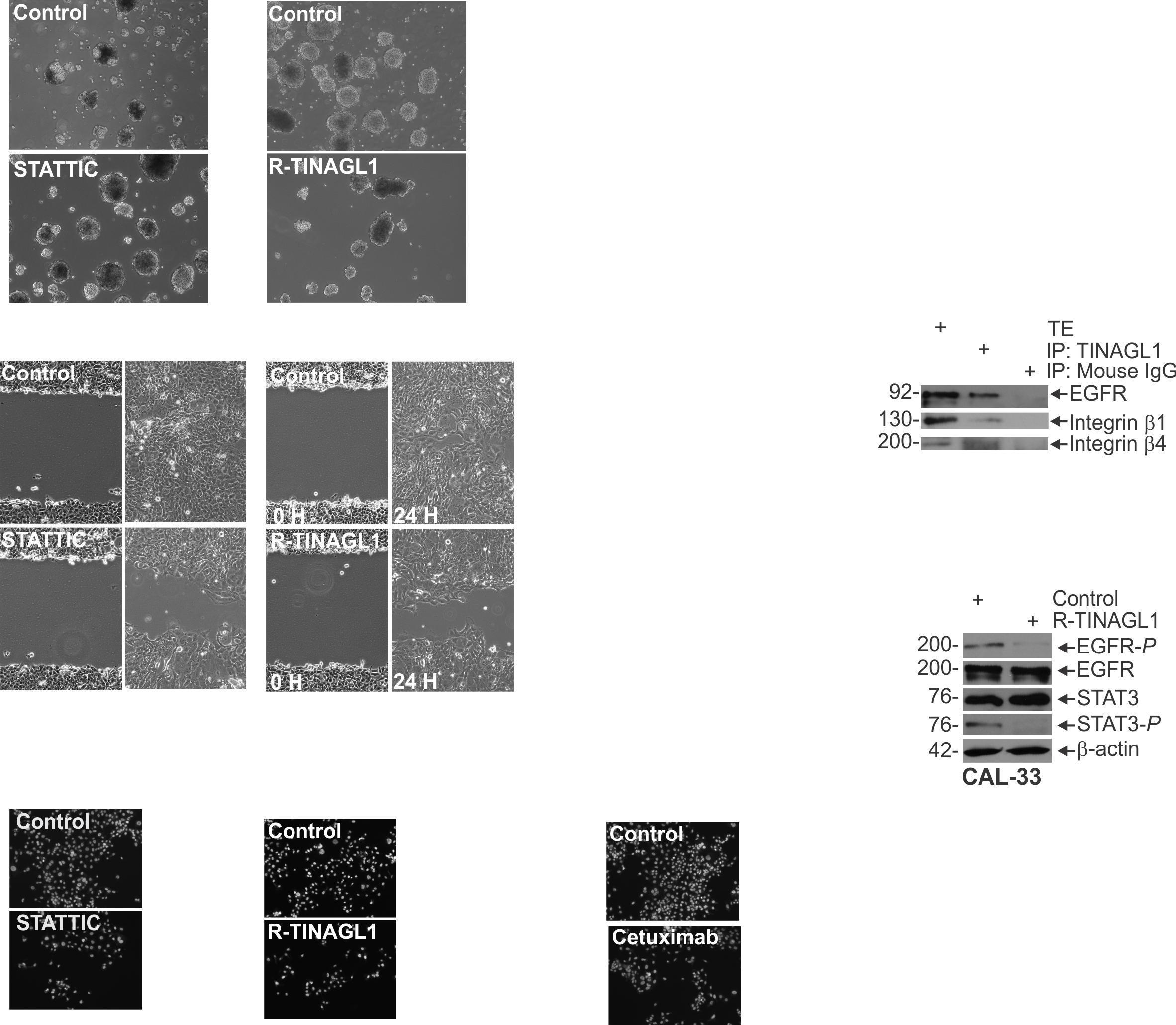
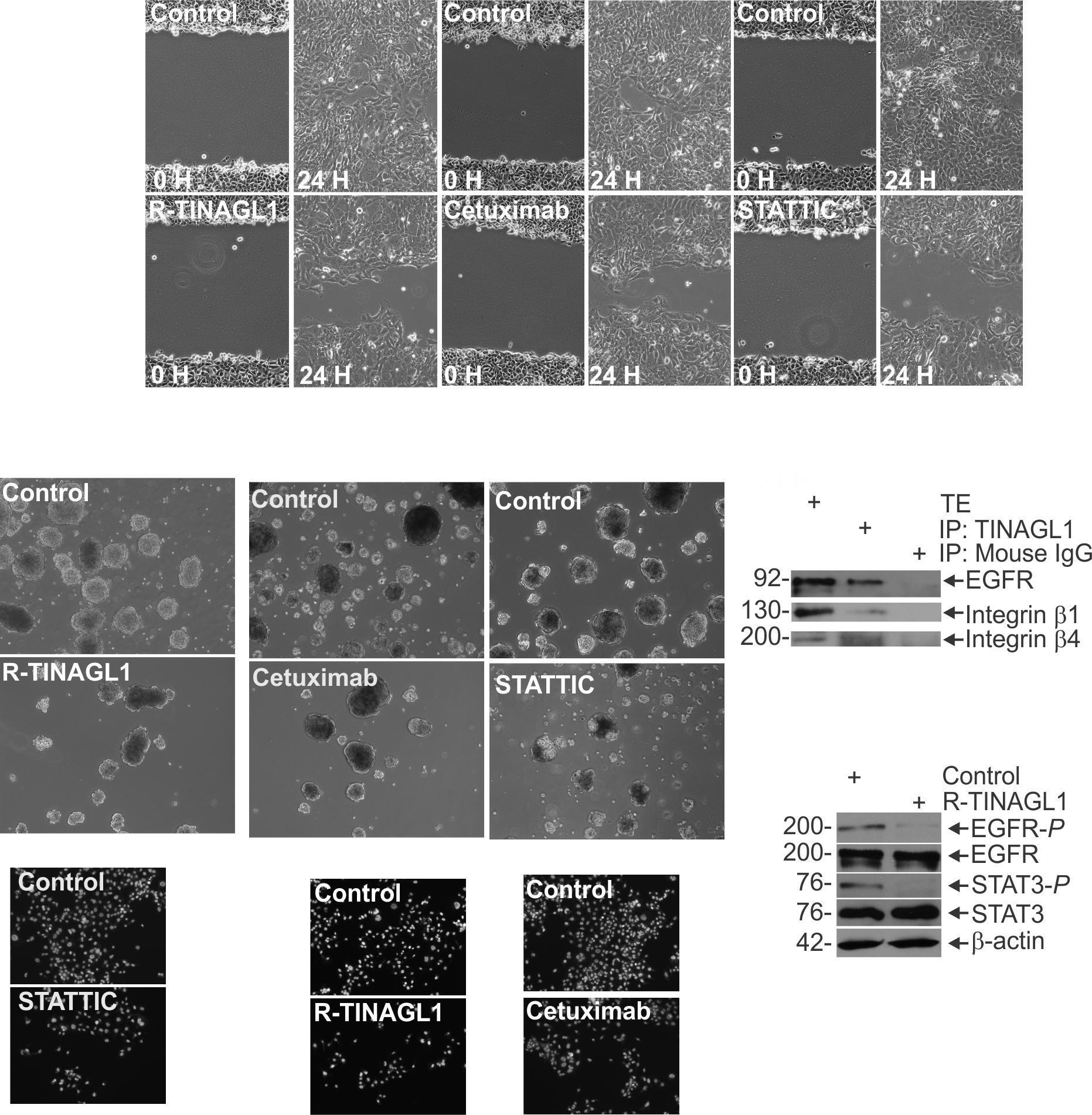
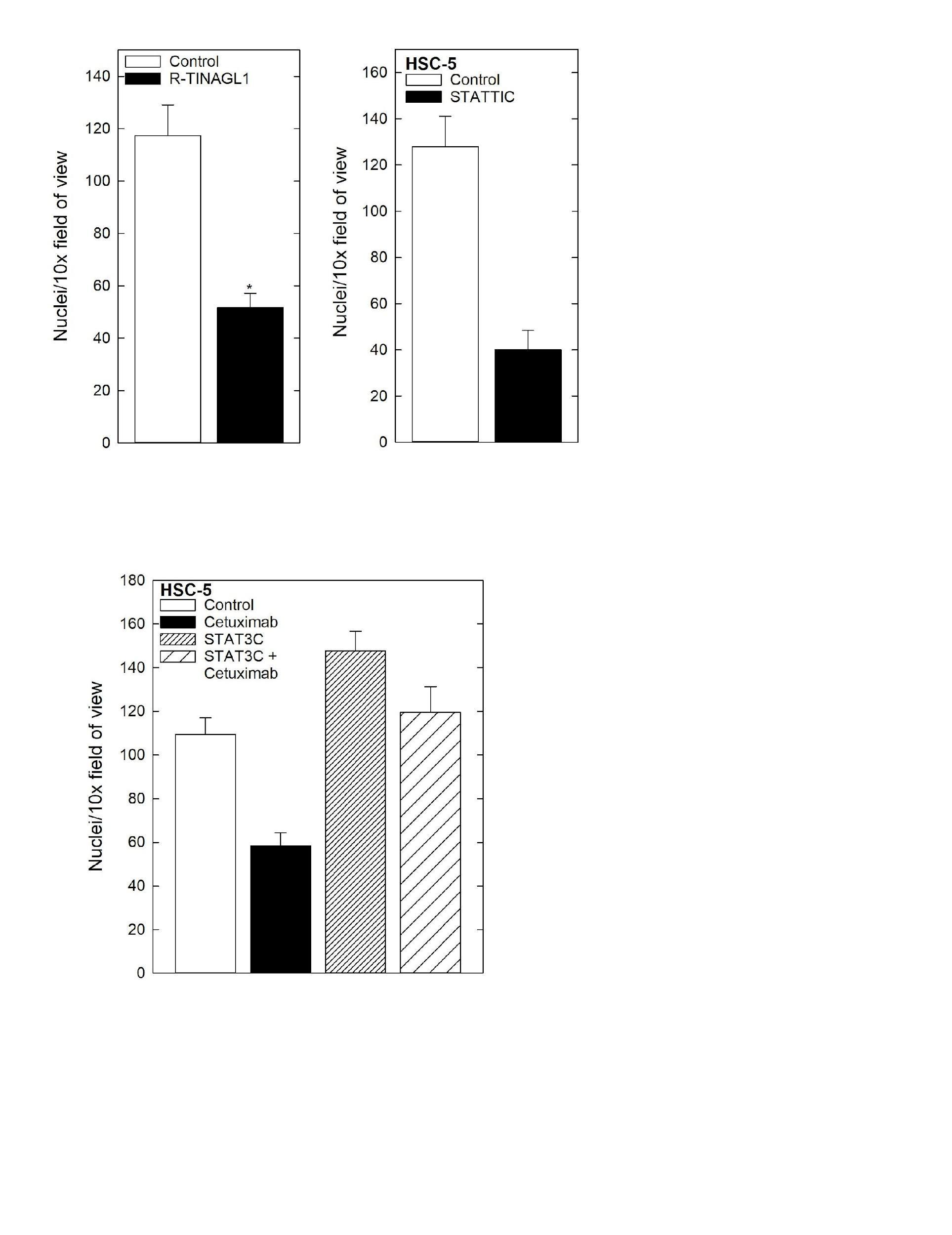
Cell Invasion Assay Matrigel was diluted in 0.01 Tris-HCL/0.7% NaCl, filter sterilized and 0.1 mLwas used to coatindividualBDBioCoatinserts(Millicell-PCF,0.4mm,12 mm, PIHP01250). Cells (2.5 x 10⁴) were plated in 100 ml growth medium supplemented with 1% FBS into the upper chambersoftheinserts.Thelowerchamberscontainedgrowth medium supplemented with 10% FBS. After invasion, membranes were harvested and the surface of upper membraneswererinsedwithPBStoremoveunattachedcells.
Membraneswerefixedin4%paraformaldehyde,stainedwith 1 mg/mL DAPI, and the underside of the membrane was photographed using an inverted fluorescent microscope and cellswerequantitated.
SpheroidFormationAssay:CAL33cellsweredissociatedwith trypsin, then centrifuged at 1000 RPM for 5 minutes. Cells were resuspended in a DMEM solution supplemented with FCS to inactivate the trypsin, then centrifuged again and washed in spheroid media to remove the FCS. These washed cells were then plated for spheroid growth in ultra-low attachment dishes containing spheroid medium consisting of (DMEM/F12 (1:1) with 2% B27 supplement, 20 ng/ml EGF, 0.4% bovine serum albumin and 4 μg/ml insulin. Spheroids numberwasmonitoredover9daysofgrowth.


