Non-gynae cytopathology
Pathology Spheres
FRCPath Part 1 Course
January 2025
Dr Casey Powell
Lecture outline
• RCPath syllabus
• Reporting systems
• Example questions

Pathology Spheres
FRCPath Part 1 Course
January 2025
Dr Casey Powell
Lecture outline
• RCPath syllabus
• Reporting systems
• Example questions
Technical aspects
Demonstrates knowledge of preparation and staining techniques for common specimen types
Demonstrates knowledge of use of special techniques; eg. immunocytochemistry
Recognises faults and artefacts of preparation; eg. air-drying
Describes panels of antibodies for particular diagnostic applications; eg. mesothelioma
Areas of learning Knowledge Skills
Morphology
Demonstrates knowledge of cell components
Demonstrates knowledge of various stains used in air-dried and fixed preparations
Identifies the nuclear features used to diagnose malignancy
Identifies features of malignancy in sites commonly investigated with cytopathology
Identifies features of specific nonmalignant diagnoses; eg. infection
Recognises normal cell populations
Recognises the differences in cell morphology in air-dried and fixed preparations
Demonstrates the ability to diagnose malignancy with confidence in specimens from breast, gastrointestinal tract, respiratory tract, urinary tract, head and neck, lymphoreticular system, serous fluids and thyroid
Demonstrates the ability to integrate clinical information and histology or other investigations into diagnosis
Demonstrates the ability to recognise when definitive diagnosis is beyond capability
Areas of learning Knowledge Skills
Report writing Identifies requirements for a report
Demonstrates ability to recall relevant datasets Identifies nationally recognised coding systems
Demonstrates the ability to write an accurate report that gives clinicians the information they need
Demonstrates knowledge of the likely outcome in terms of further investigation or management of the patient
Areas of learning Knowledge
General principles and cancer screening programme (CSP)
Applies rationale, methodology and organisation of the CSP
Demonstrates a basic understanding of the roles of component organisations, failsafe
Skills
Identifies features to determine the adequacy of a cervical sample
Demonstrates the ability to source information on the CSP
Demonstrates understanding of the difficulties in producing rigid criteria for adequacy
Ability to recognise inadequate specimens
Normal and benign changes
Identifies normal cellular components in cervical specimens
Identifies features of infections in cervical samples
Recognises typical morphological appearances of specific organisms commonly seen in cervical specimens; eg. trichomonas, candida, herpes simplex, HPV, actinomyces
Borderline nuclear changes (BNC)
Demonstrates understanding of criteria for diagnosis of BNC
Recognises the morphological features of BNC
Dyskaryosis
Demonstrates understanding of criteria for diagnosis and grading of squamous and glandular dyskaryosis
Recognises typical examples of mild, moderate and severe squamous dyskaryosis and endocervical cellular abnormalities

• Bethesda systems: Cervix, thyroid
• Yokohama: breast
• International system for reporting serous fluid cytopathology
• Milan reporting system for salivary gland cytopathology
• Paris system for urothelial neoplasia

https://tumourclassification.iarc.who.int/
A 56 year old female with a history of cirrhosis of unknown aetiology presents with increasing lethargy and abdominal distension. Clinical examination reveals tense ascites which is drained and a sample submitted for cytological examination. The consultant asks for a cell count on the specimen. The count of which type of cell provides the most diagnostic information?
(a) Eosinophil
(b) Macrophage
(c) Lymphocyte
(d) Neutrophil
(e) Basophil
A medical student undertaking a pathology study module is assigned to cytology. He notices that alcohol is put on the slide before a Papanicolaou stain is applied. He asks you to explain the purpose of this alcohol.
Choose the most appropriate response.
(a) To ‘glue’ the cells onto the slide so that the cells are not lost during staining.
(b) To fix the cells so that cellular detail is preserved.
(c) To ensure the cells take up orange G allowing easy recognition of squamous cells.
(d) To remove water from the cells so that the mountant remains transparent.
(e) To lyse red blood cells.
A 48 year old female is discussed at the breast MDT meeting. She presented with a firm, mobile mass in the left breast which had been present for 8 weeks. Mammography revealed a well defined, ovoid mass considered to be benign (M2) and ultrasound revealed a slightly lobulated, well defined hypoechoic mass again considered to be benign (U2). A fine needle aspirate was reported as carcinoma (C5).
A core biopsy was performed due to the discrepancy. A single palpable core was taken. This revealed fat necrosis and scattered breast lobules with no evidence of malignancy (B2).
The most appropriate conclusion is:
(a) The correct diagnosis is fat necrosis.
(b) The correct diagnosis is a fibroadenoma.
(c) The correct diagnosis has not been established.
(d) The correct diagnosis is carcinoma.
(e) The correct diagnosis is an abscess.
C1 - inadequate
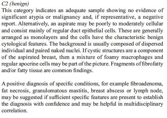
C2 - benign
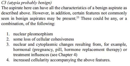
C3 - atypia probably benign
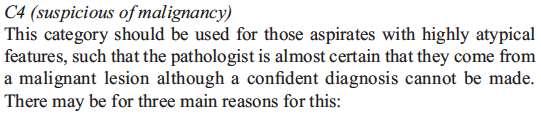
C4 - suspicious of malignancy
C5 - malignancy
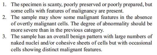
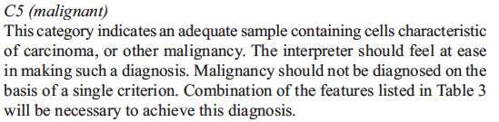


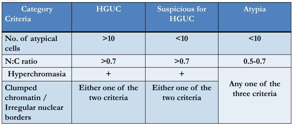
You are reporting a urine cytology sample from a 75 year old male who presented to the urology clinic with haematuria and urinary frequency. The sample contains plentiful benign urothelial cells along with blood, acute inflammatory cells and necrotic debris. You note three urothelial cells within the sample show nuclear hyperchromasia, clumped chromatin and markedly increased nuclear:cytoplasmic ratios (>0.7).
Which of the following optionsmost appropriately summarizes your report?
(a) Consistent with high grade urothelial carcinoma
(b) Consistent with low grade urothelial carcinoma
(c) Atypia
(d) Suspicious for high grade urothelial carcinoma
(e) Non-diagnostic
Standardizes terminology for reporting urine cytology, similar to the Bethesda systems for reporting cervical and thyroid cytology
Seven diagnostic categories:
I. Non-diagnostic or unsatisfactory
II. Negative for high grade urothelial carcinoma
III. Atypia
IV. Suspicious for high grade urothelial carcinoma
V. Low grade urothelial neoplasia
VI. High grade urothelial carcinoma
VII. Other malignancies (primary and metastatic)
Low grade urothelial neoplasm
‘Low grade urothelial neoplasm’ encompasses urothelial papilloma, papillary urothelial neoplasia or low malignant potential (PUNLMP), low grade papillary urothelial carcinoma AND flat, low grade intraurothelial neoplasia
Three-dimensional papillary cell clusters with nuclear overlapping and fibrovascular cores
A 79 year old male is referred to the urology department with microscopic haematuria. He has a past history of ischaemic heart disease and diabetes mellitus. Investigations performed in the clinic confirm haematuria on dipstick testing, and ultrasound reveals a mass measuring 5x3cm in the left kidney.
A sample of urine is submitted for cytological evaluation. This contains mature and anucleate squamous cells, variably degenerate urothelial cells, macrophages, neutrophils and corpora amylacea.
The most likely diagnosis is:
(a) Renal calculus
(b) Prostatic adenocarcinoma
(c) Renal cell carcinoma
(d) Transitional cell carcinoma
(e) Malakoplakia
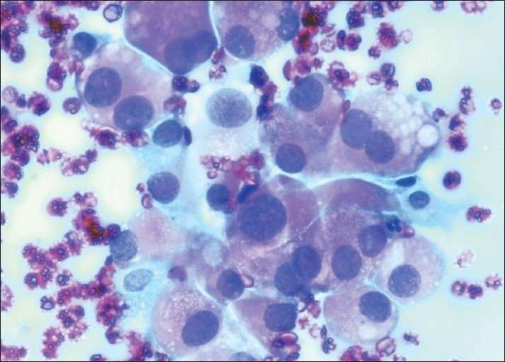
A 24 year old male presents with a mass in the right side of the neck, which has been growing for the previous 3 months. Ultrasound scan of the neck reveals a solitary mass in the right thyroid which is of high vascularity.
Enlarged lymph nodes are also present in the right carotid chain. Fine needle aspiration of the thyroid mass includes numerous cells with moderate amounts of slightly granular cytoplasm and eccentric nuclei. The nuclei are round and oval with finely stippled chromatin and inconspicuous nucleoli.
Immunocytochemistry reveals strong expression of cytokeratins and calcitonin.
The most appropriate classification for this aspirate is:
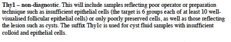
(a) Thy1
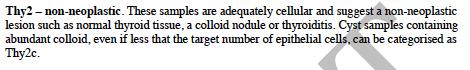
(b) Thy2
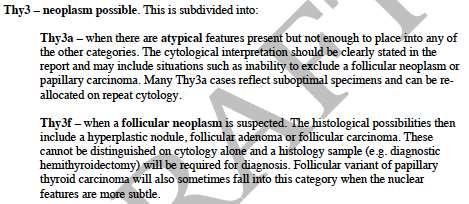
(c) Thy3f
(d) Thy4
(e) Thy5
A 24 year old male presents with a mass in the right side of the neck, which has been growing for the previous 3 months. Ultrasound scan of the neck reveals a solitary mass in the right thyroid which is of high vascularity.
Enlarged lymph nodes are also present in the right carotid chain. Fine needle aspiration of the thyroid mass includes numerous cells with moderate amounts of slightly granular cytoplasm and eccentric nuclei. The nuclei are round and oval with finely stippled chromatin and inconspicuous nucleoli.
Immunocytochemistry reveals strong expression of cytokeratins and calcitonin.
The most appropriate classification for this aspirate is:
(a) Thy1
(b) Thy2
(c) Thy3f
(d) Thy4
(e) Thy5
http://www.british-thyroidassociation.org/Guidelines/Docs/BTA_DTC_guidlines.pdf
A retired shipyard worker presents to the respiratory clinic with weight loss and recurrent pleural effusions. Imaging reveals diffuse pleural thickening. An aspirate of pleural fluid is obtained and cytological examination reveals a dispersed population of mesothelial cells, some showing mild nuclear enlargement. No clustering of cells is seen.
Immunocytochemistry reveals expression by the mesothelial cells of calretinin, CK5/6, WT-1 and BAP1. They do not express EMA. No BerEP4 or TTF-1 positive cells are seen. Which of the following statements is most applicable?
(a) There is no cytological evidence of malignancy and the patient should be discharged.
(b) There is no cytological evidence of malignancy but further investigation is advised.
(c) The cytological features are suggestive of mesothelioma and a tissue biopsy should be taken for confirmation of the diagnosis.
(d) The cytological features are diagnostic of mesothelioma.
(e) The cytological features are diagnostic of metastatic adenocarcinoma.
• High cellularity
• Mesothelial cells in sheets, ‘mulberry’ clusters or papillae
• Cell enlargement
• Variable nuclear atypia - can appear cytologically bland or show marked nuclear pleomorphism, or anything in between
• Cell engulfment
• Possible psammoma bodies
• Malignant mesothelioma vs. reactive mesothelial proliferation
• Malignant mesothelioma vs. adenocarcinoma (metastatic or locally invasive)
• Calretinin
• CK5/6
• WT-1
• D2-40 (podoplanin)
• CD141 / thrombomodulin
• In all malignant effusions:
• BerEP4 or MOC31: specific for adenocarcinoma
• TTF-1: specific for primary lung adenocarcinoma
• Other markers may be used as guided by the clinical history, eg:
• CK7, CK20
• CDX2 (GI tract)
• PAX8 (female genital tract, renal)
• GATA3, ER (breast)
• Diagnosing mesothelioma by cytology alone remains controversial
• Sensitivity 30-75%
• Sampling error probably accounts more for low sensitivity than interpretation error; sarcomatoid mesothelioma rarely sheds into pleural cavity
• Some centres always perform confirmatory tissue biopsy
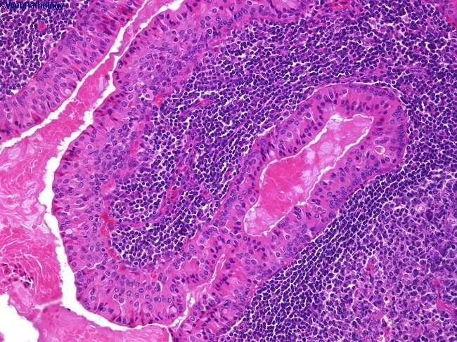
You are assessing a fine needle aspirate from a parotid mass in a 68 year old female. The aspirate contains mucoid material, plentiful lymphoid cells of mixed type and papillary groups of epithelial cells with copious, granular cytoplasm and round nuclei with prominent nucleoli.
Select the most likely diagnosis.
(a) Pleomorphic adenoma
(b) Mucoepidermoid carcinoma
(c) Warthin’s tumour
(d) Non-Hodgkin’s lymphoma
(e) Low grade polymorphous carcinoma
You are assessing a fine needle aspirate from a parotid mass in a 68 year old female. The aspirate contains mucoid material, plentiful lymphoid cells of mixed type and papillary groups of epithelial cells with copious, granular cytoplasm and round nuclei with prominent nucleoli. Select the most likely diagnosis.
(a) Pleomorphic adenoma
(b) Mucoepidermoid carcinoma
(c) Warthin’s tumour
(d) Non-Hodgkin’s lymphoma
(e) Low grade polymorphous carcinoma
(a) CD45
(b) CD9
(c) CD38
(d) CD29
(e) CD3
(f) CD30
(g) CD56
(h) CD82
(i) CD20
(j) CD99
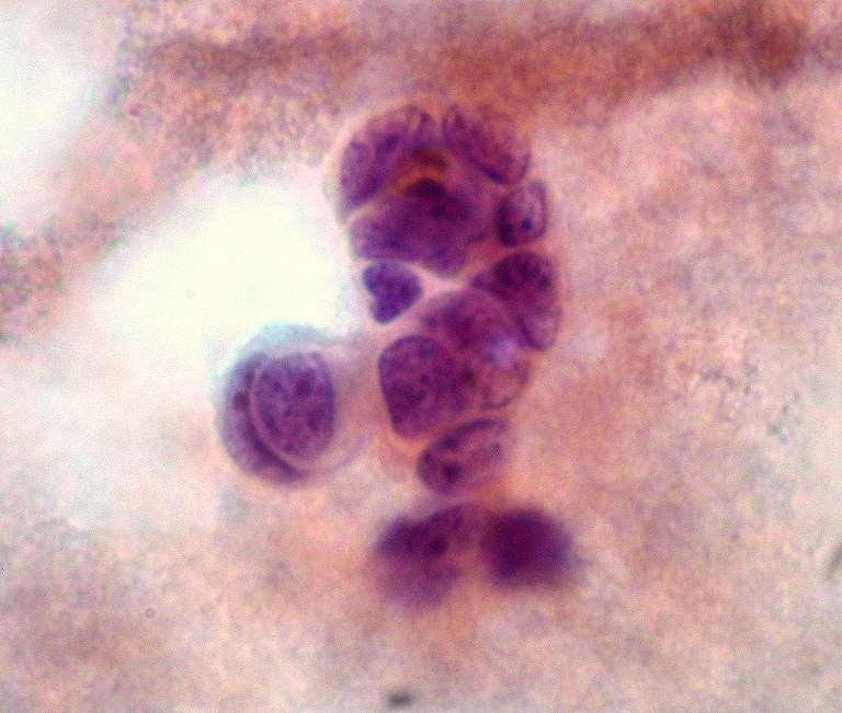
A 73 year old male presents with increasing shortness of breath. He has smoked cigarettes for many years and has a history of chronic obstructive pulmonary disease. Investigations reveal a left sided pleural effusion. A sample of this is submitted for cytological examination which reveals large numbers of mature, small lymphocytes with scattered macrophages and reactive mesothelial cells. There are also medium sized cells with scanty cytoplasm, hyperchromatic nuclei and stippled chromatin. Nuclear moulding is present. Which antibody, from the list given above, would you expect to be expressed by the medium sized cells?
A 73 year old male presents with increasing shortness of breath. He has smoked cigarettes for many years and has a history of chronic obstructive pulmonary disease. Investigations reveal a left sided pleural effusion. A sample of this is submitted for cytological examination which reveals large numbers of mature, small lymphocytes with scattered macrophages and reactive mesothelial cells. There are also medium sized cells with scanty cytoplasm, hyperchromatic nuclei and stippled chromatin. Nuclear moulding is present. Which antibody, from the list given above, would you expect to be expressed by the medium sized cells?
(a) CD45
(b) CD9
(c) CD38
(d) CD29
(e) CD3
(f) CD30
(g) CD56
(h) CD82
(i) CD20
(j) CD99
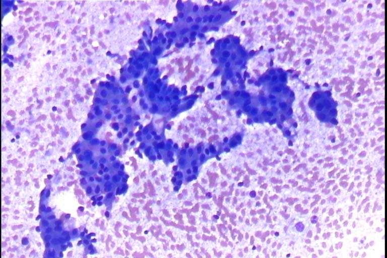
A 68 year old male presents with increasing shortness of breath and cervical lymphadenopathy. He has smoked cigarettes for many years and has a history of chronic obstructive pulmonary disease. Investigations reveal a left sided pleural effusion. A fine needle aspiration of the palpable neck lymph nodes is performed. This contains abundant material composed of cells with scanty cytoplasm with many of the cells being disrupted with streaking of the DNA. Small nucleoli are present. A colleague suggests these are from a poorly differentiated carcinoma, but you believe them to be lymphoid cells. Which antibody, from the list given above, is most likely to be expressed in this case if you are correct?
A 68 year old male presents with increasing shortness of breath and cervical lymphadenopathy. He has smoked cigarettes for many years and has a history of chronic obstructive pulmonary disease. Investigations reveal a left sided pleural effusion. A fine needle aspiration of the palpable neck lymph nodes is performed. This contains abundant material composed of cells with scanty cytoplasm with many of the cells being disrupted with streaking of the DNA. Small nucleoli are present. A colleague suggests these are from a poorly differentiated carcinoma, but you believe them to be lymphoid cells. Which antibody, from the list given above, is most likely to be expressed in this case if you are correct?
(a) CD45
(b) CD9
(c) CD38
(d) CD29
(e) CD3
(f) CD30
(g) CD56
(h) CD82
(i) CD20
(j) CD99
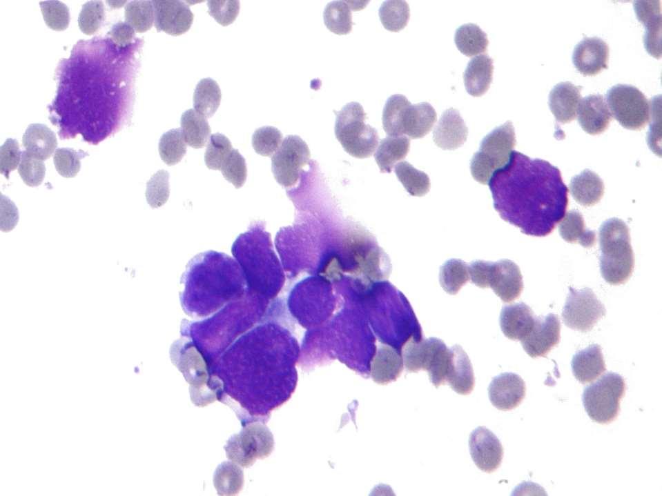
A 17 year old male presents with night sweats and weight loss. Enlarged cervical lymph nodes are noted on clinical examination. Fine needle aspiration of the lymph nodes is performed. The aspirate includes abundant material composed of a dispersed mixture of cells of varying type including eosinophils. There are several large cells with pale staining cytoplasm, large nuclei and prominent nucleoli. Some of the large cells are binucleate.
Which antigen, from the list given above, is most likely to be expressed by the large cells?
A 17 year old male presents with night sweats and weight loss. Enlarged cervical lymph nodes are noted on clinical examination. Fine needle aspiration of the lymph nodes is performed. The aspirate includes abundant material composed of a dispersed mixture of cells of varying type including eosinophils. There are several large cells with pale staining cytoplasm, large nuclei and prominent nucleoli. Some of the large cells are binucleate. Which antibody, from the list given above, is most likely to be expressed by the large cells?
(a) CD45
(b) CD9
(c) CD38
(d) CD29
(e) CD3
(f) CD30
(g) CD56
(h) CD82
(i) CD20 (j) CD99
A 63 year old female presents with a rapidly enlarging mass in the left side of the neck. An ultrasound scan of the neck reveals the mass to be formed of several large, rounded lymph nodes which lack a fatty hilum. Fine needle aspiration of this is performed. The aspirate includes numerous large cells with plentiful cytoplasm, irregular nuclei, irregular chromatin and multiple nucleoli.
Several mitotic figures are noted many of which are morphologically abnormal. Plentiful tingible body macrophages are present giving a ‘starry sky’ appearance. Which antibody, from the list given above, is most likely to be of diagnostic value in this case?
A 63 year old female presents with a rapidly enlarging mass in the left side of the neck. An ultrasound scan of the neck reveals the mass to be formed of several large, rounded lymph nodes which lack a fatty hilum. Fine needle aspiration of this is performed. The aspirate includes numerous large cells with plentiful cytoplasm, irregular nuclei, irregular chromatin and multiple nucleoli. Several mitotic figures are noted many of which are morphologically abnormal. Plentiful tingible body macrophages are present giving a ‘starry sky’ appearance. Which antibody, from the list given above, is most likely to be of diagnostic value in this case?
(i) CD20
(a) CD45
(b) CD9
(c) CD38
(d) CD29
(e) CD3
(f) CD30
(g) CD56
(h) CD82
(i) CD20
(j) CD99
A 17 year old male presents with a mass on the left chest wall. A chest X-ray reveals a mass expanding the 7th rib with an ‘onion skin’ appearance. Fine needle aspiration of the mass is performed. Numerous cells with scanty cytoplasm and round, hyperchromatic nuclei are present. Frequent mitotic figures are noted.
Which antigen, from the list given above, is most likely to be expressed by the cells?
A 17 year old male presents with a mass on the left chest wall. A chest X-ray reveals a mass expanding the 7th rib with an ‘onion skin’ appearance. Fine needle aspiration of the mass is performed. Numerous cells with scanty cytoplasm and round, hyperchromatic nuclei are present. Frequent mitotic figures are noted.
Which antibody, from the list given above, is most likely to be expressed by the cells?
• BSCC codes of practice, exfoliative and FNA
• RCPath tissue pathways for cytology
• RCPath guidelines for thyroid cytology
• The Bethesda System for Reporting on Thyroid Cytopathology
• Cytology Diagnostic Principles and Clinical Correlates; Cibas
• Comprehensive Cytopathology Bibbo and Wilbur
• Diagnostic Pathology Cytopathology Mody Thrall Krishnamurthy
• The International System for Serous Fluid Cytopathology
• IAC cytopathology: https://www.cytology-iac.org/links-toother-educational-resources