
ABSTRACT: Malignant melanoma is the deadliest form of skin cancer. Dermoscopy is a noninvasive high-resolution imaging technique that assists physicians for more accurate diagnoses of skin cancers. Melanoma is a fast-growing aggressive type of skin cancer. Due to this feature, malignant melanoma remains one of the fastest growing cancers worldwide. After it metastasizes from its origin into other tissues, the response rate to treatment declines as low as 5%, and its 10-year survival rate is only about 10% After it metastasizes, there is no surgical rem detection of malignant melanoma is critically important. Among many types of skin cancers, melanoma has the highest false negative ratio.Therefore, this thesis proposes three methods for early detection of malignant melanoma. More specifically, this thesis,Introduces a novel approach of texture-based abrupt cutoff quantification method . In current clinical practice, abrupt cutoff evaluation is subjective and errorprone. In our method, we introduce a novel approach to objectively and quantitatively measure abrupt cutoff.To achieve this, we quantitatively analyzed the texture features of a region within the skin lesion boundary using level set propagation (LSP) method.Then, we built feature vectors of homogeneity, standard deviation of pixel values, and mean of the pixel values of the region of interest between the contracted border and the original border of a skin lesion. These vectors were then classified using neural networks (NN) and support vectormachines(SVM)classifiers.
Keyword: Deep Learning,Malignant,Melenoma,Learning,TransferDermoscopy,SLIC,SkinCancer and Communication Engineering, Institute of Engineering for
ditoaccurateextractionimproveforvisualare;melanomainexperiencedmalignancy.However,featuresofdiagnosepresencevisualsubsurfaceerrorscancer-relatedmelanomaROI.Misdiagnosisopticallymagnifiesmelanoma.becomenoninvasivecost,thespotlighteddiagnosisannualandCaucasianbutbecome3%mortalityrateduetomelanomaincreasedbyintheUSA.SkincanceroccurrencehasmorecommonnotonlyintheUSAalsoindifferentcountrieswithpeoplemajoritysuchastheUKCanadawith10,000diagnosesandmortalityof1,250people.Earlyofthemelanomahasbeenduetothepersistentelevationofnumberofincidents,thehighmedicalandincreaseddeathrate.Dermoscopy,whichisoneoftheskinimagingtechniques,hasakeymethodinthediagnosisofDermoscopyisthemethodthattheregionofinterest(ROI)andtakesdigitalpicturesoftheorunderdiagnosisofisthemainreasonforskinfatalities.Thecauseoftheseisusuallyduetothecomplexityofthestructuresandthesubjectivityofinterpretations.Expertclinicianslookfortheofexclusivevisualfeaturestoskinlesionscorrectlyinalmostalltheclinicaldermoscopymethods.Theseareevaluatedforirregularitiesandinthecaseofandermatologist,diagnosisofcanbeverychallenging.Theproblemsaddressedinthisthesishowtoeliminatethesubjectivityoninterpretationofdermoscopyimagesborderirregularity/abruptness;howtotheperformanceoffeaturealgorithmsbyprovidingmoreskinlesionsegmentation;andhowreducethenumberoffalse-negative iagnosis. ISSN 2394-3777 (Print) ISSN 2394-3785 (Online) Available online at www.ijartet.com International Journal of Advanced Research Trends in Engineering and Technology (IJARTET) Vol. 9, Issue 6, June 2022 11
NITHYA D5 AP/DepartmentAP/ECEofElectronics
dnithiece@mail.comWomen,Deviyakurichi.
INTRODUCTION: The occurrence of malignant melanoma, which is the deadliest form of skin cancers, has been elevated in the last decade. Between 2009 and 2010, the
ADVANCED SKIN DISEASE DIAGNOSIS USING IMAGE PROCESSING KAVITHA V1,KAYATHRI K2,MOHANAPRIYA V3,NANDHINI M4 Department of Electronic and Communication Engineering , Bharathiyar Institute of Engineering for mohana200154@gmail.comkayathiri2002@mail.com,kavik8708@gmail.com,Women,Deviyakurichi,nandhini200110@gmail.com
Bharathiyar


that In surgical planning and cancer treatment, it is crucial to segment and measure a liver tumor's volume accurately. Because it would involve automation, standardisation, and the incorporation of complete volumetric information, accurate automatic liver tumor segmentation would substantially affect the processes for therapy planning and follow-up reporting. Based on the Hidden Markov random field, Automatic liver tumor detection in CT scans is possible using hiddenMarkovrandomfields (HMRF-EM).
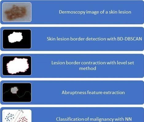
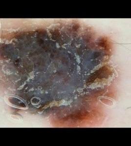
consistsofthedifferentlayers.Theback-propagationalgorithm can be divided into two phases: propagation and weight update. In the first phase of this algorithm, an input vector is propagated forward through the neural network, and the output valueisgenerated DEEP LEARNING AND TRANSFER LEARNING: Deep learning, also known as Deep Structured Learning, is a subdivision of ML supported by mass of algorithms.Deep learning can extract useful features directly from images, text and sound in supervised and/or unsupervised manners which makes it different than standard machine learning techniques. In fact, feature extraction with this approach is considered as a part of the learning process. Transfer learning is a ML technique where a model that is trained on one task is repurposed on another related taskAbruptness of pigment patterns at the periphery of a skin lesion is one of themost important dermoscopic features for detection of malignancy.In the current clinical setting, abrupt cutoff of a skin lesion is determined by an examination performed byadermatologist.Thisregionwas bounded by an interior determined using level set propagation (LSP) method.This method provides a fast bordercontraction without a need for extensive boolean operations.Then, we built feature vectors of homogeneity, standard deviation of pixel values, and mean of the pixel values of the region between the contractedborder andtheoriginal border. The data set for this part of the thesis was obtained from ISIC 2016: Skin Lesion Analysis Toward Melanoma Detection [59], which has 900 dermoscopic images with 727benign and 173 malignant lesions, and Edra Interactive Atlas of Dermoscopy [60], which has 73 benign and 27 malignant lesions.[3]discussed
Then, we considered the offset of a continuous function of whole lesion border border line of the lesion boundary which is viia i constant velocity level sets and contracted the lesion border using these
Images used in this thesis are obtained from the International Skin Imaging CollaborationsArchive.
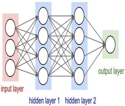
OVERVIEW OF MACHINE LEARNING: Machine learning (ML) is an area that aims to construct new algorithms to make predictions based on given data. ML generates general models using training data so thatthese models can detect the presence or the absence of a pattern in test (new) data.Patterns can be a low-level or a highlevel.Biological neural network is an important part of the human brain. It is a highly complex system and has an ability to process different tasks simultaneously. Neural network (NN) is a classifier that simulates the human brain and neurons.Instead of neurons, “perceptron” is used as a basic unit of NN .NN architecture
pixel
pixels
ixiiel
As lower homogeneity indicates sharp cutoffs, suggesting melanoma, we carried out our experiments on two dermoscopy image datasets, which consisted of 800 benign and 200 malignant melanoma using texture homogeneity at the periphery of a lesion border determined by LSP, as a classification results, we obtained 87% f1score and 78% specificity; that we obtained better results than in the previous study. We also compared the performances of two different and process by first finding the super Super are one ofthe most popular images many super algorithms, the choice of super pi algorithm in this thesis is Simple
NN classifiers
support vector machineclassifier.Westart segmentation
cases.By
over-segmentationalgorithms.Among
NN Paramete rs S V M rateLearning 0.001 functionKernel amonyloPil iterationsNumberof 1000 Polynomialorder 3 runsNumberof 20 Kernelscale otuA layershiddenNumberof 1 constraintBox nIf layerhiddenNumberofnode 4 Standardize eurT used)NNmultilayerlayershiddenNumberof(Ifis 4 fractionOutlier 500. Feature ExtractionClassification Precisi on R e c a l l Sen siti vit y F1Sc or e PerceptronLSP-MultilayerNN 0.82 180 0.75 0.8 PerceptronDS-MultilayerNN 0.77 670 0.56 0.74 LSP-SVM 0.69 460 0.66 0.66 DS-SVM 0.62 160 0.61 0.61 multilayerconnectedLSP-Fully-NN 0.86 780 0.78 0.87 hiddenconnectedDS-Fully-multi-layerNN 0.76 570 0.61 0.75 levelsets. Figure: Fully-connected multi-hidden layer NN architecture. We empirically determined the iteration numbers as 600, 750, and 1000 without constraining a stoppage criterion. Then, we added the learning rate of 0.0001 to exit the iteration between two consecutive epochs.We ran both NN methods and SVM on the same set of image data however different feature vectors based on the differentfeatureextractionmethodsused
pixels.
After the resizing step, we randomly split images into training and testing subsets.2,086 malignant and 2,086 benign images were in training set, and 200 malignant and 200 benign images were on testing sets. Notice that now the data is balanced.
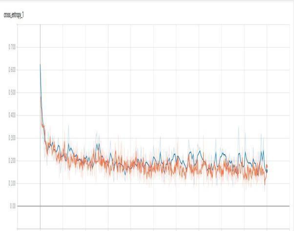
Thesedistinctclasses.results also indicate that thereis no over fitting or under fittingon the transfer learning model.Also, we examined the cross-entropy loss (log loss) which measures the performance of a classification model whose output is a probability value between0and 1.
[5] discussed that Liver tumor division in restorative pictures has been generally considered as of late, of which the Level set models show an uncommon potential with the advantage of overall optimaandfunctional effectiveness.
Cl ass Nu m be r r e c i s i o P R e c a ll n F 1S c o r e S u p p o r t -0gn)en(Bi .097 290. 49.0 002 )antgnali(M1 .092 790. 59.0 002 talTo/geeAvra 590 490. 490 004
CLASSIFICATIONRESULT:
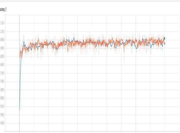
The classification results of experiment four were the best overall in all categories.Similar to the first two experiments, malignant class’ f1-score was again higher than the benign class. Training and validation iteration results are illustrated in. .This plot indicates that results are reproducible, and the algorithm is robust and reliable with high confidence for accurately classifying lesions as benign or Malignant. [8] discussed about diabetic retinopathy from retinal pictures utilizing cooperation and information on state of the art sign dealing with and picture preparing. The Pre-Processing stage remedies the lopsided lighting in fundus pictures and furthermore kills the fight in the picture. Although the Disease Classifier step was used to identify arising wounds and other data, the Division stage divides the image intotwo
REFERENCES: [1]."Cancer Facts and Figures," 8].201Junefactsandfigures-2010..CancerFactsFigures/cancer-.[Online].Available:http://www.cancer2010.org/Research/CancerFactsFigures/[Accessed [3].[2]."Melanoma,"[Online].Available:https://www.cancer.org.au/about-cancer/types-of-cancer/skin-cancer/melanoma.html.[AccessedJune2018].
This thesis proposed creative and effective methods to eliminate the subjectivity in visual interpretation of dermoscopy images and decrease the number of false- negative/false-positive diagnoses by introducing a new method for measuring abrupt cutoff and increasing the performance of feature extraction algorithms.Abruptness of pigments on the skin is one of the most important dermoscopic features for detection of malignancy. In the current clinical setting, abruptness is determined by an examination performed by a dermatologist. This process is subjective, nonquantitative, and error-prone. We presented an improved computational model to quantitatively measure abruptness of a skin lesion by quantitatively analyzing the texture features of a region within the lesion boundary.These vectors were then classified using neural networks (NN) and SVM classifiers.Diagnosis of malignant melanoma is the real reason of fatality due to skin cancer. Even though there are imaging and diagnosis techniques used commonly for melanoma like dermoscopy, automatic recognition is still challenging due to the difficulty of segmenting accurate lesion areas, similarity between melanoma and non-melanoma lesions and thevariation ofskinconditions.
CONCLUSION: Skin cancer is increasing and affects many peopleevery day. Thiscancer can be treated successfully if it is detected in early stages. Early diagnosis and treatment will lead to an increased survival chance and reduced mortality rates.. However, current clinical techniques used for the diagnosis of malignant melanoma are prone to human error due to thesubjectivity andnovicephysicians.
Besides these problems, medical images are not easy to find while protecting the anonymity of the patients.The sum up,the objectives of this thesis were to eliminate the subjectivity on visual interpretations of dermoscopy images for abrupt cutoff and to reduce the number of false-negative/false-positive diagnosis of malignancyclassifications.
Christo Ananth, S. Amutha, K. Niha, Djabbarov Botirjon Begimovich, Segmentation“EnhancingApproaches from Super Pixel Division Algorithm to Hidden Markov Random Fields with Expectation Maximization (HMRF-EM)”, International Journal of Early Childhood Special Education, Volume 14, Issue 05, 2022,pp. 2400-2410. [4]. M.Celebi,Y.Aslandogan,W.Stoecker,H.Iyatomi,H.OkaandX.Chen,"Unsupervisedborderdetectionindermoscopyimages," Skin Research and Technology, pp. 454-462,vol.13,no 4,2007. [5]. Christo Ananth,MKameswari, IssueAlgebraicExpectedGaussianBasedApproaches“EnhancingDensyJohnVadakkan,Dr.Niha.K.,SegmentationfromFuzzy-MPSOLiverTumorSegmentationtoMixtureModelandMaximization”,JournalOfStatistics,Volume13,2,June2022,pp.788-797.
With this motivation, we studied skin cancer malignancy detection to classify skin lesions and identify malignant cases.Finally, we were able to classify skin lesions with 94% average f1-score.. Also, the malignant class skin classification f1- score (95%) was higher than benign classf1-score.
[6]. G. Argenziano, H. Soyer and e.al.,"Dermoscopy of pigmented skin lesions:” Journal of the AmericanAcademy of Dermatology, vol. 48, no.5, pp. 679[7].693,2003.R.Kenet and T. Fitzpatrick," Reducing mortality and morbidity of cutaneous melanoma: a six year plan.b). identifying high and lowriskpigmented lesions usingepiluminescence microscopy," TheJournal of Dermatology, vol.21,no.11,pp.881-884 [8].1994.Christo Ananth, D.R. Denslin Brabin, Jenifer Darling Rosita, “A Deep Learning Approach To Evaluation Of Augmented Evidence Of Diabetic Retinopathy”, Turkish Journal of Physiotherapy and Rehabilitation, Volume 32,Issue 3, December 2021,pp.11813-11817. [9]. H. Pehamberger, A. Steiner and K.Wolff, " In vivo epiluminescence microscopy of pigmented skin lesions. i. pattern analysis of pigmented skin lesions," Journal of the American Academy of Dermatology, vol.17, no. 4, pp. 571-583, [10].1987.G. Argenziano, G. Fabbrocini, P. Carli, V.DeGiorgi, E.Sammarco and "EpiluminescenceM.Delfino,microscopy for the diagnosis of doubtful melanocytic skin lesions: Comparison of the ABCD fule of dermatoscopy and a new 7-point checklist based on pattern analysis," Archives of Dermatology, vol.134, no. 12, pp.1563[11].1570,1998."International Skin Imaging CollaborationArchive," [Online]. Available: archive.com/.[Accessedhttps://isic- 2018]. [12].C. Szegedy, V. I. S. Vanhoucke, J. Shlens and Z. Wojna, "Rethinking the Inception Architecture for Computer Vision," arXiv:1512.00567, 2015. [13]."World Health Organization,"
http://www.who.int/en/..[Ac[Online].Available:cessedJune2018].[14]."CancerProjectedToBecomeLeadingCauseOfDeathWorldwideIn2010,"[Online].Available:https://www.sciencedaily.com/releases/2008/12/081209111516.htm.[AccessedJuly2018]. 16