
International Research Journal of Engineering and Technology (IRJET) e-ISSN: 2395-0056
Volume: 11 Issue: 06 | Jun 2024 www.irjet.net p-ISSN: 2395-0072

International Research Journal of Engineering and Technology (IRJET) e-ISSN: 2395-0056
Volume: 11 Issue: 06 | Jun 2024 www.irjet.net p-ISSN: 2395-0072
Assale Malka Kussiaa* , Melkamu Debelo Geletaa
aTongji University, College of Environmental Science and Engineering, Shanghai, 200092, P. R. China; a* Tongji University, Shanghai 200092, China
Abstract - Tiny plastic particles known as microplastics are a major environmental concern due to their wide presence and harmful Impact on aquatic ecosystems. To tackle this issue, efforts have been made to improve waste management, adjust policies, and develop new technologies. One effective solution is to use biodegradable plastics instead of traditional ones, but the environmental impact of these biodegradable microplastics needs further investigation to address potential risks. The investigation focused on studying the toxicological impact of polylactic acid (PLA) microplastics on freshwater microalgae, specifically C. pyrenoidosa. During a 96-hour co-incubation experiment, the effects of various concentrations of PLA microplastics (ranging from 0 to 100 mg/L) on the growth of C. pyrenoidosa were examined. Different concentrations of PLA microplastics had varying effects on the growth of C. pyrenoidosa over the 96-hour period. For instance, a promotion rate of 11.68% and 5.4% was observed at a PLA concentration of 100 mg/L after 72 hours of incubation. Additionally, a growth promotion effect of 5.4% was noted at a concentration of 20 mg/L during the same incubation period. The study also found that Chl-b levels decreased significantly across all incubation periods, particularly at higher concentrations of PLA, with fluctuations observed at different time points. In PLA-treated samples, there were slight increases in Chl-a levels at specific concentrations. The fluorescence efficiency of C. pyrenoidosa samples treated with PLA microplastics was notably lower compared to the control treatment, especially at concentrations of 20 mg/L and 50 mg/L. As PLA concentrations increased, protein content decreased in C. pyrenoidosa. The research highlighted the varied effects of PLA microplastics on the fluorescence efficiency of different microalgae species, with C. pyrenoidosa showing greater susceptibility. The response of nitrate concentration varied, with MDA levels decreasing as PLA microplastic concentrations increased. The diverse reactions observed among different time and concentrations underscore the necessity for further investigation in order to fully comprehend the ecological consequences of biodegradable microplastics in water environments. This highlights the pressing requirement for additional research on the environmental risks associated with biodegradable microplastics in aquatic ecosystems.
Key words: Microplastics,BiodegradablePlastics,Microalgae,Plasticpollution.
Plastic items are often used in human production and daily activities because of their excellent mechanical properties, light weight, strong durability, and cost-effectiveness.[1]. The most commonly used plastics, such as polyethylene, polypropylene, and polystyrene, are primarily derived from petroleum. These plastics have high molecular weights and strong chemical bonds[2] Plastic particles originating from petroleum can persist in the environment for many centuries, or even millennia, andarerecognizedasemergingpollutantsthatposeathreattotheaquaticecosystem[3] Biodegradable plasticsderivedfrom sustainable sources such as starch, cellulose, bioethanol, and lignin serve as a viable substitute for conventional petroleumbased plastics[4]. These biodegradable plastics possess the ability to completely decompose into carbon dioxide (CO2) and water(H2O) withinspecificenvironmentslike water,soil,or compost, thereby allowing them to reintegrate intothe natural cycle [5] Polylactic acid (PLA), polybutylene adipate terephthalate, polyhydroxyalkanoates, polycaprolactone, and polybutylene succinate are the most prevalent types of biodegradable plastics. Among these, PLA stands as the most extensively utilized variant, finding applications across diverse industries including waste bags, agricultural coverings, food packaging,medicalsutures,andmaterialsfor3Dprinting.AsreportedbyZhangetal.(2021),PLAaccountsforapproximately 45%oftheglobalbiodegradableplasticsmarket[6].
This study investigates the impacts of PLA microplastics on the cultivation of Chlorella pyrenoidosa, an extensively studied microalgaespeciesthatplaysapivotalroleinaquaticecosystems.Microalgaeplayacriticalroleintheglobalcarboncycleby absorbing carbon dioxide through photosynthesis and converting it into biomass, which supports various trophic levels in
International Research Journal of Engineering and Technology (IRJET) e-ISSN: 2395-0056
Volume: 11 Issue: 06 | Jun 2024 www.irjet.net p-ISSN: 2395-0072
marine and freshwater environments. However, the growing presence of plastic pollution poses a significant threat to these vitalmicroorganisms.Microplastics,inparticular,candisruptthegrowth,reproduction,andmetabolicfunctionsofmicroalgae. Research indicates that microplastics can diminish the photosynthetic efficiency of microalgae, resulting in reduced biomass production and altered nutrient cycling in aquatic systems [7]. This disturbance can have far-reaching consequences throughoutthefoodchain,affectinghighertrophiclevelssuchasfishandothermarineorganismsthatdependonmicroalgae asaprimaryfoodsource.
The presenceofPLAmicroplasticsinaquaticenvironmentshasraisedconcerns regarding their effectson microalgae,which can affect their growth, metabolic functions, and ecological roles [8]. This portion of the study methodically analyzes the impactofPLAmicroplasticsonthe physiologyand biochemistryof C. pyrenoidosa usingspecificexperimentssuchasOD680, chlorophyll levels, chlorophyll fluorescence, and a range of biochemical assessments. The findings of this research provide significantunderstandingintothewiderecologicalconsequencesofmicroplasticcontamination.
2.1 Materials used in the study
The microalga Chlorella pyrenoidosa (strain no. FACHB-9) was obtained from the Institute of Hydrobiology, Chinese Academy of Sciences. Microalgae were grown in liquid SE medium which consisted of (KNO3 1.25 g/L, KH2PO4 1.25 g/L, MgSO4 .7H2O1g/L,EDTA0.5g/L,H3BO30.1142g/L,CaCl2 .2H2O0.111g/L,FeSO4 .7H2O0.0498g/L,MnCl2 .4H2O0.0142g/L, MoO30.0071 g/L CuSO4 .5H2O0.0157g/L,Co(NO3).6H2O0.0049g/L andZnSO4 .7H2O0.0882g/L) 5000ml deionizedwater addedtoa5Lbeakerwithamagneticstirreronthestirringmachine,thentheabovereagentswereaddedaswerecordactual mass measured. The beaker was covered with a foil while adding the reagents to minimize contamination. After all the reagentshavebeenaddedweleavethemediumfor30minutestostiruptothereagentsfullydissolved.ThenthemediumpH adjustedto6.1andtransferredittoaVerticalHighPressureSteamPressureovernighttoautoclave.Theplasticwehaveused to study the effect on microalgae was polylactic acid microplastic (PLA MPs). We have used the following instruments for differentpurposesthroughourstudytime:
Table-1 Apparatususedintheexperiment
Apparatus
BluePardIncubator
CimoGZX-300BS-IIIincubator
CenceH1850RCentrifugemachine
VerticalHighPressureSteamSterilizerLDZF-50L-II
UltrasonicCellCrusherNoiseIsolatingChamber
DoubleBeamUV-VisibleSpectrophotometer
MettlerToledoElectricMeasuringScale
2.2.1 Determining the optical density (OD680) test
Purpose
Storethecultivated C. pyrenoidosa
Storethe C. pyrenoidosa growthassay
Centrifugethesamples
SterilizethebasalmediumcultureandtheErlenmeyerflasks
Carryoutthecelllysateprocessofthealgalsamples
Measuresamplesabsorbances
Measurethereagentsandmicroplasticstobeused.
The determination of microalgae growth using an OD680 test is crucial as it provides a quick and convenient meanstoquantifythebiomassconcentrationofthealgaeculture. Thismethodenablesreal-timemonitoringofexperimental progress, allowing for timely adjustments to culture conditions. Moreover, it offers a non-invasive approach to assess microalgae growth without the need for cell harvest or disruption. In essence, the OD680 test serves as a valuable tool for investigatingmicroalgaeculturedynamicsandenhancinggrowthconditionsforvariousapplications.Onthiswork tomeasure
International Research Journal of Engineering and Technology (IRJET) e-ISSN: 2395-0056
Volume: 11 Issue: 06 | Jun 2024 www.irjet.net p-ISSN: 2395-0072
theODvalueofalgaeliquid,a3mlsampleoftheliquidiscollectedanditsopticaldensityvalueismeasuredusingaUV-visible spectrophotometeratawavelengthof680nm.
2.2.2 Chlorophyll content measurement
Inthisworkweutilizedabsoluteethanoltoquantifychlorophylla,chlorophyllb,andcarotenoids.Theprocedureinvolved taking 5 ml of algal liquid, centrifuging it at 10,000 rpm for 2 minutes, discarding the supernatant, and retaining the algal sludge.Thesludgewasthensubjectedtothreefreeze-thawcycles.Afterfreezingfor15minutesandthawingthreetimes,5mL ofabsoluteethanolwasaddedtothealgalslurry,mixedthoroughly,andstoredinthedarkinarefrigeratorfor24hours.The extract was then centrifuged at 10,000 rpm for 2 minutes to separate the supernatant, which was transferred to a new test tube.Theabsorbanceoftheextractatwavelengthsof663,645,and470nmwasmeasuredusingaUVspectrophotometerto determinethepigmentcontentof C. pyrenoidosa and M. aeruginosa,calculatedaccordingtoaspecificformula[9]. ( ) ( ) ( ) 0 3 03 ( ) ( )( ) (3) ( ) ( 000 3 0 ) ( )
Where,A470,A663,A645 aretheextractinabsorbanceat470nm,663nmand645nmrespectively
2.2.3 Chlorophyll fluorescence activity
A1mLaliquotofthealgaesolutionwasintroducedintoacentrifugetubewithavolumeof10mL.Subsequently,thealgae were diluted by a factor of 20 using deionized water. Following this, 1mL of deionized water was added to the same tube, whileaseparatetubereceivedanadditionof4.5mLofdeionizedwater.Subsequently,0.5mLofthedilutedalgaesolutionwas transferredfromtheinitialtubeandaddedtothenewtube.Theresultingmixtureinthenewtubewasthenstoredindarkness for a duration of 20 minutes. To assess the chlorophyll fluorescence activity, the Aqua Pen PSI was employed. The aforementioned steps were repeated, with the only variation being that the tubes were stored in a light rather than dark adapted.
2.2.4 Protein test
Lipid peroxidation tests are useful in assessing the levels of oxidative stress caused by microplastics, which is a common indicator of cellulardamage.In thisexperimental work, thetestutilized thesupernatantobtained from both the control and treatment samples during the Lysate step. To create a 'work solution', the Coomassie brilliant blue dye was diluted with deionized water at a ratio of 1:4. The samples and reagents were then added in a specific sequence as outlined in Table 2.4. After allowing the samples to incubate for 20 minutes, the protein content was determined using a Double Beam UV-visible spectrophotometeratawavelengthof595nm.
The assessment of Superoxide Dismutase (SOD) in microalgae toxicity studies involving microplastics is crucial for evaluatingthe oxidative stressthat mayoccur dueto exposure to microplastics.SODis an essential antioxidant enzyme that plays a vital role in safeguarding cells against damage caused by reactive oxygen species (ROS), which are produced during cellularmetabolismandcanaccumulateunderstressfulconditionslikeexposuretomicroplastics Theinstructionsstatedthat for the SOD (Superoxide Dismutase) test in this work were, 10mL centrifuge tubes corresponding to the samples should be obtained induplicates,with twoadditional tubesfor a Blank anda Standard. Itwasadvisedtoaddreagentsin thefollowing sequence.
International Research Journal of Engineering and Technology (IRJET) e-ISSN: 2395-0056
Volume: 11 Issue: 06 | Jun 2024 www.irjet.net p-ISSN: 2395-0072
Table-2 ReagentsforSODtest Reagents
2.2.6 Malondialdehyde (MDA) Test
The MDA Test kit was utilized to conduct this test, employing the supernatant obtained from both the control and treatment samples during the Lysate step. The addition of reagents and samples followed a specific sequence as outlined in Table2.6,withthetubesbeingvortexedaftereachaddition.Subsequently,thesamplesweretransferredtoawaterbathsetat 90oC for a duration of 30 minutes. After cooling down, the absorbance was measured at 532nm using a Double Beam UVvisiblespectrophotometer.TheMDAlevelwasdeterminedusingtheprovidedformulas.
Table- 3 MDATestreagentsandsamplessequence
50%Glaceticaceticacid
The algal control and treatment samples underwent filtration and were subsequently transferred into 10mL centrifuge tubes.Topreparethesamplesforanalysis,100µLofeachsamplewastransferredinto10mLglasstesttubesinduplicate,and thenfilleduptothe10mLmarkwithdeionizedwater.Followingthis,200µLofHydrochloricacid(HCl)and200µLofSulfamic acid (H3NSO3) were added to the samples. It is important to thoroughly mix the samples after the addition of each reagent. Thesampleswerethenallowedtostandforadurationof15minutesatroomtemperature,afterwhichtheirabsorbancewas measuredusingaUVspectrophotometeratwavelengths220nmand275nm.Thenitratelevelsweresubsequentlydetermined usingthecalibrationcurveequation.
International Research Journal of Engineering and Technology (IRJET) e-ISSN: 2395-0056
Volume: 11 Issue: 06 | Jun 2024 www.irjet.net p-ISSN: 2395-0072
absorbance x= concentration
2.3 Data collection and analysis methods
The statistical analysis of the results was performed utilizing OriginPro 9 software, in addition to Microsoft Office Excel 2021forfurtherdatamanipulationandvisualization.Thesignificanceofthefindingsderivedfromthecompletelyrandomized experimental design was evaluated through one-way analysis of variance (ANOVA), employing a predetermined significance level of p < 0.05. This approach facilitated the comparison of average values across replicated datasets. The variability was assessedbysystematicallycalculatingthestandarddeviation(SD)values,whichweresubsequentlyrepresentedaserrorbars in graphical presentations. By employing advanced statistical tools in conjunction with Excel, a comprehensive and accurate analysisoftheexperimentaldatawasconducted,therebyenhancingthereliabilityofthestudy'sfindings.
3.1 Infrared and particle size of PLA Microplastics
3.1.1 Infrared structural analysis
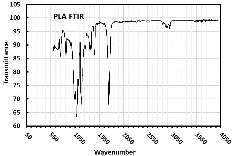
ThephysicalandchemicalattributesofPLAmicroplasticparticlessignificantlyinfluencetheirinteractionwithfreshwater microalgae during toxicity assessments, ultimately affecting the potential toxic effects on these vital aquatic organisms. The FTIRspectrumconfirmedthechemicalstructureofPLA.IntheIRspectrumofpoly(lacticacid)(PLA)(Fig-1),theC=Obond stretching is represented by a strong band at 1757.33 cm−1. Additionally, the bands at 2996.37 cm−1 and 2945.82 cm−1 correspondtotheC-Hstretchingof-CH3.ThemostnotableabsorptionforesterC-Ostretchingoccursat1187.45cm−1.The FTIRspectrumofPLAalignswiththeIRspectradocumentedintheliterature[10]
International Research Journal of Engineering and Technology (IRJET) e-ISSN: 2395-0056
Volume: 11 Issue: 06 | Jun 2024 www.irjet.net p-ISSN: 2395-0072
The size of particles plays a crucial role in ecology, as it is considered the primary factor influencing how microplastics interactwithorganismsandtheirimpactontheenvironment [11].Thedistributionofmicroplasticsinnaturalenvironments, wastewater,andaquaculturesystemsvariessignificantly,ofteninfluencedbythesourcesandpathwaysthroughwhichthese particlesentertheecosystems.Innatural waters,microplasticsprimarilyresultfromthedegradationoflarger plasticdebris caused by physical and chemical weathering. The size distribution of these particles can vary greatly, with a substantial portionmeasuringlessthan5mmindiameter.Researchhasshownthatparticlesizescommonlyrangefrom0.5mmto4mm, withnotableconcentrationsfoundinthe3–4mmand0.5–0.99mmranges[12,13]
Wastewatertreatmentplants(WWTPs)playamajorroleinintroducingmicroplasticsintoaquaticenvironments.Despite their high removal efficiencies, significant quantities of microplastics still find their way into water bodies through effluent. Microplasticsinwastewaterexhibitadiverserangeofsizes,reflectingthevarioussourcesfromwhichtheyoriginate,such as personalcareproducts,syntheticfibersfromlaundry,andindustrialwaste.Typicalsizesofmicroplasticsinwastewaterrange fromseveralmicrometerstoafewmillimeters[12].Inaquaculturesystems,microplasticsoftenarisefromthedegradationof equipmentlikenetsandropes.Theparticlesizedistributionintheseenvironmentsissimilartothatinnaturalwaters,witha predominanceofparticlesmeasuringunder5mm.However,duetospecificsourceslikefeedandequipment,theremaybea higheroccurrenceofcertainsizesandtypesofmicroplastics,suchasfibersandfragments[13]
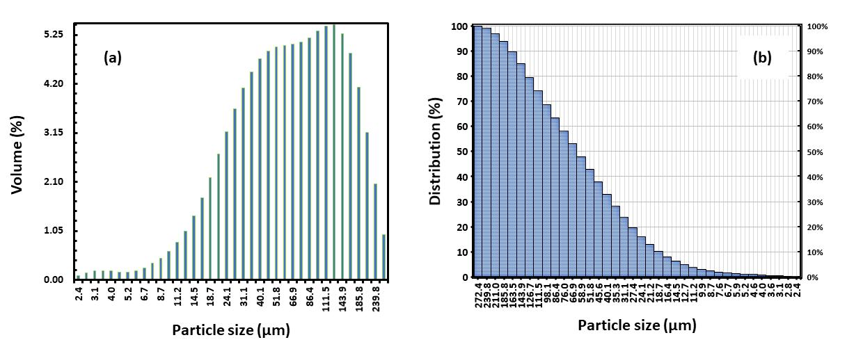
ThedistributioncurvefortheparticlesizeofPLAmicroplasticisillustratedinFig-2(a)and(b). Theanalysisrevealedthat theparticlesmeasuredfellwithinthesizerangeof2.4 -272.4μm,with90%ofthemhavinganaveragesizebetween26 -163 μmandbelow165μm.Homogeneitywasassessedbycalculatingthespanvaluesforeachformulation,wherethespanvalue was determined using the formula: Span value = (D90 – D10)/ D50. Here, D90, D10, and D50 represent the particle distribution at 90%, 10%, and 50% respectively. A span value lower than 1 indicates a homogeneously dispersed size distribution [14]. The results suggest that the particle distribution of microspheres falls within an acceptable size range for parenteralinjection.
3.2
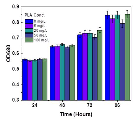
The OD680 generally mirror the microalgae cell development and biomass focus [15]. The OD680 upsides of Chlorella pyrenoidosa under various growth of polylactic acid microplastic focuses are displayed in Fig -3. At the initial stage of the cultureperiod,withthegrowthoftime,theadditionofmicroplasticcouldpromotethegrowthof Chlorella,asshowntheOD680
International Research Journal of Engineering and Technology (IRJET) e-ISSN: 2395-0056
Volume: 11 Issue: 06 | Jun 2024 www.irjet.net p-ISSN: 2395-0072
curvein(Fig-3).PLAMPswithconcentrationof100mg/Lpresentedthemostpromotingeffectattheculturetimeof96hours on C. pyrenoidosa with rate of 11.68% than control treatment. Whereas, the concentration of 20 mg/L shows the lowest growth promoting effect on the microalgae at exposure time of 72 hours with rate of 4.8 but, 5 mg/L PLA microplastic concentrationtreatmentwasthelowestpromotingeffectof C. pyrenoidosa growthatsametogrowthtimeof100mg/Lwhich is 96 hours. The graph analysis demonstrates the influence of Polylactic acid (PLA) microplastics on the growth of C. pyrenoidosa withina span of96 hours. The optical densityat 680 nm (OD680) is used asa measure of algal biomass,where higherODvaluesusuallyindicategreateralgalgrowth[16].
Throughoutthe24-hourperiod,theOD680valuesforall concentrationsofPLAmicroplastics,includingthecontrolgroup (0 mg/L), exhibit a similar trend. This observation suggests that the presence of microplastics does not have an immediate adverse impact on algae growth. This phenomenon could be attributed to the resilience of the algae or a delay in the manifestation of toxic effects induced by the microplastics. Moving forward to the 48-hour mark, the OD680 values remain consistent across the different concentrations of PLA, with a slight increase and decrease noted at 20 mg/L and 50 mg/L, respectively,incomparisontothecontrolgroup.However,thesignificanceofthesefluctuationsimpliesthatthealgaemaystill beabletothriveinthepresenceofPLAmicroplastics(Fig-3).Subsequentto72hours,acontinuousdecreaseisobservedata PLA concentration of 50 mg/L, while other treatments show minor increases, except for the 100 mg/L concentration where thegrowth-promotingeffectofPLAismostpronouncedcomparedtothecontrol.Bythe96-hourmark,anotabledeclineinOD values is evident at a concentration of 50 mg/L of PLA microplastics, followed by 5 mg/L, indicating that PLA microplastics mayhaveamultifacetedimpactonalgaegrowth,withtheseverityofnegativeeffectspotentiallydependentonthedurationof exposure. Studies have showed that, PE, PA, and PS concentrations ranging from 50mg/L to 100mg/L had no significant inhibition effectson C. pyrenoidosa growthuntil 96h exposureinthe microplastictoxicity experiment [17]the samething is here the PLA microplastic insignificant positive effect (P > 0.05) on C. pyrenoidosa. Farther more the previous research indicated that MPs (PE, PET, PVC) stimulated growth in C. pyrenoidosa, while hindering the growth of Phaeodactylum tricornutum[18].Conversely,itwasdiscoveredthatmPA,mPLA,andmPBSexhibitedthehighestinhibitionrateat100mg/L, withmPLAshowingthemostsignificantinhibitionrateat47.95%after11daysofexposurefor C. vulgaris microalgae[19]
PLA microplastics can have the potential to both stimulate growth and exhibit toxicity towards microalgae biomass for a variety of reasons. Firstly, in terms of growth promotion, PLA microplastics offer a surface area for biofilm development, therebyimprovingnutrient accessibilityandfosteringgrowthatlowerconcentrations[20] Additionally,thebiodegradability of PLA allows for the release of nutrients during degradation that can be beneficial for microalgae growth under specific circumstances[21].Ontheotherhand,toxicitycanarisefromhighconcentrationsofPLAmicroplasticscausingphysicalstress byobstructinglight penetrationnecessaryforphotosynthesis.Furthermore,chemicalleachingduringdegradationcanresult inthereleaseoflacticacidandotherharmfulby-productsthatmaynegativelyimpactthemetabolicprocessesofmicroalgae. Moreover, the production of reactive oxygen species (ROS) induced by PLA microplastics can lead to oxidative stress in microalgae, ultimately damaging cellular structures and impeding growth [20]. These contrasting effects underscore the intricate relationship between PLA microplastics and microalgae, which can be influenced by varying concentrations and environmentalfactors.
International Research Journal of Engineering and Technology (IRJET) e-ISSN: 2395-0056
Volume: 11 Issue: 06 | Jun 2024 www.irjet.net p-ISSN: 2395-0072
Chlorophyll,anessentialcompoundforphotosynthesisinhigherplants,servesastheprimarypigmentingreenplants.Chla and Chl-b are recognized as the inherent pigments responsible for absorbing and transmitting light energy during the processofphotosynthesis[22,23].Chl-anaturalgreenpigment,holdssignificantcommercialvalueasitplaysacrucialrolein plantsandalgaebyabsorbinglightenergyandconvertingitintochemicalenergythroughtheprocessofphotosynthesis [24] Theinterruptionofchlorophyllresultsinadirectdecreaseinthecapacityforphotosynthesis,consequentlyimpactingtherate atwhichplantsgrow[25].TheabsorptionandconversionoflightenergyheavilyrelyonphotosyntheticpigmentssuchasChla, Chl-b, and carotenoids. In this study, during the C. pyrenoidosa microalgae exposure to PLA, the results were presented in (Fig-4a-c).Chl-aexhibitedaslightincreaseinPLAtreatedsamples[26]comparedtothecontrolatconcentrationsof50mg/L after 96 hours, 100 mg/L after 24 hours, and 5 mg/L after 24 hours, with rates of 3.77%, 3.45%, and 2.87% respectively. Conversely,therewasanegligibleinhibitionobservedat100mg/Lafter24hours,20mg/Lafter48hours,and100mg/Lafter 72hours,withratesof5.78%,4.4%,and2.92%respectively(Fig.3.4a).Ontheotherhand,Chl-bdisplayedinhibitionrates[27] at20mg/Lafter24hours,100mg/Lafter24hours,100mg/Lafter72hours,and5mg/Lafter24hours,withinhibitionrates of10.94%,10.49%,6.88%, and6.4%comparedtothecontrol,respectively.Furthermore,carotenoidsin C. pyrenoidosa were depictedin(Fig-4c),whereinhibitionofcarotenoidswasobservedatPLAMPsconcentrationsof100mg/Lafter24hours,20 mg/Lafter48hours,and50mg/Lafter48hours,withinhibitionratesof3.4%,2.17%,and1.42%respectively.Conversely,an insignificantpositiveimpactofPLAon C. pyrenoidosa microalgaecarotenoidscontentwasnotedatconcentrationsof50mg/L after 72 hours, 20 mg/L after 72 hours, and 5 mg/L after 72 hours of incubation, with rates of 3.5%, 2.33%, and 1.2% respectively.
Microplastics have been found to have a detrimental impact on microalgal pigmentation primarily by inducing oxidative stress and physically interfering with photosynthetic processes. This interference can lead to a reduction in chlorophyll content and photosynthetic efficiency, ultimately resulting in decreased pigmentation. Research has demonstrated that exposure to microplastics, such as polystyrene and PVC, can lead to lower chlorophyll concentrations and impaired photosynthetic activity in microalgae species like Chlorella vulgaris and Skeletonema costatum. Additionally, the physical
International Research Journal of Engineering and Technology (IRJET) e-ISSN: 2395-0056
Volume: 11 Issue: 06 | Jun 2024 www.irjet.net p-ISSN: 2395-0072
presence of microplastics can obstruct light penetration and cause damage to cellular structures, further exacerbating the negativeeffectsonpigmentationandoverallhealthofmicroalgae[28].
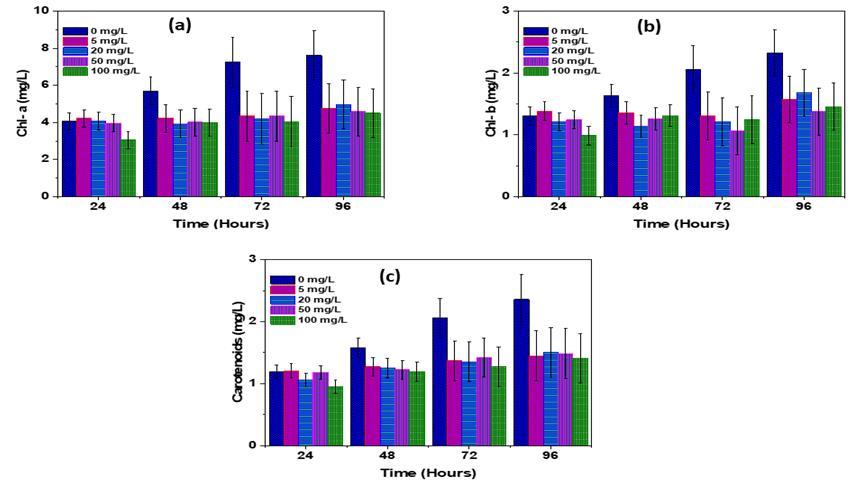
-4 EffectsofPLA-MPonthecontentsofChlorophyllconcentrationforC.pyrenoidosamicroalgae(a)chl-a,(b)chl-b,and(c) carotenoids.
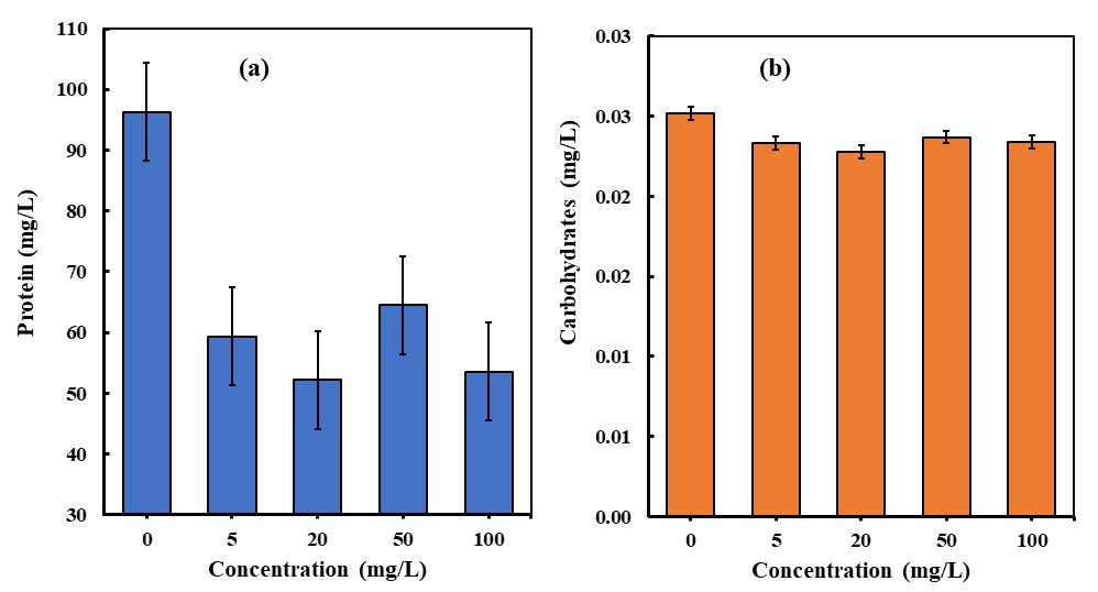
The accumulation of metabolites in microalgal biomass is significantly affected by the conditions in which they are cultivated. Microalgae have the ability to adjust their metabolic pathways in response to various stresses, leading to the productionofbiomoleculestriggeredbydifferentenvironmentalfactors [29].Thestudyconsistedofacontrolgroupwithno PLA concentration (0 mg/L) and experimental groups with varying concentrations of PLA, including 5 mg/L, 20 mg/L, 50 mg/L,and100mg/L.For C. pyrenoidosa microalgae,theproteincontentexhibitedadecline(seeFig-5a)astheconcentration ofPLAmicroplasticsincreased[30].Specifically,at100mg/L,therewasadecreaseof44.37%inproteincontentcomparedto the control treatment. Similarly, at 5 mg/L, the protein content decreased by 38.38%, while at 50 mg/L and 20 mg/L, the protein content decreased by 33.03% and 12.08% respectively, in comparison to the control treatment. These findings indicate that higher levels ofPLA microplasticsresulted in a more significant reduction in protein content for C. pyrenoidosa [31]
Additionally, a carbohydrate test on C. pyrenoidosa revealed a slight decrease at through the incubation period at all PLA MPs concentrations (see Fig -5 b). Overall, the effects of PLA microplastics on protein and carbohydrate content differed between different microalgae species [32]. The impact of polylactic microplastics on the biochemical composition of
© 2024, IRJET | Impact Factor value: 8.226 | ISO 9001:2008 Certified Journal | Page990
International Research Journal of Engineering and Technology (IRJET) e-ISSN: 2395-0056
Volume: 11 Issue: 06 | Jun 2024 www.irjet.net p-ISSN: 2395-0072
microalgae can be better understood by examining the changes in proteins and carbohydrates. These changes may indicate alterations in metabolic processes, stress responses, or nutrient uptake mechanisms in microalgae following exposure to polylacticmicroplastics.An increasein proteins and carbohydrates couldsuggest a cellularresponsetostress caused by the presence of microplastics, potentially leading to changes in energy allocation, growth, or defense mechanisms. Conversely, a decrease in these biomolecules may indicate disruptions in normal cellular functions or nutrient availability due to the interaction with microplastics. Analyzing the variations in proteins and carbohydrates in microalgae exposed to polylactic microplastics is essential for comprehending the physiological and biochemical effects of microplastic pollution on these organisms.
3.4
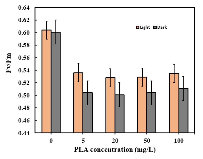
Photosyntheticparametersinmicroalgaeencompassarangeofquantifiablefactorsthatshedlightontheeffectivenessand vitality of the photosynthetic process occurring within the cells. These parameters serve as indicators of the efficiency and overallhealthofphotosynthesis.AmongthecommonlymeasuredparametersisFv/Fm,whichstandsforMaximumQuantum Yield of Photosystem II. This particular parameter gauges the utmost efficiency at which light absorbed by Photosystem II (PSII)isutilizedforphotochemistrywhileinadark-adaptedstate[33].AdecreaseintheFv/Fmvaluesignifiesthepresenceof stressordamage tothephotosyntheticapparatus.By monitoringchangesinthese parameters,itbecomespossibleto detect stress in microalgae. Environmental stressors such as variations in light intensity, nutrient availability, temperature, or the presence of pollutants like microplasticscan be identified throughalterations in these parameters. For instance,a decline in Fv/Fmsuggeststhatthemicroalgaeareunderstress[34],potentiallyleadingtoharmtothephotosystemII.
Thefluorescenceactivityof C. pyrenoidosa samplestreatedwithPLAMPswaslowercomparedtothecontroltreatment[35] (see Fig -6). Specifically, ata concentration of 20 mg/L of microalgae under light-adapted conditions, there was a significant decrease in fluorescence parameters. The decrease rates for Fv/Fm were measured at 12.58% to the control treatment. Similarly, at a concentration of 50 mg/L of PLA MPs, the decrease rates for Fv/Fm were 12.42%, with other treatments showing similar decrease rates to the control. Additionally, dark-adapted treatments also exhibited decreased levels of
International Research Journal of Engineering and Technology (IRJET) e-ISSN: 2395-0056
Volume: 11 Issue: 06 | Jun 2024 www.irjet.net p-ISSN: 2395-0072
fluorescence parameters. At a concentration of 20 mg/L, the decrease rates for Fv/Fm were 16.64%. Furthermore, at concentrations of 5 mg/L and 100 mg/L, the decrease rates for Fv/Fm showed a consistent decrease rate of 16.14% to the controltreatment.
The study findings recommend that the fluorescence efficiency of C. pyrenoidosa samples treated with PLA MPs was consistentlylowerthanthatofthecontrol treatment [35].TheFv/Fmratio, whichmeasuresthemaximumquantumyieldof photosystemII,could potentiallydecrease in the presenceof polylacticmicroplastics,indicating potential photoinhibition or stressinthephotosyntheticapparatus[36].Monitoringthesefluorescenceparameterscanenhanceourcomprehensionofthe impactsofmicroplasticexposureonmicroalgae,providinginsightsintotheecologicalimplicationsofmicroplasticpollution in aquaticenvironments.
3.5 Oxidative stress in microalgae
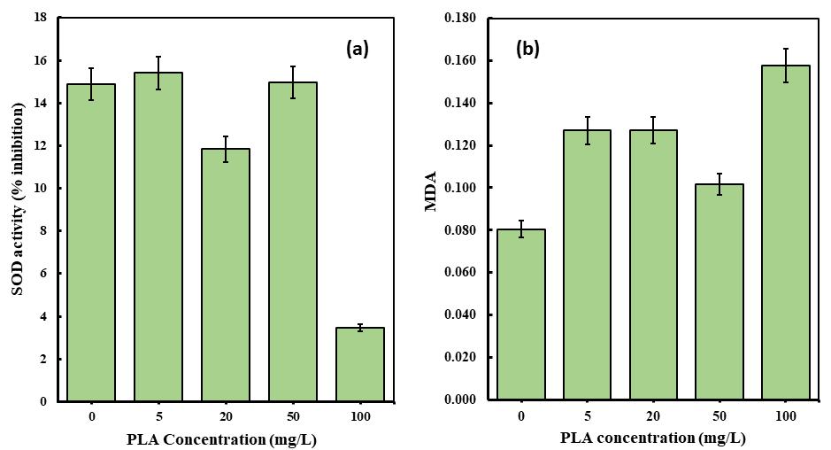
The presence of pollutants can trigger microalgal cells to produce antioxidant enzymes like SOD, which are essential in eliminating reactive oxygen species (ROS) during the antioxidant defense mechanism[17]. In this study, the impact of BMP (PLAMPs)onSODlevelsinmicroalgalcells(C. pyrenoidosa)wasexamined,revealingasignificantdecreaseinSODcontentata concentration of 100 mg/L of polylactic acid microplastic treatment (Fig -7a and b). Furthermore, the measurement of MDA content can indicate oxidation and lipid damage induced by microplastics on microalgae [37] SOD, as a critical enzyme in cellular antioxidant defense, responds to oxidative stress triggered by microplastic exposure in microalgae. Monitoring SOD levelsprovidesinsightsintotheantioxidantcapabilityofmicroalgaeandtheirabilitytocombatthedetrimentaleffectsof ROS generatedbymicroplasticexposure.
On the other hand, MDA is a marker for lipid peroxidation, which is a common consequence of oxidative stress. Elevated levels of MDA in microalgae exposed to microplastics suggest that the cells are experiencing oxidative damage to their cell membranes.BymeasuringMDAlevels,wecanassesstheextentoflipidperoxidationandtheseverityofoxidativestressinthe microalgaepopulationthatmaycausebytheeffectsofmicroplastictoamicroalga.Overall,inourcurrentstudySODandMDA measurements,in C. pyrenoidosa microalgaetoxicitystudiescausedbyPLAmicroplasticexposurecanhelpustoevaluatethe
International Research Journal of Engineering and Technology (IRJET) e-ISSN: 2395-0056
Volume: 11 Issue: 06 | Jun 2024 www.irjet.net p-ISSN: 2395-0072
impact of oxidative stress on cellular health and integrity. These biomarkers provide valuable information about the mechanismsoftoxicityandcanguideeffortstomitigatetheharmfuleffectsofmicroplasticsonaquaticecosystemsaswell.
InthepresenceofPLAmicroplastics,theMDAlevelsof C. pyrenoidosa exhibitedasignificantincrease.Atconcentrationsof 100mg/L,20mg/L,and5mg/L,theMDAlevelsroseby95.78%,57.84%,and57.74%respectively(seeFig-7b).Thisnotable increment suggests a substantial elevation in oxidative damage to the cellular membranes of the algae[38]. However, at a concentration of 50 mg/L, there was a decrease of 26.29% in MDA levels compared to the control treatment. Significant variations were observed in the inhibition of SOD activity. In the case of C. pyrenoidosa, the findings revealed a noteworthy decrease in the rates of SOD activity at PLA microplastic concentrations of 100 mg/L and 20 mg/L, with rates of 76.8% and 20.55%respectively(Fig–7a).Thisdeclinecanbeattributedtothesuppressionofthemicroalgalantioxidantsystem,whichis aconsequenceofreducedenzymeactivity,suchasSOD,thateffectivelyscavengesROS,particularlyinstressfulconditions[17]. Conversely,atconcentrationsof5mg/Land50mg/LofPLAmicroplastics,therewasaninsignificantincreaseinSODactivity inhibitionwhencomparedtothecontroltreatment.
Microalgae heavily depend on nitrogen absorption to support their fundamental growth and metabolic functions, as nitrogen plays a vital role in the synthesis of amino acids, proteins, nucleic acids, and chlorophyll. These minute organisms have the ability to utilize different nitrogen forms, including nitrate, nitrite, ammonium, and urea, with distinct species displaying varying preferences in terms of uptake mechanisms. Nitrate is commonly taken up into the cell via nitrate transporters, where it is transformed into nitrite and subsequently reduced to ammonium within the chloroplasts for integrationintoaminoacids.Thepresenceofnitrogensignificantlyinfluencesthegrowthrate,biomassyield,photosynthetic efficacy, and lipid accumulation in microalgae [39]. Intriguingly, heightened nitrogen uptake in microalgae exposed to microplastics can induce physiological reactions, such as compensatory responses, improved growth and metabolism, and modified metabolic pathways, potentially resulting in enhanced overall growth in specific circumstances despite the general harmfulimpactsofmicroplastics[40].
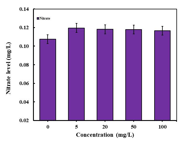
The changes in nitrate content at different concentrations of polylactic microplastics (PLA MPs) were examined, and the resultsarepresentedin(Fig-8).Inthecaseof C. pyrenoidosa microalgae,thenitratecontentat5mg/L,20mg/L,50mg/L,and 100 mg/L showed enhancements of 11.17%, 9.77%, 9.58%, and 8.4%, respectively. The fluctuations in results observed duringtoxicitytestsexaminingtheinfluenceofmicroplasticsonmicroalgaenitrogenuptakecanprovidevaluableinsights.A
International Research Journal of Engineering and Technology (IRJET) e-ISSN: 2395-0056
Volume: 11 Issue: 06 | Jun 2024 www.irjet.net p-ISSN: 2395-0072
reduction in nitrogen uptake by microalgae when exposed to microplastics may suggest an inhibition in their capacity to absorb nitrogen, which is essential for their growth and survival. Conversely, an increase in nitrogen uptake could indicate thatmicroalgaearetryingtocounteractthestresscausedbymicroplasticsbyabsorbingmorenutrients.
These findings contribute to understanding the potential repercussions of microplastics on marine ecosystems and formulatingapproaches to minimize their harmful impacts.Thevariationsinnitrogen levelsdetected in toxicitytestsdueto the impact of microplastics on microalgae can be affected by several factors. These factors encompass physical interference, where microplastics hinder the absorption of vital nutrients such as nitrogen by adhering to microalgae cell surfaces or disruptingtheirfeedingmechanisms.Moreover,chemicalleachingfrommicroplasticsmayintroduceharmfulsubstancesinto theenvironment,potentiallyinfluencingthenutrientabsorptionprocessesofmicroalgae.Indirectconsequencesmayemerge as microplastics modify the composition of the microbial community, leading to changes in nutrient availability and competition for resources that can impact nitrogen levels in microalgae. Additionally, exposure to microplastics can induce stressresponsesinmicroalgae,resultinginalterationsintheirmetabolicprocesses,includingnitrogenuptakeandutilization Understandingthesefactorsallowsresearcherstoassesstheeffectsofmicroplasticsonmicroalgaeanddevelopstrategiesto mitigatetheirimpactsonmarineecosystems.
This study focused on examining the influence of polylactic acid (PLA) microplastics on the green microalga Chlorella pyrenoidosa.TheresultsdemonstratedthattheeffectsofPLAmicroplasticsonalgaegrowthvariedovertime.Initially,after24 hours,allconcentrationsofPLAexhibitedsimilargrowthpatterns,indicatingnoimmediatenegativeconsequences.However, after48hours,slightfluctuationswereobserved,withthemostsignificantgrowthenhancementobservedataconcentration of 100 mg/L. Subsequently, at 72 hours, a continuous decline in growth was observed at a concentration of 50 mg/L, suggesting potential adverse effects. The protein content decreased as the concentration of PLA increased, with the most
International Research Journal of Engineering and Technology (IRJET) e-ISSN: 2395-0056
Volume: 11 Issue: 06 | Jun 2024 www.irjet.net p-ISSN: 2395-0072
substantial reduction observed at 100 mg/L. Additionally, there was a slight decrease in carbohydrate content across all concentrationsofPLA.ThesefindingsemphasizetheintricateimpactofPLAmicroplasticsonthebiochemicalcompositionof microalgae,indicatingpotentialstressresponsesandmetabolicchanges.Thestudyalsorevealedlowerfluorescenceactivityin C. pyrenoidosa samples treated with PLA microplastics compared to the control group. Notably, under light-adapted conditions, at a concentration of 20 mg/L, the fluorescence parameters Fm/Fo, Fv/Fo, and Fv/Fm decreased by 16.01%, 26.53%,and12.58%,respectively.Similarreductionsinfluorescenceparameterswereobservedindark-adaptedtreatments. Monitoring fluorescence parameters is crucial for comprehending the effects of microplastic exposure on microalgae. Furthermore, the levels of superoxide dismutase (SOD) significantly decreased with a treatment of 100 mg/L PLA microplastics, indicating oxidative stress. The levels of malondialdehyde (MDA), a marker for lipid peroxidation, notably increasedin C. pyrenoidosa exposedtoPLAmicroplastics,suggestingoxidativedamagetocellmembranes.Thenitratecontent varied at different concentrations of PLA, with enhanced nitrogen uptake observed in microalgae. These findings offer valuable insights into the potential impacts of microplastics on marine ecosystems and emphasize the importance of mitigatingtheiradverseeffects.
IwouldliketoexpressmysinceregratitudetoProfessorZhangYalei,mysupervisorduring thiswork,forhisunwavering support.Hispatience,motivation,andextensiveknowledgehavebeeninvaluableinassistingmewithmy paperwriting.Iam trulyappreciativeofhismentorship.Furthermore,Iwishtorecognizethesignificantcontributionofmylaboratoryassistant, Mr. YaZhou Xu, whose guidance and support were essential in the successful completion of my work within the required standardsandtimeframe.Hiscommitmenttoconductingexperiments withmeand providingaccesstolaboratoryresources were pivotal to this paper process, I am grateful for his assistance. I also extend my thanks to my classmate, Mss. Gosego BiotumeloMoreri,forherhelpduringmylaboratorywork.Additionally,Iamthankfultotheentire researchgroupatTongji University,ledbyProfessorZhangYalei,fortheirunwaveringsupportwheneverIrequiredassistanceduringmyexperiments.
Lastly but not the least, I want to show my appreciation to my family and the local government for their encouragement and backing, which enabled me to pursue my academic endeavors. I am also grateful to my classmate Mr. Melkamu Debelo Geletaforhiscompanionshipandsupport.
[1] W. Huang, J. Deng, J. Liang, and X. Xia, "Comparison of lead adsorption on the aged conventional microplastics, biodegradablemicroplasticsandenvironmentally-relevanttirewearparticles," Chemical Engineering Journal, vol.460, p.141838,2023/03/15/2023,doi:https://doi.org/10.1016/j.cej.2023.141838.
[2] A. Ashrafy et al., "Microplastics Pollution: A Brief Review of Its Source and Abundance in Different Aquatic Ecosystems," Journal of Hazardous Materials Advances, vol. 9, p. 100215, 2023/02/01/ 2023, doi: https://doi.org/10.1016/j.hazadv.2022.100215.
[3] S. Khan, M. Naushad, M. Govarthanan, J. Iqbal, and S. M. Alfadul, "Emerging contaminants of high concern for the environment: Current trends and future research," Environmental Research, vol. 207, p. 112609, 2022/05/01/ 2022, doi:https://doi.org/10.1016/j.envres.2021.112609
[4] T. D. Moshood, G. Nawanir, F. Mahmud, F. Mohamad, M. H. Ahmad, and A. AbdulGhani, "Biodegradable plastic applications towards sustainability: A recent innovations in the green product," Cleaner Engineering and Technology, vol.6,p.100404,2022/02/01/2022,doi:https://doi.org/10.1016/j.clet.2022.100404
[5] S. Lambert and M. Wagner, "Environmental performance of bio-based and biodegradable plastics: the road ahead," Chemical Society Reviews, 10.1039/C7CS00149Evol.46,no.22,pp.6855-6871,2017,doi:10.1039/C7CS00149E.
[6] X.Zhang et al.,"Photolyticdegradationelevatedthetoxicityofpolylacticacidmicroplasticstodevelopingzebrafishby triggering mitochondrial dysfunction and apoptosis," Journal of Hazardous Materials, vol. 413, p. 125321, 2021/07/05/2021,doi:https://doi.org/10.1016/j.jhazmat.2021.125321
International Research Journal of Engineering and Technology (IRJET) e-ISSN: 2395-0056
Volume: 11 Issue: 06 | Jun 2024 www.irjet.net p-ISSN: 2395-0072
[7] L. G. A. Barboza and B. C. G. Gimenez, "Microplastics in the marine environment: Current trends and future perspectives,"(ineng), Mar Pollut Bull, vol.97,no.1-2,pp.5-12,Aug152015,doi:10.1016/j.marpolbul.2015.06.008.
[8] J. Yuan, J. Ma, S. Yiran, T. Zhou, Y. Zhao, and F. Yu, "Microbial degradation and other environmental aspects of microplastics/plastics," Science of The Total Environment, vol. 715, p. 136968, 05/01 2020, doi: 10.1016/j.scitotenv.2020.136968.
[9] Z. Yan et al., "Comparative toxic effects of microplastics and nanoplastics on Chlamydomonas reinhardtii: Growth inhibition, oxidative stress, and cell morphology," Journal of Water Process Engineering, vol. 43, 2021, doi: 10.1016/j.jwpe.2021.102291.
[10] P. Singla, R. Mehta, D. Berek, and S. N. Upadhyay, "Microwave Assisted Synthesis of Poly(lactic acid) and its Characterizationusing Size ExclusionChromatography," Journal of Macromolecular Science, Part A, vol.49, no.11,pp. 963-970,2012,doi:10.1080/10601325.2012.722858.
[11] E.Besseling,J.T.Quik,M.Sun,andA.A.Koelmans,"Fateofnano-andmicroplasticinfreshwatersystems:Amodeling study," Environmental pollution, vol.220,pp.540-548,2017.
[12] X. Li et al., "Occurrence, Bioaccumulation, and Risk Assessment of Microplastics in the Aquatic Environment: A Review," Water, vol.15,no.9,p.1768,2023.[Online].Available:https://www.mdpi.com/2073-4441/15/9/1768
[13] S. Xiang, Y. Xie, X. Sun, H. Du, and J. Wang, "Identification and Quantification of Microplastics in Aquaculture Environment," (in English), Frontiers in Marine Science, Review vol. 8, 2022-January-06 2022, doi: 10.3389/fmars.2021.804208.
[14] A. Ismail, F. Mohamed, L. Rosli, M. Shafri, M. Haris, and A. Adina, "Spectrophotometric Determination of Gentamicin Loaded PLGA Microparticles and Method Validation via Ninhydrin-Gentamicin Complex As A Rapid Quantification Approach," Journal of Applied Pharmaceutical Science, pp.007-014,2016,doi:10.7324/japs.2016.600102.
[15] K. Miao et al., "Cultivation of Chlorella pyrenoidosa with different phosphorus forms under photoautotrophic and mixotrophic modes: Biochemical component synthesis and phosphorus bioavailability appraisement," Journal of Cleaner Production, vol.359,p.132058,2022/07/20/2022,doi:https://doi.org/10.1016/j.jclepro.2022.132058
[16] M. Rinawati, L. Sari, and K. Pursetyo, "Chlorophyll and carotenoids analysis spectrophotometer using method on microalgae," in IOP Conference Series: Earth and Environmental Science, 2020, vol. 441, no. 1: IOP Publishing, p. 012056,doi:10.1088/1755-1315/441/1/012056.
[17] W. Yang et al., "The combined toxicity influence of microplastics and nonylphenol on microalgae Chlorella pyrenoidosa," Ecotoxicology and Environmental Safety, vol. 195, p. 110484, 2020/06/01/ 2020, doi: https://doi.org/10.1016/j.ecoenv.2020.110484.
[18] C. Song, Z. Liu, C. Wang, S. Li, and Y. Kitamura, "Different interaction performance between microplastics and microalgae:Thebio-eliminationpotentialofChlorellasp.L38andPhaeodactylumtricornutumMASCC-0025," Science of The Total Environment, vol. 723, p. 138146, 2020/06/25/ 2020, doi: https://doi.org/10.1016/j.scitotenv.2020.138146
[19] Y. Su et al., "Biodegradable and conventional microplasticsposed similar toxicity to marine algaeChlorella vulgaris," Aquat Toxicol, vol.244,p.106097,Mar2022,doi:10.1016/j.aquatox.2022.106097.
[20] E.Cavalletti et al.,"CopperEffectonMicroalgae:ToxicityandBioremediationStrategies," Toxics, vol.10,no.9,p.527, 2022.[Online].Available:https://www.mdpi.com/2305-6304/10/9/527
International Research Journal of Engineering and Technology (IRJET) e-ISSN: 2395-0056
Volume: 11 Issue: 06 | Jun 2024 www.irjet.net p-ISSN: 2395-0072
[21] R. Jimenez, G. Markou, S. Tayibi, A. Barakat, C. Chapsal, and F. Monlau, "Production of Microalgal Slow-Release Fertilizer by Valorizing Liquid Agricultural Digestate: Growth Experiments with Tomatoes," Applied Sciences, vol. 10, no.11,p.3890,2020.[Online].Available:https://www.mdpi.com/2076-3417/10/11/3890.
[22] N.V.GaleandS.C.Thomas,"Dose-dependenceofgrowthandecophysiologicalresponsesofplantstobiochar," Science of The Total Environment, vol. 658, pp. 1344-1354, 2019/03/25/ 2019, doi: https://doi.org/10.1016/j.scitotenv.2018.12.239.
[23] Q. Zhang et al., "The combined toxicity effect of nanoplastics and glyphosate on Microcystis aeruginosa growth," Environmental Pollution, vol. 243, pp. 1106-1112, 2018/12/01/ 2018, doi: https://doi.org/10.1016/j.envpol.2018.09.073.
[24] V. da Silva Ferreira and C. Sant'Anna, "Impact of culture conditions on the chlorophyll content of microalgae for biotechnological applications," World J Microbiol Biotechnol, vol. 33, no.1, p. 20, Jan 2017,doi: 10.1007/s11274-0162181-6.
[25] M.AshrafandP.J.C.Harris,"Photosynthesisunderstressfulenvironments:Anoverview," Photosynthetica, vol.51,no. 2,pp.163-190,2013,doi:10.1007/s11099-013-0021-6.
[26] D. Rede, C. Delerue-Matos, and V. C. Fernandes, "The Microplastics Iceberg: Filling Gaps in Our Understanding," (in eng), Polymers (Basel), vol.15,no.16,Aug102023,doi:10.3390/polym15163356.
[27] Z. Li, S. Dong, F. Huang, L. Lin, Z. Hu, and Y. Zheng, "Toxicological Effects of Microplastics and Sulfadiazine on the Microalgae Chlamydomonas reinhardtii," Frontiers in Microbiology, vol. 13, p. 865768, 04/01 2022, doi: 10.3389/fmicb.2022.865768.
[28] Z. Li, S. Dong, F. Huang, L. Lin, Z. Hu, and Y. Zheng, "Toxicological Effects of Microplastics and Sulfadiazine on the MicroalgaeChlamydomonasreinhardtii,"(inEnglish), Frontiers in Microbiology, OriginalResearchvol.13,2022-April282022,doi:10.3389/fmicb.2022.865768.
[29] C. Paliwal et al., "Abiotic stresses as tools for metabolites in microalgae," Bioresource Technology, vol. 244, pp. 12161226,2017/11/01/2017,doi:https://doi.org/10.1016/j.biortech.2017.05.058
[30] C. Cunha et al., "The effect of microplastics pollution in microalgal biomass production: A biochemical study," Water Res, vol.186,p.116370,Nov12020,doi:10.1016/j.watres.2020.116370.
[31] Y. Lu et al., "Uptake and Accumulation of Polystyrene Microplastics in Zebrafish (Danio rerio) and Toxic Effects in Liver,"(ineng), Environ Sci Technol, vol.50,no.7,pp.4054-60,Apr52016,doi:10.1021/acs.est.6b00183.
[32] H.T.NairandS.Perumal,"TrophicTransferandAccumulationofMicroplasticsinFreshwaterEcosystem:RisktoFood Security and Human Health," International Journal of Ecology, vol. 2022, p. 1234078, 2022/11/15 2022, doi: 10.1155/2022/1234078.
[33] K. Roháček, "Chlorophyll Fluorescence Parameters: The Definitions, Photosynthetic Meaning, and Mutual Relationships," Photosynthetica, vol.40,pp.13-29,01/032002,doi:10.1023/A:1020125719386.
[34] Y. Xu et al., "The aging of microplastics exacerbates the damage to photosynthetic performance and bioenergy production in microalgae (Chlorella pyrenoidosa)," Water Research, vol. 259, p. 121841, 2024/08/01/ 2024, doi: https://doi.org/10.1016/j.watres.2024.121841
[35] C.Qingsheng et al.,"ThetoxiceffectsofpolystyrenemicroplasticsonfreshwateralgaeChlorellapyrenoidosadepends on the different size of polystyrene microplastics," Chemosphere, vol. 308, p. 136135, 08/01 2022, doi: 10.1016/j.chemosphere.2022.136135.
International Research Journal of Engineering and Technology (IRJET) e-ISSN: 2395-0056
Volume: 11 Issue: 06 | Jun 2024 www.irjet.net p-ISSN: 2395-0072
[36] E. H. Murchie and T. Lawson, "Chlorophyll fluorescence analysis: a guide to good practice and understanding some newapplications," Journal of Experimental Botany, vol.64,no.13,pp.3983-3998,2013,doi:10.1093/jxb/ert208.
[37] T. Ding, J. Zhang, W. Ni, and J. Li, "Combined toxicity of arsenite and dimethylarsenic acid on the freshwater diatom Nitzschiapalea," Ecotoxicology, vol.26,no.2,pp.202-210,2017/03/012017,doi:10.1007/s10646-016-1755-2.
[38] D.V.Vavilin,J.-M.Ducruet,D.N.Matorin,P.S.Venediktov,andA.B.Rubin,"Membranelipidperoxidation,cellviability andphotosystemIIactivityinthegreenalgaChlorellapyrenoidosasubjectedtovariousstressconditions," Journal of Photochemistry and Photobiology B: Biology, vol.42,no.3,pp.233-239,1998.
[39] R.Huerlimann,R.deNys,andK.Heimann,"Growth,lipidcontent,productivity,andfattyacidcompositionoftropical microalgae for scale-up production," (in eng), Biotechnol Bioeng, vol. 107, no. 2, pp. 245-57, Oct 1 2010, doi: 10.1002/bit.22809.
[40] S.Zhang, W. Gao,K. Cai,T. Liu,and X. Wang,"Effectsof MicroplasticsonGrowthandPhysiological Characteristicsof Tobacco (Nicotiana tabacum L.)," Agronomy, vol. 12, no. 11, p. 2692, 2022. [Online]. Available: https://www.mdpi.com/2073-4395/12/11/2692
© 2024, IRJET | Impact Factor value: 8.226 | ISO 9001:2008 Certified Journal | Page998