
International Research Journal of Engineering and Technology (IRJET) e-ISSN: 2395-0056
Volume: 11 Issue: 07 | July 2024 www.irjet.net p-ISSN: 2395-0072

International Research Journal of Engineering and Technology (IRJET) e-ISSN: 2395-0056
Volume: 11 Issue: 07 | July 2024 www.irjet.net p-ISSN: 2395-0072
Md. Ashikur Rahman1, Ishtiak Ahmed2, Md. Abdullah Al Humayun3
1 Department of Petroleum and Mining Engineering, Chittagong University of Engineering and Technology, Chattogram, Bangladesh.
2Mechanical and Production Engineering, Ahsanullah University of Science and Technology, Dhaka, Bangladesh.
3Deptartment of Electrical and Electronic Engineering, Eastern University, Dhaka, Bangladesh
Abstract - Melanoma represents a particularly aggressive form of skin cancer, necessitating prompt detection to mitigate mortality rates and minimize the invasiveness of treatment. Contemporary advances in computer-aided diagnosis (CAD) have leveraged sophisticated imaging techniques to enhance early-stage diagnosis of skin malignancies. This research aims to construct a web-based platform for theautomated analysis of dermoscopic images to ascertain early melanoma presence. The implementation employs the Inception V3 model, aConvolutionalNeuralNetwork (CNN)architecture renowned for its efficacy in image classification tasks, specifically in medical imaging domains such as dermoscopy. This model facilitates the processes of image acquisition,preprocessing,segmentation,featureextraction, and classification. The web application, developed using Python, HTML, and CSS, showcases a streamlined interface for clinical application. Empirical evaluations reveal that the model achieves an accuracy range of 90-93%, underscoring its potential utility in clinical settings. This platform empowers users to swiftly identify skin abnormalities, thereby enhancing early diagnosis and preventive care, significantly contributing to advancements indermatologicaloncology.
Key Words: Convolutional Neural Network (CNN), Computer-aided diagnosis (CAD), Dermoscopic images, Empirical evaluations, Image classification, Inception V3 model,Melanoma
Melanoma is recognized as the deadliest form of skin cancer,withsignificantglobalimpact.Ithasbeenreported recently that new melanoma cases reported in 2020 was 325,000 may increase up to 510,000 by 2040. Likewise, deaths due to melanoma mayrise byapproximately68%, from57,000in2020to96,000in2040[1]
Early detection of melanoma can lead to a complete cure throughsimpleexcision ofthemalignanttissue.However, late-stage detection often results in metastasis, leading to apoorprognosis[2].Melanomaoriginatesinmelanocytes, the skin cells responsible for producing pigments that determine skin color. The patterns of melanoma
occurrence vary geographically; in Western countries, melanomas typically arise in sun-exposed areas such as the chest, forehead, and limbs. Conversely, in Asia, melanomas more commonly develop on the hands and feet, which are less exposed to the sun, as well as on mucous membranes, including the lining of the mouth, throat, gastrointestinal tract, and the vaginal tract in women[4]
Excessive exposure to ultraviolet (UV) radiation from sunlight or artificial tanning is a major risk factor for the development of melanoma. Reducing UV exposure can significantly lower the risk of melanoma, achievable through measures such as consistent use of sunscreen, wearinglong-sleevedclothing,andusinghatstoblockthe sun. Additionally, genetic mutations in cancer-causing genes, particularly BRAF and NRAS, CDKN2A, play a crucialroleinmelanomadevelopment[2][4]
In the past decade, malignant melanoma has become one of the most dangerous cancers, spreading rapidly worldwide.Eachyear,morethanonemillioncasesofnonmelanoma skin cancer and over 250,000 cases of melanoma are reported [2]. In the United States, melanomawasrankedfifthforexpectednewcancercases in both males and females in 2019 Error! Reference source not found..TheincidenceofmelanomaintheU.S. has exhibited a concerning upward trend, significantly contributing to cancer-related mortality over the past decades. To address this, a web-based deep learning system has been developed for melanoma detection, employingtheInceptionmodeltoachievefasteroperation and higher accuracy. In response, a sophisticated webbaseddeeplearningsystemusingtheInceptionmodelhas been developed, offering rapid and highly accurate melanoma detection with innovative approach aims to enhanceearlydiagnosis.
BeforeSkincancer,particularlymelanoma,representsa pressing global health concern. A new study by scientists from the International Agency for Research on Cancer (IARC)andpartnerspredictsthatthenumberofnewcases of cutaneous melanoma per year will increase by more
International Research Journal of Engineering and Technology (IRJET) e-ISSN: 2395-0056
Volume: 11 Issue: 07 | July 2024 www.irjet.net p-ISSN: 2395-0072
than 50% from 2020 to 2040. The study, published in the journal JAMA Dermatology, provides global patterns of cutaneous melanoma in 2020 as well as projections of the numbersofnewcasesanddeathsfor2040[7]
Early detection plays a pivotal role in addressing this challenge. It has been acknowledged that localized melanoma boasts a remarkable five-year survival rate of 95%.Furthermore,itexhibitsmoreaggressivebehaviorin transplant recipients, with a higher risk of metastasis and mortality. Thisstarkcontrastemphasizestheurgentneed foraccurateandefficientdetectionmethods[8]
However, the intricacies of melanoma pose substantial diagnostic challenges. Research reveals significant variations in dermatologists' diagnostic accuracy, with sensitivity rates for early-stage melanomas up to 78.3%. Such discrepancies underscore the necessity for a standardized,technology-drivenapproach[9]
The impact of melanoma is further highlighted by its potential formorbidityandmortality.Although melanoma accounts for only about 1% of skin cancer cases, it is responsible fora significantportion of skin cancer-related deaths. In the United States, for instance, it is estimated that in 2019, about 7,410 people died to melanoma. Early detection and timely treatment are crucial factors in improving outcomes. The five-year survival rate for localized melanoma is impressively high at around 95%. However, when melanoma progresses to advanced stages and spreads to distant organs, the five-year survival rate drops dramatically [8]. This underlines the importance of not only raising awareness about the risks of melanoma but also implementing effective prevention strategies and earlydetectionmeasures.
Considering the current scenario in this field of research this research is devoted to provide cutting-edge artificial intelligencetechniquestofacilitatemelanomadetection.By harnessing the power of machine learning, aim of this researchistoestablishaconsistent,accurate,andefficient method for identifying and classifying melanoma lesions. This approach has the potential to facilitate early interventionandsignificantlyimprovepatientoutcomesin the battle against this formidable disease. The foundation ofthisresearchworkisrootedinstatisticaldata,reflecting the urgency and potential of the proposed innovative approachtorevolutionizemelanomadetection.
This section presents the details description of different models and techniques with their process of integration withAItechnologyinvolvedintheresearchmethodology.
Deep Learning models have revolutionized various fields by learning complex patterns from raw data. In computer vision, they excel in image classification, object detection, and image segmentation, with applications like facial recognition and medical diagnostics. In Natural Language Processing (NLP), models such as Recurrent Neural Networks (RNNs) and Transformers have transformed tasks like sentiment analysis and language translation. In healthcare, Deep Learning enhances medical imaging for early disease detection and drug discovery. Autonomous systems, like self-driving cars, also benefit from these modelsforreal-timedecision-making.Additionally,digital platforms utilize Deep Learning for personalized recommendationsystems.
AI, particularly Deep Learning, has significantly improved melanoma detection. Convolutional Neural Networks (CNNs) analyze dermatoscopic images to identify melanoma, aiding early detection and enhancing diagnosticaccuracy.Thesemodelsproviderobustsupport to dermatologists, offering reliable second opinions. However, their efficacy depends on high-quality data, meticulousannotation,andethicalconsiderations[10][11].
Machine learning techniques, such as transfer learning with models like Inception V3, are crucial for melanoma detection.Transferlearningleveragespre-trainedmodels, reducing training time and optimizing resources. Data augmentation techniques expand datasets, enhancing model generalization and classification accuracy for variousskindiseases.
Deep Learning, especially CNNs, has redefined melanoma detection by extracting complex features from medical images. Techniques like transfer learning and data augmentation enhance model performance and adaptability [12]. This integration promises earlier detection, reduced false negatives, and personalized treatment strategies, advancing melanoma diagnosis and patientcare.
Deep learning (DL) involves computer algorithms that improve automatically through experience and data, building models based on training data to make predictionsordecisionswithoutexplicitprogramming.DL is used in various applications, enabling computers to perform tasks by learning from provided data. While simple tasks can be programmed step-by-step, advanced
International Research Journal of Engineering and Technology (IRJET) e-ISSN: 2395-0056
Volume: 11 Issue: 07 | July 2024 www.irjet.net p-ISSN: 2395-0072
tasks require DL models Error! Reference source not found.. DListhemostimportantpartoftheprogramasit dictatescreatingthebackboneoftheinterface.
For studying image classification with DL, the following stepsareinvolved:
1. Developing the Inception Model using machine learningalgorithms,
2. Selectingmelanomadataforimplementation,
3. ApplyingInceptionforhyper-parametertuning,and
4. Validatingthemodelwithmelanomadata.
Deep learning algorithms, a subset of machine learning techniques, have revolutionized numerous fields by enabling computers to learn patterns directly from data. These algorithms, based on artificial neural networks designedtosimulatethehumanbrain'sstructure,excelat automatically learning hierarchical data representations through multiple layers, or "deep" architectures. This capability allows deep learning algorithms to perform tasks such as image and speech recognition, language translation, and strategic game playing with state-of-theart performance. Convolutional Neural Networks (CNNs) are particularly effective for image-related tasks, while Recurrent Neural Networks (RNNs) are suited for sequencedatalikelanguage.
Despite their impressive achievements, deep learning algorithms require substantial computational resources and extensive datasets. Continuous advancements are enhancing their efficiency and applicability, solidifying theirroleinshapingmodernAIcapabilities.
This research work aims to leverage these advancements to improve melanoma skin malignancy classification. The dataset includes two classes: melanoma and nonmelanomaError! Reference source not found..Usingthe publicly accessible ISIC database for training and testing images,theAIarchitectureInception V3wasemployed to train the dataset. Unlike previous studies that compared melanomawithspecificorotherskin-relateddatasets,this approach includes all other skin-related diseases as nonmelanoma for comprehensive comparison and improved accuracy.Thismethodaimstodistinguishmelanomafrom otherskindiseases.
This significance of this research lies in its potential to enhance melanoma detection accuracy, improving early diagnosisandtreatmentoutcomes.Theprimaryobjectives are to develop an AI-based framework for testing melanomaskindiseases,discussthechallengesmelanoma poses, explore preventive measures, and highlight
proposed solutions by various researchers. The methodology,materials,andresultsobtainedaredetailed, culminating in an evaluation of the system and its outcomes.
This research work follows a structured and iterative development approach. The methodology includes collecting a melanoma detection dataset from Kaggle, splitting it into training and testing sets, and cleaning the training data. The Inception deep learning model is then importedandtrainedusingtheprepareddata.Themodel's accuracy is tested with the test data, and the results are analyzed using a confusion matrix. The system design encompasses data preprocessing, encoding, feature selection, model training, and evaluation, highlighting its advantages,disadvantages,andfeatures[12].
This diagram provides a detailed overview of the program's architecture and the user-admin interface specificallydesignedformelanomadetection.Itshowcases the structural components and the interaction flow between the user and administrative functionalities, highlighting how the system facilitates efficient and accuratemelanomadetection.
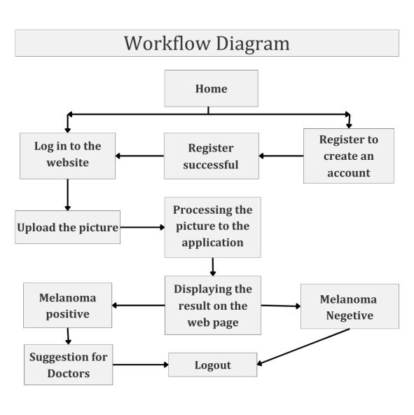
Figure 1: Workflowdiagramofmelanomadetection program
The interface diagram is intentionally kept simple to ensure comprehensibility for all users, given its role in handlingmedicaldatawithinadatascienceframework.
International Research Journal of Engineering and Technology (IRJET) e-ISSN: 2395-0056
Volume: 11 Issue: 07 | July 2024 www.irjet.net p-ISSN: 2395-0072
Melanoma Detection Application
The application features two primary roles: "User" and "Admin."
User Interactions:
Login: Secureaccesstopersonalizedaccounts.

Figure 2: UserloginPageofthewebsite
Registration: Createnewaccounts.

Figure 3: Usersignuppageofthewebsite
Interact with Blog: Explore melanoma awarenesscontent.
Access Detection Service: Utilize melanoma detectionfunctionality.
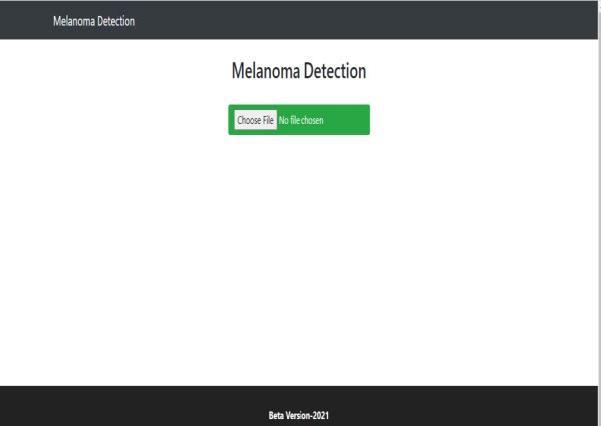
Figure 4: Pictureuploadpageofthewebsite
Browse Gallery: View images to aid visual understanding.
Explore About Us: Learn about the application's missionandpurpose.
Positive Detection: Receivedoctorsuggestionsif melanomaisdetected.
Admin Interactions:
Login: Authenticatetoaccesstheadminpanel.
Manage Blogs: Createandupdateblogcontent.
Gallery Management: Maintain and update imagegalleries.
Positive Detection Support: Provide doctor suggestionsforpositivedetections
2.3
2.3.1
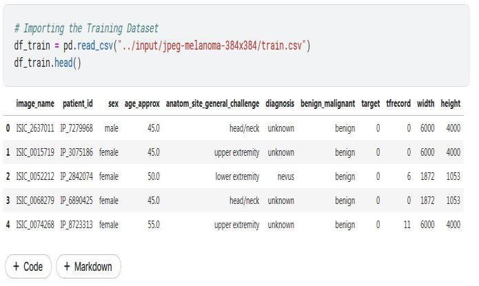
The datasets used in this study are sourced from Kaggle, comprising information on 1180 patients aimed at identifyingPulmonaryAbnormalities.Thedatasetincludes 16 different attributes per patient. Of the patients, 34.7% are classified as Normal, while 65.3% exhibit Pulmonary Abnormalities. The dataset is divided into training (80%) andtesting(20%)sets.Thetableprovidesasampleofthe dataset,listingtheattributesandtheirrespectivescope.
Figure5showsthesampleofthetablewiththeattributes andthepossiblescopeofthedataset.
International Research Journal of Engineering and Technology (IRJET) e-ISSN: 2395-0056
Volume: 11 Issue: 07 | July 2024 www.irjet.net p-ISSN: 2395-0072
For the task of melanoma detection, data sourced from Kaggle underwent rigorous analysis focusing on image quality. Images with low pixel counts or blurred quality were excluded to eliminate low-quality data. Erroneous data points, such as outliers in age, were corrected by replacing them with average values to enhance accuracy. Furthermore, data standardization was carried out using the standard scalar process in Python, leveraging the Pandas library for normalization. These preprocessing stepswerecrucialtoensurethereliabilityandconsistency of the dataset, essential for training and evaluating machinelearningmodelsinmelanomadetectionresearch.
This study highlights the robust performance of the Deep Learning System (DLS) across various skin conditions throughanextensivedevelopmentandvalidationprocess. The DLS not only produces primary diagnoses but also provides differential diagnoses, marking a significant advancement in supporting clinical decision-making. Moreover, its ability to handle a varying number of input imagesandmetadatavariablesdemonstratesitsflexibility andadaptabilitytoawiderangeofclinicalscenarios.This versatileapproachensuresthatthesystemcaneffectively cater to diverse clinical needs, enhancing its practical utilityindermatologicalpractice[12].
In this study, transfer learning has been employed to enhance melanoma classification accuracy. Transfer learning leverages pretrained models, such as Inception V3, initially trained on extensive datasets for solving similar tasks. By retraining the final layer of Inception V3 with the specific dataset, its previously learned features and patterns have been effectively utilized. Hence, optimizing performance without starting from scratch. Thedatasetisdividedintotraining,validation,andtesting sets, often in a 70-15-15 ratio. The model is then trained using transfer learning, initialized with pre-trained weights from the ImageNet dataset Error! Reference source not found.. This approach significantly reduces
both training time and the computational resources needed[14].
Dataset of the current research has been partitioned into training and test sets, comprising 80% and 20% of the images,respectively.Toarguewiththedataset,techniques including shearing, zooming, flipping, and brightness adjustments have been applied, effectively doubling its originalsize.Inceptionhasbeenfine-tunedV3byadapting its final layer to the classification task, training the model over6epochswithabatchsizeof32.
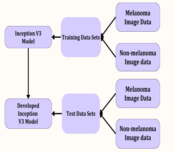
In addition to melanoma images, diverse non-melanoma skin diseases such as acne fulminans, acne nodules, eczema, Taksim,black spot, fungal acne,herpes, pityriasis versicolor, papule, pustular, rosacea, and whitehead have been incorporated Error! Reference source not found.. This approach has ensured the proposed model's robustness and ability to generalize across various skin conditions. The overall methodology underscores the commitment to leveraging advanced deep learning techniques for accurate and efficient melanoma detection in clinical settings. The overall process of training the DeeplearningmodelisshowninFigure6.
This process includes importing necessary libraries, loading and preprocessing dataset, and creating and training the Inception V3 model in Jupyter Notebook. A Jupyter Notebook is an interactive tool for creating and sharing documents with live code, equations, visualizations, and narrative text. Popular in data science, research, and education, it combines code execution, data analysis, and explanations in one place. It's used for data science, statistical modeling, machine learning, and more Error! Reference source not found.. Additionally, the performanceofthemodelisevaluatedandvisualized.
International Research Journal of Engineering and Technology (IRJET) e-ISSN: 2395-0056
Volume: 11 Issue: 07 | July 2024 www.irjet.net p-ISSN: 2395-0072
The first step involves importing essential libraries and packages required for data manipulation, preprocessing, andmodel building.KeylibrariesincludeTensorFlowand Kerasfordeeplearning,aswellasPandasandNumPyfor data handling. The dataset, publicly available on Kaggle, contains melanoma records from the USA dated September 2018. Kaggle is an online platform that brings together data scientists, machine learning practitioners, and enthusiasts from around the world. It's a playground for exploring, learning, and competing in the exciting realmofdatascience[17].
The dataset is loaded and described to understand its features. Data preprocessing involves handling missing values, data augmentation, and normalization to ensure thedatasetissuitablefortrainingthemodelusingPandas library.
AninstanceoftheInceptionV3modeliscreatedforimage classification.Theinputimagesareresizedtothespecified IMAGE_SIZE with 3 color channels (RGB). The model is initialized with pre-trained weights from the ImageNet dataset. The top fully connected layers of the model are excludedtoaddcustomlayersforclassificationwhichwill eventually enhance the overall performance of the designedsystem.
Model Compilation
Themodelisconfiguredfortrainingusingthe'categorical crossentropy'lossfunction,'adam'optimizer,andtracking theaccuracymetricduringtrainingandvalidation.
Data Generators
Data generators are set up using ImageDataGenerator from TensorFlow/Kerasto handledata augmentation and preprocessing. This includes rescaling pixel values and applyingshear,zoom,andhorizontalfliptransformations.
Training the Model
The model is trained using the fit_generator method, which allows for efficient handling of large datasets by generatingbatchesofdataonthefly.
Evaluating and Visualizing Model Performance
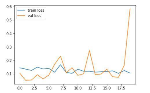
Figure 7: PlotOfthetrainingloss(error)andvalidation lossacrossepochs
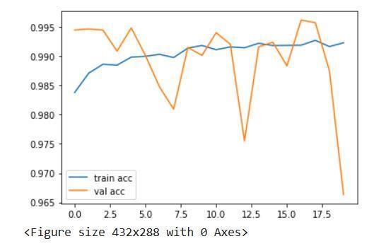
Figure 8: Plotofthetrainingaccuracyandvalidation accuracyacrossepochs
The training and validation loss, as well as accuracy over thetrainingepochs,areplottedinFigure7andFigure8to visualize the model's performance. From the comparative analysisoftheseplots,itisascertainedthatstructuredand iterative approach ensures a robust methodology for predicting melanoma using deep learning techniques, leveraging the power of the Inception V3 model for accurateandefficientclassification.
Quality assurance (QA) ensures software systems meet specifiedrequirementsandbehaveasexpected.Itinvolves defining test parameters in the technical specification document and identifying defects during development to preventpost-releasebugs[18].Intraditionalsoftware,QA focuses on verifying system functionality under various conditions. However, in machinelearning, especiallydeep learning, the focus shifts to data quality, model performance, and system robustness. QA in ML involves validating and verifying both software artifacts and behavior, providing an objective assessment to
International Research Journal of Engineering and Technology (IRJET) e-ISSN: 2395-0056
Volume: 11 Issue: 07 | July 2024 www.irjet.net p-ISSN: 2395-0072
understand and mitigate implementation risks. Comprehensive QA is crucial for reliable and accurate machinelearningsystems.
3.1.1
Unit testing involves breaking down the program into individual units or components and testing each separately to ensure they perform as expected. Each unit, such as a function, method, procedure, module, or object, isisolatedandverifiedforcorrectness.Keybenefitsofunit testinginmodeldevelopmentinclude:
EarlyIssueDetection:Identifiesproblemsearlyin thedevelopmentcycle.
IsolationofIssues:Pinpointsspecific components whereissuesarise.
Code Refactoring Documentation: Provides a reliable foundation for making code improvements.
3.1.2
Integration testing focuses on how different components or modules of the system interact and work together. In the context of this research work, integration testing ensures that the various steps such as data preprocessing, Inception V3 model implementation, and result evaluation function cohesively and correctly. Integration among every section of the program reduces theerrorsandelevatestheperformanceofthesystem.
3.1.3
System testing verifies the functionality and performance of the entire system as a whole. This involves testing the complete data processing pipeline, model training, prediction, and result evaluation to ensure the model operatesasintended[19].
3.1.4
Performance testing evaluates the model's efficiency, speed, and resource consumption. This ensures that the model performs well even with large datasets and can deliverresultswithinacceptabletimeframes.
3.1.5
Securitytestingaddressespotentialvulnerabilitiesorrisks inthemodel'simplementation,particularlywhenhandling sensitive medical data. This ensures that the system is secureandprotectspatientinformation.
These comprehensive testing approaches ensure that the machine learning system is robust, reliable, and meets all specifiedrequirements.
3.2
The developed model in this research work achieves an average accuracyof 99.01% withthe experimental values used for training. While this accuracy is sufficient for implementation on the website, there is potential for further improvement. Increasing the dataset size and the number of training epochs could enhance the model's performance.
This figure demonstrates a positive detection from the testingdata,validatingthemodel'saccuracy.
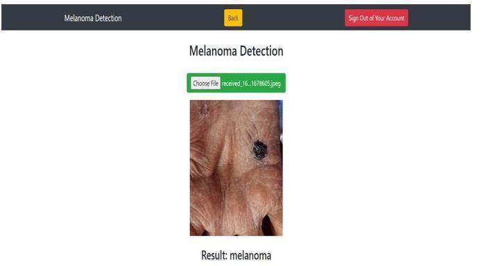
Figure 9: AnalysisResultonwebpage
In recent years, significant research efforts have been directed towards improving the diagnosis of melanoma skin diseases. These advancements have led to the development of more accurate and reliable diagnostic models,contributingtobetterclinicaloutcomes.
Table -1: SampleTableformat
A comprehensive array of testing methodologies has been meticulously applied to rigorously evaluate the efficacy, robustness, and reliability of carefully crafted deep learning model. This pivotal phase of the development process is instrumental in validating the model's
International Research Journal of Engineering and Technology (IRJET) e-ISSN: 2395-0056
Volume: 11 Issue: 07 | July 2024 www.irjet.net p-ISSN: 2395-0072
performanceacrossvariousdimensions, ensuringit meets thehigheststandardsofaccuracyandgeneralization.
The proposed model achieves an impressive average accuracyof99.01%withtheexperimentalvaluesusedfor training. While this level of accuracy is sufficient for implementation on the website, there is potential for further improvement. Increasing the dataset size and the number of training epochs can enhance the model's performance further, allowing for even greater precision andreliability.
In recent years, significant research efforts have been directed towards improving the diagnosis of melanoma skin diseases. These advancements have led to the development of more accurate and reliable diagnostic models, contributing to better clinical outcomes. The integration of cutting-edge machine learning techniques hasplayeda pivotal rolein these improvements, enabling the detection and classification of melanoma with higher accuracy.
Acomprehensivearrayoftestingmethodologieshasbeen meticulously applied to rigorously evaluate the efficacy, robustness, and reliability of our carefully crafted deep learning model. This pivotal phase of the development process is instrumental in validating the model's performanceacrossvariousdimensions.Ensuringthatthe proposed model meets the highest standards of accuracy andgeneralizationiscrucial foritssuccessful deployment inclinicalsettings.Byrigorouslytestingandvalidatingthe model, its ability to perform reliably in real-world applications, ultimately contributing to better patient outcomes and advancing the field of melanoma diagnosis hasbeenacknowledged.
Thenewlydevelopedmodeldemonstratesacommendable accuracy of 99.01% despite being trained on a relatively limited dataset. This research employed a deep convolutional neuralnetwork,specificallyInceptionv3,to classify melanoma and other skin diseases. The userfriendly website interface allows patients to identify melanomawithjustafewclicks.
The outcomes of this research show proper utilization of very deep convolutional neural networks, combined with transfer learning and fine-tuning on dermoscopic images, achieve better diagnostic accuracy compared to the existing technologies used by expert doctors and clinicians. Unlike other studies, this paper introduces a new method for melanoma detection using a variety of skin disease tests and non-melanoma datasets. This approach significantly enhanced the accuracy of the appliedAIalgorithm,Inceptionv3.
Thewebsiteofferspeopleacrosstheglobetheopportunity to detect early signs of melanoma and take preventive measures.Dermatologists in thecountrywill benefitfrom thistoolandcanuseittoaidinpatienttreatment.
Looking ahead, the dataset can be expanded to further improve the model's accuracy, particularly for nonmelanoma skin types. Additionally, this model can be integrated seamlessly with the website, which still requires further enhancements on both the front-end and back-end.
[1] Z. Babic, et al. “Trends in Melanoma Mortality in Serbia: A 22-Year Population-Based Study”, Iran J Public Health, vol. 53, no. 4, pp.828-836, 2024. doi: 10.5826/dpc.1301a8
[2] M. Kamińska, “Healthcare problems of a patient with melanomaskincancer”,GenMedHealtSci, vol. 30, no. 2, pp. 112-115, 2024. DOI: https://doi.org/10.26444/monz/189438
[3] S.Fazelpour,S.C.Deverapalli,B.Nguyen,“Skincancerassociatedgenodermatosesinskinofcolorpatients:a review”,ArchDermatolRes,vol316,no.6,282,2024. DOI:10.1007/s00403-024-03087-w
[4] H.Y.Lee, W. Y.Chay, M.B. Tang,M.T.Chio, S.H. Tan, “Melanoma: differences between Asian and Caucasian patients”, Ann Acad Med Singapore, vol. 41, no. 1, pp.17-20,2012.DOI:10.1201/b12510-3
[5] F. Bray, J. Ferlay, I. Soerjomataram, R. L. Siegel, L. A. Torre, A. Jemal, “Global cancer statistics 2018: GLOBOCAN estimates of incidence and mortality worldwide for 36 cancers in 185 countries”, CACancer J Clin, vol. 68, no. 6, pp. 394-424, 2018. DOI: https://doi.org/10.3322/caac.21492
[6] F. Bray, J. Ferlay, I. Soerjomataram, R. L., Siegel, L. A., Torre, A. Jemal, “Erratum: Global cancer statistics 2018: GLOBOCAN estimates of incidence and mortalityworldwidefor36cancersin185countries”, CA-CancerJClin, vol.70,no.4,p.313.
[7] M. Arnold, D. Singh, M. Laversanne, et al. “Global Burden of Cutaneous Melanoma in 2020 and Projections to 2040”, JAMADermatol., vol. 158, no. 5, pp.495–503,2022
[8] S.Sol, F. Boncimino, K. Todorova, S. E. Waszyn, A. Mandinova, “Therapeutic Approaches for NonMelanoma Skin Cancer: Standard of Care and Emerging Modalities”, IntJ MolSci, vol. 25, no. 13, p. 7056,2024.
International Research Journal of Engineering and Technology (IRJET) e-ISSN: 2395-0056
Volume: 11 Issue: 07 | July 2024 www.irjet.net p-ISSN: 2395-0072
[9] L.Fried, A. Tan, S.Bajaj,T. N.Liebman,D. Polsky, J. A.Stein, “Technological advancesfor the detection of melanoma: Advances in diagnostic techniques”, J Am AcadDermatol, vol.83,no.4,pp983-992,2020.
[10] T. M. Cover and P. E. Hart, “Nearest Neighbor Pattern Classification,” IEEE Trans. Inf. Theory, vol. 13, no. 1, pp.21–27,1967,doi:10.1109/TIT.1967.1053964.
[11] D. Steinberg, "CART: Classification and regression trees”, In ‘The Top Ten Algorithms in Data Mining’, (Eds.XWu,VKumar)pp.179–202,2009.
[12] M. A. Albahar, “Skin Lesion Classification Using Convolutional Neural Network with Novel Regularizer,” IEEE Access, vol. 7, pp. 38306–38313, 2019,doi:10.1109/ACCESS.2019.2906241.
[13] T. B. Trafalis and O. O. Oladunni, “Support Vector DeepsandApplications,” RecentAdv.DataMin.Enterp. Data Algorithms Appl., pp. 643–690, 2008, doi: 10.1142/9789812779861_0014.
[14] V. Pugazhenthi, S. K. Naik, A. D. Joshi, S. S., Manerkar, V.U.,Nagvekar,K.P.Naik,C.G.Palekar,K.Sagar,“Skin diseasedetectionandclassification”,IntJAdvEngRes Sci,vol.6,no.5,pp.396-400,2019.
[15] M. A. Albahar, “Skin Lesion Classification Using Convolutional Neural Network with Novel Regularizer,” IEEE Access, vol. 7, pp. 38306–38313, 2019.
[16] V. Bhavya Sai, G. Narasimha Rao, M. Ramya, Y. Sujana Sree, and T. Anuradha, “Classification of skin cancer images using TensorFlow and inception V3,” Int. J. Eng. Technol., vol. 7, pp. 717–721, 2018, doi: 10.14419/ijet.v7i2.7.10930.
[17] E. Vocaturo, E. Zumpano, and P. Veltri, “Features for melanomalesionscharacterizationincomputervision systems,”Feb.2019,doi:10.1109/IISA.2018.8633651.
[18] E.Vocaturo,E.Zumpano,andP.Veltri,“Ondiscovering relevant features for tongue colored image analysis,” Jun.2019,doi:10.1145/3331076.3331124.
[19] M.Wati,Haviluddin,N.Puspitasari,E.Budiman,andR. Rahim, “First-order Feature Extraction Methods for ImageTextureandMelanoma SkinCancerDetection,” in JournalofPhysics:ConferenceSeries, Sep. 2019, vol. 1230, no. 1, p. 12013, doi: 10.1088/17426596/1230/1/012013.
2024, IRJET | Impact Factor value: 8.226 | ISO 9001:2008