
International Research Journal of Engineering and Technology (IRJET) e-ISSN: 2395-0056 p-ISSN: 2395-0072

Volume: 11 Issue: 08 | Aug 2024 www.irjet.net


International Research Journal of Engineering and Technology (IRJET) e-ISSN: 2395-0056 p-ISSN: 2395-0072
Volume: 11 Issue: 08 | Aug 2024 www.irjet.net
Anup Dange, Tanuja Thakar, Rajshri Lomate , Satyajit Bahir
Department of Computer Engineering, GH Raisoni Institute of Engineering and Technology, Pune Maharashtra, India
Abstract - Recent developments in medical imaging have brought attention to how critical it is to diagnose brain cancers like cerebral carcinoma as soon as possible. In this context, convolutional neural networks (CNNs), a type of deep learningalgorithm,havebecomea very usefultool.Inorderto detect the existence of tumors, these algorithms automate the processof evaluating MRI scans, extracting relevantdata,and classifying them. CNN-based models provide a rapid and accurateway to identify brain tumors by eliminatingthe need for manual interpretation. This model is designed to classify MRI scans, enabling healthcare professionals to swiftly and correctly detect the existence of brain malignancies. By automating the diagnostic process and reducing the reliance onmanualinterpretation,thisapproachoffersthepotentialto revolutionize the field of cerebral carcinoma diagnosis, makingitmore efficientandlesssusceptible tohumanerror.
Key Words: CNN(Cerebral Neoplasm Detection), Brain Tumor, Medical Imaging
1. INTRODUCTION
A brain tumor is a lethal ailment, characterized by a high fatality rate, originating from the abnormal growth of one or more brain tissues. It not only disrupts the brain's typical operations but also impacts surrounding tissues. Detecting tumors on their size and location within the brain. Our challenge lies in automating the early-stage identification of brain tumors from MRI images, a formidabletask.
Brain tumors, viewed as a severe ailment, affect all age groups significantly. They represent 85- 90% of primary CNS tumors, with about 11,700 new cases yearly. This underscores the imperative for enhanced research and treatment.
Variousbraintumortypes,suchasbenign,malignant, and pituitary tumors, require distinct categorization. Prolonging patient lives demands meticulous care, thoughtful strategies, and precise diagnostics. Among these, MagneticResonanceImaging(MRI)standsoutas the most dependable means of detecting brainmalignancies. These MRI scans yield vast sets of image data, which are meticulouslyreviewedbyradiologists.
Therefore,suggestingtheimplementationofasystem that utilizesParticularly,convolutionalneuralnetworks(CNNs) are deep learning algorithms, for the purpose of detection andclassificationcouldprove highly beneficialformedical professionalsworldwide.

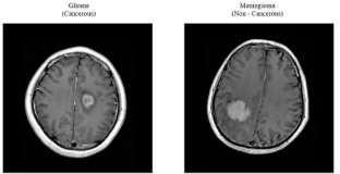
The tumor-affected area of the brain is shown in Fig- 1, which additionally determines the precise position of the infectionwithinthebrain.
1. Automating early-stage detection of brain tumors usingCNNsandMLmodels.
2. Improving accuracy and efficiency in brain tumor diagnosis.
3. EvaluatingtheperformanceofCNN-basedmodels fortumordetection.
4. Advancing deep learning applications in medical imaging for better diagnosis and patient outcomes.
[1]Choudhury and colleagues proposed a method for brain tumor detection and classification using Convolutional Neural Networks (CNN) and Deep Neural Networks (DNN). Their work serves as a foundation for applying deep learning techniques to brain tumor diagnosis.
[2] Inthisstudy,aliteraturesurveyreviewsrelatedworks in brain tumor detection from medical images usingdeep learning, including CNN and SVM methods, providing valuableinsightsforourresearch.
[3] A deconvolution network for semantic segmentation was introduced by Noh et al. (2015). Their work made an important contribution to the field of

Volume: 11 Issue: 08 | Aug 2024 www.irjet.net
International Research Journal of Engineering and Technology (IRJET) e-ISSN: 2395-0056 p-ISSN: 2395-0072
computer vision by learning the inverse mapping of convolutional neural networks, which is now a key idea in contemporary semantic segmentation models, to address pixel-wiselabelingchallenges.
[4]Abd-Ellah et al. (2019) conducted a comprehensive review on brain tumor diagnosis from MRI images, highlighting practical implications, key achievements, and lessons learned in the field. This review offers valuable insights into the state of the art in brain tumor diagnosis, servingasafoundationalresourceforourresearch."
[5]Using deep c r detection and classification. Their work advances the field of medical image analysis by showing how CNNs can reliably diagnose lung cancer, which is useful for our research on related deep learning applicationsinmedical onvolutional neural networks (CNN), "Sasikala,Bharathi,andSowmiya(2018)publishedastudy on lung cance imaging.
[6] IslamandZhang(2017)developedaground-breaking deep learning-based multiclass classification method for Alzheimer's disease detection utilizing brain MRI data. Theirworkmakesmajorstridesintheuseofdeeplearning to medical diagnostics, and it also influences our investigation of related approaches to disease identificationinmedicalimaging.
[7]Using the R programming language, Kiranmayee, Rajinikanth,andNagini(2017)performedexploratorydata analytics on brain tumor data. Their work serves as an exampleofthevalueofdataanalyticsinmedical research by providing insights that direct our own data analysis approaches with regard to the detection and diagnosis ofbraintumors.
With the help of neural network design and implementation,thehuman brainismimicked.This paper offer MRI pictures of particular brain regions are used by CNNtodetectbraintumors.Brainregionsareextractedon the first level of the MRI image, and each slice in that region is divided to acquire malignancies. CNN architecture is employed to partition the tumor areas. To evaluatethepatientimages,CNNisemployed.Thisstudy's main objective is to find brain tumors. whether a patient hasabenignormalignanttumorintheirbrain.
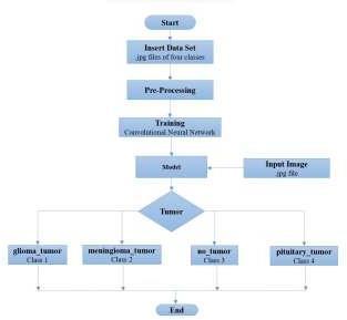
1) Collect Brain Tumor Dataset
We obtained a large number of MRI images of brain tumors during the dataset gathering stage of our cerebral neoplasm identification study. Since these photographs came from multiple medical sources, a variety of tumor forms, sizes, and patient demographics are guaranteed. For training effective CNNandmachinelearningmodelsto precisely identify brain neoplasms, a large and diverse datasetisessential.
2)Image Preprocessing
Every image underwent the pre-processing processes listedbelow:1.Cutoffaportionofthebrainthatcontains the images. 2. Due to the fact that the photos are from various sources and range in size across the dataset, it is best to transform them into the shape of (150, 150,3). Therefore,allphotosmustbeinthesameformatinorderto provideinformationtoaneuralnetwork.
3)
Training(CNN)
Convolutional Neural Network (CNN) is createdduring the training stage of a brain neoplasm detection project from MRI utilizing CNN and ML models. It receives a tagged collection of MRI pictures with cancers identified in them. The CNN develops an understanding of the patterns and characteristicslinkedtoneoplasms.Toreduceclassification mistakes, the model tunes its internal parameters using backpropagation and optimization algorithms during training.Toassessthemodel'seffectiveness,Thedatasetis frequently split into training and training and validation sets.Theprocessiterativelycontinuesashyperparameters are adjusted until the model exhibits precise tumor detection abilities on unobserved data, enabling it to predicttumorpresenceorabsenceinnewMRIpictures.
International Research Journal of Engineering and Technology (IRJET) e-ISSN: 2395-0056 p-ISSN: 2395-0072
Volume: 11 Issue: 08 | Aug 2024 www.irjet.net
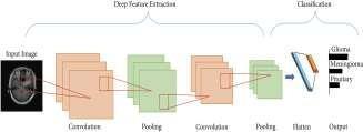
Fig(3).Convolutional Neural Network
Theinputimageswerereducedinsizeto150x150pixels and processed using a first layer that included a Convolutional layer having 32 Filters with size 3x3 also RectifiedLinearUnit(ReLU)activation.Low-levelpicture characteristicswerecapturedbythislayer.
Convolutional layers with 64 filters and ReLU activation weresuccessivelyaddedtotwomorelayers,improvingthe model's capacity for feature extraction. The feature maps were down-sampled using a MaxPooling layer with a 2x2 pool size, which decreased computational effort and aided with spatial abstraction. To avoid dropout layers set at a rateof0.3wereaddedaftertheselayers.
Two further Convolutional layers with 64 filters each madeupthe followinglayers,which were then proceeded by a Max Pooling layer and another Dropout module. To capture more intricate patterns and enhance the model's capacity to identify intricate details in MRI images, this cyclewasrepeated.
Themodelcomprisedtwosetsofconvolutionallayerswith 128 filters each to further boost the depth and feature representation. Max Pooling layers, Dropout layers, and layers toregularize the network were then included. This was crucial for spotting complex cerebral neoplasmrelatedstructures.
AConvolutionallayerwith256filtersandReLUactivation, another Max Pooling layer, and a Dropout layer were addedtothemodel later.Thenetwork was able to collect high-level and abstract properties because to this architecture.
The feature maps were then converted into a 1D vector, flattened, and fed into two dense layers: the first, a Fully Connected(Dense)layerconsistingof512unitswithReLU activation, and the second, another Dense Layer featuring 512 units and ReLU activation. Forclassifyingthedataand understanding complicated relationships within it, these completelyconnectedlayerswerecrucial.
Theoutputlayerwasmadeupof4neuronswithsoft max activation,andafinalDropoutlayersetatarateof0.3 was added to reduce over fitting. This allowed themodel to output probability distributions for the fourclasses linkedtocerebralneoplasmidentification.
We assessed the models' performance during the testing stage of our brain neoplasm detection research utilizing CNN and machine learning models. Our MRI The dataset wasdivided intotrainingandtesting portions. To test the models' capacity to correctly identify brain abnormalities, newMRIpictureswereshowntothem.Weevaluatedtheir efficiency in categorizing neoplastic and non-neoplastic patientsbymeasuringtheiraccuracy,precision,recall,and F1 score. For added robustness, we also performed crossvalidation. These tests supported the validity of our models' dependability and showed how well-suited they were to helping doctors make confident and accurate brainneoplasmdiagnoses.
OurtrainedCNNandmachinelearningmodelsareusedin the last stage of our brain neoplasm detection project to examine MRI data. These models analyze the photos and look for probable brain tumors after receiving rigorous training. Their output helps doctors diagnose patients quicklyandcorrectly,whichenhancespatientcare.
The evaluation of our proposed CNN-based model for cerebral neoplasm detection involved rigorous testing and analysis. In this section, we present the performance metrics along with visual representations of accuracy and loss, providing insights into the effectiveness of our approach.
To assess the efficacy of our model, we utilized standard performance metrics including accuracy, precision, recall, andF1score. Themodel was trained on a diversedataset consisting of MRI images of brain tumors, ensuring robustness and generalization. Table I summarizes the performancemetricsobtainedduringtesting.
Table I: Performance Metrics of CNN-Based Model for CerebralNeoplasmDetection
Metric Value
Accuracy 095
Precision 0.94
Recall 096
F1Score 095
Volume: 11 Issue: 08 | Aug 2024 www.irjet.net
International Research Journal of Engineering and Technology (IRJET) e-ISSN: 2395-0056 p-ISSN: 2395-0072
The high accuracy and balanced precision-recall scores demonstrate the effectiveness of our model in accurately identifyingcerebralneoplasmsfromMRIimages.
B. Visualization of Accuracy and Loss
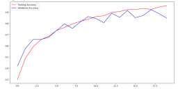
Fig-4:Training&ValidationAccuracy
Thegraphillustratestheprogressionoftrainingaccuracy, indicating the model's learning capability over successive epochs. A steady increase in accuracy is observed, demonstrating effective convergence towards optimal performance.
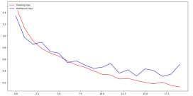
Fig-5:Training&ValidationLoss
Theplotdisplays thevariationintrainingloss throughout thetrainingepochs.Aconsistentdecrease in loss signifies the model's ability to minimize errors and improve predictiveaccuracyovertime.
6.
The paper presents an existing method that can address various challenges, such as detecting tumors accurately and the time it takes to detect them. The combination of the CNN and ML models can provide a moreaccurateand timely diagnosis of cerebral tumors. The CNN technique is idealforachievinghighaccuracywith low error rates. Our objective is to see how this research can lead to advancements in medical imaging and the utilization of deeplearninginhealthcare.
7. REFERENCES
[1]Choudhury, C.L., Mahanty,C .,Kumar, R.,&Mishra,
B. K. (2020). Brain Tumor Detection and Classification
Using Convolutional Neural Network and Deep NeuralNetwork. 2020 International Conference on ComputerScience,EngineeringandApplications(ICCSEA)
DOI:10.1109/ICAIS50930.2021.9395920
[2]Wu, Wentao, et al. ”An Intelligent Diagnosis Method of BrainMRITumorSegmentationUsingDeepConvolutional Neural Network and SVM Algorithm.” Computational and MathematicalMethodsinMedicine2020(2020)
[3]Noh, Hyeonwoo, Seunghoon Hong, and Bohyung Han. "Learning deconvolution network for semantic segmentation." In Proceedings of the IEEE international conferenceonComputerVision,pp.1520-1528.2015.
[4] Abd-Ellah,MahmoudKhaled,etal.”A reviewon brain tumor diagnosis from MRI images: Practical implications, key achievements, and lessons learned.” Magnetic resonanceimaging61(2019):300-318.
[5]Sasikala, S., M. Bharathi, and B. R. Sowmiya. ”Lung Cancer Detection and Classification Using Deep CNN.”, International Journal of Innovative Technology and ExploringEngineering(IJITEE)8.2S(2018).
[6] Islam, Jyoti, and Yanqing Zhang. ”A novel deep learning based multiclass classification method for Alzheimer’s disease detection using brain MRI data.” International conference on brain informatics. Springer,Cham,2017.
[7]Kiranmayee, B. V., T. V. Rajinikanth, and S. Nagini. ”Explorative Data Analytics of Brain Tumour Data Using R.” 2017 International Conference on Current Trends in Computer, Electrical, Electronics and Communication (CTCEEC).IEEE,2017.
[8] Yang,Guang,etal.”Discretewavelettransform- based whole-spectral and subspectral analysis for improved braintumorclusteringusingsinglevoxelMRspectroscopy.” IEEE Transactions on Biomedical Engineering 62.12 (2015):2860-2866
[9]Demirhan, Ays¸e, Mustafa Tor¨ u, and ¨ ˙Inan Guler. ”Segmentation of tumor ¨and edema along with healthy tissuesofbrainusingwaveletsandneuralnetworks.”IEEE journal of biomedical and health informatics 19.4 (2014): 1451-1458
[10] Guo, Lei, et al. ”Tumor detection in MR images using one-class immunefeature weighted SVMs.” IEEE TransactionsonMagnetics47.10(2011):3849-3852..