
International Research Journal of Engineering and Technology (IRJET) e-ISSN:2395-0056
Volume: 11 Issue: 08 | Aug 2024 www.irjet.net p-ISSN:2395-0072

International Research Journal of Engineering and Technology (IRJET) e-ISSN:2395-0056
Volume: 11 Issue: 08 | Aug 2024 www.irjet.net p-ISSN:2395-0072
1Student BIT Dept of CSE Bangalore, India
2Assistant Professor BIT Dept of CSE Bangalore, India
Abstract The project classifies brain tumors from MRI images using advanced deep learning. It employs CNNs with attention mechanisms and GANs for data augmentation. The dataset includes glioma, meningioma, pituitary tumors, and no tumors. DenseNet and custom Attention CNN improve detection accuracy. This assists medical professionals in early diagnosis and treatment planning.
Keywords Convolutional Neural Networks (CNNs), Generative Adversarial Networks (GANs), DenseNet, AttentionCNN
I. INTRODUCTION
Detecting and diagnosing brain tumors through MRI scans is critical for patient care and treatment planning. Traditionalmethodsrelyheavilyonradiologists,whichcan be time-consuming and prone to human error. Deep learning offers a promising alternative, with CNNs, GANs, DenseNet, and Attention Mechanisms enhancing detection accuracy. This project uses a comprehensive MRI dataset categorizedintoglioma,meningioma,pituitarytumors,and no tumors. Models, including DenseNet for its feature extraction capabilities and Attention CNN to highlight relevant image parts, are trained on this data. GANs are usedtogeneratesyntheticimages,addressingdatascarcity and class imbalance. The goal is to develop a highly accurate and reliable model that assists medical professionals in diagnosing brain tumors, improving patient outcomes, and streamlininghealthcareservices.By integrating these advanced techniques, the project aims to enhance the accuracy, efficiency, and interpretability of braintumordetection.
A. Objectves:
Enhance Detection Accuracy Develop a model to significantlyimprovetumordetectionaccuracy.
Increase Diagnostic Efficiency Streamline the diagnostic processtoreducetimeandresourceconsumption.
Improve Model Interpretability Implement tools like Grad-CAMtomakemodeldecisionstransparent.
Address Data Limitations Use GANs for dataset augmentationtoovercomedatascarcity.
Validate Model Generalizability Ensure robust performance across diverse data sets with rigorous validationtechniques.
Facilitate Clinical Integration Design the system for easy adoptioninexistingmedicalworkflows.
Contribute to Medical Research Provide actionable insightstoadvancemedicalimagingandoncology.
II. LITERATURE SURVEY
AnuragGoswami,ManishD.[3],aimstoreplacethemissing parts of the facial photos using Recurrent Generative Adversarial Network. Feature Representation Net also regarded as the semantic feature extractor. Three feature extractors with multiple scales are in use. The use of dilated convolution has increased the receptive field. FeatureTransformationNetregardedasmappingfunction overthefeaturedomain.Theinputisthefeatureextracted from FTN and output will be transferred features. This is able to be rebuilt face images. Here ConvLSTM has been adopted. The image reconstruction net predicts the facial pictureateachscalebyusingthecharacteristicsproduced bytheFTN.Despitethefactthattherearemanyofoptions for upsampling or downsampling, they adopt the simple bilinearinterpolationforalltheexperimentsintheirwork. To fullyexploit thecorrection of adjacentlevel featuresof theirmodel,theydesignanovelshortlinkstructuretofuse the multi-scale features. Discriminator network has global andlocal discriminator. Zhang,Mingming et al.[5],aims to construct an improved Generative Adversarial Networks based on the context encoder and proposes a selflocalizationocclusionmethodforrestoringfacesinimages algorithm. The generator in this paper adopts the convolution network with the structure of Variational Autoencoder.Theencoderuses12convolutionallayers,1 fullyconnectedlayer.Thedecoderuses12deconvolutional layerandonefullyconnectedlayer.LeakyReLuisused as activation function in the first 25 layers and Tanh is used as activation in the last layer. The discriminator in this paperisbasedonVGG19with13convolutionallayerand5 pooling layers. CelebA was the dataset used, and the trainingAdamoptimizerhadalearningrateof0.002.Jiang, Yi, Jiajie Xu et al.[8], presents an approach for face image inpainting using skip connection layers between the encoderandthedecoder.Inpaintingispartofalargesetof image generation problems. To solve this problem, an
International Research Journal of Engineering and Technology (IRJET)
Volume: 11 Issue: 08 | Aug 2024 www.irjet.net
auto-encoderasthegeneratorofmodel.Theauto-encoder contains two networks: an encoder and a decoder. Different from the typical AutoEncoder architecture, they add skip-connection between the corresponding layers of the encoder and decoder sections to prevent the network layer from deteriorating due to the deepening of the network layers. Skip-connection can make sure that the decoding stage can utilize the output of the low-level coding stage of the corresponding resolution to supplementthedecoderwithpartofthestructuralfeature information lost during the encoder downsampling phase, and enhance the structure prediction capability of the generator. The encoder uses a multi-layer convolution layer architecture. Similarly, the architecture of the decoder is symmetric to the encoder with transposed convolution layers. Between the encoder and the decoder, we employ four layers of dilated convolution instead of fully connected layers. Wang, Chen et al.[11], aims to create a two stage generation network for face image restoration that integrates contextual attention and multiscale joint attention. The first stage of the generator consistsofthreeparalleldecoder-encoderbrancheswhich are used to extract feature information of different scales from input image X with mask. Then these units are upsampled to the original resolution through deconvolution operation, and three groups of features are merged into the feature map. The second stage contextual attention module consists of two parallel encoders. The upper part is the contextual propagation layer and the lowerpartistheattentionpropagationlayer.Thefeatures are being extracted and the merged into the shared decoder. Restore the original input image through upsampling and finally output the fine reconstructed result. In the training, the WGAN-GP discriminator is used to compare the ground truth T and the output Y to obtain the adversarial loss. There are total four losses used here: spatial attenuation, reconstruction loss, adversarial loss andMRF-likeloss.Shah,Riya,AnjaliGautametal.
Existing System and Drawbacks: Theexistingsystemfor brain tumor classification primarily relies on traditional Convolutional Neural Networks (CNNs) like DenseNet, which use standard layers such as convolutional, pooling, and fully connected layers to extract features and classify images. While these models are effective in general image recognitiontasks,theyexhibitlimitationsinmedicalimage analysis due to their uniform processing of all image regions. This uniformity can lead to suboptimal performance in identifying subtle and localized features crucial for accurate tumor detection. Additionally, standardCNNsmayrequireextensivelabeleddatasetsand computational resources, limiting their applicability in real-worldmedicalscenarioswhereannotateddatacanbe scarce and computational efficiency is critical. These drawbacksunderscore the need for enhanced models that
e-ISSN:2395-0056
p-ISSN:2395-0072
can dynamically focus on important image regions to improvediagnosticaccuracyandefficiency.
Problem Statement: To compare advanced deep learning techniques for enhancing the detection and diagnosis of brain tumorsinMRIscans,focusingonaccuracy,efficiency, and interpretability to improve clinical outcomes and streamlineneuro-oncologicaldiagnostics.
Detecting brain tumors in MRI scans is challenging and traditionally relies on radiologists, which can be timeconsuming and prone to errors. Deep learning, especially CNNs, offers automation and improved accuracy but struggles with capturing complex patterns in MRI images. AdvancedmodelslikeDensenet121arebeingdevelopedto enhancedetection,aiming to improve patient outcomes by providing more precise and efficient diagnostics.As shown inFigure1.
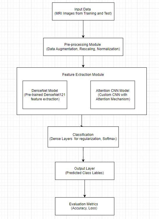
Figure1:Architecturediagram
Proposed Modules:
DataPreProcessingModule
FeatureExtraction
Classification/SegmentationModule
Evaluationmodule
International Research Journal of Engineering and Technology (IRJET) e-ISSN:2395-0056
Volume: 11 Issue: 08 | Aug 2024 www.irjet.net p-ISSN:2395-0072
Algorithms Used:
Convolutional Neural Networks (CNNs) arethebackbone of the image classification tasks in this project. They are particularly effective for processing and analyzing visual data due to their ability to automatically and adaptively learn spatial hierarchies of features from input images. CNNs consist of several layers, including convolutional layers, pooling layers, and fully connected layers. In this project, CNNs are used to classify MRI images of brain tumors into four categories glioma tumor, meningioma tumor,notumor,andpituitarytumor.
Attention Mechanism in CNN Theattention mechanismis a powerful enhancement to traditional CNN architectures, enablingthemodeltofocusonthemostrelevantpartsofthe input image when making predictions. In this project, the attentionmechanismisintegratedintoacustomCNNmodel to improve the classification of brain tumor types. This mechanismworksbyassigningdifferentweightstodifferent regionsofthefeaturemap,effectivelyallowingthenetwork to prioritize certain areas over others. For example, in an MRI scan, the attention mechanism can help the model concentrate on areas with tumor-like structures while ignoring irrelevant background information. This selective focus improves the model’s ability to detect and classify tumorsaccurately.
Generative Adversarial Networks (GANs) Generative Adversarial Networks (GANs) are a critical component of this project, used primarily for generating synthetic MRI imagestoaugmentthetrainingdataset.GANsconsistoftwo neuralnetworksthegeneratorandthediscriminator,which compete in a game-theoretic framework. The generator creates fake images, attempting to mimic real MRI scans, while the discriminator evaluates the authenticity of these images,distinguishingbetweenrealandfakeones.
DenseNet (Dense Convolutional Network) DenseNet, or Dense Convolutional Network, is a type of CNN that connects each layer to every other layer in a feed-forward fashion. For each layer, the feature maps of all preceding layers are used as inputs, and its own feature maps are passed on to all subsequent layers. This dense connectivity pattern improves the flow of information and gradients throughoutthenetwork,makingiteasiertotrainverydeep networks.Inthisproject,DenseNet121,apopularvariantof DenseNet, was employed to leverage its strengths in handling complex image classification tasks. By reusing features across layers, DenseNet reduces the number of parameters, mitigates the vanishing gradient problem, and enhancesfeaturepropagation.
Dataset Used:
ThedatasetusedintheprojectaretakenfromBrainTumor MRI Dataset (kaggle.com).It consists of four different categoriesasshownbelow.Thetotalnumberofimagesused
are 2000 images with each category containing approximately 500 images. The different categories of the datasetarementionedbelow.
Categories: Glioma Tumor, Meningioma Tumor, Pituitary Tumor,NoTumor
Data Structure: The dataset is divided into two main directories: Training and Testing. Each directory contains subdirectories for each of the four categories, with the imagesstoredwithinthesesubdirectories.
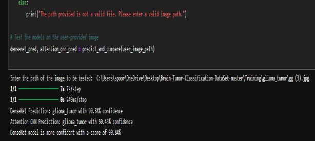
As shown in the Figure 2, the user inputs the path of an MRI image to test the models. The DenseNet model predicts the image as a glioma tumor with 98.40% confidence, while the Attention CNN predicts the same with 94.58% confidence. DenseNet's prediction is deemed moreconfidentwithahigherscoreof99.94%.
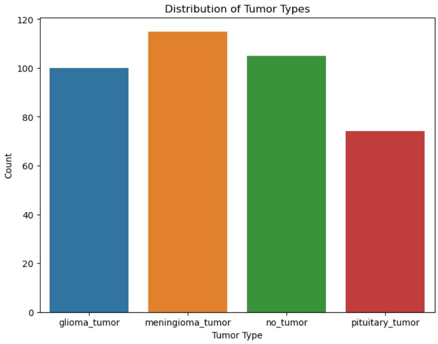
Figure3:Visualizethedistributionoftumortypesinthe trainingdataset
International Research Journal of Engineering and Technology (IRJET) e-ISSN:2395-0056
Volume: 11 Issue: 08 | Aug 2024 www.irjet.net p-ISSN:2395-0072
AsshownintheFigure3,thedistributionoftumortypesin thedatasetispresented.Meningiomatumorsarethemost prevalent, followed by images with no tumor and glioma tumors. Pituitary tumors have the least representation in thedataset.
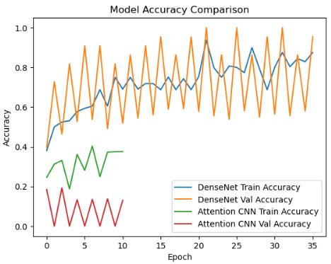
AsshownintheFigure4,thegraphcomparesthetraining and validation accuracies of two models: DenseNet and Attention CNN, over 35 epochs. DenseNet exhibits a relatively stable training accuracy with fluctuating validation accuracy, while Attention CNN shows high variability in both training and validation accuracies. DenseNet's performance appears more consistent and potentially more reliable for this dataset compared to AttentionCNN.
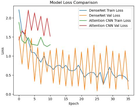
AsshownintheFigure5,thegraphcomparesthetraining andvalidationlosses ofDenseNetandAttention CNN over 35epochs.DenseNetexhibitsaconsistentdecreaseinboth training and validation losses, suggesting effective learning.Incontrast,AttentionCNNshowshighvariability in losses, indicating unstable learning behavior. DenseNet
demonstrates more reliable performance with lower and morestablelossvaluescomparedtoAttentionCNN.
WORK Conclusion
Inthisproposedmethod,wedevelopedandcomparedthe performance of DenseNet and Attention CNN models for brain tumor classification using MRI images. Both models were trained on a labeled dataset with data augmentation techniquesto improve generalization. Whilethe DenseNet model demonstrated better performance and higher confidence in its predictions, the overall confidence levels were below thedesired thresholdof 90%. Theintegration of GANs for synthetic image generation and various techniques like hyperparameter tuning and learning rate adjustments showed potential for enhancing model accuracy.
Advanced Data Augmentation Experimenting with more sophisticateddataaugmentationtech.
Hyperparameter Optimization Implementing automated hyperparametertuningmethods.
Attention Mechanism Enhancements Investigating more advancedattentionmechanisms
GAN Improvements Enhancing the quality and diversity ofGAN-generatedsyntheticimages
Larger and Diverse Datasets Acquiring more comprehensivedatasetstotrainmodels,capturingawider
VI. REFERENCE
[1] Anurag Goswami, Manish Dixit,“An Analysis of Image SegmentationMethodsforBrainTumourDetectiononMRI Images”,IEEE.2020
[2] S´ergio Pereira, Adriano Pinto, Victor Alves and Carlos A. Silva, “Brain Tumor Segmentation using Convolutional NeuralNetworksinMRIImages”,IEEE.2020
[3] Hamza Rafiq Almadhoun and Samy S. Abu Naser, “DetectionofBrainTumorUsingDeepLearning”,Scientific Reports.2020.
[4]MasoumehSiar,MohammadTeshnehlab“BrainTumor Detection Using Deep Neural Network and Machine LearningAlgorithm”Elsevier.2020
[5] Neelum noreen , sellappan palaniappan, muhammad imran and muhammad Shoaib. “A Deep Learning Model Based on Concatenation Approach for the Diagnosis of BrainTumor.”IEEE.2021
International Research Journal of Engineering and Technology (IRJET) e-ISSN:2395-0056
Volume: 11 Issue: 08 | Aug 2024 www.irjet.net p-ISSN:2395-0072
[6] Laura Boldúa, Anna Merinoa, Andrea Acevedoa, Angel Molina, and José Rodellar. “A Robust Approach for Brain Tumor Detection in Magnetic Resonance Images Using FinetunedEfficientNet.”IEEE.2021
[7] Arkapravo Chattopadhyay, Mausumi MaitraRodellar “MRI-based brain tumour image detection using CNN baseddeeplearningmethod..”Elsevier.2021
[8] Rajeev Ratan, Sanjay Sharma “Brain tumor detection based on multi parameter MRI image analysis” Elsevier. 2021
[9] T M Shahriar Sazzad ,Kawsar Ahmmed, Mahmuda Rahman.“Development of Automated Brain Tumor IdentificationUsingMRIImages.”IEEE.2022
[10] Mohammad shahjahan majib, md. Mahbubur rahman ,t.M.Shahriarsazzad,nafizimtiazkhan,andsamratkumar dey“AVGGNet-BasedDeepLearningFrameworkforBrain TumorDetectiononMRIImages.”Elsevier.2022
[11] M.O. Khairandish, M. Sharma, V. Jain, J.M. Chatterjee, N.Z. Jhanjhi “A Hybrid CNN-SVM Threshold Segmentation Approach for Tumor Detection and Classification of MRI BrainImages”Elsevier.2022
[12] Rohini Paul Joseph, C. Senthil Singh, M.Manikandan “Deep learning identifes Acute Promyelocytic Leukemia in bonemarrowsmears”Elsevier.2020
[13] Tanzilal Mustaqim, Chastine Faticah, and Nanik Suciati. “ Brain Tumor Detection Using MRI Images Review”IJRET.2020
[14] Abdul Hannan Khan,Sagheer Abbas,Muhammad Adnan Khan,Umer Farooq,Wasim Ahmad Khan, Shahan Yamin Siddiqui, and Aiesha Ahmad “ Intelligent Model for BrainTumorIdentificationUsingDeepLearning.”Hindawi. 2020
[15] Rohini Paul Joseph, C. Senthil Singh, M.Manikandan “ BrainTumorSegmentationtoCalculatePercentageTumor UsingMRI”IES-KCIC.2020
[16] Sohaib asif, wenhui yi , qurrat ul ain, jin hou, tao yi, and jinhai si “ Improving Effectiveness of Different Deep Transfer Learning- Based Models for Detecting Brain Tumors From MRImages”IEEE.2020
[17] Maram fahaad almufareh, muhammad imran ,abdullah khan , mamoona humayun , and muhammad asim “ Automated Brain Tumor Segmentation and Classification in MRI Using YOLO-Based Deep Learning” MDPI.2023
[19]T. Logeswari and M. Karnan “ An improved implementation of brain tumor detection using segmentationbasedonsoftcomputing”JCREOI.2023
[20]T. Dina Aboul Dahab, Samy S. A. Ghoniemy, Gamal M. Selim. “Automated Brain Tumor Detection and Identification Using Image Processing and Probabilistic NeuralNetworkTechniques.”IJRET2023