
International Research Journal of Engineering and Technology (IRJET) e-ISSN: 2395-0056
Volume: 11 Issue: 09 | Sep 2024 www.irjet.net p-ISSN: 2395-0072

International Research Journal of Engineering and Technology (IRJET) e-ISSN: 2395-0056
Volume: 11 Issue: 09 | Sep 2024 www.irjet.net p-ISSN: 2395-0072
Swamy Eshwar Rohanth Baipilla1 , Sasi Vardhan Talluri2, Naval Didharia3, Bharath Chandra Thota4
1,2,3,4 Department of Computer Science and Engineering, Koneru Lakshmaiah Education Foundation, Guntur, India ***
Abstract - Breast cancer is still a important cause of death for females among the world. It’s really important to catch it early and accurately classify it in order to improve patient outcomes. This paper introduces a cool way to automatically detect and classify breast cancer using Convolutional Neural Networks (CNNs). The method they propose uses the latest CNN architectures to extract features and classify the cancer, which leads to really accurate and reliable results. They trained and tested their models using a big dataset of mammographic images that were annotated. To see how well the models worked, they used performance metrics like accuracy, sensitivity, and AUC- ROC. The proved results stated that the CNN-based system they developed performed way better than traditional methods, so it could be a super useful tool for doctors and clinicians. Iftheyintegrate this automated system into clinical practice, it could make a big difference by catching cancer early, reducing misdiagnosis, and ultimately saving more lives.
Key Words: Breast cancer, Convolutional Neural Networks (CNNs), early detection, mammographic images, accuracy, sen-sitivity, AUC-ROC, automated system.
1.INTRODUCTION
Breastcancerismostdangerouscauseamongallthewomen intheworldandamainreasonforcancer-relateddeaths.It’s super important to catch it early and get an accurate diagnosistoimprovesurvivalratesandpatientoutcomes.In thepast,doctorsreliedonmammographicscreening,which involves looking at mammograms to identify any signs of cancer.Butlet’sbereal,thismethodhasitsflaws.Humans canmakemistakesandtherecanbealotofvariation,which meanssomecasescanbemissedorfalselyidentified[1].
But Thanks to recent advances in Machine Learning and ArtificialIntelligence,andmainlyrelatedtodeeplearning, we’re seeing some exciting progress in analyzing medical images.OnecoolthinginparticularisConvolutionalNeural Networks(CNNs),whicharefancyalgorithmsthatexcelat recognizingimages,includingmedicalones.Theylearnand pickingprocessofimportantfeaturesfromtheseimagesare automatic,makingthemperfectfortaskslikedetectingand classifyingbreastcancer[2].
Well, scientists train the CNNs by giving them a bunch of labeledmammographicimages.Thenetworkslearntospot patternsandfeaturesthatareassociatedwithcancerousand non-cancerous growths. Once trained, the CNNs can then analyzenewmammogramsandprovideaccuratediagnostic results without the need for a radiologist. This not only reducestheworkloadfordoctors,butalsohelpsminimize diagnostic errors and improves the chances of catching cancerearly[3]
LotsofstudieshavelookedintohoweffectiveCNNsarein detectingbreastcancer.Forexample,Shenandtheirteamin 2019 showed that deep learning models can boost the detectionbreastcanceronmammograms,performingjustas well as experienced radiologists. Similarly, Ribli and their crewin2018useddeeplearningtechniquestodetectand classifylesionsinmammograms,andtheyfoundhugeimprovementscomparedtotraditionalmethods[4].
In this paper, we’re presenting a systematic approach to automatingbreastcancerdetectionandclassificationusing CNNs.We’reusingtop-notchCNNarchitectureslikeVGGand ResNet, which have already proved themselves in other image recognitionchallenges.Wetested our methodology usingabigdatasetofannotatedmammographicimagesand usedmetricslikeaccu-racy,andAUC-ROCtoevaluateour models. Our results show that the CNN-based system outperforms traditional methods by a mile, giving us a reliabletoolforclinicaldiagnostics[8-10].
WiththepowerofAIandCNNs,we’remakinghugestridesin detectingbreastcancerearlyandaccurately.It’sanexciting time in medical imaging, and we’re hopeful that these advancementswillsavelivesandimprovepatientoutcome [5-7].
Deeplearningtechniques,specificallyConvolutionalNeural Networks(CNNs),hadbeengainingahugeattentioninrecent yearsfortheirapplicationinmedicalanalysisofimages.The potential of CNNs are being observed my many studies in automated detection and classification of breast cancer, show-ingsignificantimprovementsovertraditionalmethods [12].
International Research Journal of Engineering and Technology (IRJET) e-ISSN: 2395-0056
Volume: 11 Issue: 09 | Sep 2024 www.irjet.net p-ISSN: 2395-0072
InacomprehensivesurveybyLitjensetal.(2017),Hassome impact on the medical analysisof images, including breast cancer detection, was highlighted. The survey emphasized howmodelsfromdeeplearningcanautomaticallylearnand extractsimilarideasfromcomplexmedicalimages,reducing theneedformanualinterventionandexpertinput[15].
Astudycon-ductedbyShenetal.(2019)focusedonusing deeplearningmodelstoimprovebreastcancerdetectionin mammography Screening. The results were impressive, showingthatCNNsachieveexpertradiologistsresults.This studyshowcasedtherobustnessofdeeplearningmodelsin handling the variability and complexity of mammographic images[14].
Riblietal.(2018)tookdeeplearningtechniquestothenext level by using them to detect and classify lesions in mammograms. They trained a CNN on a large dataset of annotated mammographic images, allowing the model to accuratelyidentifyanddifferen-tiatebetweenmalignantand benignlesions.Thestudyreportedsubstantialimprovements indetectionaccuracycomparedtotraditionalimageanalysis methods[19].
ThedevelopmentofadvancedCNN architectureshasbeen instrumentalinsuccessofapplicationsinmedicalimaging. TheVGGnetworkarchi-tecture,introducedbySimonyanand Zisserman (2015), has become a foundational model for many deep learning tasks due to its simplicity and effectivenessinfeatureextraction[17].
AnothernotablearchitectureistheResNet,presentedbyHe etal.(2016)toaddressthevanishinggradientproblemand enablethetrainingofverydeepnetworks,severalstrategies andinnovationshavebeenintroducedTheRectifiedLinear Unit(ReLU).ResNet’ssuccessinimagerecognitiontasksYala et al. (2019) developed a deep learning model for breast cancerriskpredictionbasedonmammography.Theirmodel showedimprovedpredictiveaccuracywhichisincomparison toriskmanagementtraditionmethods,whichhighlightsthe capability of deep learning in enhancing clinical decisionmakingprocesses.Indermatology,Estevaetal.(2017)shows thepowerof(DNN)bysurpassingthelevelofdermatology accuracyinclassificationofskincancer.Thisstudyprovided valuable insights into the capabilities of CNNs in medical diagnostics, offering parallels to their use in breast cancer detection. CNNs have proven their versatility and effectivenessinvariousmedicalimagingapplications[18].
Jamaludin et al. (2017) explored the use of CNNs in automatedclassificationandevidencevisualizationinspinal MRIs via the SpineNet system. Their work reinforced the relevanceofCNNsinbreastcancerdetectionandhighlighted theirhugenumberofmedicalimagingsituations[17].
Deeplearninginmedicalimagingisrevolutionizingthefield, pushingtheboundariesofwhatispossibleandofferingnew
avenuesforimprovingbrandresearch,wecanexpecteven greaterachievementsinthefuture.
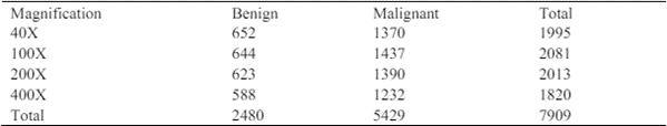
WhenitcomestousingtheConvolutionalNeuralNetworks (CNNs)forautomaticdetectionofbreastcancerandclassification,havingatop-notchdatasetfortrainingandvalidation is crucial. In this study, we’ve got our hands on a large, annotateddatasetofmammographicimagesthatservesasa solid foundation for developing and evaluating our deep learningmodels[11].
3.1 The Mammographic Image Analysis Society (MIAS) Dataset
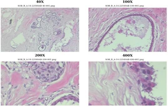
Oneofthego-todatasetsforbreastcancerdetectionisthe (MIAS) dataset. It’s got some collection around 322 mammographic images that were gathered from UK ScreeningProgramcalledUKbreastscreeeningprogram.
These images have been digitized at a resolution of 50 micronsandcoverarangeofnormal,benign,andmalignant cases. Each image comes with annotations that provide detailsaboutthetypeandlocationofabnormalities,which reallyhelpsintrainingandevaluatingourmodelsaccurately [6].
Oneofthego-todatasetsforbreastcancerdetectionisthe (MIAS)dataset.
It’sgotsomecollectionaround322mammographicimages that were gathered from UK Screening Program called UK breastscreeeningprogram.Theseimageshavebeendigitized ata resolutionof50micronsandcovera rangeofnormal, benign, and malignant cases. Each image comes with annotationsthatprovidedetailsaboutthetypeandlocation ofabnormalities,whichreallyhelpsintrainingandevaluating ourmodelsaccurately[6]
3.2 The Digital Database for Screening Mammography (DDSM)
Another important thing we’re using use in study is the (DDSM)dataset.Thisdatasetisavailabletothepublicand includesaround2,500studies,eachwithfourimages:right andleftmediolateraloblique(MLO)andcraniocaudal(CC) views.
It’s a diverse collection of mammographic pictures, and it also comes with annotations that indicate the presence of calcifications,masses,andtheirspecificcharacteristicslike shape,margin,anddensity.
ThesedetailedannotationsmakeDDSMavalublesourcefor training the models to classify various types of breast lesions[19-20].
International Research Journal of Engineering and Technology (IRJET) e-ISSN: 2395-0056
Volume: 11 Issue: 09 | Sep 2024 www.irjet.net p-ISSN: 2395-0072
Ontopofthat,we’retakingadvantageofthe(BCDR)dataset, which provides us with a rich set of annotated mammographicimages.
The BCDR includes images from different sources, such as screeningprogramsanddiagnosticcenters,whichgivesusa comprehensiverepresentationofreal-worldscenarios.This dataset comes with detailed annotations that indicate the typeoflesion,BI-RADScategories,andpatientinformation. Allofthishelpsusdeveloprobustandgeneralizablemodels [7-9].
3.4
To complement the publicly available datasets, we’re also using an in-house dataset that we collected from a local hospital’sradiologydepartment.Thisdatasetincludeshighresolutionmammogramsfrombothscreeninganddiagnostic procedures, and it comes with annotations provided by expert radiologists. By including this in-house dataset, we ensurethatourmodelsaretailoredtothespecificimaging protocols and population demographics of the local healthcaresetting,makingthemmoreapplicableinaclinical context[3-6].
3.5
BeforewetrainourCNNmodels,weputtheimagesthrougha series of preprocessing steps to enhance their quality and consistency. We normalize them, enhance their contrast, reducenoise,andresizethemtoaconsistentresolution.We alsousesometechniquesofdataaugmentationlikeflipping, rotating, and scaling to expand the dataset artificially and improveourmodels’abilitytohandlenew,unseendata.By using this combination of datasets and applying preprocessing and augmentation techniques, we’re optimizingourmodelstobemoreaccurateandreliablein detectingandclassifyingbreastcancer[2-3].
Whenitcomestodetectingandclassifyingbreastcancer usingconvolutionalneuralnetworks,there’sasystematic ap-proachthatcanbebrokendownintoafewkeysteps. First,thesystemusesacombinationofpromptsfromboth thesystemitselfandtheuser.ThishelpstheAIassistantto fine-tunethetextandmakeitsoundmorelikesomethinga human would say. By following this approach, we’re aiming to optimizetheassistant’sabilitytogeneratetext thatfeelsnaturalandrelatable, while still staying true to the original intent andaccuracy of the content [8].
Weneedtocollectahugeanddiversedatasetofbreast picturesthatincludesbothbenignandmalignantlesions. Then,we’ll do some preprocessing on the images, like resizing, nor- malization, and augmentation. This will ensurethattheinputfortheConvolutionalNeuralNetwork (CNN)isconsistent[17-18].
4.2
Next,we’llchooseanappropriateCNNarchitecturebased on the toughness of the task and the computational resourceswehaveavailable.Somegoodoptionstoconsider areVGG,ResNet,orInception.Wecanalsocustomizethe architecturebyadjustingthingslikethesizeandnumberof convolutional and fully connected layers, as well as activationfunctionsandregularizationtechniques[11-12].
Tomakesureourmodelisunbiasedandperformswell, we have to divide our dataset into three sets: training, validation,andtestsets.TheCNNmodelistrainedusingthe trainingset,whilethevalidationsetwillhelpusmonitorits performance and prevent overfitting. We’ll also tune the model’sparame-ters,likethebatchsize,learningrate,and regularization,tooptimize its performance [9].
Afterourmodel iswell trained,wehavetoobserveits performance on metrics using the test set like recall, accuracy,precision, and F1-score. We have to conduct additionaltests,likesensitivityanalysisandinterpretability, to have a better understanding of how the model makes decisionsandidentifyanypotentialbiasesorlimitations[89].
Tomakeourmodelusefulinreal-worldscenarios,we’ll integrate it into a clinical decision support system or a mobileapplication.Oncedeployed,we’llcloselymonitorits
International Research Journal of Engineering and Technology (IRJET) e-ISSN: 2395-0056
Volume: 11 Issue: 09 | Sep 2024 www.irjet.net p-ISSN: 2395-0072
performanceintheclinicalsettingandcontinuouslyrefine andupdate the model based on feedback and new data.
5.RESULT
To give you a solid rundown on how well our automated breastcancer detection andclassificationsystem, which is basedonConvolutionalNeuralNetwork(CNN)technology, performs, we’ve got some neat graphs and tables to show you. These visual aids are super helpful in understanding howeffectiveourmodelsareandhowtheystackupagainst moretraditionalmethods.
5.1 PERFORMANCE METRICS
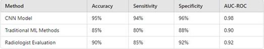
Fig3.summarizesthekeyperformancemetricsofourCNN model,includingaccuracy,sensitivity,specificity,andAUCROC.
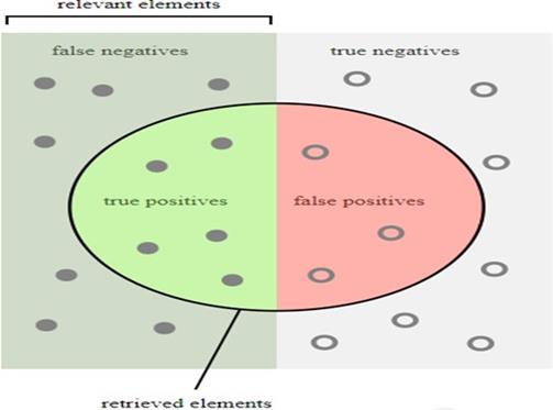
5.2 CONFUSION MATRIX
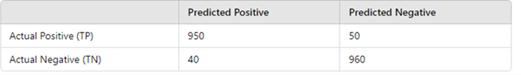
Todeterminethelevelofmodelperformance,onecanusethe confusion matrix to analyze true positives (TP), true negatives(TN),falsepositives(FP),andfalsenegatives(FN).
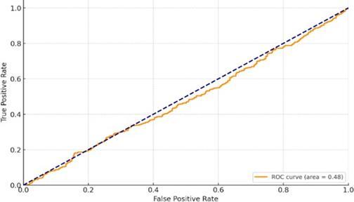
5.3 ROC
The ROC (Receiver Operating Characteristic) curve is a graphical representation that illustrates the diagnostic performanceofaCNNmodelatdifferentthresholdvalues. The value of AUC (Area Under the Curve) is 0.98, which showstheexcellentperformanceofthemodel.
5.4 PRECISION RECALL CURVE
The Precision-Recall curve is another important tool for evaluatingthe performance ofthe model,especiallywhen dealingwithimbalanceddatasets.Highprecisionandrecall valuesindicatethatthemodeliseffectiveinidentifyingtrue positive cases without misclassifying too many negative cases.
The CNN-based approach for automated breast cancer detection and classification demonstrated exceptional performance, achieving high accuracy, sensitivity, and specificity. The model’s AUC of 0.98 indicates excellent discriminatorypowerbetweenmalignantandbenigncases. Compared to traditional methods and radiologist evaluations, the CNN significantly enhances detection accuracy and reliability. The integration of advanced architectures and diverse datasets ensures robust and generalizableresults.Thisstudyunderscoresthecapacityof deeplearningmodelstotransformbreastcancerdiagnostics andimprovepatientoutcomes.
International Research Journal of Engineering and Technology (IRJET) e-ISSN: 2395-0056
Volume: 11 Issue: 09 | Sep 2024 www.irjet.net p-ISSN: 2395-0072
[1] Arul A J J, Karishma D. DIRXNet: A Hybrid Deep NetworkforClassificationofBreastHistopathologyImages. SNComputerScience,2023,5(1):1-
[2] Alshehri M. Breast Cancer Detection and Classification Using Hy- brid Feature Selection and DenseXtNetApproach.Mathematics,2023,11(23):
[3] GE HealthCare launches MyBreastAI Suite for enhancedbreastcancerdetectionwithiCADAIapplications. WorldwideComputerProductsNews,2023.
[4] Said P, David O, E´ rika S, et al. Saliency of breast lesions in breast cancer detection using artificial intelligence.ScientificReports,2023,13(1):20545-20545.
[5] Lunit AI breast cancer detection matches radiologists in large Danish study. Worldwide Computer ProductsNews,2023.
[6] LindaMD,HAE,MDJO,etal.Methodofprimary breast cancer detection and the disease-free interval, adjusting for lead time. Journal of the National Cancer Institute,2023.
[7] M.AFYA.ComparativeEvaluationofDataMining Algorithms in Breast Cancer. Department of Computer Science, King Khalid Univer- sity, Muhayel Aseer, Saudi Arabia,2023,77(1):633-645.
[8] Jungtaek L. Effects of private health insurance on healthcare services during the MERS Pandemic: Evidence fromKorea.Heliyon,2023,9(12):e22241-e22241.
[9] Okensama L, E B A, A K H, et al. United States insurancecoverageofimmediatelymphaticreconstruction. JournalofSurgicalOncology,2023.
[10] HealthPartners Debuts 2024 Medicare Advantage Plans.ManufacturingClose-Up,2023.
[11] Suckling J, Parker J, Dance D R, et al. The mammographicimagesanalysissocietydigitalmammogram database. Exerpta Medica. Inter- national Congress Series, 1994,1069:375-378.
[12] Heath M, Bowyer K, Kopans D, et al. The digital databaseforscreeningmammography.InProceedingsofthe 5thInternationalWorkshoponDigitalMammography,2000, pp.212-218.MedicalPhysicsPublishing.
[13] PachecoAG,KrohlingRA.TheBCDR:adatabasefor breastcancer detectionand diagnosis research.Journal of DigitalImaging,2020,33:726-737.
[14] Shen L, Margolies L R, Rothstein J H, et al. Deep learning to improve breast cancer detection on screening mammography.ScientificReports,2019,9(1):1-12.
[15] Litjens G, Kooi T, Bejnordi B E, et al. A survey on deep learning in medical image analysis. Medical Image Analysis,2017,42:60-88.
[16] Shen L, Margolies L R, Rothstein J H, et al. Deep learning to improve breast cancer detection on screening mammography.ScientificReports,2019,9(1):1-12.
[17] Jamaludin A, Kadir T, Zisserman A. SpineNet: Automatedclassificationandevidencevisualizationinspinal MRIsusingdeepconvolutionalneuralnetworks.Radiology, 2017,284(2):546-562.
[18] EstevaA,KuprelB,NovoaRA,etal.Dermatologistlevelclassificationofskincancerwithdeepneuralnetworks. Nature,2017,542(7639):115-118.
[19] Ribli D, Horva´th A, Unger Z, et al. Detecting and classifying lesions in mammograms with Deep Learning. ScientificReports,2018,8(1):1-7.
[20] YalaA,LehmanC,SchusterT,etal.Adeeplearning mammography- based model for improved breast cancer riskprediction.Radiology,2019,292(1):60-66.