Rit LbhÍ nr.


 Albína Hulda Pálsdóttir & Kevin P. Smith
ISSN 1670-5785 ISBN 978-9979-881-56-8
Desember 2019 Landbúnaðarháskóli Íslands
© Albína Hulda Pálsdóttir, Kevin P. Smith, Agricultural University of Iceland & IcelandicZooArch 2019 Zooarchaeological analysis of bones from Hallmundarhellir cave
Rit LbhÍ nr. 85
Publisher: Agricultural University of Iceland (Landbúnaðarháskóli Íslands) Place: Reykjavík
ISSN 1670 5785 ISBN 978 9979 881 56 8
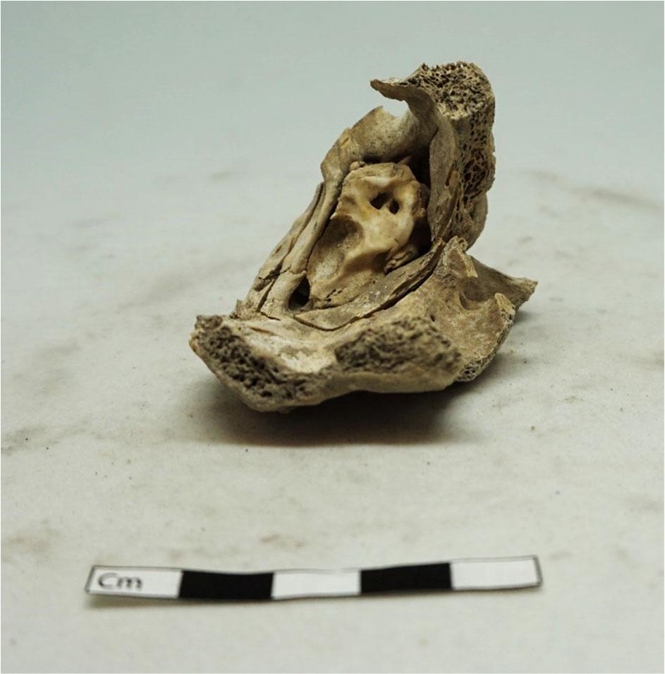
Picture on front page: Sample 2 (ÞJMS no.2017 37 002). Sheep mandible with teeth. Photo by: Albína Hulda Pálsdóttir
List of figures
List of tables
Hallmundarhellir and the 2017 investigation
Analysis methods
Overview of the animal bones and teeth from Hallmundarhellir
Detailed information about each sample/bone
Sample 1 (ÞJMS no. 2017 37 001)
Sample 2 (ÞJMS no. 2017 37 002)
Sample 3 (ÞJMS no. 2017 37 003)
Sample 4 (ÞJMS no. 2017 37 004)
Sample 5 (ÞJMS no. 2017 37 005)
Sample 6 (ÞJMS no. 2017 37 006)
Aging 22
Bones discernible from photos from the cave
Bones recovered previously from the cave
Discussion
Further work at the site
Funding
Bibliography
Figure 1: Hallmundarhellir, following 2017 mapping by Kevin Smith and Guðmundur Ólafsson, with the locations of faunal samples marked in. ©Kevin P. Smith 2019
Figure 2: Dog skeleton with major bone elements labeled (Davis, 1987, p. 54; Reitz & Wing, 2008, p. 364).
Figure 3: The elements from a sheep skeleton collected in Hallmundarhellir cave are coloured in orange, elements coloured blue were recognisable from photos taken at the site in 2017 but those bones were not recovered at the time.
Figure 4: Sample 1 (Þjms no. 2017 37 001). Sheep/goat cervical vertebra, unfused, seen from two sides. Scale 3 cm. Photographer: Albína Hulda Pálsdóttir.
Figure 5: Sample 2 (ÞJMS no.2017 37 002). Sheep mandible with teeth, lingual side. Scale 10 cm. Photographer: Albína Hulda Pálsdóttir
Figure 6: Sample 2 (ÞJMS no.2017 37 002). Sheep mandible with teeth, buccal side. Some possible knife marks visible as well as possible rodent gnawing marks. Scale 10 cm. Photographer: Albína Hulda Pálsdóttir
Figure 7: Area with possible knife marks and possible rodent gnawing in circle in the mandible (ÞJMS no. 2017 37 002). Scale 5 cm. Photographer: Albína Hulda Pálsdóttir 13
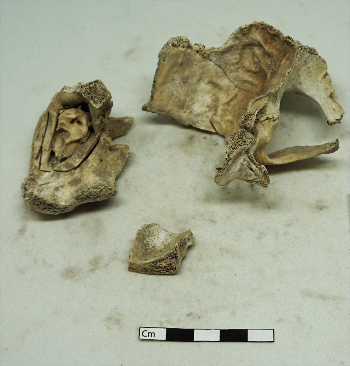
Figure 8: Sample 3 (ÞJMS no. 2017 37 003). Half a sheep skull including petrous bone. View from the posterior of the skull. The skull has been chopped roughly in half and the occipital has also been chopped through where the arrow points. Scale 5 cm. Photographer: Albína Hulda Pálsdóttir 15
Figure 9: Sample 3 (ÞJMS no. 2017 37 003). Half a sheep skull including petrous bone. View from inside the skull. Scale 5 cm. Photographer: Albína Hulda Pálsdóttir 16
Figure 10: Sample 3 (ÞJMS no. 2017 37 003). The skull after the petrous bone was cut out. Scale 5 cm.
Photographer: Albína Hulda Pálsdóttir 17
Figure 11: Sample 3 (ÞJMS no. 2017 37 003). The petrous bone after having been cut out of the skull. This part of the skull is in the ancient DNA laboratory of the University of Oslo. Scale 5 cm.
Photographer: Albína Hulda Pálsdóttir.
Figure 12: Sample 4 (ÞJMS no. 2017 37 004). Sheep mandible with teeth. Scale 5 cm. Photo by: Albína Hulda Pálsdóttir.
Figure 13: Sample 5 (ÞJMS no.2017 37 005). Sheep/goat maxilla with teeth, broken in three fragments. Scale 5 cm. Photographer: Albína Hulda Pálsdóttir.
Figure 14: Sample 6 (ÞJMS no 2017 37 006). Proximal humerus of a sheep/goat. Has been chopped through the shaft. Scale 5 cm. Photographer: Albína Hulda Pálsdóttir.
Figure 15: Sample 6 (ÞJMS no 2017 37 006). Closer view of the chopped shaft of the humerus. Scale 5 cm. Photographer: Albína Hulda Pálsdóttir. 21
Figure 16: Close up of bone concentration in feature 4. Towards the bottom left a pelvis fragment and phalange, in the concentration on the lower left is the unfused distal epiphysis of a sheep/goat metapodial. In the middle of the photo an axis, calcaneus, thoracic vertebra and a number of ribs. 25
Figure 17: The south bench in Hallmundarhellir cave. Towards the lower right side, a fully fused metapodial from sheep/goat can be seen as well as some ribs. 26
Table 1: Speciation of the mandible( ÞJMS no. 2017 37 002) following Halstead et al. (2002). 13
Table 2: Tooth wear of the mandible ( ÞJMS no. 2017 37 002) following Grant (1982). 14
Table 3: Measurements of the mandible ( ÞJMS no. 2017 37 002) following von den Driesch (von den Driesch, 1976) 14
Table 4: Measurements of the petrous bone following (Mallett & Guadelli, 2013) 15
Table 5: Speciation of mandible ÞJMS no. 2017 37 004 following Halstead et al. (2002). .................... 18
Table 6: Tooth wear of the sheep mandible ( ÞJMS no. 2017 37 004) following Grant (1982). ............ 18
Table 7: Measurements of Sample 4 (ÞJMS no. 2017 37 004) following von den Driesch (1976). ....... 19
Table 8: Measurements of Sample 5 (ÞJMS no.2017 37 005) following von den Driesch (1976). 20
Table 9: Aging of the bones from Hallmundarhellir cave 22 Table 10: Zooarchaeological analysis data from Hallmundarhellir. Recording ollows the NABONE system 9thedition (North Atlantic Biocultural Organization Zooarchaeology Working Group, 2010) with the addition of texture (Harland et al., 2003). Zones following Dobney & Rielly (1988). Columns with no recorded data were removed (pathology, burning). Measurements are in the chapter about each sample.
Hallmundarhellir is a cave site with man made stone structures, located beneath the surface of the Hallmundarhraun in the interior of Borgarbyggð, West Iceland. The cave was discovered in 1956 by Kalman Stefánsson of Kalmanstunga and was previously explored by Gísli Gestsson in 1958 (Gestsson, 1960).
In July 2017 Kevin P. Smith, Brown University, Guðmundur Ólafsson, National Museum of Iceland and Magnús Sigurðsson, The Cultural Heritage Agency of Iceland documented the condition of the archaeological remains within the cave with a crew from the TV show “Expedition Unknown”. During this visit to the cave six animal bones were removed from the cave for radiocarbon dating and ancient DNA analysis. The locations of these bones were mapped precisely within the cave as part of the effort to improve understanding of this site (Figure 2). More detailed information about the 2017 investigation can be found in the site report (Smith, Ólafsson, Pálsdóttir, & Sigurðsson, 2019).
The investigation research permit number from The Cultural Heritage Agency of Iceland (Minjastofnun Íslands) is 201707 0072. The National Museum of Iceland´s reference number for material collected from the site is 2017 37 XXX.


Zooarchaeological analysis of the bones and teeth from Hallmundarhellir took place in the Icelandic ZooArch laboratory at the Agricultural University of Iceland in Keldnaholt, Reykjavík on August 9th to 11th 2017 and was done by zooarchaeologist Albína Hulda Pálsdóttir. The Icelandic ZooArch reference collection was used for identification (Pálsdóttir & Skúladóttir, 2016).
Basic data was recorded through the NABO Zooarchaeology working group’s NABONE system (9th edition) which combines an Access database with specialized Excel spreadsheets. The NABONE package allows application of multiple measures of abundance, taphonomic indicators, and skeletal element distribution (North Atlantic Biocultural Organization Zooarchaeology Working Group, 2010).
Figure 2: Dog skeleton with major bone elements labeled (Davis, 1987, p. 54; Reitz & Wing, 2008, p. 364).


All teeth and bones were identified to species and element (Figure 2), when possible, and basic taphonomic indicators were recorded. The bones’ texture was recorded following the York system (Harland, Barrett, Carrott, Dobney, & Jaques, 2003) to get an overview of the general state of preservation of the collection. All bones and teeth were photographed from multiple angles. Zones were recorded following the standards of Dobney & Rielly (1988).
Land mammal element measurements were done according to the metrical standards of von den Dreisch (1976), recorded to the millimeter level using digital callipers, with additional measurements taken that are specific to sheep (Ovis aries) bones (Mallett & Guadelli, 2013; Popkin, Baker, Worley, Payne, & Hammon, 2012).
Bones from sheep (Ovis aries) and goat (Capra hircus) are often difficult to identify confidently to species. For distinctions between sheep (Ovis aries) and goat (Capra hircus) bones, the standards of Zeder & Lapham (2010) were followed with aid from other publications (Boessneck, 1969; Halstead, Collins, & Isaakidou, 2002; Mallett & Guadelli, 2013).
The majority of the elements recovered from Hallmundarhellir had been put moist into closed plastic bags in the field which caused some mold growth on the bones in the month that passed from their recovery to analysis. The elements were handled with gloves and lightly brushed to remove loose soil and mold and to make sure that all butchery marks and taphonomic indicators could be well observed. They were allowed to dry slowly and put in bags with holes to prevent further mold growth.
Due to the limitations of the project’s research permit and the landowners’ requirements, only six animal bones and teeth were removed from Hallmundarhellir cave. These represent a small sample of the bones and teeth found in the cave (Smith et al., 2019). Field observations and photographs taken within the cave in 2017 and by previous visitors (Smith et al., 2019) document bones scattered throughout the cave, with an apparent abundance of ribs and vertebrae, a smaller number of long bone fragments, and a few heavier skeletal elements (e.g. pelvis, scapulae) or lower limb elements. All of the bones collected from Hallmundarhellir come from sheep (Ovis aries) or caprines (sheep/goat). Full results of the zooarchaeological analysis are in Table 10 at the end of the report.
Based on radiocarbon dating of the bones, they were deposited in at least two separate events representing two phases of activity in the cave, the first phase dating to the 12th and early 13th century and the second phase of activity to the latter part of the 13th century (Smith et al., 2019).
Figure 3: The elements from a sheep skeleton collected in Hallmundarhellir cave are coloured in orange, elements coloured blue were recognisable from photos taken at the site in 2017 but those bones were not recovered at the time.
Goat (Capra hircus) bones are relatively common in Viking Age archaeofaunas in Iceland but they become increasingly rare after the 13th century (Baldurssdóttir, Pálsdóttir, & Hallsson, 2017; McGovern, 2009; McGovern, Harrison, & Smiarowski, 2014). The age of the archaeofaunal material

from Hallmundarhellir falls towards the end of the time period when goat bones are relatively common in Icelandic archaeofaunas. Therefore, it seems likely that the bones identified here as sheep/goat actually came from sheep. However, few comparative archaeofaunas have been analysed from western Iceland so far. During the large scale excavations of Reykholt in Borgarfjörður very little animal bone was recovered as bone preservation was bad due to the acidity of the soil; there, the total number of fragments was 667 (TNF) and the number of identified specimens was only 150 (NISP). A total of 57 caprine bones were identified in the Reykholt archaeofauna, including 13 sheep bones and a single goat bone (McGovern, 2012). The goat bone dates to c 12th 14th century (phase 2.1 of the site) (McGovern, 2012; Sveinbjarnardóttir, 2012, pp. 48–49) which is roughly contemporary with the Hallmundarhellir material and leaves open the possibility that goats could be represented within the assemblage from this cave.
Sample 1 (ÞJMS no. 2017 37 001)
Figure 4: Sample 1 (Þjms no. 2017 37 001). Sheep/goat cervical vertebra, unfused, seen from two sides. Scale 3 cm. Photographer: Albína Hulda Pálsdóttir.

Sample number 1 (Þjms no. 2017 37 001) from Hallmundarhellir cave is a cervical vertebra from a sheep/goat. There were no butchery marks observed on the vertebra and it is nearly complete. When the bone was removed from the bag there was white mould all over the surface of the bone. The bone was allowed to dry and the mould was gently brushed off.
Both the anterior and posterior epiphyses of the vertebra were unfused indicating a fairly young animal but there are no published sources for the fusion of cervical vertebrae epiphyses in sheep/goats. According to Silver (1969, p. 285) the body and arch of sheep/goat vertebrae fuse at 3 6 months so the this individual was likely at least 3 months old.


Sample 2 (2017 37 002) is a right mandible identified as sheep (Table 1).

Table 1: Speciation of the mandible( ÞJMS no. 2017 37 002) following Halstead et al. (2002)

Premolar 3 form square and angular Sheep
Premolar 4 missing n/a
Molar 1 shape rounded and symmetrical Sheep
Molar 2 shape rounded and symmetrical Sheep
Molar 3 shape rounded Sheep MD.1 no foramen visible Sheep or goat MD.2 no hollow visible Sheep
There is a difference in texture depending on the side: on the buccal side texture was recorded as 2 or good (Figure 6) but 4 or poor on the lingual side (Figure 5). The lingual side clearly faced up in the cave and was not nearly as well preserved. On the buccal side below the M1 and M2 there are some marks which could be knife marks and/or rodent gnaw marks, see Figure 7. However, due to the geology of the cave and its rough lava surface it is also possible that these scratches were simply caused by the bone rubbing up against the floor of the cave. To determine the origins of the marks with certainty a microscopic analysis would have to be performed to determine the shape of the marks in detail.
The tooth wear score of the mandible is 38 39 following Grant (1982), see Table 2. Following O’Connor (O’Connor, 2003, pp. 160, 162) the mandible falls into the category Adult 3 to Elderly indicating that it comes from a fully adult animal probably at least 5 6 years old or possibly 6 years or older. The mandible was measured following von den Driesch (1976), see Table 3.
Table 2: Tooth wear of the mandible ( ÞJMS no. 2017 37 002) following Grant (1982).
Tooth P2 P3 P4 M1 M2 M3 Total Wear stage Missing In wear Missing g h h/j Points following Grant 12 13 14 39
Table 3: Measurements of the mandible ( ÞJMS no. 2017 37 002) following von den Driesch (von den Driesch, 1976)
Measurement no Measurements in mm 3 53,53
127,44
144,82
74,33
50,57 9 24,35 10 Length 20,38 Breadth 7,55
70,65
67,87
101,19
a 37,84
b 23,48
c 19,17
Sample 3 (ÞJMS no. 2017 37 003)
Figure 8: Sample 3 (ÞJMS no. 2017 37 003). Half a sheep skull including petrous bone. View from the posterior of the skull. The skull has been chopped roughly in half and the occipital has also been chopped through where the arrow points. Scale 5 cm. Photographer: Albína Hulda Pálsdóttir
This sample is a partial skull of a sheep with a small horn core. The species identification is based on the shape of the petrous bone (Mallett & Guadelli, 2013) and the shape of the cranial sutures (Boessneck, 1969). The skull has been roughly chopped in half as part of traditional svið preparation. Usually the horn has been sawn off as part of the svið preparation but that does not seem to be the case here. The skull has also been chopped through the occipital, to remove the head from the spinal cord. The surface of the skull is well preserved but there are no visible knife or gnawing marks, nor signs of burning. This sample is the most unequivocal example of butchery in the Hallmundarhellir archaeofauna.


The petrous bone was measured following Mallet & Guadelli (2013) (Table 4).
Table 4: Measurements of the petrous bone following (Mallett & Guadelli, 2013)
All the sutures are still clearly visible on the skull and there is no bone growth on the back of the skull which indicates that the animal was young, similar to what is found in fall slaughtered lambs in the AUI reference collection so it is likely the animal was 4 8 months old at death although the effect of castration on skull fusion in sheep is unknown.

The petrous bone was removed from the skull for aDNA sampling for the project “The Horses and Sheep of the Vikings: Archaeogenomics of Domesticates in the North Atlantic” with permission from the National Museum of Iceland (see Figure 10 and Figure 11). As the petrous bone had to be removed before the skull was sent for radiocarbon dating the petrous bone had to be cut out at the AUI lab and then sampled at the ancient DNA laboratory at the Centre for Ecological and Evolutionary Synthesis (CEES), the University of Oslo. The bone had excellent DNA preservation (unpublished data).

Figure 10: Sample 3 (ÞJMS no. 2017 37 003). The skull after the petrous bone was cut out. Scale 5 cm. Photographer: Albína Hulda Pálsdóttir

Figure 11: Sample 3 (ÞJMS no. 2017 37 003). The petrous bone after having been cut out of the skull. This part of the skull is in the ancient DNA laboratory of the University of Oslo. Scale 5 cm. Photographer: Albína Hulda Pálsdóttir.

Sample 4 (ÞJMS no. 2017 37 004)
Sample 4 is a mandible fragment identified as sheep based on criteria in Halstead et al. (2002); see Table 5.

Table 5: Speciation of mandible ÞJMS no. 2017 37 004 following Halstead et al. (2002)
Premolar 3 form square and angular Sheep
Premolar 4 form square and angular Sheep
Molar 1 shape rounded and symmetrical Sheep
Molar 2 shape rounded and symmetrical Sheep
Molar 3 shape rounded Sheep
The bone looks very fresh and retains yellow color. Some white mold flecks were visible on the surface of the bone from being put in a closed plastic bag before it was dry. The mandible had possibly been roughly chopped behind and below the tooth row for marrow extraction but the signs are not unequivocal. No knife marks were observed on the mandible. On the lingual side of the mandible there are possible puncture marks which could have been done by dog/fox teeth but an opposing mark is missing, so this could not be determined with any certainty.
Table 6: Tooth wear of the sheep mandible ( ÞJMS no. 2017 37 004) following Grant (1982).
stage
Points following Grant
In
j
12 40
The tooth wear score of the mandible is 40 following Grant (1982), see Table 6. Following
(O’Connor, 2003,
160,
the mandible falls into the category Adult 3 indicating a fully adult animal probably 5 6 years old. The mandible was measured following von den Driesch (1976),


Sample 5 is a maxilla of sheep/goat with a complete tooth row. The maxilla is broken into three pieces. There are no speciation criteria available for maxillary teeth so it was not possible to identify the bone definitively to either sheep or goat based on visual analysis. All the teeth in the maxilla are erupted and worn (details see Table 10). This gives a minimum age of 18 24 months (Silver, 1969, p. 297). None of the maxillary teeth are very heavily worn so it is unlikely that this was a very old animal. Table 8: Measurements of Sample 5 (ÞJMS no.2017 37 005) following von den Driesch (1976).
no. Measurement in mm
6 (ÞJMS no. 2017 37 006)
Figure 14: Sample 6 (ÞJMS no 2017 37 006). Proximal humerus of a sheep/goat. Has been chopped through the shaft. Scale 5 cm. Photographer: Albína Hulda Pálsdóttir.
Sample 6 is a sheep/goat proximal humerus (Figure 14). Only the distal end of the humerus can be confidently identified as either sheep or goat according to published studies (Boessneck, 1969; Salvagno & Albarella, 2017; Zeder & Lapham, 2010) and, therefore, it is not possible to identify this bone to either sheep or goat based on visual analysis. However, this bone is quite robust and more

similar to sheep proximal humerus bones in the AUI reference collection than to the single goat humerus from an adult female goat (M047) that is in the collection.

Flecks of white mould are visible on the articulation of the bone as well as on the side that was buried in the cave. The side that faced up has some visible green moss/algae growth.
Only one measurement (von den Driesch, 1976, p. 77) could be taken of the proximal humerus, the BP (greatest breath of the proximal end), which was 51,8 mm.
The proximal end of the humerus is fully fused. The proximal humerus is one of the last bones in the sheep skeleton to fuse; in ewes it fuses between 16 and 31 months but in castrated males between 28 and 52 months (Popkin et al., 2012, p. 1784). This individual was therefore certainly over 16 months old at death.
The humerus has clearly been butchered with a chop through the middle of the shaft of the bone, see Figure 15. This type of butchery is common in archaeofaunal collections from Iceland based on my experience; however, very limited work has been done on butchery marks in Icelandic archaeofaunas.
The preservation of the elements found in Hallmundarhellir is generally excellent.
Two of the elements have clear butchery marks, the chopped half skull (Sample 3) and the chopped proximal humerus (Sample 6). Two further elements have possible butchery marks, knife marks on the sheep mandible (Sample 2) and possible chopping on the partial sheep mandible (Sample 4). None of the elements have been burnt. Possible rodent gnawing marks were observed on sheep mandible 2017 37 002 (Sample 2) and what could be puncture marks from canine (dog or fox) gnawing was observed on the partial sheep mandible (sample 4).
Based on the information presented in Table 9 the material from Hallmundarhellir collected so far seems to mostly represent adult animals apart from the cervical vertebra (Sample 1) which is unfused and likely from a relatively young animal, the partial skull (Sample 3) The ages presented in Table 9 are minimum ages for each element. In some cases, the known age of fusion of an element has a large range as is the case with the proximal humerus discussed above.
Table 9: Aging of the bones from Hallmundarhellir cave
1 Cervical vertebra 3 6 months Probably a relatively young animal
2 Mandible 5 6 years
3 Skull 4 8 months
4 Mandible 5 6 years
5 Maxilla with teeth 18 24 months Probably at least on the upper end of the range if not somewhat past it 6 Proximal humerus 16 months
Recent research has shown that various factors such as nutrition, sex and castration have an impact on the fusion age of sheep bones and this impact varies depending on element and timing (Popkin et al., 2012). Castration can delay epiphyseal fusion of late fusing elements by 12 21 months (Popkin et al., 2012) but it is very difficult to identify castrated sheep in the archaeological record with any certainty using methods available today (Binois Roman, 2016). Nutritional state has also been shown to have an effect on dental eruption in sheep (Worley, Baker, Popkin, Hammon, & Payne, 2016).
Castrating sheep had many benefits for managing sheep in the past: less aggression, controlled breeding, higher fat content in meat and better quality wool (Binois Roman, 2016). In Iceland the practice has been largely associated with wool production and based on historical documents castration of male sheep was very common in Iceland at least from the medieval period onwards (e.g. Karlsson, 2009, p. 126). Little is known about the details of sheep castration in Iceland,
especially timing and castration methods, which increases the ambiguity of aging sheep bones from Icelandic archaeofaunas. It is likely that previous zooarchaeological research in Iceland has underestimated the impact of castration on age estimations from archaeofaunal material and this is something requiring further study within Icelandic archaeology.
A number of bone elements can be observed in photos taken during the 2017 visit to the cave. These all seem likely to come from sheep/goats although of course clear identification cannot be done from photos alone. Elements visible in the photos include at least one axis, ulna, unfused distal metapodial, fully fused metapodial, calcaneus, phalanges, pelvis, thoracic vertebrae and sacrum (Figure 16, Figure 17). This adds significantly to the six samples recovered from the cave and discussed above. The archaeofauna from Hallmundarhellir likely has at least a few hundred elements based on the photos from the cave. At this time it is unclear if the archaeofauna represents provisioning of the site with animals slaughtered elsewhere or primary butchery with whole animals being brought to Hallmundarhellir and slaughtered on site. This can’t be determined unless more of the archaeofauna from the site is analysed and by doing a detailed analysis of the minimum number of individuals (MNI) represented in the archaeofauna.
Figure 16: Close up of bone concentration in feature 4. Towards the bottom left a pelvis fragment and phalange, in the concentration on the lower left is the unfused distal epiphysis of a sheep/goat metapodial. In the middle of the photo an axis, calcaneus, thoracic vertebra and a number of ribs.


In Sarpur, the Icelandic collective cultural history collection management system, there is one entry of bones that were previously recovered from Hallmundarhellir. On October 19th 1993, Árni Björn Stefánsson, cave explorer from Kalmanstunga, removed eight bones from Hallmundarhellir and turned them over to the National Museum of Iceland on October 20th 1993. They are registered under number 1994 66. The bones were found right behind the big wall right by the cave entrance. The bones were two ribs, part of a skull, a femur and a vertebra and were all presumed to come from sheep (“Sarpur.is—Kindarbein,” n.d.).
Hallmundarhellir is one of eight known sites located within the caves of the Hallmundarhraun lava field in the interior of western Iceland (Hróarsson, 1991, 2006). Gísli Gestsson felt, on the basis of two short visits to the site and without the benefit of radiocarbon dating, that the site represented a Viking Age outlaw shelter (1960). The 2017 reconnaissance clarified the complexity and age of the
site, demonstrating that it is a fortified site built and used during the so called Sturlungaöld (the Sturlung Period) of the late 12th and 13th centuries in a location that was hidden below the surface of the lava field but not far from one of the major paths that led across the Icelandic interior from the Borgarfjörður district (Smith et al., 2019). The site is, so far, unique and is also enigmatic. Analysis of this small sample of faunal remains helps to answer some questions about the faunal resources used at the site but leaves many questions open that would have to be answered through a more complete analysis of the site´s fragile zooarchaeological record.
The six bones collected from Hallmundarhellir on July 27th 2017 are all well preserved and ideal for zooarchaeological analysis. From this limited sample of bones from Hallmundarhellir, the clear butchery marks observed on some of the bones show that they are not likely to be from sheep that had wandered into the cave and died. Carnivore activity also would not account for the accumulation of bones in this manner; if the bones came from a fox den they would be more fragmented and exhibit multiple gnaw marks. It is certain that the bones were in the cave as a result of human activity.
All of the bones that could be confidently identified to species came from sheep (Ovis aries) with a few further elements that could be identified as sheep/goat. In Gísli Gestsson’s record of his visit to Hallmundarhellir in 1958 there is mention of sheep bones and horse teeth being found in the cave (Gestsson, 1960). During the 2017 expedition to the cave only bones from sheep/goat were observed (Smith et al., 2019). Based on photos from the 2017 visit to the cave (Smith et al., 2019) the Hallmundarhellir archaeofauna is likely to contain at least a few hundred bones.
The butchery patterns observed in this small sample of the Hallmundarhellir archaeofauna fit well with what is normally observed in Icelandic archaeofaunas. The sheep skull chopped in half (Sample 3) is an example of svið. Skulls with similar butchery are common in Icelandic archaeofaunas, e.g. from the oldest phase of the Alþingisreitur excavation (871 1226) (Pálsdóttir, 2013), from Kotið in Skagafjörður (871 to 1104) (Cesario, n.d.), from the fishing station at Gufuskálar in Snæfellsnes (mid 15th century) (Feeley, 2012), and from Möðruvellir (late 16th to late 18th century) (Harrison, 2008). This method of preparing sheep skulls seems to date back to the settlement period and continues today.
However, very limited research has been done on butchery in Icelandic archaeofaunas so far.
Little has been published about animal bones found in cave sites in Iceland so far. A large archaeofauna was recovered from Vígishellir cave which is part of the Surtshellir/Stefánshellir cave complex in Hvítársíða, western Iceland in 2001 (McGovern, 2003). The Vígishellir archaeofauna includes thousands of tiny fragments of bone which were very intensely processed and has elements from cattle, pig, sheep, goat and horse, most of which come from fully mature individuals although some juvenile animals were also consumed at the site (McGovern, 2003). During research of
Surtshellir in 2012 and 2013 the interpretation of the site changed and remains of seven separate bone piles in Vígishellir were documented. These bone piles were clearly intentional and all put down at the same time according to radiocarbon dates from the site (pers. comm Kevin Smith, November 17th 2019). Material from the excavations in 2012 and 2013 in Surtshellir is still being analysed.
Leynir cave in the Snæfellsnes peninsula was found by explorers in 2014 (Magnússon, 2014) and investigated by The Cultural Heritage Agency of Iceland but the results of the investigation have not been published. In the cave there was a hearth with cooking stones and an area for sleeping. A number of horse long bones, from a mare, were found in the cave and the cave seems to have been in use for a brief period during the Viking Age. The bones come from a single horse which was likely eaten. Horse bone from the site are radiocarbon dated to cal. AD 1022 1155 AD and 983 1151 (Nistelberger et al., 2019; Supplement).
Based on the archaeofaunas from the two Viking Age cave sites, Vígishellir and Leynir, and the material from Hallmundarhellir which is a few hundred years younger it seems clear that cave sites in Iceland were used in a wide variety of ways and over differing time spans. There is still much we do not understand about these sites but a full analysis of archaeofaunas from Icelandic cave sites will be vital to their interpretation.
If any further research is conducted at Hallmundarhellir all bone material should be collected with detailed location information. A zooarchaeologist should participate in designing the recovery strategy for the archaeofauna in the cave to maximize the information recovered about this unique site (Baker & Worley, 2019). Any archaeological remains in the cave, including animal bones and teeth, would be under great threat if people started to visit the cave in any number. If that happens, the animal bones and teeth should be collected in an appropriate manner before too many visits occur in order to preserve the information they can give about the use and occupation of this unique archaeological site.
Funding for Smith´s reconnaissance of Hallmundarhellir and AMS dating of the bones recovered from it was provided by Circle the Globe Productions. The zooarchaeological analysis and ancient DNA analysis of material from Hallmundarhellir was undertaken as part of the project “The Horses and Sheep of the Vikings: Archaeogenomics of Domesticates in the North Atlantic Research” funded by
the Icelandic Research Fund 2016 2019. Grant No. 162783 051. The Icelandic ZooArch reference collection has been funded by grants from the Archaeological Heritage Fund no. 201401 0051, 201602 0099.
Baker, P., & Worley, F. (2019). Animal Bones and Archaeology: Recovery to Archive. Swindon: Historic England.
Baldurssdóttir, B. K., Pálsdóttir, A. H., & Hallsson, J. H. (2017). Geitfé á Íslandi uppruni, staða og framtíðarhorfur [Goats in Iceland origins, current status and future potential]. Skrína Rit
Um Auðlinda , Landbúnaðar‐ Og Umhverfisvísindi [Skrína Journal about Natural Resources, Agricultural and Environmental Science], 3(2). Retrieved from http://skrina.is/?q=is/Skr%C3%ADna_2017_grein_2
Binois Roman, A. (2016). To cut a long tail short: The tail docking and gelding of lambs in Western Europe. A confrontation of archaeological and historical sources. ARGOS, 54, 132–139.
Boessneck, J. (1969). Osteological Differences between Sheep (Ovis aries Linné) and Goat (Capra hircus Linné). In Science in Archaeology: A Survey of Progress and Research (pp. 331–358).
New York: Prager Publishers.
Cesario, G. M. (n.d.). Skagafjörður Church and Settlement Survey: Archaeofauna from Kotið, 2016 and 2017 [CUNY NORSEC Laboratory Report]. Retrieved from CUNY Northern Science and Education Center, NORSEC The Fiske Center at the University of Massachusetts, Boston website: https://www.nabohome.org/uploads/gracecesario/kotid report FINAL final.pdf
Davis, S. J. M. (1987). The Archaeology of Animals New Haven: Yale University Press.
Dobney, K. M., & Rielly, K. (1988). A method for recording archaeological animal bones: The use of diagnostic zones. Circaea, 5(2), 79–96.
Feeley, F. (2012). Mammal Consumption at the Medieval Fishing Station at Gufuskálar A Preliminary Zooarchaeology Report from the 2011 Excavation (NORSEC Zooarchaeology Laboratories
Report No. 71). Retrieved from CUNY Doctoral Program in Anthropology CUNY Northern
Science & Education Center (NORSEC) Brooklyn College Zooarchaeology Laboratory Hunter College Bioarchaeology Laboratory website:
https://www.nabohome.org/uploads/ffeeley/Feeley_2012_Interim_Zooarch_Report_Gufuska lar_Trench_8_Mammals.pdf
Gestsson, G. (1960). Hallmundarhellir. Árbók hins íslenzka fornleifafélags, 57(1960), 76–82.
Grant, A. (1982). The use of tooth wear as a guide to the age of domestic ungulates. In B. Wilson, C. Grigson, & S. Payne (Eds.), Ageing and sexing animal bones from archaeological sites (pp. 91–
108). Oxford: British Archaeological Reports British Series.
Halstead, P., Collins, P., & Isaakidou, V. (2002). Sorting the Sheep from the Goats: Morphological
Distinctions between the Mandibles and Mandibular Teeth of Adult Ovis and Capra. Journal of Archaeological Science, 29(5), 545–553. https://doi.org/10.1006/jasc.2001.0777
Harland, J. F., Barrett, J. H., Carrott, J., Dobney, K., & Jaques, D. (2003). The York System: An integrated zooarchaeological database for research and teaching. Internet Archaeology, 13.
Retrieved from http://intarch.ac.uk/journal/issue13/harland_toc.html
Harrison, R. (2008). Status Report on the faunal analysis from the 2007 Midden excavation at Möðruvellir, Eyjafjörður, N Iceland Retrieved from CUNY Northern Science and Education Center website: https://www.nabohome.org/publications/labreports/MOO_07_Status_Report.pdf
Hróarsson, B. (1991). Hraunhellar á Íslandi Reykjavík: Mál og menning. Hróarsson, B. (2006). Íslenskir hellar Reykjavík: Vaka Helgafell : Edda. Karlsson, G. (2009). Lífsbjörg Íslendinga frá 10. Öld til 16. Aldar Reykjavík: Háskólaútgáfan. Magnússon, Þ. (2014, February 5). Hellirinn Leynir í Neshrauni á Snæfellsnesi. Retrieved September 27, 2016, from Umhverfisstofnun website: http://www.ust.is/einstaklingar/frettir/frett/2014/03/05/Hellirinn Leynir i Neshrauni a Snaefellsnesi/
Mallett, C., & Guadelli, J. L. (2013). Distinctive features of Ovis aries and Capra hircus petrosal parts of temporal bone: Applications of the features to the distinction of some other Caprinae (Capra
ibex, Rupicapra rupicapra). PALEO Revue D’Arcahélogie Préhistorique, 24, 173–191.
McGovern, T. H. (2003). Animal Bones from Vígishellir Cave, W Iceland (No. 8; p. 15). Retrieved from CUNY Northern Science and Education Center website: http://www.nabohome.org/publications/labreports/Norsec8Vigishellir.pdf
McGovern, T. H. (2009). Chapter 4: The Archaeofauna. In G. Lucas (Ed.), Hofstaðir: Excavations of a Viking Age Feasting Hall in North Eastern Iceland (pp. 168–252). Reykjavík: Fornleifastofnun Íslands.
McGovern, T. H. (2012). 6.4 Animal Bone. In G. Sveinbjarnardóttir, Reykholt: Archaeological investigations at high status farm in Western Iceland (pp. 257–259). Reykjavík: Þjóðminjasafn Íslands og Snorrastofa.
McGovern, T. H., Harrison, R., & Smiarowski, K. (2014). Sorting Sheep and Goats in Medieval Iceland and Greenland: Local Subsistance, Climate Change, or World System Impacts? In R. Harrison & R. A. Maher (Eds.), Human ecodynamics in the North Atlantic: A collaborative model of humans and nature through space and time (pp. 153–176). Lanham, Maryland: Lexington Books.
Nistelberger, H. M., Pálsdóttir, A. H., Star, B., Leifsson, R., Gondek, A. T., Orlando, L., Boessenkool, S. (2019). Sexing Viking Age horses from burial and non burial sites in Iceland using ancient DNA.
Journal of Archaeological Science, 101, 115–122. https://doi.org/10.1016/j.jas.2018.11.007
North Atlantic Biocultural Organization Zooarchaeology Working Group. (2010). NABONE Zooarchaeological Recording Package 9th edition (9th ed.). Retrieved from https://www.nabohome.org/products/manuals/fishbone/NABONE9thEditionFeb10.pdf
O’Connor, T. P. (2003). The Analysis of Urban Animal Bone Assemblages: A Handbook for Archaeologists York: Council for British Archaeology.
Pálsdóttir, A. H. (2013). Dýrabeinin frá Alþingisreit IV. fasi (871 1226): Uppgröftur 2008 2012 (No. 2013–1). Reykjavík: Íslenskar fornleifarannsóknir ehf.
Pálsdóttir, A. H., & Skúladóttir, E. (2016). Samanburðarsafn í dýrabeinafornleifafræði við
Landbúnaðarháskóla Íslands: Staða árið 2016 og framtíðarhorfur (No. 71). Retrieved from Landbúnaðarháskóli Íslands website: https://rafhladan.is/handle/10802/12517
Popkin, P. R. W., Baker, P., Worley, F., Payne, S., & Hammon, A. (2012). The Sheep Project (1): Determining skeletal growth, timing of epiphyseal fusion and morphometric variation in unimproved Shetland sheep of known age, sex, castration status and nutrition. Journal of Archaeological Science, 39(6), 1775–1792. https://doi.org/10.1016/j.jas.2012.01.018
Reitz, E. J., & Wing, E. S. (2008). Zooarchaeology (2nd ed.). New York: Cambridge University Press. Salvagno, L., & Albarella, U. (2017). A morphometric system to distinguish sheep and goat postcranial bones. PLOS ONE, 12(6), e0178543. https://doi.org/10.1371/journal.pone.0178543
Sarpur.is—Kindarbein. (n.d.). Retrieved November 17, 2019, from Sarpur.is website: http://sarpur.is/Adfang.aspx?AdfangID=339917
Silver, I. A. (1969). The ageing of domestic animals. In D. Brothwell & E. Higgs (Eds.), Science in Archaeology: A Survey of Progress and Research (Revised and enlarged edition, pp. 283–302).
New York: Praeger Publishers.
Smith, K. P., Ólafsson, G., Pálsdóttir, A. H., & Sigurðsson, M. A. (2019). Hallmundarhellir: Report of Investigations, 2017 (No. 5). Haffenreffer Museum of Anthropology, Brown University. Sveinbjarnardóttir, G. (2012). Reykholt: Archaeological Investigations at a High Status Farm in
Western Iceland Reykjavík: Þjóðminjasafn Íslands og Snorrastofa.
von den Driesch, A. (1976). A Guide to the Measurement of Animal Bones from Archaeological Sites
(Vol. 1). Cambridge: Peabody Museum of Archaeology and Etnhology, Harvard University.
Worley, F., Baker, P., Popkin, P., Hammon, A., & Payne, S. (2016). The Sheep Project (2): The effects of plane of nutrition, castration and the timing of first breeding in ewes on dental eruption and
wear in unimproved Shetland sheep. Journal of Archaeological Science: Reports, 6, 862–874. https://doi.org/10.1016/j.jasrep.2015.10.029
Zeder, M. A., & Lapham, H. A. (2010). Assessing the reliability of criteria used to identify postcranial bones in sheep, Ovis, and goats, Capra. Journal of Archaeological Science, 37(11), 2887–2905. https://doi.org/10.1016/j.jas.2010.06.032
Table 10: Zooarchaeological analysis data from Hallmundarhellir. Recording ollows the NABONE system 9thedition (North Atlantic Biocultural Organization Zooarchaeology Working Group, 2010) with the addition of texture (Harland et al., 2003). Zones following Dobney & Rielly (1988).
Columns with no recorded data were removed (pathology, burning). Measurements are in the chapter about each sample.
Þjms no Sample no Date Other comments Species Bone End Count Fragment size Zone Texture Fusion Butchery Gnaw Side Sex Comments P2 P3 P4 M1 M2 M3
2017 37 001 1 27.07.2017 White mold all over surface of bone
Ovis/Capra Cervical vertebra Whole 1 5 1 Unfused no no b
2017 37 002 2 27.07.2017 Sheep (Ovis aries)
Mandible Whole 1 11 1 6 4 Fused Knife? Rodent? r The lingual side of the bone is quite weathered and also has some green moss/algae growth, butchery marks will not have been preserved on that side. Knife marks visible below M1 and M2 on buccal side. P4 is missing.
2017 37 003 3 27.07.2017 Sheep (Ovis aries)
2017 37 004 4 27.07.2017 White mold flecks visible on bone
Skull Fragment 1 10 2 Fused Svið/ chopped no l Female? Petrous, frontal, horn core fragment, parietal, temporal, occipital
Missing In wear Missing g h h/j
Sheep (Ovis aries)
Mandible Fragment 1 10 1 1 Chopped? ? l Bone looks very fresh. Missing In wear k g g
2017 37 005 5 27.07.2017 Ovis/Capra Maxilla Fragment 1 10 2 no no l Maxilla and zygomatic, complete tooth row, broken in 3 pieces.
2017 37 006 6 27.07.2017 White mold flecks visible on bone as well as some green moss/algae growth
Ovis/Capra Humerus Proximal 1 10 1, 2, 11, 9 1 Fused Chopped no r Bone has clearly been chopped through the shaft