13 minute read
Rehabilitation of Acute Ankle Sprain in Football


FEATURE / CIAN GORMLEY
Introduction
Advertisement
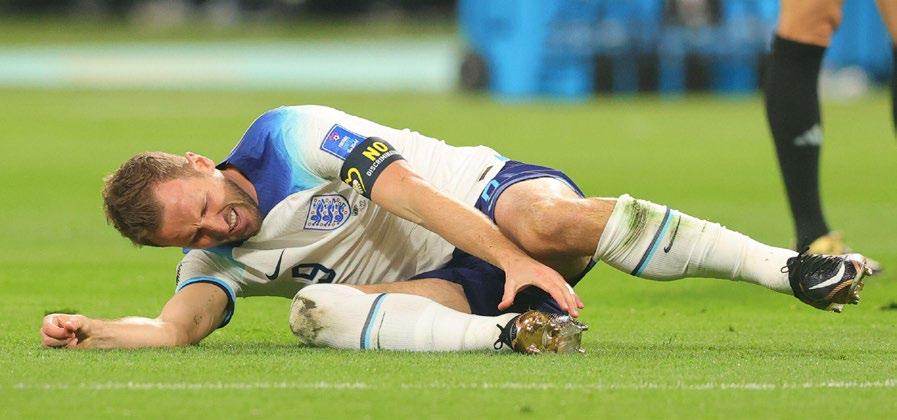
Acute ankle sprains are one of the most commonly incurred musculoskeletal injuries in team sport athletes1. The Football Association Medical Research Programme conducted an audit of injuries in professional football across two seasons7. They found that ankle ligament sprains accounted for 11% of total injuries in football, with 77% of sprains involving the lateral ligament complex. Ankle inversion, with the foot in various degrees of plantar flexion, is the most common mechanism for Ankle Sprains 3 .
Mechanical stability of the ankle joint is provided by ligament integrity, while functional stability is maintained by ankle proprioception provided by muscles, tendons, ligaments, and capsular innervation3 . Acute ankle sprains occur when there is an overreliance on the ankle’s mechanical stability, due to the shortcomings of the ankle’s functional stability.
Acute ankle sprains have the highest reinjury rate of all lower limb musculoskeletal injuries4. Athletes who suffer acute ankle sprains have a twofold increase in reinjury risk in the year after the initial injury5 . Reinjury of the ankle is just one of the many symptoms that defines Chronic Ankle Instability (CIA)6. Other symptoms include pain, persistent swelling, feelings of instability, “giving way” of the ankle and reduced functional capacity.
In footballers, acute ankle sprains can occur in both contact and non-contact scenarios, with the frequency influenced by playing position. Contact mechanisms (59%) were more common than non-contact mechanisms (39%) in outfield players, while the opposite was true for goal keepers where non-contact were more common than contact (79% vs. 21%, respectively)7 .
When looking at the most common mechanisms for contact injuries, tackling (36%) carried a greater risk than being tackled (18%). Alternatively, when analysing non-contact mechanisms, 77% of non-contact sprains were caused during landing, twisting, turning, and running. As practitioners, this information provides a great insight into areas that can be focused on to reduce the risk of both contact and non-contact acute ankle sprains. Tackling technique and landing mechanics, as well as running and change of direction mechanics should be incorporated into athletic development programs to reduce the initial injury risk and subsequent reinjury risk.
While 83% of ankle sprains had a rehabilitation period of less than one month, the research suggests that most acute ankle sprains are not severe. However, due to the nature of the fixture list in football – particularly in the 2022/23 EFL Championship Season for example – there is a potential for a player in the starting 11 to miss 3 fixtures in 7-days and up to 9 fixtures in one-month which can be detrimental to a team’s standings in the league at the end of the season.
As practitioners it is important to address the high incidence and subsequent re-injury rates of acute ankle sprains and appropriately return athletes to full performance post-injury. To do this, a Return to Sport framework should be defined, and subsequently adapted, to maximise specificity for footballers. Similarly, by applying elements of this framework, and implementing them into a team’s athletic performance program, the initial injury risk can be reduced in the entire team.
Mechanosensory Impairments
Mechanosensory impairments are the dysfunction in the transduction of mechanical stimuli into neural signals. As practitioners, it is important to identify where these impairments are post-injury. To do this the qualities that are affected must be defined before they can be assessed.

Over-Pronation
By assessing an athlete along the forcevelocity continuum, prior to assessing whether there is any apprehension in more football specific movements, a short- to long-term rehabilitation plan can be created to address the athlete holistically. This approach ensures the player isn’t rushed back by strapping their ankle or putting them on a course of anti-inflammatory medication, without addressing the underlying mechanosensory impairments. A robust testing strategy ultimately helps to reduce the risk of players developing chronic ankle instability.
So, what should be assessed?
In essence, it must be identified whether a player can: 1. Produce high levels of force 2. Produce high levels of force quickly 3. Produce high levels of force quickly in specific environments
The goal is to return a player’s injured ankle to within 10% difference of the uninjured
Neutral Over-Supination
side, or to baseline levels if the data is available. In addition to the quantitative data, an athlete’s qualitative symmetry should be assessed by analysing movement patterns across the controlchaos continuum8 .
Outlined below are some tests that assess the aforementioned areas. These can be used to identify any asymmetries caused by the mechanosensory impairments after an acute ankle sprain.
1. Producing Force
The purpose of the following assessments are to measure an athletes peak force and Rate of force development capabilities. This is the foundation of the functional stability discussed earlier.
In order to provide a holistic picture of where an athlete is at after an acute ankle sprain, the key muscles around the ankle must be assessed. The position of the foot during force production must also be noted – reducing over-pronation or oversupination. This ensures the ankle is in the most mechanically advantageous position.
The calf muscles – the soleus and the gastrocnemius – are the two largest muscles in the lower leg. These muscles work most in a flexed and extended knee position, respectively. Their strength qualities should be tested accordingly. The two easiest ways to assess the strength of these muscles is through the use of Isokinetic Dynamometry (IKD) to assess isotonic strength or by using force plates to assess isometric strength.
Three common strength tests used are outlined below.
In addition to assessing the calf muscles, it is also important to assess an athlete’s eversion strength. The athlete can be tested in both a neutral foot position, as well as in a plantarflexed and inverted position, to replicate a position specific to the injury mechanism.
Table 1: Plantarflexion and Dorsiflexion Strength Assessments
Warm Up Reps
Sets X Reps
Target
Prone Ankle IKD @ 30deg/sec Standing Calf Isometric
5 (60-100%) 2 (80 & 90%)
2 X 5 1 X 3 (5sec PUSH)
Seated Calf Isometric
2 (80 & 90%)
1 X 3 (5sec PUSH)
180-200% (Peak Torque/BW) 2.5 X Bodyweight (N/kg) 1.5 X Bodyweight (N/kg)
Table 2: Eversion Strength Assessment
Warm Up Reps
Sets X Reps
Target
Eversion Isometric
2 (80 & 90%)
1 X 3 (5sec PUSH)
<10% difference compared to non-injured limb
2. Producing Force Quickly
Once an athlete is capable of producing high levels of force, the athlete’s explosive and reactive strength should be assessed as these qualities will be more applicable to the on-pitch demands during football training and games.
In addition to returning players to 90% limb symmetry in both explosive and reactive strength, it is also important to gradually reintroduce frontal plane plyometrics too. As previously discussed, 77% of noncontact sprains were caused during landing, twisting, turning, and running. Before reintroducing twisting and turning through both planned change of direction work and reactive agility work, ensuring athletes can both absorb and produce force in the sagittal plane, but most importantly – the frontal plane, due to the mechanism of the injury – should be assessed. In the early stage of rehab, it is advantageous to expose players to sagittal plane hop and stick exercises before progressing to frontal plane variations. Initially, alternate leg variations can be used before progressing to unilateral variations.

In order to work the full SSC continuum, it is important to utilise both explosive strength through single jumps for height/distance while challenging the landing mechanics, however it is also important to work reactive strength in multiple planes too. By introducing repeated hops with a focus on reducing contact time, an athlete can be progressed towards more specific movements that they will be exposed to during training and matches.
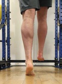
Finally, by adding both visual and aural stimuli to replicate the psychological and ecological demands of the game, all bases can be covered to ensure a successful return to performance. Image 2: Banded Single Leg Calf Raise – Peroneal Bias
Table 3: Explosive and Reactive Strength Assessments
Warm Up Reps
Sets X Reps
Target
Single Leg CMJ
2 each leg (80 & 90%)
1 X 3 each leg
>50% of Double Leg CMJ <10% difference compared to non-injured limb
Single Leg DJ
2 each leg (80 & 90%)
1 X 3 each leg
> 0.8 Reactive Strength Index <250ms Contact Time >20cm Jump Height <10% difference compared to non-injured limb
Repeated Hop
10 (80-90%)
2 X 10 each leg
<250ms Contact Times <10% difference in RSI compared to non-injured limb
Table 4: Reactive Strength Progressions
Reactive Strength
Level 1
Level 2
Level 3
Level 4
Level 1 Level 2 Level 3
Linear Ankling Lateral Ankling Extensive Skater Hops (Focus – Reduce Contact Time)
Linear Penguin Taps Lateral Penguin Taps Intensive Skater Hops
Linear Double Leg Pogos Lateral Double Leg Pogos Repeated Double Leg Hurdle Hops (Medial & Lateral)
Linear Single Leg Pogos Medial/Lateral Single Leg Pogos Repeated Single Leg Hurdle Hops
Table 5: Explosive Strength Progressions
Explosive Strength
Level 1
Level 2
Level 3
Level 4
Level 1 Level 2 Level 3
Linear Alternate Leg Hop and Stick Lateral Alternate Leg Hop and Stick Multidirectional Leg Hop and Stick
Linear Alternate Leg Hurdle Hop and Stick Lateral Alternate Leg Hurdle Hop and Stick Multidirectional Leg Hop and Stick
Linear Single Leg Hop and Stick Lateral Single Leg Hop and Stick Alternate Leg Hop and Stick with Rotation
Linear Single Leg Hurdle Hop and Stick Lateral Single Leg Hurdle Hop and Stick Single Leg Hop and Stick with Rotation
3. Producing Force Quickly in Specific Environments
Once players have sufficient force production capabilities and both explosive and reactive strength capacities, their ability to utilise these qualities in football specific environments should be assessed.
Typically, with acute ankle sprains, once an athlete has achieved 85% limb symmetry on their single leg drop jumps and repeated hops, they can return to linear running. Once an athlete returns to linear running they can gradually return to match and supra-maximal match demands of both High-Speed Running Distance (>55% of Player’s Max Speed) and Sprint Distance (>85% of Player’s Max Speed). By exposing players to these running zones the risk of hamstring injuries upon their return to both training and matches can be mitigated.
More importantly however, when planning the end stage rehab for football players, is replicating both the positional demands of planned change of direction and reactive agility that each player is exposed to during a game. By gradually reintroducing both linear and multidirectional jumping into the program early on in the rehab, a player’s ability to absorb and produce force in multiple planes is sufficiently improved. This lays the foundation for more specific movements on the pitch.
Alongside the qualitative analysis of landing and change of direction mechanics, where a player’s ability to land in a hip dominant landing pattern is assessed, prior to exposing them to the more chaotic movements experienced in games, the number of accelerations – and particularly decelerations – that they must do in a game must also be quantified. These numbers will typically be dictated by both position and team tactical demands. When looking at positions, typically, a centre-half will typically make more intense (>3metres/second/second) accelerations and decelerations than a striker. Alternatively, a team that employs a high-press strategy will be required to complete more intense accelerations and decelerations than teams that prefer to utilise a low-block for example.
Thankfully, with the availability of GPS units, an individual player’s typical game demands can be quantified – particularly focusing on both the count of accelerations and decelerations, as well as the distance covered during both of these movements. The latter is typically referred to as Explosive Distance by GPS companies (distance covered during accelerations and decelerations >3metres/ second/second). The intensity of these movements can be assessed by relating back to a per minute value, ie. Accelerations and Decelerations per minute. Finally, including a measure of density can also be utilised where the count of these accelerations and decelerations is measured as a distance per effort value.
The above GPS metrics can be influenced by pitch area and drill time. Similarly, they can be made more complex by adding players in. Due to the 360-degree nature of football, a player can be progressed through a box or rondo drill by starting them as an outside player where they are required to make lateral movements while challenging their passing accuracy, before progressing them to being an inside player where they are required to work both in possession and out of possession. The same principle can be applied to small sided games where the complexity of the drill is increased in line with the player’s return to performance plan.
To ensure specific movements are incorporated into the end-stage rehab, the Performance Analysis team can also be utilised if they are available to us, to look at movements that the player is expected to undertake during the three moments of football games – attacking, defending and transitioning. This ensures the multidisciplinary team is utilised in the rehab process. This ensures all the player’s bases are covered and they can return fully to performance, while reducing the risk of the player experiencing reoccurrences and subsequently chronic ankle instability.

Conclusion
1. Can the player produce high levels of force? • Ensure the player is strong and symmetrical in plantar flexion with both a flexed and extended knee, in addition to eversion in both a neutral and plantarflexed foot position.
2. Can the player produce high levels of force quickly? • Assess and identify any deficits in both single leg explosive strength and reactive strength by analysing single leg countermovement jumps, drop jumps, and repeated hop tests.
3. Can the player produce high levels of force quickly in specific environments? • Progress from planned change of direction drills to reactive agility drills in both attacking, transitioning and defending movements as appropriate.
1. Gribble PA, Bleakley CM, Caulfield BM, et al. Evidence review for the 2016 International Ankle Consortium consensus statement on the prevalence, impact and long-term consequences of lateral ankle sprains. Br J Sports Med 2016;50:1496–505. 2. Nery, C., Raduan, F., & Baumfeld, D. (2016). Foot and ankle injuries in professional soccer players: diagnosis, treatment, and expectations. Foot and ankle clinics, 21(2), 391-403. 3. Hintermann, B. (1999). Biomechanics of the unstable ankle joint and clinical implications. Medicine and science in sports and exercise, 31(7 Suppl), S459-69. 4. Anandacoomarasamy A, Barnsley L. Long term outcomes of inversion ankle injuries. Br J Sports Med 2005;39:e14. 5. Verhagen EA, van Tulder M, van der Beek AJ, et al. An economic evaluation of a proprioceptive balance board training programme for the prevention of ankle sprains in volleyball. Br J Sports Med 2005;39:111–5. 6. Docherty CL, McLeod TC, Shultz SJ. Postural control deficits in participants with functional ankle instability as measured by the balance error scoring system. Clinical journal of sport medicine. 2006 May 1;16(3):203-8. 7. Woods, C., Hawkins, R., Hulse, M., & Hodson, A. (2003). The Football Association Medical Research Programme: an audit of injuries in professional football: an analysis of ankle sprains. British journal of sports medicine, 37(3), 233-238. 8. Taberner, M., Allen, T., & Cohen, D. D. (2019). Progressing rehabilitation after injury: consider the ‘control-chaos continuum’. British journal of sports medicine, 53(18), 1132-1136.