1 minute read
Correlating SEM and AFM In-Situ
By Dr Cameron Chai and Dr. Kamran Khajehpour
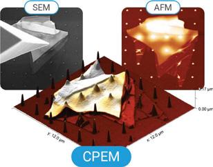
Correlating datasets from complimentary analytical/ imaging techniques is growing in popularity as the combined datasets result in a deeper understanding of the materials being investigated. Being able to collect these individual datasets in-situ saves time, and eliminates the possibility of changes such as oxidation, as all measurements are taken under the same experimental conditions at the same time. Scanning Electron Microscopy (SEM) and Atomic Force Microscopy (AFM) are two such complimentary techniques that allow us to probe materials down to the nanoscale. SEM offers high resolution 2D imaging with rapid sample navigation that can be augmented with chemical analysis using EDS or WDS (Energy Dispersive and Wavelength Dispersive Spectroscopy). AFM adds atomic resolution imaging capabilities as well as the ability to translate these images into 3D. Through a range of different modes, AFM uniquely affords the user the ability to measure mechanical, magnetic and electrical properties, all of which can be correlated with structural data. Adding all these complimentary datasets makes highly detailed nanostructural studies possible. Nenovision has made these studies a reality with the development of LiteScope, a compact AFM designed for use in your SEM, using a process called Correlative Probe and Electron Microscopy or CPEM. The clever system scans your sample, simultaneously using both techniques, with a known offset between the AFM tip and SEM focal spot. By subtracting this offset from the two datasets, they can be precisely overlaid providing improved insights into the structure and properties of your materials. LiteScope is compatible with most existing SEMs, and can be easily installed in a matter of minutes. The software allows SEM and AFM channels to be selected, viewed, and recorded at the same time.
CPEM enables sample analysis in a way that was difficult, or impossible, using SEM and AFM modes individually. By marrying the two techniques resulting in advanced correlative imaging new possibilities become a reality in fields such as materials science, nanotechnology, semiconductors, life sciences and more.


