


If you had your time again—today—would you still choose medicine?

It’s interesting to note that even back in the olden times—say, the 1990s—the path to becoming a doctor was pretty much consistent, with the same established social contract. Study hard at high school, get top grades, gain admission to medical school and, if you make it all the way to the end, earn the title ‘Doctor.’
In Britain at least, even if you had to have another best part of a decade to work ridiculously long hours as a ‘junior’ doctor, the prize at the end of the process was a consultant-level position—with power, money and status. You could drive an Aston Martin and live in a splendid home.
People would listen to your judgment, trust it, and... also bring passport applications to be countersigned by you. You, like judges, company directors or members of parliament, were the right kind of person for this important task.
Today, however…Man, that's a tougher decision. The pay, relative to the good ol' days, is lower—yet the demand for your services (mostly thanks to baby boomers) has never been higher. (Aside: I don't know how this can square with basic economic theory about supply and demand, but I suspect political lobbying by health insurers is involved somewhere.)
You're not respected nearly enough for the work you've put in to become a doctor—thanks to the prevalence of unverified content on the Internet.
If a patient decides they don't want surgery for that early and treatable pancreatic tumor, and instead pursue a macrobiotic vegan diet for a few months (the tragic Steve Jobs gambit), they can find a forum or a Telegram group that will help them believe that.
Others won't believe your intervention is effective and will want to argue the toss— all while the waiting room gets ever more crowded. D'oh, the humanity!
Then there are the glorified Markov models-in-a-box coming for your jobs. Who would enter radiology as a specialty today, when companies are heralding the arrival of software that reads the patient's history, records the conversation with the patient, picks the imaging protocols, reads the images, writes the report, and sends them to referring physicians?
At this point, you're probably better off training to be the technician that runs the instruments, as surely that's going
to be better paid than the ‘person who talks to the patient.’ Assuming the patient doesn't just talk to a screen with a doctor’s avatar.
Given that medical retina shares many similarities with eye radiology, if an optical coherence tomography (OCT) that can automatically adjust the chinrest and center on the eye is ever developed—we’re all in trouble.
In refractive surgery, the value of experienced surgeons lies in their ability to interpret topography maps and other data, using their knowledge and judgment to plan the best laser ablation possible. However, if the artificial intelligence (AI)-powered nomograms become sufficiently refined and predictive, then the value of this experience could diminish, too.
Did you know they're developing cataract surgery robots, too?
My son is almost 14 years old. He's now at the point where, when given homework from school, he either does it and learns the lessons intended—or uses ChatGPT. Or doesn’t do it at all. From a societal perspective, this is profoundly concerning.
What will the future pipeline of medical students look like soon? Still predominantly the academic elite, but how many of them will there be? Are the young ophthalmologists (YOs) of today among the last to learn medicine the... human way? Who can operate without Silicon support?
Naturally, I work with some YOs, so I asked one of them about her take on this. Her response: We won't be replaced for many years yet, as all of these technologies will require human validation and supervision. In the meantime, they may even make our work easier, too.
Unlike myself, she doesn't perceive this as an existential threat to the future of medicine. With every fiber of my being, I hope she's right.

Mark Hillen, PhD Director of Communications ELZA Institute, Zurich, Switzerland Editor-At-Large | CAKE
The Lean, Green Sophi Phaco Machine
From Nightmare to Dream Vision
Transforming impossible vision cases into dream outcomes with artistic precision and a patient-first philosophy
Eyeing the Options


A Vision Realized Appasamy Associates unveils new HQ and international expansion
Rethinking Refraction
Research shows prioritizing 'functional vision' and slight myopia with monofocal IOLs improves daily life for cataract patients
A Foldable Fix for Glaucoma?
Ophthalmologists share practical advice for young surgeons on navigating combined cataract and glaucoma procedures 22
A novel glaucoma device promises to revolutionize surgical interventions with its minimally invasive technique
The Topography Revolution
Bridging AI and Eye Care
A pioneer in biomedical informatics, Dr. Sally Baxter seamlessly integrates ophthalmology with Big Data analytics to reshape patient care
A new topography-guided laser vision correction system is set to redefine refractive surgery
Next-Gen Eye Defenders
Armed with AI, MIGS and digital training, a new generation of ophthalmologists is transforming glaucoma care

Tech Takes on Glaucoma
A new wave of technological innovation brings new hope to the fight against glaucoma


The Crux of Glaucoma Disparities Experts reveal stark disparities in glaucoma care and propose innovative solutions for equitable vision for all

Dr. Harvey S. Uy
University of the Philippines; Peregrine Eye and Laser Institute, Manila, Philippines harveyuy@gmail.com

Dr. William B. Trattler
Center For Excellence In Eye Care Miami, Florida, USA
wtrattler@gmail.com
Prof. Burkhard Dick
University Eye Hospital Bochum Bochum, Germany
burkhard.Dick@kk-bochum.de

Dr. Francis Mah
Scripps Clinic Medical Group La Jolla, California, USA
Mah.Francis@scrippshealth.org

Dr. Cathleen McCabe
The Eye Associates Sarasota, Florida, USA
cmccabe13@hotmail.com
Prof. Dr. Sorcha Ní Dhubhghaill Brussels University Hospital (UZ Brussel) Brussels, Belgium
nidhubhs@gmail.com

Matt Young CEO & Publisher
Gloria D. Gamat Chief Editor
Mapet Poso Editor
Matt Herman Associate Editor
Maricel Salvador Graphic Designer
Writers
April Ingram
Chow Ee-Tan
Diana Truong
Hazlin Hassan
Tan Sher Lynn
Contributors
Dr. Arun Gulani
Dr. Johannes Weisensee
Dr. Fathi Nouira
Hannah Nguyen COO
Travis Plage CFO
Ruchi Ranga Society Relations & Conference Manager
International Business Development
Brandon Winkeler
Robert Anderson
Sven Mehlitz


by Diana Truong
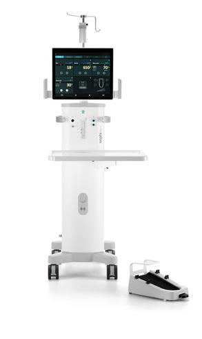
In the heart of the Philippines, The Medical City (TMC) is pioneering a greener approach to cataract surgery. By adopting Sophi (Rayner; Worthing, UK), a phacoemulsification system designed for sustainability and efficiency, the hospital has drastically cut its operating room waste while setting a new benchmark for OR productivity.

Eye care is in a race against time—not just to save sight, but to save the planet. Operating rooms (ORs), while essential, are among the most resource-intensive parts of any hospital, producing staggering amounts of waste.
At the forefront of this challenge is cataract surgery, where traditional phacoemulsification machines with single-use cassettes have long been the norm. Each surgery leaves behind piles of discarded plastic and packaging, adding up to an enormous environmental footprint.
Enter Sophi, a phaco machine designed with sustainability at its core. Unlike conventional systems, Sophi replaces disposable cassettes with “day-pack” alternatives, drastically reducing waste. But its impact doesn’t end with the environment—it’s also a gamechanger for OR efficiency.
At TMC, the shift to Sophi is transforming the way cataract surgeries are performed. Beyond slashing waste, the hospital has seen smoother workflows, quicker turnarounds and a renewed sense of purpose among its surgical team. Could this be the model for the sustainable future the eye care industry so desperately needs? Let’s take a closer look.
Operating rooms are often described as the beating heart of a hospital, but their pulse comes with an environmental cost. For cataract surgeries, conventional phacoemulsification machines with single-use cassettes are a major culprit.
“The prescribed practice… is to change cassettes, tubings and hand pieces with each case,” said Dr. Victor Caparas, chair of TMC’s
ophthalmology department. “With multiple cases day after day, week after week, this will pile up and create a tremendous amount of waste.”
The numbers back this up. A 2024 observational study conducted in Belgium revealed that machines using single-use cassettes produced 306.7 kilograms more plastic waste per 1,000 surgeries than their multiuse counterparts—a 75.3% increase. The environmental toll didn’t stop there; these systems required 67.7% more storage space, translating into higher transport emissions.*
This troubling reality inspired Dr. Isabela Bondoc, a resident at TMC, to delve deeper. “What inspired me to conduct this research was being in the OR every day,” she shared. “I saw the significant amount of single-use items used in the operating room to treat our patients.”
Dr. Bondoc’s study is rigorous, tracking power consumption, staff travel, pharmaceutical use and surgical waste. Her preliminary findings reveal a startling truth: in November, TMC performed 240 phacoemulsification cases, each generating waste equivalent to 3.4 kilograms of CO2 emission. In total, phaco surgeries produced 816 kilograms of CO2 equivalent of waste in that month alone. “To put this into perspective, it would take 656 square meters of forest or 82 medium to large-sized trees one year to absorb this amount,” she explained.
In the bustling operating rooms of TMC, where cataract surgeries dominate the surgical calendar, the need for innovation was clear. For many years, the hospital relied on conventional phacoemulsification machines—functional but far from sustainable. When the time came to choose a new system, Sophi emerged as the clear winner, meeting the hospital's twin goals of environmental responsibility and operational efficiency.
“Cataract surgery is by far the most performed procedure [here],” explained Dr. Caparas. “Whatever can be done in reducing waste, as the Sophi system does, would definitely be in line with TMC’s vision.” The decision wasn’t solely about cutting
back on waste; Sophi’s unique design promised a smoother, more efficient workflow, an essential feature in the fast-paced environment of TMC’s operating rooms.
Conventional machines require disposable cassettes and timeconsuming priming before each procedure. Sophi’s day-pack system, by contrast, eliminates much of the hassle. The Belgium study highlighted a seven-minute priming advantage for every 10 procedures with day-pack systems, a small but impactful time-saving that adds up in busy ORs.1
The system’s innovative design also drew praise from TMC surgeons. “You have [its] ergonomic design, its customization option and a very sensitive response,” noted TMC surgeon Dr. Abbey De Guia. “These are the major advantages… especially when it comes to both user experience and surgical precision.”
In TMC’s operating rooms, the introduction of Sophi has sparked a quiet revolution—not just in reducing waste, but in redefining efficiency and enhancing patient care. For senior residents like Dr. Michelle Talusan and Dr. Rexelle Piad, the day-to-day benefits of Sophi are palpable.
“The learning curve with Sophi has been surprisingly smooth,” shared Dr. Talusan. “It has an interface that caters to various levels of surgical experience…The transition was much less daunting than we anticipated.”
Efficiency gains have been a standout feature, with Sophi’s lean cassette system leading the charge. “The process of loading the cassette is intuitive and hassle-free,” explained Dr. Piad. The cassette slips into the machine with ease and…the machine has a lot of fail-safe mechanisms that ensure correct loading each time.” This streamlined process has translated to fewer interruptions and quicker turnaround times in the OR.
Sophi is equipped with a high-power battery that can handle up to 20 surgeries a day, has also won praise for its design and functionality. “It’s free from any disturbing cables or tubes, making it very flexible for surgeons during surgery,” noted Dr.
Piad. “The wireless dual-linear foot pedal showcases a lot of switches, and those switches are very customizable.”
The system’s surgical performance is equally impressive. “Sophi’s IOP control pump maintains stable intraocular pressure throughout the procedure,” said Dr. Talusan. “This is significant for me as a beginning surgeon since it allows me to focus on my technique.”
Dr. De Guia emphasized how Sophi benefits both surgeons and patients, particularly in challenging cataract cases. “Its advanced features, such as finely tuned power modulation, enhanced vacuum control and realtime feedback, allow for precise management of difficult procedures while minimizing risks,” she explained.
She also highlighted Sophi’s innovative safety measures. “The Clean Venturi Pump has a special patented ceiling foil,” Dr. De Guia noted. “Patient liquids are safely sealed between a foil and a cassette, preventing them from escaping into the environment.” This design adds an extra layer of protection against contamination, ensuring a safer surgical experience.
environmental consciousness at the forefront.
“Minimizing medical waste from surgical procedures without compromising patient care or outcomes would significantly enhance the long-term sustainability of the healthcare system,” Dr. Bondoc emphasized. “Such efforts would also encourage policymakers to advocate for more sustainable methods in conducting surgery, benefiting not only our patients but the environment as well.”
The financial implications are equally compelling. “Increasing efficiency, lowering costs and reducing waste all translate into savings for both the hospital and the patient.” noted Dr. Caparas. These savings are particularly critical in highvolume surgical settings like TMC, where resource optimization directly impacts operational budgets.
Sophi’s success at TMC offers a blueprint for other institutions striving to align with global environmental and healthcare goals. It challenges the industry to think differently about sustainability—beyond compliance, toward proactive leadership in environmental stewardship.
The story of Sophi at TMC extends beyond its walls, illustrating the transformative potential of sustainable innovations in eye care. By proving that waste reduction and efficiency can coexist, Sophi paves the way for hospitals in the Philippines—and globally— to reimagine surgical care with
*Kallay O, Sadad R, Zafzafi A, et al. Cataract surgery and environmental sustainability: A comparative analysis of single-use versus reusable cassettes in phacoemulsification. BM J Open Ophthalmol. 2024;9(1):ee001617.
A version of this article was first published on cakemagazine.org
Disclaimer: Environmental, Social, and Governance (ESG) factors are complex and subject to interpretation. The information provided here is based on available data and industry standards at the time of publication. It is important to note that ESG ratings and metrics are estimates and may not accurately reflect future performance. For more information, please refer to Rayner’s ESG Commitment Statement available at rayner. com.
Rayner and Sophi are proprietary marks of Rayner, 10 Dominion Way, Worthing, West Sussex, BN14 8AQ. Registered in England: 615539. © 2025 Rayner Group, all rights reserved.


by Dr. Arun Gulani
More than just science, cataract surgery is an art. I’ve spent decades honing my Gulani KLEAR™ System, a blend of cutting-edge technology and artistic surgical skill, to tackle the most challenging vision cases. My approach prioritizes minimally invasive techniques and meticulous planning, ensuring patients achieve not just clear vision, but their best possible vision.
Every day in my practice, I get referrals from around the world—patients who have often been told they’re ‘impossible’ cases, ‘not candidates’ for surgery, or just plain ‘nightmares’ to deal with.
For more than three decades, I have helped these patients by not only
providing hope but also delivering results—unaided visual outcomes. This allowed them to return to their productive lives and professions. To achieve this, I have developed the Gulani KLEAR™ System—a full spectrum of Kerato-LenticuloRefractive techniques—and integrated cutting-edge technology. This approach overcomes surgical
challenges and turns complex cases into visual successes, using minimally invasive surgical artistry, all of which is visually oriented.
This mindset and approach embody my philosophy, Gulanism: “A doctor’s inability should not result in a patient’s disability.”
“A doctor’s inability should not result in a patient’s disability.”
It is disheartening to see surgical demonstrations at conferences that prioritize surgical acrobatics over patient outcomes, often showcasing gory displays of blood, vitreous and stitches. These presentations usually end with the same statement, “patient did well.” Most of these patients come to me extremely disappointed, visually disabled and looking for help.
My mindset, coupled with meticulous planning and surgical artistry, allows me to transform ‘vision nightmares’ into success stories, often with single or staged techniques. This isn’t just a surgical philosophy—it’s a paradigm mindset shift, and one that
I’m passionate about sharing with colleagues in the industry.
When I see patients from countries far and wide—often after enduring years of rejections, failed surgeries and declining hope—I see more than just their charts, topography and optical coherence tomography (OCT). I see their potential.
Each case is an opportunity to push the boundaries of what’s possible in vision correction. My approach is about raising the bar for our profession and redefining how surgeons think—shifting from surgical acrobatics to artistic precision with actual vision outcomes.
Besides a mindset shift, this also requires a detailed and in-depth understanding of refractive surgery and visual optics, corneal and lenticular anatomy, and surgical skills that involve the cornea, anterior segment and lens
Mastering surgical skills and technologies is akin to building with Lego pieces or composing musical minuets. Approaches can be assembled forward, backward, sideways or in combination to correct complex cases—using what I call the Gulani Planning System (GPS) to achieve unaided vision endpoints.
To illustrate this mindset, and to inspire colleagues in the industry— as documented in thousands of patients’ journeys on our YouTube channel—I will share two prototype cases: One demonstrating a ‘forward’ approach and another a ‘backward’ approach to correcting poor outcomes.
This case involved a 75-year-old nurse who had undergone hexagonal
keratotomy (Hex K) decades ago for hyperopia. This outdated procedure left her with severe corneal instability, high irregular astigmatism and a host of other complications. She came to me with vision reduced to counting fingers, a staggering 23.50 D of irregular astigmatism, 89.90 D keratometry, and a history of being dismissed by other surgeons as ‘untreatable.
For me, cases like these are not challenges; they are opportunities to demonstrate what’s possible with the right mindset.
My philosophy: My work always begins with the GPS. I first define the target—the patient’s ‘unaided’ vision potential—before deciding on the technique and technology. Every step of the surgery is meticulously planned to maximize vision while using the least interventional techniques.
How I Solved This Case: The solution here involved two brief, topical stages:
1. Step 1: Stabilizing the Cornea I performed a no-stitch INTACS procedure to stabilize the cornea. This reduced the keratometry and astigmatism from 23.50 D to 1.4 D while maintaining corneal integrity despite Hex K cuts and Fuchs’ dystrophy. I call this step making the cornea ‘sensible’ or ‘measurable,’ which lays the foundation for future intraocular lens (IOP) precision.
2. Step 2: Cataract Surgery with Precision
Once the cornea was stable, I performed LenzOplastique®based cataract surgery with a toric IOL implant to correct residual astigmatism. I ensured that every optical element was perfectly aligned—like ‘pins before
bowling’—to achieve a ‘strike’ in visual outcomes.
The Outcome: This patient’s life changed in ways words can’t describe. For the first time in 60 years, she could see her face in the mirror. She returned to her life’s passion of caring for others. My mantra has always been, ‘Once your mind is decided, your hands follow.’ That’s exactly what happened here.
Another prototype case involved a 75-year-old male patient who came to me after unsuccessful premium cataract surgery and lost his pilot’s license. His case had resulted in hyperopia, presbyopia, anterior corneal scarring and high keratometry from keratoconus, compounded by a YAG capsulotomy. His vision was 20/200, and he had been told by multiple surgeons that the only option was an invasive lens implant exchange with vitrectomy.
Why I Took a Different Approach: I believe that complex cases demand innovative thinking, not more invasive surgery. Using my 5S system, I developed a staged solution that prioritized functional vision over anatomical fixes. So in this case, the single surgery that would address all these issues of anterior corneal scarring and high keratometry along with emmetropia would be myopic surface laser surgery.
The question then was how do I turn this hyperopic patient into a myopic patient and then proceed with that single, least interventional surgery?
1. Step 1: Inducing Myopic Astigmatism
I implanted a piggyback lens on the existing pseudophakic lens.


This altered the eye’s refractive state from hyperopia to myopic astigmatism, setting the stage for my planned myopic laser surgery.
2. Step 2: Surface Laser Surgery
After confirming stability, I performed LaZrPlastique® laser surgery. This refractive laser technique flattened the corneal center, reduced keratometry, removed the scar and increased the optical zone while bringing the patient to emmetropia.
The Outcome: This patient’s vision progressed to unaided 20/15 and he resumed his pilot’s license. Seeing his reaction to achieving a perfect vision was incredibly rewarding. Cases like this reinforce why I’m committed to thinking differently. By carefully staging the procedures, I addressed all impacting factors while avoiding invasive techniques. This patient’s journey from hopelessness to clarity exemplifies my belief that every complex case deserves a tailored, least interventional approach.
From ‘midzone’ to ‘endzone’
The above two prototype cases are examples of those I have helped using this mindset. However, in many referred cases, I must point out that achieving their ‘dream vision’ from the so-called ‘nightmare vision’ was just a simple step. In fact, I believe their surgeons had actually done a good job, but left the outcome ‘midzone.’ All I needed to do was guide these patients to the vision ‘endzone’ and ‘claim the trophy.’
Among the most common cases I encountered involved surgeons
who celebrated technical surgical success, like clear corneal grafts, symmetric stitches or topographical changes, but completely neglected the patients’ request for unaided vision endpoints.
These cases often involve keratoconus patients who have undergone successful INTACS, CAIRS or other intracorneal implants, DALK or penetrating corneal transplants.
These surgeons did not perform bad or incorrect surgery. However, had they only believed in their own abilities and the patients’ potential for unaided vision, they would have experienced a greater sense of achievement. Not to mention extremely happy patients.
reactions and life changes after their vision restorative work.
This will then inspire every eye surgeon to perform for that relentless goal, turning into surgical artists in the process. Not only will so-called ‘routine’ patients achieve beyond 20/20 vision endpoints, but those that are discarded and neglected as ‘impossible’ and ‘nightmares’ will truly achieve their ‘dream’ vision.

These cases are more than just surgical successes; they represent a new way of thinking about vision correction. My Vision a La Carte, ‘3T’ concept—“Target first, Technique second, Technology last”—is at the core of my philosophy.
“Target first, Technique second, Technology last.”
I'm confident that all our colleagues can achieve this level of surgical expertise and technological adaptation if only they would change their mindset from presenting chart data and colorful topographies to actual unaided visual endpoints. And more importantly, show the patient's
Dr. Arun C. Gulani is a world-renowned LASIK, cataract, and corneal surgeon. He performs the entire spectrum of advanced vision surgeries to reduce dependence on glasses and contacts, customising vision correction surgery to meet each patient’s unique goals. He has extensive experience in a wide variety of eye surgery techniques and technology. He was formerly the Chief of Cornea and Assistant Professor of Ophthalmology in the University of Florida’s School of Medicine before founding the Gulani Vision Institute in 2003, where he receives a global clientele and acts as a consultant to eye surgeons and the eye care industry as well. With an eye of an artist, his passion is to make people see; and with his no-hype, one-on-one personalized care, he has turned Jacksonville, Florida, into a vision destination for the world.
gulanivision@gulani.com
by Tan Sher Lynn
When cataracts and glaucoma collide, ophthalmologists face a crucial treatment dilemma: To combine or not to combine? The answer, as leading experts Dr. Chelvin Sng and Dr. Iftekher Iqbal shared, lies in a delicate balance of patient-specific factors, surgical expertise and a deeper understanding of potential complications.
The decision to perform combined cataract and glaucoma surgery is far from a simple one. It's a high-stakes balancing act, where surgical expertise, patient conditions and potential complications weigh heavily.
Dr. Chelvin Sng, adjunct associate professor at the National University of Singapore and the medical director of her clinical practice at Mount Elizabeth Novena Hospital in Singapore, and Dr. Iftekher Iqbal, a glaucoma specialist, MIGS pioneer and accomplished researcher from Bangladesh, shed light on the intricacies of this decision-making process.
From navigating intraoperative challenges to optimizing postoperative care, they offer invaluable insights for young ophthalmologists (YOs) seeking to master this complex field.
In deciding whether to perform combined or separate cataract and glaucoma surgery, Dr. Sng emphasized the importance of tailoring the approach based on the patient’s condition.
“If the patient has visually significant cataracts as well as glaucoma, I would be inclined to offer combined glaucoma and cataract surgery,” she said. “Phaco-minimally invasive glaucoma surgery (MIGS) would be appropriate for patients with mild-tomoderate and medically controlled glaucoma. For patients with medically uncontrolled advanced glaucoma, I often offer combined phaco-tube rather than phaco-trab, as cataract surgery may reduce the success rate of trabeculectomy.”
Dr. Iqbal echoed the individualized approach. “I would consider combined surgery in patients with advanced glaucoma under the following conditions: If the glaucoma is progressing or not controlled with maximum medications; in patients with systemic diseases, such as uncontrolled diabetes; those with increased health risks for multiple surgeries or limited resources for further intervention; and those with poor patient compliance with antiglaucoma medications, postoperative regimens or multiple

Alternatively, he would choose standalone cataract surgery for patients with mild to moderate glaucoma and well-controlled intraocular pressure (IOP) with medications and stable visual field changes
Surgical options: Phaco, trabs, MIGS or tubes
Surgical options for patients with coexisting cataracts and glaucoma depend on multiple factors.
“The choice depends on the stage of glaucoma,
Control IOP. If there are no safety concerns, add prostaglandin analogs or aqueous suppressants to maintain target IOP—if required.
Manage Inflammation. A gradual tapering of steroids (prednisolone 1%) over a month effectively controls inflammation most of the time. For patients prone to steroid-induced IOP elevation, we may need to use alternative anti-inflammatory agents or closely monitor them.
Enhance Visual Recovery. Adopt a conservative approach when selecting IOLs for glaucoma patients to ensure a more predictable visual outcome. Use extended depth of focus (EDOF) lenses over multifocal lenses to enhance visual outcomes without compromising contrast sensitivity. The choice of IOL is also influenced by whether the patient undergoes standalone cataract surgery or a combined procedure with trabeculectomy or MIGS.
Use Eye drops. Use postoperative steroid eye drops to reduce inflammation and minimize subconjunctival scarring for bleb-forming procedures, Conversely, some patients are steroid responders and a prolonged duration of topical steroids may increase intraocular pressure.
Monitor IOP. For glaucoma patients, especially those with advanced glaucoma, closely monitor IOP after surgery. For patients with end-stage glaucoma and at a high risk of wipeout, home monitoring of IOP with iCare tonometers may be advisable so that any increase in IOP can be promptly treated.
Prescribe Proper Medication. For bleb-forming procedures, prescribe oral non-steroidal anti-inflammatory drugs (NSAIDs), which can reduce the duration and frequency of topical steroids required. When the ocular inflammation is better controlled, consider switching to loteprednol, which is less likely to induce a steroid response.
IOP levels with more predictability than trabeculectomy.”
She further explained that factors that influence her choice include the severity of glaucoma, the target IOP, the number of pre-operative glaucoma medications, the health of the fellow eye, as well as financial considerations as implants may be costly for some patients.
According to Dr. Iqbal, a shallow anterior chamber is a common intraoperative challenge when performing combined cataract and glaucoma surgery, particularly in patients with primary angle-closure glaucoma (PACG). This makes various steps—from paracentesis to rhexis, nucleus management and IOL implantation—more difficult.
“This issue can be mitigated with preoperative mannitol infusion and adequate ocular massage when using peribulbar or retrobulbar anesthesia,” he noted.
target IOP, medication burden and disease progression. For mild to moderate glaucoma, I prefer phaco with MIGS for effective IOP control and quicker recovery,” Dr. Iqbal shared. “In advanced or uncontrolled cases, phaco-trabeculectomy offers more aggressive pressure reduction. If glaucoma is stable on minimal medication, phaco alone may suffice. Each decision is tailored to the patient’s needs and long-term outcomes.”
Meanwhile, Dr. Sng shared her preference for tube shunt implantation in advanced glaucoma cases. “I prefer phaco-tube rather than phaco-trab for patients with medically uncontrolled advanced glaucoma and a visually significant cataract. Cataract surgery is likely to reduce the success rate of trabeculectomy more than it does for tube implant surgery,” she said. “The Paul Glaucoma Implant (PGI) is my tube of choice, which can achieve low
“In
advanced or uncontrolled cases, phacotrabeculectomy offers more aggressive pressure reduction. If glaucoma is stable on minimal medication, phaco alone may suffice. Each decision is tailored to the patient’s needs and long-term outcomes.”
- Dr. Iftekher Iqbal
To prevent sudden decompression of the anterior chamber, Dr. Iqbal emphasized the importance of meticulous technique. “Performing slow paracentesis is crucial to avoid damaging the iris or lens capsule. It’s also important to avoid overfilling the anterior chamber with ocular viscoelastic devices (OVDs) and to proceed with phacoemulsification at a slower pace,” he added.
As for Dr. Sng, one of her common intraoperative challenges for angle surgery is obtaining a good view of the angle—especially if the device is implanted in the inferior quadrant. “The patient needs to be cooperative in moving their head and eye in the right direction to facilitate proper positioning,” she said.
A prerequisite for combined procedures
Before attempting combined procedures, Dr. Sng stressed the importance for young ophthalmologists to first be competent cataract surgeons.
“Glaucoma patients may have shallow anterior chambers (AC), small pupils, loose zonules and floppy
irises. These make cataract surgery rather challenging. Furthermore, cataract surgery complications affect the success of glaucoma surgery. So aim to get the cataract out safely! For angle surgeries, YOs should practice intraoperative gonioscopy with a direct goniolens before their first angle surgery,” she suggested.
“Patient selection is important for the best outcomes, so choose the most appropriate glaucoma surgery for each patient. Comprehensive preoperative counseling is required to ensure that the patient understands all the surgical options for glaucoma. It is also important to have realistic expectations of each surgery’s efficacy,” she added.
Meanwhile, Dr. Iqbal emphasized that mastering the combined procedures requires dedicated practice and mentorship, as the learning curve for combined procedures is significant. Engaging in hands-on training and seeking guidance from experienced surgeons can facilitate skill development.
and monitoring for potential complications like bleb failure or infection.
“When doing combined procedures, I suggest first creating a trabeculectomy flap without entering the AC, followed by either temporal or superior cataract surgery. I also recommend using different ports for cataract surgery and trabeculectomy, and using corneal traction sutures instead of holding the superior rectus muscle. Be careful when using antimetabolites like MMC or 5-FU and leave a little air in the AC to keep it from collapsing during suturing and prevent shallowing in the early postoperative period,” he further explained.

To improve their skills in combined cataract and trabeculectomy surgery, he advised YOs to focus on the following:
• Develop proficiency in creating a scleral flap, performing sclerostomy and ensuring proper conjunctival closure to facilitate effective aqueous humor drainage and IOP control.
• Pay meticulous attention to wound construction to prevent postoperative complications such as wound leaks or hypotony.
• Possess a detailed knowledge of anterior segment anatomy to navigate surgical planes accurately and avoid damage to adjacent structures.
• Cultivate the ability to make realtime decisions, such as adjusting the scleral flap or applying antifibrotic agents, to optimize surgical outcomes.
• Be prepared to manage postoperative care, including using anti-inflammatory medications
Dr. Chelvin Sng is an adjunct associate professor at the National University of Singapore and the medical director of her clinical practice at Mount Elizabeth Novena Hospital. She graduated from the University of Cambridge, UK, with triple first class honors and distinctions. Dr. Sng is the co-inventor of the Paul Glaucoma Implant, which is in clinical use in Europe, United Kingdom, Asia and Australia. She is the earliest surgeon in Asia to implant several novel MIGS devices, including the XEN Gel Implant and the iStent Inject. She has also published the earliest data on the iStent and the XEN Gel Implant in angle closure eyes. As the Convenor of the AsiaPacific Glaucoma Society (APGS) MIGS Interest Group, Dr. Sng has organized MIGS wetlabs and training courses for surgeons in the Asia-Pacific region. She is the co-editor of an open-access book on Minimally Invasive Glaucoma Surgery, which has more than 80,000 downloads worldwide. Dr. Sng was voted on the global Ophthalmologist Power Lists in 2017, 2021, 2022, 2023 and 2024. She has received international awards from ASCRS, AAO, ARVO and APAO.
chelvin@gmail.com
To illustrate his point, Dr. Iqbal shared a case study where he performed a routine combined trabeculectomy and cataract surgery on a 52-year-old lady with PACG. Upon entering the anterior chamber with a phaco probe, the patient's cataract immediately subluxated for approximately 3 to 9-clock hours under the flow of a balanced salt solution (BSS). However, no zonular weakness or subluxation was noticed during the capsulorhexis procedure. He had to put a Cionni ring to fix the capsular bag, place the single-piece intraocular lens within the capsular bag, and finish the surgery without further incidents.
“One of the critical learning points from this case was that patients with PACG have zonulopathy most of the time, and one should keep this in mind while operating on such cases, especially with an extremely shallow anterior chamber,” he concluded.

Dr. Iftekher Iqbal is a distinguished glaucoma specialist, MIGS pioneer, and accomplished researcher from Bangladesh, holding a consultant position at Ispahani Islamia Eye Institute and Hospital. He also consults at Bangladesh Eye Hospital and Institute, where he introduced MIGS, especially TrabEx+ (now TrabEx Pro), Gonioscopy-Assisted Transluminal Trabeculotomy (GATT), and Bent Ab-interno Needle Goniectomy (BANG), significantly advancing regional glaucoma care. He is passionate about teaching and mentoring emerging ophthalmologists and glaucoma specialists through surgical training and fellowship programs. His research, published in internationally indexed journals, enhances glaucoma management practices in Bangladesh. With expertise in complex surgical cases, he is dedicated to innovation, education, research, and the ongoing evolution of eye care in Bangladesh and beyond.
dr.iftekher.iqbal@gmail.com

by Diana Truong
With the inauguration of its expansive 25,000-square-foot corporate headquarters in Chennai, India, Appasamy is poised for its most ambitious chapter yet: expanding its global footprint while remaining firmly rooted in its ethos of affordability and quality.
The journey of Appasamy Associates, India’s largest indigenous ophthalmic equipment manufacturer, began in 1978 with a bold mission: to fill a critical void in vision care. At a time when imported intraocular lenses were a luxury beyond the reach of most Indians, Appasamy stepped in with groundbreaking solutions. Their low-cost alternatives didn’t just meet the need—they redefined accessibility to ophthalmology.
The company’s story of innovation took off with its affordable cryosurgical equipment, which was offered at a fraction of the cost of imported versions. This ethos of
pharmaceuticals and microsurgical instruments. Milestones like the creation of the world’s first nonelectric vitrectomy unit and India’s first ophthalmic Nd:YAG and 532 nm green lasers have firmly established Appasamy as a pioneer across clinical subspecialties.
Today, Appasamy operates eight state-of-the-art manufacturing facilities across India and exports to 80 countries, including Russia, Indonesia, Brazil, Vietnam and the United States. Yet, despite its international reach, the company remains deeply rooted in India, with 80% of its $120 million annual revenue generated from the domestic market.
“India is a very challenging market, given the high sensitivity in terms of pricing and nononsense attitude towards quality.”

‘affordable innovation’ became the foundation of Appasamy’s ascent, transforming it from a modest familyowned manufacturer into a global leader.
“India is a very challenging market, given the high sensitivity in terms of pricing and no-nonsense attitude towards quality,” noted Arvind Kasthuri, president of the equipment franchise division. “So we don’t cut corners in terms of quality in order to achieve pricing.”
Over the decades, Appasamy has broadened its product range to encompass microscopes, ophthalmic lasers, surgical systems,
- Arvind Kasthuri
Now ranked as the third-largest ophthalmic company in India, trailing only global giants Alcon and ZEISS, Appasamy is ready to bring its unique approach to the world stage. Its ability to meet the diverse demands of both emerging and high-end markets has positioned the company as a formidable competitor in global vision care.
The past few years have not been without challenges. The COVID-19 pandemic brought unforeseen hardships, including the passing of key members of the Appasamy family. Yet, this period of adversity also marked the rise of a new generation of leadership. With fresh perspectives and bold ideas, they have steered the company through these turbulent times, emerging stronger and more determined than ever.
For much of its history, Appasamy Associates has operated under the radar, letting its products and services speak for themselves. Now,
with a high-quality and visionary management team carrying forward the same ethos, the company is stepping into a new era—one of expansion and visibility. The grand unveiling of Appasamy’s state-of-theart headquarters marks the first step in sharing its progressive vision with the world.
The gleaming new facility symbolizes the company’s forward-thinking vision. Housing APEX—a 1,200-square-foot glasswalled experience center—the headquarters reflect Appasamy’s commitment to progress. APEX isn’t just a showcase for the latest in ophthalmic technology; it’s a hub of collaboration and education. It invites ophthalmic professionals to engage hands-on with innovations, fostering deeper connections and a greater understanding of their potential.
At the heart of Appasamy’s success lies its dedication to addressing real-world needs through customerdriven innovation. “We have learned from our customers,” shared CEO Senthil Kumar. “We have taken their feedback, and we are able to [make] products which address their needs and patient needs. That’s something they’ve always looked forward to from Appasamy.”
“We have taken their feedback, and we are able to [make] products which address their needs and patient needs. That’s something they’ve always looked forward to from Appasamy.”

- Senthil Kumar
This philosophy of customer-driven innovation fuels a robust product pipeline. “We are currently working on 21 projects,” Mr. Kumar revealed. “We are trying to launch correctly, with the right evaluations to make
sure the product is robust, and the customers get value for money from the product.”
The company’s R&D efforts have already delivered significant breakthroughs, including INFOCUS—an extended-depthof-focus lens, SWISSPHOB— an advanced monofocal lens introduced in 2021 with its toric version launched two months ago, and Regen—a trifocal IOL released earlier this year. To further validate their effectiveness, these lenses are currently undergoing clinical trials. “We do periodical clinical studies and several publications have been made with newer developments and products,” noted IOL Franchise President Dr. K. Rengasamy.
Appasamy is also investing heavily in next-generation technologies to narrow the gap with leading global players. A prime example is the development of swept-source optical coherence tomography (SS-OCT) engines, which will power advanced diagnostic devices like OCTs and optical biometers.
“We do periodical clinical studies and several publications have been made with newer developments and products.”

- Dr. K. Rengasamy
But Appasamy’s vision doesn’t stop at developing high-performance devices. The company’s focus extends to creating interconnected solutions that streamline workflows and improve outcomes for eye care providers. “We don’t believe in a walled garden. We would like to have an open-source architecture, which makes us a very customer-friendly company,” emphasized Mr. Kasthuri. “We would like the doctor to get the best out of our products… to be able to connect our products to other products.”
As Appasamy steps into this new phase of its journey, its
global ambitions are rooted in a steadfast commitment to quality and affordability. Its ability to meet stringent regulatory standards while adapting to diverse healthcare environments has been a key driver to its success.
“If a particular product meets a very highly demanding Indian customer, it also means that the same product, with minor modifications, can be suited for very high regulatory barrier countries like the U.S. or Europe, and also to very price-sensitive markets like Africa, Southeast Asia, West Asia and Latin America,” Mr. Kasthuri explained.
This adaptability has allowed Appasamy to forge relationships with international markets, often providing products under OEM brands to meet specific regional needs. By balancing high standards with costeffectiveness, the company has carved out a unique position in the global ophthalmic industry.
From its humble beginnings in 1978 to its current evolution as a key player in the industry, Appasamy Associates continues to push the boundaries of affordability and quality. The inauguration of its headquarters marks a new chapter, one filled with opportunities to bring transformative vision care solutions to the world.
“We welcome all our customers, if they are visiting Chennai, to come see us in our office and our factories,” Mr. Kumar concluded. “We are happy to have them over with us and show them around.”
As the world watches, one thing is clear: Appasamy Associates is ready to redefine the future of ophthalmology—one affordable innovation at a time for a healthy vision.
Appasamy's new international HQ in Chennai, India, was unveiled on December 7, 2024. Reporting for this story took place during the event. A version of this article was first published on cakemagazine.org.


Research shows prioritizing 'functional vision' and slight myopia with monofocal IOLs improves daily life for cataract patients
by Dr. rer. med. Johannes Weisensee
For years, perfect distance vision was the gold standard in cataract surgery. But what about reading a menu or working on a computer? A growing body of research suggests that 'functional vision’ is what truly matters. By aiming for slight myopia with monofocal IOLs, surgeons can achieve remarkable visual outcomes for their patients.
It has been common practice to target emmetropia at distance as a refractive outcome in cataract surgery. However, satisfactory distance visual acuity (VA) alone is insufficient for a good quality of life. Patients require adequate vision at all distances to carry out daily activities. 1
Over the years, the focus has shifted toward a more logical goal: functional vision.1 This prioritizes the visual ability of patients to perform various daily activities at intermediate and near distances, including working with computers, watching television or reading the price of groceries.
For the typical elderly cataract patient, it is even more important to preserve visual function at these ranges since their lifestyle mostly involves indoor recreational activities.
Several intraocular lens (IOL) optics have been designed to preserve this functional range of vision. Multifocal (MF) and extended depthof-focus (EDoF) IOLs utilize roughly discrete foci for different distances and an elongated range of focus, respectively, to achieve increased depth of focus.2
While MFIOLs are associated with photic phenomena and decreased contrast sensitivity, EDoF IOLs— though minimizing these issues due to reduced addition power—provide less satisfactory near vision.2
Newer extended range of vision IOLs further reduce addition power to achieve visual quality comparable to monofocal IOLs with improved intermediate and distance vision. However, they provide worse near vision compared to EDoF IOLs.3
Although the search is still on for novel IOLs that can achieve monofocal-like visual quality over an extended distance range, further reductions in addition power to reduce photic phenomena will also constrict the depth of focus.
The key question is whether we should solely rely on the pursuit
of new IOL technologies or solve the needs of cataract patients with already established technologies. At least for now, the answer lies in optimizing the proven optics of monofocal IOLs with the application of binocular summation.
Depth of field (DOFi) in object space corresponds to depth of focus (DOF) in image space. While DOF is symmetric, DOFi is asymmetric, with the range distal to the object target being larger than the proximal range.4
This means that for emmetropic refraction where the distal DOFi falls behind the far point, most of the DOF is lost. But a slightly myopic target will shift DoFi proximally to lie more within the functional vision distance range, thereby increasing DOF.
Indeed, cumulative defocus was found to be minimal with slightly myopic postoperative refractions from 0.25 DS to 0.5 DS.5 Thus, slight myopia could be the ideal refractive state to achieve improved outcomes with monofocal IOLs. However, would we lose quality in distance vision with this myopic target?
I believe bilateral mild myopic targets would still achieve satisfactory distance vision due to binocular summation, which is the phenomenon of superior visual performance with binocularity compared to monocular vision— enhancing contrast sensitivity by a factor of √2 and VA by a factor of approximately 1.1.6,7
To test this theory, we recruited patients who had undergone bilateral cataract surgery with the aberrationneutral CT Asphina 409MP monofocal IOL (Carl Zeiss Meditec AG; Jena, Germany) at our clinic in Germany.
In our retrospective study recently published in Current Eye Research, 70 eyes of 35 patients (mean age 74.0±7.9 years) were recruited six weeks post cataract surgery in the second eye. Biometric measurements were retrospectively obtained from patient records and data collected on prospective examinations included postoperative pupil diameter, spherical aberration, and VA at far, intermediate (67 cm) and near (40 cm) distances. Refraction was assessed monocularly and binocularly, with and without a

0.5 D addition on top of subjective refraction for far distance. We then evaluated various parameters to generate a predictive model that could estimate the effect of residual refractive error on VA.8
Pupil diameter (mean: 3.62±0.70 mm) and spherical aberration (mean: 0.41± 0.15 µm) were not found to have a significant effect on VA in this dataset. This could be because the impact of pupil diameter on VA is known to be clinically significant when values are greater than 5.5 mm.8
Spherical equivalent (SEQ, mean: 0.43±0.51 D) and absolute defocus equivalent (DEQ abs, mean: 0.89±0.65 D) were calculated from residual refraction using equations detailed in the literature.8 When correlated with VA loss, DEQ abs emerged as the best predictor, providing the following handy predictive formula: VA loss = 0.23 ⋅ DEQ abs.
Based on this equation, we created a prediction model for binocular VA at varying distances with Plano and 0.5 D refractive targets and generated hypothetical defocus curves [Figure 1] .
In this simulator, emmetropia showed a VA gain of roughly 1.5 lines for a distance beyond 2 m. However, binocular VA at 6 m distance with 0.5 D target was still adequate (0.05 logMAR). For distances ≤ 2 m, slight myopia achieved higher VA. Additionally, a DOF gain of 16 cm was estimated for near vision with slight myopia. Hence, 0.5 D target refraction was predicted to provide superior intermediate and near vision, as expected, while also providing reasonable distance vision due to binocular summation.
We then compared this simulation to real-world binocular refraction data obtained from our study population (n=35) when assessed with distance correction (emmetropic target) and with 0.5 D addition on top of distance

Figure 2. Real-world defocus curves obtained for emmetropic (green) and 0.5 D myopic (blue) refractive targets
correction (slight myopic target) [Figure 2]
Real-world defocus curves obtained were similar to the simulated results, with slight myopic target showing higher VA at 2–3 m distance and closer, achieving a depth of focus of nearly 2.25 D. VA (logMAR) with 0.5 D target at 67 cm intermediate (1.5 D defocus point) and 40 cm near (2.5 D defocus point) working distance was 0.16 and 0.37, respectively, (compared to 0.28 and 0.53, respectively, with emmetropic target).
Remarkably, real-life binocular summation had an even greater influence on distance vision, with an above-average VA of 0.02 obtained with 0.5 D target (compared to 0.05 predicted by the simulator).
Thus, better intermediate and near visual outcomes were achieved without compromising distance vision. Most patients (75%) gained 1 line of VA with binocular summation, translating to 23 cm more room in DOF. Although impressive VA gains were seen with binocular summation in our study, it is prudent to keep in mind that the enhancing effect of binocular summation on VA is reduced beyond 0.35 D DEQ, which
is approximately equivalent to 1 line of VA loss.7,9
Thus, surgeons must ensure that interocular VA difference does not exceed 1 line of VA in order to reap the maximum benefits of binocular summation.
Bilateral implantation of a monofocal IOL with a slightly myopic refractive target of 0.5 D achieves an increased range of vision. This strategy combines the better light transmission and optical quality of monofocal IOLs with the extended range of presbyopia-correcting IOLs. It also protects patients if they go on to develop ocular pathologies affecting the quality of vision where presbyopia-correcting IOLs may worsen visual outcomes.
1. Ribeiro F, Cochener B, Kohnen T, et al. Definition and clinical relevance of the concept of functional vision in cataract surgery ESCRS Position Statement on Intermediate Vision. J Cataract Refract Surg. 2020;46(1):S1-S3.
2. Rampat R, Gatinel D. Multifocal and Extended Depth-of-Focus Intraocular Lenses in 2020. Ophthalmology. 2021;128(11):e164-e185.
3. Fernández J, Rocha-de-Lossada C, Zamorano-Martín F, Rodríguez-Calvo-deMora M, Rodríguez-Vallejo M. Positioning of enhanced monofocal intraocular lenses between conventional monofocal and extended depth of focus lenses: a scoping review. BMC Ophthalmol. 2023;23(1):101.
4. Wang B, Ciuffreda KJ. Depth-of-Focus of the Human Eye: Theory and Clinical Implications. Surv Ophthalmol. 2006;51(1):75-85.
5. Naeser K, Hjortdal J. Optimal refraction with monofocal intraocular lenses: no beneficial effect of astigmatism. Acta Ophthalmol. 2011;89(2):111-115.
6. Sabesan R, Zheleznyak L, Yoon G. Binocular visual performance and summation after correcting higher order aberrations. Biomed Opt Express. 2012;3(12):3176.
7. Martino F, Castro‐Torres JJ, Casares‐López M, Ortiz‐Peregrina S, Ortiz C, Jiménez JR. Effect of interocular differences on binocular visual performance after inducing forward scattering. Ophthalmic Physiol Opt. 2022;42(4):730-743.
8. Weisensee J, Ringhofer OM, Langenbucher A. Prediction of Visual Acuity in Pseudophakic Cataract Population Based on Residual Refraction. Curr Eye Res. Published online June 3, 2024:1-7. doi:10.1080/0271 3683.2024.2359981
9. Pérez GM, Archer SM, Artal P. Optical Characterization of Bangerter Foils. Invest Ophthalmol Vis Sci. 2010;51(1):609.
Financial disclosure
The author has no financial interests to disclose.

We recommend this target refraction for cataract patients in general—and those who are unable to afford or are unfit for more complex IOL systems in particular—to provide significant DOF gains for maximal postoperative patient satisfaction.
Dr. rer. med. Johannes Weisensee is a freelance consultant optometrist currently affiliated with Saarland University, Germany. He specializes in the field of intraocular lens optics.
jw@optics.de


A novel glaucoma device promises to revolutionize surgical interventions with its minimally invasive technique
by Diana Truong
In the relentless battle against glaucoma, where every millimeter of pressure counts, a new contender has emerged, defying traditional surgical norms. Born from the urgency of pandemic-era healthcare, the eyePlate-300, with its ultra-thin, foldable design, is challenging the status quo of glaucoma drainage devices.
Glaucoma remains a leading cause of irreversible blindness worldwide, and effective surgical interventions are crucial for managing severe cases.
Traditional glaucoma drainage devices (GDDs) like the Ahmed Glaucoma Valve (New World Medical; California, USA) and Baerveldt Glaucoma Implant (Abbott Medical Optics; California, USA) have long been relied upon to reduce intraocular pressure (IOP). But their limitations—such as large incisions, long healing times and risks of diplopia—have driven the search for more refined solutions.
Lockdown innovation: The eyePlate-300
Enter the eyePlate-300, a novel, non-valved GDD developed by Rheon Medical (Lausanne, Switzerland). It was originally designed as the default drainage device for the eyeWatch system—the first flow-controllable GDD.
The eyePlate-300 first caught the attention of Dr. Faisal Ahmed (United Kingdom), a senior glaucoma ophthalmic consultant surgeon and head of specialty at the Imperial College Health NHS Trust, during
to minimize operating time, reduce aerosol-generating procedures, and hence lessen staff exposure to COVID-19, the eyePlate-300’s streamlined implantation process became an asset.

the 2020 International Society of Glaucoma Surgery (ISGS) meeting in London—just weeks before the COVID-19 pandemic led to global lockdowns.
During discussions with Rheon’s executives, Dr. Ahmed recognized the potential of the eyePlate-300. Its ultra-thin, foldable design stood out as a key advantage, allowing for implantation through a smaller incision.
"The reason I was really excited about the eyePlate is that it has a number of properties that I thought may make it better than current devices," Dr. Ahmed explained. "It was the first GDD that was very flexible—I could fold it in half or even roll it—which meant that it could be implanted through a smaller incision.”
As the pandemic took hold, Dr. Ahmed found himself treating emergency glaucoma cases at Western Eye Hospital, one of the few UK centers still performing urgent ophthalmic surgeries during lockdown.
The eyePlate-300 quickly became his alternative to the Baerveldt-350 implant, which required more extensive surgery. Given the need
The eyePlate-300 features a convex shape, allowing it to conform naturally to the curvature of the eye. Its plate width is 18.5 mm, compared to Baerveldt’s 32.0 mm, so it can be positioned between the recti muscles (as opposed to beneath them), reducing muscle manipulation and lowering the risk of diplopia.1
Additionally, its plate thickness(0.8 mm) is slimmer than the Baerveldt-350 (0.95 mm) and significantly thinner than the Ahmed FP-7 (2.1 mm), reducing bulk and improving patient comfort.1
“The large surface area and flatter plate were great because they’re both associated with lower eye pressure,” Dr. Ahmed explained.
This is all good news hypothetically, but how does the eyePlate-300 hold up under the careful watch of researchers? Good question.
To evaluate the eyePlate-300’s performance, Dr. Ahmed launched a one-year pilot study involving 16 eyes from 15 patients who had previously undergone unsuccessful IOPlowering procedures. Results were encouraging.
Mean IOP decreased from 31.5 mmHg preoperatively to 10.7 mmHg at one year, reflecting a 67% reduction. Patients also experienced a dramatic decrease in medication use, with the average number of glaucoma drops falling from 3.1 to 0.7. Notably, no patients required additional IOP-lowering surgeries during the 12-month follow-up.1
The study reported an 87% success rate, as defined by the World Glaucoma Association Guidelines on
Glaucoma Surgical Trials, with 47% of cases achieving complete success— meaning IOP was controlled without medication.1,2
“From our initial results, this is promising as lower eye pressures are associated with better success rates and, therefore, more chance of sight preservation for our patients,” Dr. Ahmed noted.
For context, the landmark Ahmed versus Baerveldt (AVB) study of 238 patients reported one-year success rates of 58% for the Ahmed GDD and 72% for the Baerveldt-350—both lower than the rates observed with the eyePlate-300.3
Minimizing trauma, maximizing results with MITS
While the device itself showed promise, Dr. Ahmed sought to further optimize its implantation. Traditional GDD implantation requires a large limbal peritomy, which can be particularly challenging in patients with scarring from previous surgeries. Additionally, the recti muscles must be identified and often tied, adding further complexity.
Dr. Ahmed’s minimally invasive tube surgery (MITS) technique, designed specifically for the eyePlate-300, addresses these challenges by allowing the device to be implanted through a smaller linear incision away from the limbus.4
"As the eyePlate can be folded, it only requires a much smaller conjunctival and Tenons incision,” Dr. Ahmed explained. "The eyePlate GDD is not as wide as the Baerveldt 350, so the recti muscles, although need to be identified, do not need tying up.”
By avoiding a large limbal peritomy and reducing muscle dissection, MITS should offer several advantages, including less surgical trauma, faster recovery times and a lower risk of wound reopening and GDD exposure. Being the scientist that he is, Dr. Ahmed put his MITS technique to the test.
A second study assessed the outcomes of 13 eyes undergoing
eyePlate-300 implantation with the MITS technique. Again, the results were promising.4

Mean preoperative IOP was 35.7 mmHg, which dropped to 11.1 mmHg at 12 months. Patients required significantly fewer glaucoma medications, with the average number of drops decreasing from 3.5 to 0.85. No intraoperative complications were reported, and no patients required additional surgeries.4
This study reported a 92% success rate, with most patients maintaining stable best-corrected visual acuity (BCVA).4
While these early results are encouraging, larger, long-term studies are needed to validate these findings. Ongoing two- and three-year follow-ups in Dr. Ahmed’s study will provide further insights into longterm safety and efficacy.
"I believe we need to raise awareness of the device as well as the new MITS technique, which will allow good results and, hopefully, better recovery for our patients," Dr. Ahmed emphasized.
With its pandemic-driven origins, innovative design and minimally invasive implantation, the eyePlate-300—paired with MITS— may offer a promising, less invasive alternative to traditional drainage devices. If longer-term results continue to support its benefits, this combination could reshape how glaucoma surgeons manage refractory cases, offering a new path to better patient care.
Dr. Faisal Ahmed is a senior consultant at Imperial College Healthcare NHS Trust and is an honorary senior lecturer at Imperial College, London. Dr. Faisal was previously the head of Specialty and IT lead for Ophthalmology. He also works at the London Clinic and Nuffield Parkside Hospital, London. He trained as a glaucoma research fellow at Moorfields Eye Hospital and undertook a clinical glaucoma fellowship at the Western Eye Hospital. Dr. Faisal is an experienced and innovative surgeon and was the first in the world to use a new glaucoma drainage device, the eyePlate. His passion for glaucoma surgery teaching led to the creation of the Apple trabeculectomy (the Trapplectomy) simulation model. His work was first presented at the World Glaucoma Congress in 2017 and is now used globally. Dr. Faisal co-authors the award-winning Emerging Laser and Surgical Treatments for Glaucoma national website, founded the Imperial College Ophthalmic Research Group (ICORG), and actively participates in surgical trials and reviews for journals regularly. He also served as the UK national coordinator for World Glaucoma Day. As a dedicated volunteer with Humanity First, Dr. Faisal has made significant contributions. He created and authored the Medical Disaster Response course, serving as the lead author for the original course manual. Notably, the UK government adopted this manual and course for its inaugural medical disaster response course.
faisal.ahmed5@nhs.net
1. Ahmed F, Normando E, Ahmed S, et al. Evaluating the safety and efficacy of a novel glaucoma drainage device in high-risk adult glaucoma patients: A one-year pilot study. J Clin Med. 2024;13(17):4996.
2. Shaarawy, T. World Glaucoma Association (Eds.) Guidelines on Design and Reporting of Glaucoma Surgical Trials. Amsterdam, The Netherlands: Kugler, 2009.
3. Christakis PG, Kalenak JW, Zurakowski D, et al. The Ahmed versus Baerveldt study: One-year treatment outcomes. Ophthalmol. 2011;118(11):2180-2189.
4. Singh B, Swampillai AJ, Utukuri M, et al. Minimally invasive tube surgery (MITS)-a novel method in glaucoma drainage device implantation. J Clin Med. 2024;13(21):6590.
by Dr. Fathi Nouira
Laser vision correction has long aimed for spectacle independence, but true satisfaction extends beyond mere sharpness. A new era dawns with topography-guided treatments, addressing both low- and high-order aberrations for optimal visual quality. OCTAVIUS, a CE-marked system, offers customizable ablation profiles, bringing surgeons closer to achieving complete patient contentment.

Laser vision correction is among the most commonly performed surgeries in the world,1 which makes it imperative for surgeons to continue striving for greater safety and efficacy.
The laser corneal ablation procedure has undergone several refinements since its introduction.2 Early ablation profiles were plagued with deficiencies in laser design and delivery that induced higher-order aberrations (HOAs) and caused visual disturbances postoperatively.
The current standard for laser vision correction is wavefront-optimized treatment, which utilizes populationbased ablation profiles to maintain a prolate corneal shape, thereby decreasing treatment-induced HOAs.
However, wavefront-optimized ablation only treats lower-order aberrations (spherical and cylindrical errors) without correcting preexisting HOAs.2 Uncorrected HOAs can degrade visual quality— especially in dim light—by reducing image contrast and sharpness,3 and producing optical phenomena like glares and halos.
This can lead to a ’20/20 unhappy’ patient who is dissatisfied with their postoperative visual outcome despite 20/20 vision, due to a deterioration in optical quality.
To correct preexisting HOAs, ablation profiles need to be tailored to each patient’s preoperative corneal or ocular aberrations. The advent of wavefront-guided treatment introduced the concept of individualized corneal ablations, with treatment profiles based on patients’ unique ocular aberrometry measurements that corrected both low- and high-order aberrations while inducing minimal HOAs.2
Although a wavefront-guided approach has theoretical advantages over wavefront-optimized treatment, real-life outcomes with both techniques have not been radically different.2
While wavefront-guided ablation induced significantly lesser HOAs in
eyes with preoperative root-meansquare (RMS) HOAs > 0.3 µm, no statistically significant differences were noted in eyes without considerable preoperative HOAs (RMS HOAs < 0.3 µm).4
This can be attributed to less precise ablation resulting from the inherent variability of wavefront aberration measurements, which are influenced by factors such as tear film distribution, accommodation state, pupil size and pupil center shifts.5-7 Aberrometry measurements are also difficult to acquire in eyes with highly irregular corneas or media opacities.
Compared to wavefront-guided treatment that corrects total ocular aberrations, topography-guided ablation solely corrects corneal aberrations, smoothening corneal irregularities to fit an ideal curve centered around the corneal vertex. This makes topography-guided ablation highly repeatable and predictable, as measurements are not pupil-dependent and are unaffected by accommodation or intraocular aberrations.8
Accuracy is further increased by analyzing more points on the cornea and treating more peripheral aberrations compared to wavefrontguided ablation.8 Topographyguided ablation is also effective in eyes with significant aberrations or corneal opacities, such as eyes with keratoconus, postsurgical corneal ectasias or traumatic corneal scars.
In previous studies, topographyguided treatment has been observed to achieve predictable outcomes and improve visual symptoms,9 with significantly lesser HOAs induced compared to wavefront-optimized treatment.10
The OCTAVIUS Customized Corneal Treatment, recently CE-marked and introduced to the TENEO™ 317 Model 2 excimer laser platform (Bausch & Lomb Technolas, Munich, Germany) [Figure 1], represents a leap forward in the optimization of laser vision correction.


OCTAVIUS allows surgeons to customize treatment via two topography-guided ablation strategies: (a) OCTAVIUS CT anterior, based on anterior corneal topography, and (b) OCTAVIUS CT total, based on the total (anterior + posterior) corneal topography. The CT total strategy is particularly useful in eyes with discrepancies in anterior and posterior corneal surface profiles, as incorporating the contribution of posterior corneal astigmatism can increase treatment accuracy.11
OCTAVIUS offers further treatment customization by allowing surgeons to adjust the intensity of ablation on a sliding scale from 0% to 100%, making it a uniquely tissuepreserving procedure that can be especially valuable in eyes with limited corneal stroma postrefractive surgery.
Real-world results with
I had the opportunity to perform corneal ablations with OCTAVIUS at my clinical practice in Sousse, Tunisia. Corneal treatments were easy to perform without the need for complex calculations, as ablation profiles were customized by the software by integrating the manifest refraction and the unique topographic data measured for each patient [Figure 2]. The results were presented at the 42nd Annual Congress of the European Society of Cataract and Refractive Surgeons (ESCRS),12 and briefly recapitulated here.
Visual and refractive outcomes were studied in 60 eyes of 39 patients (21 bilateral, 18 unilateral) treated with OCTAVIUS CT anterior strategy with 100% ablation intensity.
Patients were followed up for onemonth post-procedure. Although topography-guided ablation is most useful in highly aberrated eyes, I decided to include all patients, regardless of corneal aberrations, to study the utility of this technology in a population similar to that likely to be encountered in routine practice.
One month postoperatively, mean manifest spherical equivalent (SE) reduced from -3.46±1.35 D at baseline to 0.09±0.21 D, while manifest cylinder reduced from -0.83±0.62 D to -0.05±0.15 D [Table 1]
Binocular uncorrected distance visual acuity (UDVA) of 20/20 or better was achieved in 100% eyes; 86% achieved UDVA of 20/16 or better (compared to 5% with corrected distance visual acuity (CDVA) of 20/16 or better at baseline), and 24% eyes achieved UDVA of 20/12.5 or better (compared to zero eyes with CDVA of 20/12.5 or better at baseline). So, with OCTAVIUS, most patients achieved better uncorrected binocular vision than their best spectacle-corrected preoperative vision.
The procedure also showed high predictability with 98.3% of eyes within ±0.5 D of their intended postoperative SE and 62% being within ±0.13 D. Immediate refractive improvements were observed on the first postoperative day (mean SE: 0.13±0.24), stabilizing by one week (mean SE: 0.08±0.24) and remaining stable at one month of follow up (mean SE: 0.09±0.21). OCTAVIUS also showed high safety, with no eyes showing loss of one or more lines of CDVA. A gain of one and two lines of binocular CDVA was seen in 57% and 43% eyes, respectively. Improvements in CDVA could be attributed to the correction of corneal aberrations by OCTAVIUS [Figures 3 and 4].
Beyond the quantitative visual acuity gains, what I found most gratifying was the overwhelmingly positive response from my patients regarding their quality of vision. Patients were highly satisfied and reported seeing better without glasses postoperatively than they could with glasses preoperatively, and most
Sphere [D], Manifest
Cylinder [D], Manifest
Table 1. Refractive outcomes following corneal ablation with OCTAVIUS

3a. [Pentacam scans of the left eye of a 30-year-old patient with myopia. Preoperative corneal topography shows clear superior-inferior asymmetry with corresponding best-corrected visual acuity (BCVA) of 20/20.

3b. [Pentacam scans of the left eye of a 30-year-old patient with myopia.] Following ablation with OCTAVIUS CT anterior strategy at 100% intensity, postoperative corneal topography showed regularization of the cornea with the disappearance of superior-inferior asymmetry; corresponding uncorrected visual acuity was 20/20, with BCVA of 20/16. Spherical Equivalent [D], Manifest
patients reported improvements in night vision with reduction in halos. It was clear that the customized topography-guided ablation by OCTAVIUS had not only preserved visual quality, but actually improved it.
These outcomes are in line with those reported for topographyguided ablations in the literature.8 Although further studies will be required to establish the safety and efficacy of OCTAVIUS, the sound treatment principle and uniquely customizable strategies make it likely that OCTAVIUS will improve visual outcomes in a wide range of refractive conditions.
Corneal ablation with OCTAVIUS achieves predictable and stable outcomes with high efficacy and safety, making it a promising new tool in the refractive surgeon’s armamentarium. Its highly customizable topography-guided ablation profiles may bring surgeons closer to reaching the ultimate goal of refractive surgery: Supranormal uncorrected visual acuity and optical quality, leading to complete patient satisfaction.

Dr. Fathi Nouira is a cataract and refractive surgeon at Clinique Ophtalmologique de Laser Excimer, Sousse, Tunisia.
drnouira@lasiktunisie.com
1. Sandoval HP, Donnenfeld ED, Kohnen T, et al. Modern laser in situ keratomileusis outcomes. J Cataract Refract Surg. 2016;42(8):12241234.


2. Stonecipher K, Parrish J, Stonecipher M. Comparing wavefront-optimized, wavefrontguided and topography-guided laser vision correction. Curr Opin Ophthalmol. 2018;29(4):277-285.
3. Williams D, Yoon GY, Porter J, et al. Visual Benefit of Correcting Higher Order Aberrations of the Eye. J Refract Surg. 2000;16(5):S5549.
4. Feng Y, Yu J, Wang Q. Meta-Analysis of Wavefront-Guided vs. WavefrontOptimized LASIK for Myopia. Optom Vis Sci. 2011;88(12):1463-1469.
5. Maeda N. Clinical applications of wavefront aberrometry – a review. Clin Exp Ophthalmol. 2009;37(1):118-129.
6. Dobos MJ, Twa MD, Bullimore MA. An evaluation of the Bausch & Lomb Zywave aberrometer. Clin Exp Optom. 2009;92(3):238-245.
7. Atchison DA, Mathur A. Effects of Pupil Center Shift on Ocular Aberrations. Invest Ophthalmol Vis Sci. 2014;55(9):5862-5870.
8. Ramamurthy S, Soundarya B, Sachdev G. Topography-guided treatment in regular and irregular corneas. Indian J Ophthalmol. 2020;68(12):2699-2704.
9. Stulting DR, Fant BS. Results of topographyguided laser in situ keratomileusis custom ablation treatment with a refractive excimer laser. J Cataract Refract Surg. 2016;42(1):11-18.
10. Cheng SM, Tu RX, Li X, et al. TopographyGuided Versus Wavefront-Optimized LASIK for Myopia With and Without Astigmatism: A Meta-analysis. J Refract Surg. 2021;37(10):707-714.
11. Zhou W, Stojanovic A, Utheim TP. Assessment of refractive astigmatism and simulated therapeutic refractive surgery strategies in coma-like-aberrations-dominant corneal optics. Eye Vis (Lond). 2016;3(1):13.
12. Nouira F. First Experience With Topography Guided Ablation Treatment Based On OCT Readings. Poster presented at the Annual Congress of the European Society of Cataract and Refractive Surgeons; September 6-10, 2024; Barcelona, Spain.
Financial disclosure
The author has no financial interests to declare.
Armed with AI, MIGS and digital training, a new generation of ophthalmologists is transforming glaucoma care
by Diana Truong

The next-gen heroes of glaucoma are rewriting the treatment rulebook. From cutting-edge training tech to AI-powered diagnostics and novel surgical techniques, these innovators are ensuring the future of glaucoma care is sharper, smarter and more accessible than ever.

If glaucoma were a villain, it would be the silent, shadowy mastermind lurking in the background—stealing sight without warning. But every great villain has its heroes, and in the fight against this leading cause of irreversible blindness, a new generation of ophthalmologists is donning their capes (or rather, their surgical loupes) to change the game.
Armed with cutting-edge technology, relentless determination and a knack for thinking outside the box, these young ophthalmologists are stepping into the spotlight and redefining glaucoma care. From revolutionizing surgical techniques to wielding artificial intelligence (AI) like a high-tech superpower, they are pushing the boundaries of what’s possible.
“Young ophthalmologists are well connected around the world, so they can share their views: How they're treating their patients as well as what the evidence says,” said Dr. Iftekher Iqbal (Bangladesh), an AsiaPacific Academy of Ophthalmology (APAO) Young Ophthalmologist who taught himself minimally invasive glaucoma surgery (MIGS). “We can bridge the gap between what is going on now and what will be next.”
The next generation of ophthalmologists are leveraging social media and online platforms to create a global learning ecosystem. Plus, webinars, virtual wet labs and digital mentorship programs are making high-quality training more accessible than ever before. By embracing digital learning and technological advancements, this new wave of ophthalmologists is ensuring glaucoma care continues to evolve—bringing effective
solutions to patients who need them most.
Unlike cataract surgery, which has a well-defined learning curve, glaucoma surgery is more like mastering a complex superpower— it requires meticulous precision, extensive training and an unshakable understanding of ocular anatomy.
“Glaucoma surgery is not like cataract surgery. It’s quite sophisticated,” noted Dr. Iqbal, highlighting the steep learning curve that young ophthalmologists must navigate. With the stakes so high, training methods must evolve to ensure these next-gen ophthalmic superheroes are ready for battle.
“Previously, when I was a resident, we used real pig eyes for glaucoma training,” recalled Dr. Chelvin Sng (Singapore), an ophthalmologist who co-authored a book on MIGS. “Now, we have surgical simulation technologies for MIGS wetlabs with different types of ocular training models.”
Just like Tony Stark needed his lab to perfect the Iron Man suit, young ophthalmologists now have virtual reality (VR) and AI-driven simulators to refine their skills. These cuttingedge training tools—like HaagStreit’s Eyesi® Surgical simulator (Bern, Switzerland)—allow trainees to practice complex procedures without ever touching a real eye.
“I think simulation is very innovative and practical to trial things before performing on the human eye,” said Dr. Iqbal. He emphasized the importance of mastering anterior chamber angle evaluation— essential for MIGS.

According to Dr. Sng, simulation can also help refine suturing skills and allows surgeons to become more familiar with the MIGS injectors as well as loading and re-loading MIGS devices.
But even superheroes face challenges. Access to MIGS devices remains a major hurdle for training. “For MIGS training, the cost of the devices can be significant,” Dr. Sng noted. “I hope MIGS companies can provide more free devices to train young ophthalmologists.”
Advancements in simulation could take things even further. “It would be great if technology can simulate challenging conditions, such as the uncooperative patient who cannot lie still during surgery or a poor view of the angle with a direct gonioscopy lens,” said Dr. Sng. “This would help surgeons become more competent in performing surgery in less-thanideal conditions.”
Additionally, integrating haptic feedback into simulators could enhance the realism of training, allowing surgeons to develop the delicate touch needed for precise intraocular maneuvers. AI-powered coaching tools could further personalize training by identifying common errors and providing realtime guidance.
AI is quickly becoming the ophthalmologist’s equivalent of superhuman vision, spotting what the human eye might miss. AIdriven imaging and automated analysis of visual fields and optical coherence tomography (OCT) scans are transforming early glaucoma detection, enabling more precise diagnosis and personalized treatment plans. However, ethical concerns around data privacy and overreliance on automation remain key discussion points.
A 2024 meta-analysis of 60 studies on AI in glaucoma care found that AI models demonstrated impressive accuracy in identifying glaucomatous damage, often matching the diagnostic precision of experienced eye care professionals. That said, performance varied significantly across studies due to differences in imaging technologies, population diversity and data quality.*
“When I was a young ophthalmologist more than 10
years ago and started performing MIGS in Singapore, many older ophthalmologists were very dismissive of this whole genre of glaucoma surgery.”
- Dr. Chelvin Sng

Of the 60 studies, 35 focused primarily on AI’s role in diagnosing glaucoma, while the remaining 25 explored its application in treatment and outcome prediction. These predictive models showed promise in identifying patients at higher risk of disease progression, allowing for earlier and more aggressive intervention.*
Despite these advancements, researchers highlighted key limitations, particularly the lack of large, diverse and well-annotated datasets. This restricts AI’s applicability across different patient populations and imaging modalities. Furthermore, many AI models have yet to undergo prospective validation in real-world clinical settings—an essential step before they can be safely integrated into routine practice.*
These challenges are especially pronounced in regions with limited access to specialized eye care, where AI-driven solutions have the potential to make the greatest impact.
In rural and underserved communities, AI integrated with at-home vision testing kits and smartphone-based retinal imaging devices could help to bridge the gap. “In many provinces or states [in Bangladesh], you can’t get appropriate care. If you incorporate telemedicine with AI, at least you will sort out the patients at risk of developing glaucoma or other conditions,” Dr. Iqbal explained.
However, for AI to truly revolutionize glaucoma care, continued collaboration between researchers, clinicians and policymakers is essential. Standardized data-sharing protocols, improved algorithm transparency and ethical guidelines will be crucial in ensuring AI serves as
a reliable and equitable tool in global eye care.
MIGS is the high-tech gadget every superhero ophthalmologist wants in their arsenal. Unlike traditional surgeries, MIGS offers quicker recovery times and fewer complications, making it an attractive option for patients. “It fulfills an unmet need for a safer glaucoma surgery that can be offered earlier to patients with early-tomoderate glaucoma,” noted Dr. Sng.
“When I was a young ophthalmologist more than 10 years ago and started performing MIGS in Singapore, many older ophthalmologists were very dismissive of this whole genre of glaucoma surgery,” Dr. Sng recalled. “But with time, as I repeatedly presented my surgical results at international conferences, more and more Singaporean ophthalmologists started incorporating MIGS into their surgical practices. And now, 10 years later, MIGS is an established surgical option which is widely adopted by glaucoma specialists in Singapore, and it has a distinct role in the glaucoma treatment algorithm.”
Dr. Iqbal strongly agreed. “MIGS is the most fascinating innovation in glaucoma management right now,” he concurred. “It's quite simple, though it has a steep learning curve. You have to deal with a lot of things, like the patient, the microscope and the gonioscopy.”
However, some skeptics remain. “Some glaucoma surgeons have termed MIGS as ‘Minimally Effective Glaucoma Surgery,’ but this often results from using trabecular bypass MIGS in patients with advanced glaucoma,” Dr. Sng explained. “Patient selection is essential for the success of MIGS.”
And then there’s the issue of cost. “Glaucoma surgeons now have a whole repertoire of procedures they can choose from, but the cost of MIGS devices and new tube implants can be prohibitive for some patients,” Dr. Sng added. “Fortunately, there are cheaper alternatives such as goniotomy; and for some patients,
trabeculectomy remains the most appropriate option.”
Dr. Sng added that current MIGS devices work quite well for patients with mild-to-moderate glaucoma and as a phaco-plus procedure. “I’m excited about further innovations in MIGS, especially less invasive surgical devices which are effective for patients with advanced glaucoma. I also look forward to newer imaging technologies, which may allow us to prognosticate the outcome of trabecular bypass MIGS so that we can better identify patients who might benefit from these surgeries,” she said.
Newer surgical techniques beyond MIGS are also making waves. “Tube implants such as the Paul Glaucoma Implant also improve the predictability and safety profile of tube surgery,” Dr. Sng noted. “The PGI is now my tube implant of choice as, in my hands, it is more effective than the Ahmed tube, and postoperative hypotony is less common than a Baerveldt tube.”
Combination therapies are also proving to be a valuable strategy. “In our case series on iStent implantation in angle closure eyes, we showed that combination phaco with iStent implantation was a safe and effective surgery,” she explained. “However, iris occlusion of the implant occurs more frequently in angle closure eyes compared with open angle eyes, even after a cataract surgery.”
“In collaboration with Jason Cheng’s group at Khoo Teck Puat Hospital, we published a randomized trial comparing cataract surgery alone with combined cataract surgery and one iStent G1 device,” she added. “We found that the combined procedure was associated with a higher success rate as more patients were medication-free.”
Pharmacological advancements are also transforming glaucoma care, with sustained-release drug implants like Allergan’s Durysta® (bimatoprost intracameral implant; Dublin, Ireland) leading the charge. These implants

reduce the need for daily eye drops, providing patients with a more convenient and effective treatment option.
“Anti-glaucoma medication is for the long term. There is a chance of ocular surface disorders as well as periorbitopathy,” Dr. Iqbal noted. “Slow-release or sustained-release devices could be the solution.”
Awareness remains a major hurdle. “In Bangladesh, awareness as well as early detection is quite poor right now,” Dr. Iqbal said. “We are not able to disseminate the actual knowledge or actual bad impact of glaucoma yet to the general population. More awareness will lead us to early detection of glaucoma and less chance of developing advanced glaucoma.”
poor disease control and higher rates of blindness.
Even when treatments are available, adherence remains a challenge due to cost, lack of awareness, and the chronic nature of glaucoma management. Strengthening healthcare infrastructure, reducing costs through government subsidies, and increasing training opportunities for ophthalmologists in underserved areas are essential steps toward closing the care gap.
The fight against glaucoma is far from over, but the next generation
Dr. Sng agreed. “Glaucoma awareness is still lacking in many countries and people do not schedule regular eye examinations. We need to make more people aware that early glaucoma does not have symptoms and the only way to detect it is by regular eye screening.”
Beyond awareness, the global burden of glaucoma continues to pose challenges, especially in resourcelimited settings.
“Like many developing nations, Bangladesh faces infrastructure and resource limitations. But this does not mean our patients should receive suboptimal care,” Dr. Iqbal emphasized. “My goal has always been to bring the latest advancements in glaucoma surgery and treatment to my patients, ensuring they get the same level of care as in developed countries when possible.”
Lack of access to affordable treatment is another pressing issue. Many patients in low-income countries cannot afford regular check-ups or medications, leading to

Dr. Chelvin Sng is an adjunct associate professor at the National University of Singapore and the medical director of her clinical practice at Mount Elizabeth Novena Hospital. She graduated from the University of Cambridge, UK, with triple first class honors and distinctions. Dr. Sng is the co-inventor of the Paul Glaucoma Implant, which is in clinical use in Europe, United Kingdom, Asia and Australia. She is the earliest surgeon in Asia to implant several novel MIGS devices, including the XEN Gel Implant and the iStent Inject. She has also published the earliest data on the iStent and the XEN Gel Implant in angle closure eyes. As the Convenor of the AsiaPacific Glaucoma Society (APGS) MIGS Interest Group, Dr. Sng has organized MIGS wetlabs and training courses for surgeons in the Asia-Pacific region. She is the co-editor of an open-access book on Minimally Invasive Glaucoma Surgery, which has more than 80,000 downloads worldwide. Dr. Sng was voted on the global Ophthalmologist Power Lists in 2017, 2021, 2022, 2023 and 2024. She has received international awards from ASCRS, AAO, ARVO and APAO.
chelvin@gmail.com
of ophthalmologists is stepping up like true superheroes. Through AI-powered diagnostics, MIGS advancements and groundbreaking drug delivery systems, these young innovators are not just keeping pace with progress—they’re setting the pace.
With the right training, tools and techniques, these ophthalmic champions are shaping a future where blindness from glaucoma is no longer an inevitability. The villains of vision loss better watch out—because the next-gen heroes of glaucoma are here to save the day.
*Tonti E, Tonti S, Mancini F, et al. Artificial intelligence and advanced technology in Glaucoma: A review. J Pers Med. 2024;14(10):1062.
Dr. Iftekher Iqbal is a distinguished glaucoma specialist, MIGS pioneer, and accomplished researcher from Bangladesh, holding a consultant position at Ispahani Islamia Eye Institute and Hospital. He also consults at Bangladesh Eye Hospital and Institute, where he introduced MIGS, especially TrabEx+ (now TrabEx Pro), Gonioscopy-Assisted Transluminal Trabeculotomy (GATT), and Bent Ab-interno Needle Goniectomy (BANG), significantly advancing regional glaucoma care. He is passionate about teaching and mentoring emerging ophthalmologists and glaucoma specialists through surgical training and fellowship programs. His research, published in internationally indexed journals, enhances glaucoma management practices in Bangladesh. With expertise in complex surgical cases, he is dedicated to innovation, education, research, and the ongoing evolution of eye care in Bangladesh and beyond.
dr.iftekher.iqbal@gmail.com


by Chow Ee-Tan
With a unique blend of medicine and machine learning, Dr. Sally Baxter is navigating the complexities of Big Data to open new pathways in research and patient care. From developing cutting-edge retinal imaging to championing diverse representation in AI datasets, her work is a testament to the power of interdisciplinary innovation.
Balancing clinical practice, groundbreaking research and a bustling family life, Dr. Sally Baxter—an associate professor of Ophthalmology and Biomedical Informatics at the University of California, San Diego (UCSD)— embodies the modern physicianscientist. She's not only pushing the boundaries of AI in ophthalmology but also nurturing the next generation of medical innovators.
Her story is an interesting blend of intellectual curiosity and a genuine passion for patient care.
Dr. Baxter’s interest in informatics and artificial intelligence (AI) was piqued during her master’s degree studies. Her thesis examining blindness in Belize was focused on information systems as well as structuring and standardizing data.
“My master’s thesis for a public health program involved examining a blindness registry in Belize. I even traveled to Belize to conduct fieldwork,” she shared. “I realized the importance of the flow and communication of information, both on individual patient perspectives as well as on a broader population level. Throughout my medical school training, I remained engaged in research and enjoyed using quantitative approaches via statistics and epidemiology to make sense of observational health data,” she continued.
During her ophthalmology residency, Dr. Baxter saw how broad digitization of health data offered opportunities to generate new insights. It was during that time when AI started making more headway, particularly with convolutional neural networks making large gains in accurate image-based diagnosis.
Her involvement in a few AI projects sparked a desire for further specialized training. She then pursued an informatics fellowship after her residency, which broadened her understanding of the field and its relevance to healthcare delivery.
“Information systems form the backbone of modern healthcare delivery and have become core components of our daily clinical

practices. I was fascinated by so many different facets of the field,” she enthused.
After completing her residency training at the University of California, San Diego, Dr. Baxter immersed herself in the scientific process and intellectual stimulation offered by research and an academic career.
“I like to look into how we best generate data to drive future research, as well as re-use existing data produced in the course of routine clinical care,” she shared.
Dr. Baxter is involved in Bridge2A, a multi-center initiative sponsored by the United States National Institutes of Health, as well as AI-READI, a project focused on building a dataset that can be used to drive AI/machine learning (ML) studies for diabetes.
“Our team developed a complex collection protocol to collect data on multiple facets of diabetes. We made painstaking efforts to ensure the data was collected in a standardized, comprehensive and equitable fashion. While the dataset is not massive—it will ultimately consist of data from approximately 4,000 patients—it is demographically balanced,” she explained. “It will include granular, well-phenotyped data as well as novel data types, such as cutting-edge retinal imaging modalities and home environmental sensor data. Such data have not been widely generated before.”
To improve the reuse of existing data, Dr. Baxter co-leads an international workgroup focused on data standards in ophthalmology, facilitating a better combination of data from diverse research sources.
As data from different healthcare systems have been formatted and coded differently, Dr. Baxter said there is a need to have a common data model to combine these data effectively. Her group has been working on identifying the gaps and filling them with standardized concepts to allow better use of data generated through routine clinical care or from existing research datasets.
“Conclusions from any study may not apply to populations that were not included in the study cohort, so broad representation is important for generalizability,” Dr. Baxter explained. “I have been very fortunate to have been involved with several initiatives that are aimed at improving diverse representation in research, including the Bridge2AI program as well as the All of Us Research Program,” she added.
Today, Dr. Baxter splits her time between clinical care, research and education/mentorship. With dual appointments in both ophthalmology and informatics, she has various opportunities to interact with a wide range of individuals across multiple fields.
Among her many awards and accolades, the United States Marshall Scholarship, an award from the British Government for postgraduate studies in the United Kingdom, is one of the most meaningful to her.
“It was such an honor to be one of 40 students awarded across the United States. Studying in the UK gave me a global perspective, and learning public health in addition to clinical medicine also broadened my education,” she shared.
While on the Marshall Scholarship, Dr. Baxter also met her husband, Dr. Michael Baxter, an otolaryngologist. They were classmates at the London School of Hygiene and Tropical Medicine.
Another major milestone in her career was receiving the National Institutes of Health Office of the
Director Early Independence Award. It provided funding for Dr. Baxter to start her independent research program and jumpstart her faculty career. She was recently promoted to associate professorship.
“This should allow me to take on higher-risk projects and really innovate,” she enthused. “I am hopeful I can take full advantage of this opportunity in the coming years.”
Dr. Baxter also received the Charles A. Oliver Memorial Prize for highest performance in ophthalmology at the University of Pennsylvania and the Lamont Ericson, MD Award for outstanding patient care by a resident at UCSD.
“I consider patient care my passion and calling, but it can also be the hardest part of the job. When patients sometimes lose vision despite our best efforts, our support for them is even more important. With the growing presence of technology and the use of AI and other tools, I believe it is more crucial than ever that we, as physicians,


provide humanistic, empathetic care. Even as more information becomes accessible to patients, I think they will increasingly look to us as information stewards to guide them through all of the noise. It is such a privilege to take care of patients and have them entrust us with their sight and their lives,” she reflected.
It’s all about ‘shots on goal’
Like many, Dr. Baxter’s illustrious career has had its challenges. One of them was raising a family while pursuing a career. She has four children, two girls and two boys, ranging from three months to 13 years old.
“I had two children in medical school, one during residency, and one as a faculty member. My husband also has a very demanding career. While at times it was challenging to balance home life with our professional lives, I have been extremely fortunate to have his support and that of our extended family,” Dr. Baxter shared.
“We try our best to be present wherever we are, asking for help when we need it, and giving ourselves grace when trying to balance everything.”
Her family lived in Yokosuka, Japan, for two years when her husband, an active-duty US Navy officer, was deployed there to support US military personnel. It was a wonderful opportunity for her to live abroad again, immersing in another culture.
Breaking into academia and establishing a career in research proved to be another significant challenge for Dr. Baxter.
“I did not initially have success with grant applications, and I sometimes took the rejections personally. Over

time, though, I realized that getting rejections—manuscripts, grants, etc.—is just another part of the job,” she reminisced. “My mentors encouraged me to persist and keep trying. I am glad things ultimately worked out.”
Dr. Baxter tries to pay it forward to her students and mentees by fully supporting them in their submissions and reassuring them that rejections are part of the career development process.
“As one of my mentors said, it’s all about ‘shots on goal.’ I think having strong persistence and resilience also helps a lot in establishing an academic career,” she said.
and broader research, working alongside esteemed colleagues and mentees.
“The eye is an exquisite organ, and I believe sight is our most treasured sense—our primary mode of perceiving the world. Thus, to be able to help people with their vision, whether through research or clinical care, remains my passion and motivation,” she concluded.

Beyond her professional life, Dr. Baxter excels in maintaining a worklife balance. She particularly enjoys exercising and engaging in sports with her children.
“I had formal time management coaching through my university as a collegiate athlete, and those same strategies still apply today. I keep a detailed calendar and plan out every week’s schedule in advance so I know what work tasks and family activities are anticipated,” she shared. “I plan out short-term, mid-term and longterm tasks and try to avoid waiting until the last minute for any given item.”
For Dr. Baxter, prioritization is key. “Finally, I try to minimize social media use because it’s such a distraction and time sink,” she said.
Dr. Baxter feels privileged to have a rewarding career that makes an impact on both individual patients

Dr. Sally L. Baxter , MD, MSc, is a clinician-scientist working at the intersection of ophthalmology and biomedical informatics. She is an associate professor of Ophthalmology and Biomedical Informatics (Department of Medicine) at the University of California, San Diego. She has expertise in artificial intelligence, biomedical and clinical informatics, big-data analytics and data standards. She has a particular interest in promoting diversity, equity and inclusion in clinical practice, research and training. She is the first ophthalmologist to receive the National Institutes of Health (NIH) Office of the Director’s Early Independence Award in 2020. Additionally, she was recognized in Ophthalmology Management’s “40 Under 40” list and was a recipient of the 2023 NEI Director's Award as well as the 2024 ARVO Ludwig von Sallman Clinician Scientist Award. Dr. Baxter is the chief of Ophthalmology Informatics and Data Science at the University of California, San Diego, chair of the American Academy of Ophthalmology (AAO) Data Standards Workgroup, member of the AAO Committee on Artificial Intelligence, and founder and colead of the Observational Health Data Sciences and Informatics Workgroup in Eye Care and Vision Research. She is also the PI of multiple grant-funded research studies and training programs and has over 130 scientific publications.
s1baxter@ucsd.edu

A
new wave of technological innovation brings new hope to the fight against glaucoma
by Hazlin Hassan
With millions at risk of irreversible blindness, the fight against glaucoma is more urgent than ever. Luckily, technology is stepping up with some seriously innovative solutions. Think AI that sees what doctors might miss, VR that makes eye exams a breeze, and drug delivery that frees patients from daily drops.

Glaucoma has long been a formidable challenge for ophthalmologists and patients alike. With over 80 million people affected globally, the need for innovative solutions to diagnose, manage and treat this silent thief of sight has never been more urgent.
Fortunately, the field of ophthalmology is undergoing a technological renaissance, with cutting-edge tools and approaches transforming glaucoma care. From artificial intelligence (AI) to virtual reality (VR) and groundbreaking drug delivery systems, the future of glaucoma management is here.
Let’s explore six of the most exciting innovations that are reshaping the landscape of glaucoma care today.
1
AI is no longer the stuff of science fiction—it’s a reality in glaucoma care. Case in point: The iCare DRS1 (iCare Finland Oy; Vantaa, Finland) fundus camera, paired with its cloudbased ILLUME/RetCAD (Thirona Retina/iCare Finland Oy) AI software, has shown promise as a screening tool for glaucoma.
“We were impressed with its ability as a screening tool,” said Dr. Jason Cheng, a glaucoma consultant at Sydney Eye Hospital. They are now planning to deploy this technology in Fiji to screen for both diabetic retinopathy and glaucoma in resource-limited settings.
“We were impressed with the iCare DRS' ability as a screening tool.”
- Dr. Jason Cheng
This is a game-changer for early detection, especially in regions where access to specialized care is limited
2
AI’s potential doesn’t stop there. Researchers at Moorfields Eye Hospital and UCL Institute of Ophthalmology have developed
RETFound (London, England).2 This AI model is capable of detecting glaucoma and other serious conditions, including diabetic retinopathy, before symptoms manifest by leveraging deep learning algorithms.
Trained on 1.6 million retinal images, RETFound can analyze vast datasets to identify early signs of optic nerve damage, enabling timely intervention.
“We show that adapted RETFound consistently outperforms several comparison models in the diagnosis and prognosis of sight-threatening eye diseases,” said Zhou Y et al. in their paper published in Nature 3
With AI, doctors are moving toward a future where glaucoma can be detected at its earliest stages, potentially saving millions from vision loss.
“We show that adapted RETFound consistently outperforms several comparison models in the diagnosis and prognosis of sightthreatening eye diseases.”
- Zhou, et al.
3
4

What makes the Corvis ST special? According to Dr. Cheng, it has shown superior intraobserver and interobserver repeatability compared to other tonometers, with studies reporting excellent intraclass correlation coefficients for various parameters in both normal and keratoconic eyes.
“The Corvis ST tonometer demonstrates excellent reliability and reproducibility,” he said.
Its ability to provide detailed assessments of corneal biomechanical properties can help clinicians better understand how corneal properties influence IOP readings. Furthermore, it allows diseases such as keratoconus to be detected at a very early stage.
4
making it ideal for use in diverse settings, from urban clinics to remote locations. For instance, the Sydney Eye Hospital team has successfully used the device in their Aboriginal outreach clinic in rural New South Wales.
The VR headset isn’t just a cool gadget, it’s a testament to how technology can bridge gaps in healthcare access. By bringing advanced diagnostic tools to underserved communities, Olleyes VisuALLis helping to level the playing field in glaucoma care.
5 Corvis ST Tonometer
Intraocular pressure (IOP) measurement is a cornerstone of glaucoma management, but traditional tonometers have their limitations.
Enter the Corvis ST Tonometer (OCULUS Optikgeräte GmbH; Wetzlar, Germany)4, a non-contact device that uses an air puff and a high-speed Scheimpflug camera to measure IOP with unparalleled precision.
Imagine conducting a comprehensive eye exam using a VR headset. Sounds futuristic, right? Well, the future is now, thanks to the Olleyes VisuALL VR headset (New Jersey, USA).5 This innovative device performs a range of diagnostic tests using VR technology, including visual field analysis, visual acuity, color vision and pupillometry.
“The Olleyes VR headset offers improved patient comfort and reduced testing time,” noted Dr. Cheng. It is also incredibly versatile,
Glaucoma management has long relied on eye drops, which are notoriously inconvenient.
The harsh truth: 95% of that precious glaucoma med takes a detour down the cheeks like a rogue raindrop, while the 5% that makes it into the eyes gets booted out faster than a party crasher. This obviously limits efficacy.6
Introducing colloidal drug aggregates (CDAs), a groundbreaking technology developed by researchers at the University of Toronto, Canada. By chemically modifying the glaucoma drug timolol to form colloids and embedding them in a biocompatible hydrogel, the team has created a formulation that can release
medication for up to seven weeks with a single injection.
“The ability to deliver glaucoma medication for such an extended period with a single, minimallyinvasive injection represents a significant advancement in ocular drug delivery,” said Dr. Mickael Dang, the study’s lead author.7
The team’s research, published in the journal Advanced Materials, demonstrated that this approach effectively lowered IOP in a rat model while minimizing systemic side effects.
“We’re excited about the future— we plan to advance this technology out of the lab and towards the clinic, ultimately impacting clinical care,” shared Prof. Molly Shoichet, the study’s corresponding author. If successful, this could revolutionize glaucoma treatment, offering patients a more convenient and
effective alternative to daily eye drops.
Deep learning, the brainy rebel of AI, is making waves by turning complex data into eye-opening insights with precision. AI in glaucoma primarily involves algorithms that analyze large datasets, including optical coherence tomography (OCT) images, fundus photographs and visual field tests.
“The ability to deliver glaucoma medication for such an extended period with a single, minimallyinvasive injection represents a significant advancement in ocular drug delivery.”
- Dr. Mickael Dang
These algorithms can identify subtle changes in the retinal nerve fiber layer thickness, optic nerve head structure and visual field patterns—which may not be readily apparent to human clinicians.
A study to evaluate the performance of a deep learning-based AI software for the detection of glaucoma from stereoscopic optic disc photographs found that the AI system obtained a diagnostic performance and repeatability comparable to that of
a large cohort of ophthalmologists and optometrists.8 It concluded that deep learning-based AI systems can demonstrate significant promise in the assisted detection of glaucomatous optic neuropathy.
AI models can also identify patients at higher risk of rapid progression by analyzing longitudinal data from visual fields and OCT scans, making it easier for ophthalmologists to offer personalized treatment plans.
In conclusion, the advancements in glaucoma care are nothing short of revolutionary. These technologies are transforming how we detect, manage and treat this complex condition.
These innovations are not just about improving outcomes—they’re about empowering patients and making healthcare more accessible.

References
1. iCare. iCare ILLUME. Available at https://www.icare-world.com/product/icare-illume/. Accessed on March 9, 2025.
2. Moorfields Eye Hospital, NHS Foundation Trust. Moorfields launch AI model to boost global research into reducing blindness. Available at https://www.moorfields.nhs.uk/about-us/news-andblogs/news/moorfields-launch-ai-model-to-boost-global-research-into-reducing-blindness. Accessed on March 9, 2025.
3. Zhou Y, Chia MA, Wagner SK, et al. A foundation model for generalizable disease detection from retinal images. Nature. 2023;622(7981):156-163.
4. Oculus. Corvis ST. Available at https://www.oculus.de/en/products/corvis-st/. Accessed on March 9, 2025.
5. Olleyes. Redefining the Frontiers of Vision Testing Technology. Available at https://olleyes.com/. Accessed on March 9, 2025.
6. Boddu SH, Gunda S, Earla R, Mitra AK. Ocular microdialysis: a continuous sampling technique to study pharmacokinetics and pharmacodynamics in the eye. Bioanalysis. 2010;2(3):487-507.
7. Institute of Biomedical Engineering, University of Toronto. New Drug Delivery Treatment Could Improve Glaucoma Treatment. Available at https://bme.utoronto.ca/news/new-drug-deliverysystem-could-improve-glaucoma-treatment/. Accessed on March 9, 2025.
8. Rogers TW, Jaccard N, Carbonaro F, et al. Evaluation of an AI system for the automated detection of glaucoma from stereoscopic optic disc photographs: The European Optic Disc Assessment Study. Eye (Lond). 2019;33(11):1791-1797.
Dr. Jason Cheng is a glaucoma consultant at Sydney Eye Hospital and Liverpool Hospital in Sydney, Australia, and is a senior lecturer at University of New South Wales and the University of Sydney. He completed his residency at Moorfields Eye Hospital in London, United Kingdom, followed by a glaucoma fellowship at the University of Toronto, Canada. He then worked in Singapore as a consultant for three years before moving to Australia in 2016. He was appointed as secretary of the Gateway Committee of the AsiaPacific Academy of Ophthalmology (APAO), board director of Sight for All, visiting faculty member at the Pacific Eye Institute (Fiji), editorial board member of Eye Journal, and member of the Royal Australian and New Zealand College of Ophthalmologists (RANZCO) Global Eye Health Committee.
chengophthalmology@gmail.com

Experts reveal stark disparities in glaucoma care and propose innovative solutions for equitable vision for all by April Ingram
While healthcare equity remains a global aspiration, glaucoma exposes the stark reality of persistent disparities. Renowned experts Dr. Eydie Miller-Ellis, Dr. Benjamin Xu and Dr. Joel S. Schuman explored the multifactorial nature of these inequities, highlighting the critical need for patient-centered care.
Health equity for all is a hot button topic on a global scale. But even as healthcare leaders and other stakeholders are focused on efforts to move the needle toward improving care and access, disparities remain. This is particularly true for glaucoma patients.
Despite recognized advancements in the diagnosis and treatment of glaucoma, significant disparities persist in detection, management and outcomes.
The true extent of glaucoma disparities
Dr. Eydie Miller-Ellis, professor of Clinical Ophthalmology and director of Glaucoma at the Scheie Eye Institute/University of Pennsylvania, noted that disparities in glaucoma are multifactorial—many of which are related to patient circumstances. But how we deliver care and align a management plan to best fit the individual patient needs is key.
“Glaucoma disparities stem from the differential ability of the patient to access and navigate the healthcare system. Additionally, the healthcare provider must understand that many patients have life burdens that extend beyond their eye care, such as inadequate housing, food and transportation,” Dr. Miller-Ellis shared.
Dr. Benjamin Xu, associate professor of Ophthalmology, chief of the
Glaucoma Service, and director of Data Science and Artificial Intelligence (AI) at the USC Roski Eye Institute, recognizes the importance of disparities research. This is even more critical in the context of irreversible diseases like glaucoma, when early detection and intervention strongly influence outcomes.
“Glaucoma significantly impacts health outcomes, especially among Black, Hispanic and Asian populations in the US, where disparities in access to care are driven by socioeconomic and geographic factors,” Dr. Xu shared. “For example, older adults and men often show lower participation in screenings and treatment adherence.”
Dr. Joel S. Schuman, the Kenneth L. Roper Endowed chair, vice chair for Research Innovation, and codirector for Glaucoma Service at Wills Eye Hospital, as well as co-inventor of optical coherence tomography (OCT), has investigated disparities in glaucoma evaluation and management.
“Disparities in glaucoma extend beyond access to care and the care received. Individuals at the lower end of the social determinants of health scale present with significantly more severe glaucoma. They also tend to have faster disease progression. We need to be more attentive and aggressive in treating this population,” he explained.
Visual field testing is commonly used for glaucoma diagnosis, assessment and progression monitoring. However, these tests are sometimes not done during patients’ visits.
In a study, published last year in Translational Vision Science & Technology, Dr. Schuman and colleagues identified that Black and Asian patients with glaucoma receive fewer visual function tests per visit compared to those who are White, even when socioeconomic disadvantage and disease severity were considered.1
Advancements in care and treatment options may allow for more customizable management plans. However, we must take the time to really understand the needs and lifestyle of the patient, recognizing potential barriers to ongoing care.
“Glaucoma care is so much more than what you do for a patient in the office,” noted Dr. Miller-Ellis. “For patients to adhere to an eye drop regimen, they need insurance coverage for the recommended therapy, co-pays that are affordable, and the physical ability to apply the drops. To make glaucoma treatment easier for the patient, we strive for the simplest regimen with the fewest possible drops that they can easily incorporate into their lives.”
The availability of treatments that remove or minimize patient adherence issues seem like the logical solution. However, even if they exist, they may be out of reach for many.
“For interventions that don’t rely on patient adherence, we have laser trabeculoplasty and sustainedrelease drugs. While most insurance companies and health plans cover laser treatments, coverage for intracameral injection of sustainedrelease medication or surgical implantation of drug reservoirs is less consistent,” explained Dr. Miller-Ellis. “These treatments are expensive, and the numerous products in the pipeline will also be expensive. They may not also be available to
uninsured or underinsured patients who need them the most.”
We often focus on the circumstances or characteristics of the patient when identifying disparities in glaucoma care. But is it possible that there are potential biases within our practices and ourselves that influence care delivery?
Dr. Miller-Ellis stressed the importance of recognizing and rectifying these biases.
“The economics of a clinical practice require that we have full schedules that are run efficiently. Thus, we look at a patient’s no-show rate. However, this can also lead to disparities in glaucoma care. Most electronic health records can calculate the noshow rate for each patient, even put it next to a patient’s name on your clinic schedule. Low rates may bias the provider against the patient,” she explained. “Some practices may use

Dr. Benjamin Xu , MD, PhD, received his B.S. from Yale University and M.D. and Ph.D. in neuroscience from Columbia University. He then completed his ophthalmology residency at the USC Roski Eye Institute and glaucoma fellowship at the UCSD Shiley Eye Institute. He is now associate professor of Ophthalmology, chief of the Glaucoma Service, and director of Data Science and Artificial Intelligence (AI) at the USC Roski Eye Institute. His NIH-funded research focuses on developing novel diagnostic tools to detect patients at high risk for glaucoma using non-invasive ocular imaging and AI. He also studies the impact of glaucoma on diverse patient populations and devises strategies for delivering more effective care using epidemiological and electronic healthcare data. Dr. Xu has published 80 peer-reviewed papers, received a dozen research awards, and delivered more than 50 lectures nationally and internationally.
benjamin.Xu@med.usc.edu
this to penalize patients in terms of giving them access to follow-up appointments. These are the patients who are at highest risk of going blind!”
Dr. Miller-Ellis emphasized the need to analyze the reasons patients don’t show. “It could be that their ride was canceled, and they couldn’t get another. Maybe their job doesn’t have paid time off for doctor appointments. Maybe they can’t afford the visit co-pay. Telephone calls to a doctor’s office are becoming harder, and patients are advised to use the patient portal on the computer. Perhaps they don’t have the technology to access the patient portal,” she stressed. “Despite our busy schedules, addressing social determinants of health and patient health literacy is essential for identifying and overcoming obstacles to equitable care.”
While it is clear that disparities exist, the pressing question is: What can be done to address them?
Dr. Xu and colleagues recently conducted a review of studies identifying disparities in glaucoma care, which also proposed strategies to address these disparities.2

Dr. Joel S. Schuman , MD, FACS is the Kenneth L. Roper Endowed chair, vice chair for Research Innovation and Glaucoma Service co-director at Wills Eye Hospital, professor of Ophthalmology at Sidney Kimmel Medical College at Thomas Jefferson University, and professor of Biomedical Engineering at Drexel University. He and his colleagues were the first to identify a molecular marker for human glaucoma (Nature Medicine, 2001). Continuously funded by the National Eye Institute as a principal investigator since 1995, he is an inventor of optical coherence tomography (OCT), used world-wide for ocular diagnostics. Dr. Schuman has published more than 450 peer-reviewed scientific journal articles.
jschuman@willseye.org
These strategies included teleglaucoma and communitybased screening initiatives, aimed at increasing access to eye care to facilitate detection and improve health literacy and education.
The group hoped that by shining a light on disparities in glaucoma care, they could improve awareness, motivate stakeholders to take collaborative action, reduce burden and improve outcomes.
“Addressing these issues requires interventions such as telemedicine and education initiatives, alongside efforts to overcome challenges like bias and technological literacy. Ultimately, improving patientcentered care and fostering strong provider-patient relationships are crucial to reducing glaucoma's burden in vulnerable communities,” shared Dr. Xu.
1. Davuluru SS, Jess AT, Kim JSB, et al. Identifying, understanding, and addressing disparities in glaucoma care in the United States. Transl Vis Sci Technol. 2023;12(10):18.
2. Madu CT, Lee TF, Sohn A, et al. Disparities in visual field testing frequency among subjects with glaucoma. Transl Vis Sci Technol. 2024;13(4):2.

Dr. Eydie Miller-Ellis , MD, is professor of Clinical Ophthalmology, director of Glaucoma, and vice chair of Faculty Affairs at the Scheie Eye Institute/University of Pennsylvania. She has lectured nationally and internationally on the medical and surgical management of glaucoma and training the eye care team. She has received the Distinguished Alumna Award from the Department of Ophthalmology at Yale and the University of North Carolina. At the University of Pennsylvania, she was elected to the Academy of Master Clinicians (2014) and received the FOCUS Award for the Advancement of Women in Medicine (2019). She also received the 2023 American Glaucoma Society Outstanding Educator Award.
eydie.miller@pennmedicine. upenn.edu

According to the latest study data, MIEBO delivers unprecedented, lasting dry eye symptom relief in just five minutes.
The treatment landscape for millions of dry eye sufferers in the United States just got a bit friendlier.
Newly published data demonstrates that patients using Bausch + Lomb’s (Ontario, Canada) MIEBO (perfluorohexyloctane ophthalmic solution) reported dry eye symptom relief as quickly as five minutes after initial administration, with benefits maintained throughout the 14-day study period.*
The phase IV study results, published in Ophthalmology and Therapy, build upon previous phase III findings and offer insight into how quickly patients may experience relief before the day 15 timepoint that was evaluated in earlier trials.*
"We know that the symptoms of dry eye disease, which include eye dryness, blurred vision, burning, stinging and eye pain, can have a significant impact on daily activities such as reading, driving, working on a computer and using devices," said Andrew Stewart, president of Global Pharmaceuticals and International Consumer at Bausch + Lomb (Ontario, Canada), in a news release.
For Bausch + Lomb, the results add up to one critical unmet need in the dry eye treatment landscape: speed.
"This phase IV data demonstrates just how quickly MIEBO can help patients feel relief. We saw significant symptom improvement in a matter of minutes, which was sustained at 60 minutes,” added Mr. Stewart in a statement to Media MICE. “This data should help doctors feel confident that they have a good option to get more of their evaporative dry eye patients comfortable from day one."
What are the details of the MIEBO phase IV study results?
The prospective, multicenter, openlabel study evaluated MIEBO's effects on symptom severity and frequency early in treatment. Inclusion criteria matched the phase III studies, with all patients having a history of dry eye disease and evidence of meibomian gland dysfunction. Patients rated their experiences using a visual analog scale (VAS) from 0 to 100.*
Dr. Shane R. Kannarr, study author and optometrist at Kannarr Eye Care in Kansas (USA), noted, "Rapid relief of dry eye symptoms is an important factor for adherence to treatment. These patientreported results show that MIEBO provided relief from dry eye symptoms quickly – in some cases in as little as five minutes after the patient's first use – and that relief was maintained and improved over the two-week period."
Key findings from the trial included:
• The primary endpoint was met, with mean overall symptom severity decreasing significantly from 72.1 at baseline to 27.8 at day 7.
• Significant symptom relief was observed within 5 minutes of the first administration, with VAS scores decreasing from 72.1 at baseline to 38.5, and further improving to 31.7 at 60 minutes post-administration.
• Significant reductions were seen across all symptoms including dryness, blurred vision, eye irritation, light sensitivity and eye pain.
• Patient awareness of dry eye symptoms decreased from 77.6% at baseline to 27.6% by day 14.
• Patient satisfaction ratings were high, increasing from 83.0 at day 3 to 90.0 by day 14.
• Patients most commonly described MIEBO as "silky, smooth and soothing" upon administration.
• No treatment-related adverse events were reported.*
MIEBO is the first and only prescription treatment to directly target tear evaporation and is indicated for the signs and symptoms of dry eye disease.
For more information, see Bausch + Lomb’s press release on the phase IV MIEBO results.
Editor’s Note: A version of this article was first published on cookiemagazine.org
*Bacharach J, Kannarr SR, Verachtert A, et al. Early effects of perfluorohexyloctane ophthalmic solution on patient-reported outcomes in dry eye disease: A prospective, open-label, multicenter study. Ophthalmol Ther. 2025;14:693-704.
The new eye drop formulation goes beyond treating allergic conjunctivitis.
New research has revealed that bilastine 0.6% (multidose preservative-free formulation), an allergic conjunctivitis treatment, may have a new application in corneal wounds.
A recent study published in Science Reports contains the findings about the newly developed bilastine 0.6%.1
When tested alongside preservative-free ketotifen 0.025% and azelastine 0.05%, bilastine 0.6% accelerated corneal repair within 72 hours, outperforming preserved formulations.1
While bilastine 0.6% was designed for the symptomatic treatment of allergic conjunctivitis, researchers found they do much more—helping keep the eyes hydrated and even supporting corneal wound healing.
The Science Reports study unveiled encouraging results for bilastine 0.6% and its potential use in corneal wound healing.
Spanish researchers pitted bilastine 0.6% eye drops against sodium hyaluronate (HA) against eight established antiallergic formulations in a comprehensive head-to-head comparison.
Using an ex vivo bovine cornea model, bilastine 0.6% demonstrated superior bioadhesion strength (0.025 mJ), demonstrating intriguing staying power on the ocular surface. When human conjunctival cells faced dehydration stress, the bilastine formulation acted as a protective shield, significantly outperforming competitors across multiple concentrations.
There was similarly striking data for the wound healing results.
In both immortalized and primary human corneal cells, the bilastine 0.6% formulation shined, promoting dramatic wound closure (60-100%) within 72 hours.
The secret weapon? Researchers pointed to the winning combination of bilastine with hyaluronic acid, creating a dynamic duo that enhances corneal hydration, extends retention time and accelerates healing. For patients battling the uncomfortable symptoms of allergic conjunctivitis, this innovative formulation offers a promising new horizon in ocular surface protection and repair.
Another study from 2024 has already demonstrated the treatment’s efficacy in allergic conjunctivitis. Current treatments have often been noted for their low ocular bioavailability and potential toxicity from preservatives.
The objective of the two doublemasked, vehicle-controlled clinical trials conducted in 2024 was to evaluate the efficacy of a oncedaily, preservative-free bilastine 0.6% eye drop formulation for treating allergic conjunctivitis.2
The findings were as follows:
• Efficacy was achieved for ocular itching with bilastine 0.2%, 0.4% and 0.6% at 15 minutes and 8 hours after instillation.
• Bilastine 0.6% remained effective at 16 hours, indicating prolonged relief.
• It demonstrated non-inferiority to ketotifen 0.025% at the onset of action.
These results suggest that bilastine 0.6% eye drop provides rapid and long-lasting relief for allergic conjunctivitis.
Bioadhesion ensures it stays on the ocular surface longer than eight other tested eye drops, improving retention and effectiveness. When sodium hyaluronate is added to the equation, the result is dehydration protection, keeping the eyes hydrated and reducing dryness.
Unlike many commercially available drops containing preservatives that can damage eye cells, bilastine 0.6% was found to be gentle on conjunctival cells, showing no cytotoxic effects.
Building upon the 2024 clinical trials, this study suggests that this novel bilastine 0.6% with sodium hyaluronate may offer longer-lasting relief, improved ocular surface hydration and better healing properties, making it a potentially superior option for managing allergic conjunctivitis.
Editor’s Note: A version of this article was first published on cakemagazine.org
1. Arana E, Gonzalo A, Andollo N, et al. The new bilastine eye drop formulation protects against conjunctival dehydration and promotes corneal wound healing in a comparative in vitro study. Sci Rep. 2025;15(1):7987.
2. Gomes PJ, Ciolino JB, Arranz P, Gonzalo A, Fernández N, Hernández G. Bilastine 0.6% Preservative-Free Eye Drops as an Effective Once-Daily Treatment for the Signs and Symptoms of Allergic Conjunctivitis: A Pooled Analysis of 2 Randomized Clinical Trials. J Investig Allergol Clin Immunol. 2024;34(6):385394.
