From APAO in New Delhi to APVRS in Manila— and every destination in between—this year promises a whirlwind journey through the world of ophthalmology.
The countdown to Media MICE’s conference season has begun, and our first stop is the 40th Asia-Pacific Academy of Ophthalmology Congress (APAO 2025) at the iconic IIC in New Delhi. This landmark event is held in conjunction with the 83rd Annual Conference of the All India Ophthalmological Society (AIOC 2025)—a powerhouse collaboration between two titans of the Asian ophthalmic community.
With India home to the second-largest ophthalmology network globally (just behind China)*, it’s only fitting that APAO and AIOC set the stage for an exciting year ahead.
But first, a bit of history—because Media MICE and India go way back…
Media MICE’s journey through the Indian ophthalmic world isn’t just a recent adventure—it’s a long-standing connection built over years of collaboration, innovation and storytelling.


Since 2017 (when PIE was launched at APAO Singapore), Media MICE has been making its mark in India, exploring the dynamic ophthalmic market, while our CEO, Matt Young, has been forging industry partnerships here since 2013. It was during this journey that a pivotal connection was made—when our very own Society Relations & Conference Manager, Ruchi Ranga, first crossed paths with Matt Young.


Since then, Media MICE has woven a strong network of partnerships and friendships across India’s ophthalmic landscape. From the Delhi Ophthalmological Society (DOS) to the Vitreo Retina Society of India (VRSI) and, of course, the All India Ophthalmological Society (AIOS)—we’ve connected with the very heart of Indian ophthalmology.
It’s safe to say that among our many friends and allies–from Gurugham to Delhi, from Chandigarh to Ahmedabad, from Mumbai to Chennai, some of the world’s most influential Indian ophthalmic leaders stand alongside us –from the legends to the YOs! Like us, they are dedicated to pushing boundaries, innovating in eye care and leaving a lasting impact on the global stage. Today we continue to support Indian ophthalmology because we believe in the power of collaboration—and the visionaries shaping the future of ophthalmology.
Media MICE and India are inseparable… what started as a single knock became two, then four—and before we knew it, we were many.
With every visit, every handshake and every collaboration, our connection with India has only strengthened. From a spark of curiosity to a thriving network, we’ve been welcomed time and again, and we keep coming back—not just as guests, but as family.
And now, as we make our grand return, we’re bringing the full Media MICE experience. This time, we’re rolling in with a full multimedia team, a cutting-edge digital show daily, and a special “flip issue” of our PIE and CAKE—because why offer just gulab jamun when we can serve rasgulla too?
None of this would be possible without the unwavering support of our industry partners—the true giants of ophthalmology. Their trust, collaboration and vision have lifted us higher, not just in India, but across the global stage.
Today—16-year old Media MICE, 8-year old PIE and 6-year old CAKE—with fresh stories to tell, innovations to explore and a whole lot of creative energy, we couldn’t be more thrilled for what’s ahead. Because when it comes to reshaping the future of eye care, we know that together, we see farther.
India, you had me at pink sunsets and radish paranthas.
Namaste.

Sincerely,
Gloria D. Gamat Chief Editor | PIE, CAKE and COOKIE Head, Editorial Department | Media MICE
Visualizing the Retinal Vasculature
As recommended by the Clinical Director at Heidelberg Engineering, Christopher Mody
A Retina Renaissance
From groundbreaking studies in ROP to innovative approaches for GA, experts highlight exciting progress in retina
Sharper, Smarter and Seriously Smooth
The lens that’s transforming retinal surgery
From Capture to Clarity
Retinal expert shares key tips for essential techniques in capturing and interpreting high-quality retinal images


Beyond the Scan
Latest breakthroughs in retinal imaging—from novel techniques for blood flow measurement to AI-powered diagnostics
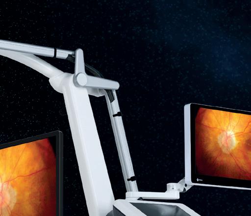
High-Tech Upgrades for High-Stakes Surgery
Cutting-edge microscope technologies and robotic systems are redefining the future of vitreoretinal surgery


Retina Rulebreaker
Prof. Dr. Şengül Özdek’s inspiring career breaks barriers and transforms pediatric retina care in Turkey


Eye-Opening Advances
New therapies for retinal diseases promise to improve long-term outcomes and enhance patient care
Spotlight on Retinal Rarities
Ophthalmologists share rare and complex retinal conditions and their treatment strategies at EURETINA 2024
Of Reunions and Retinas
NVRS 2025 showcases cutting-edge retinal advancements and fosters lasting friendships

Dr. Alay S. Banker
Banker’s Retina Clinic and Laser Centre Ahmedabad, India alay.banker@gmail.com

Dr. Arshad Khanani
Sierra Eye Associates; University of Nevada, Reno School of Medicine Nevada, USA arshad.khanani@gmail.com

Dr. Barbara Parolini
Eyecare Clinic Milan, Italy parolinibarbara@gmail.com

Prof. Gemmy Cheung
Singapore National Eye Centre (SNEC) Singapore gemmy.cheung.c.m@singhealth.com.sg

Dr. Hudson Nakamura
Bank of Goias Eye Foundation Goiânia, Brazil hudson.nakamura@gmail.com

Dr. Veeral Sheth
University Retina and Macula Associates; University of Illinois at Chicago, USA vsheth@gmail.com


In vivo fluorescein angiography (FA), first described in 1961, has enabled the visualization of the retinal vasculature for over 60 years. Highquality video recordings of vascular flow can be captured using confocal scanning laser ophthalmoscopy (cSLO). This dynamic component of angiographic imaging adds an extra dimension to image interpretation and diagnosis. FA provides a clear visualization of the foveal avascular zone (FAZ) and the outline of the foveal arcade, offering clinicians valuable insights into a patient’s visual performance and potential. Identifying these features is an essential aspect of FA studies. However, a limitation of FA is that, as a two-dimensional projection of a three-dimensional vascular structure, it does not allow for the differentiation of the distinct capillary plexuses within the retina.
Optical coherence tomography (OCT) has revolutionized imaging of retinal structures, greatly enhancing our understanding of chorioretinal pathology in vivo. OCT angiography (OCT-A) enables non-invasive imaging of blood flow and provides high-resolution transverse section (en-face) images of retinal vascular patterns. The fundamental principle behind OCT-A is that, in a static
eye, the only moving structures are the blood cells flowing within the vessels. OCT-A can differentiate moving cells from static retinal tissue, detecting flow signals through consecutive B-Scans recorded at the same retinal location, which are then displayed as transverse section images.

However, OCT-A images are susceptible to motion artifacts.

of 5.7 microns and an axial resolution of 3.9 microns per pixel, enabling detailed visualization of all four vascular plexuses. This high resolution ensures diagnostic confidence and clinical certainty.
The SPECTRALIS diagnostic imaging platform from Heidelberg Engineering addresses this issue by employing a live eye tracking system, allowing for precise follow-up scans and minimizing motion-related distortions. The retina contains four distinct vascular plexuses, each with unique morphology tailored to meet the metabolic demands of specific retinal tissues. The average diameter of a retinal capillary, including the lumen, is approximately 8 microns, with blood flow measuring around 6.4 microns in diameter. To accurately capture capillary flow and blood vessel patterns, an OCT-A system must meet or exceed this resolution.
The SPECTRALIS OCT Angiography Module achieves a lateral resolution
Another critical factor in imaging vascular flow with OCT-A is the non-linear nature of erythrocyte movement. Variations in blood cell size, shape and orientation can cause clustering at choke points, particularly at vascular bifurcations or in tortuous vessels. As a result, flow may not be detected at a given moment during sampling. Multisampling is essential for detecting this non-linear flow. The SPECTRALIS OCT Angiography Module addresses this challenge through a combination of automatic real-time (ART) multisampling and TruTrack dual-beam live eye tracking, which mitigate motion artifacts and generate accurate flow information. This technology produces exceptionally high-quality OCT-A images, ensuring consistent and reliable documentation of capillary blood flow over time.
by Tan Sher Lynn
The 24th Congress of European Society of Retina Specialists (EURETINA 2024), held late last year in Barcelona, Spain, buzzed with excitement as experts unveiled cutting-edge treatments for a spectrum of retinal conditions.
In a late-breaking session, retinal specialists revealed promising data on treatments’ long-term efficacy and safety, offering new hope for patients facing vision-threatening diseases.
Here’s a glimpse at the highlights that are poised to reshape the landscape of retinal care.
FIREFLYEYE: Aflibercept 0.4 mg for acute-phase ROP
FIREFLYEYE is the first prospective, controlled Phase IIIb study evaluating long-term efficacy and safety outcomes after treating acute-phase retinopathy of prematurity (ROP) with aflibercept 0.4 mg versus laser photocoagulation. Final results through age five are expected in 2026.
Dr. Andreas Stahl (Germany) shared the interim results. “Through three years of age, efficacy outcomes were well sustained, and no safety concerns (ocular or systemic) were identified. Disease reactivation after aflibercept 0.4 mg was rare,

and those that did reactivate, did so before 50 weeks of age. By two years, retinal vascularization was complete in 80% of eyes, visual function was age-appropriate, and myopia was both less frequent and less severe in the aflibercept 0.4 mg group compared to the laser group. Additionally, we did not observe any aflibercept-related growth or safety concerns,” he explained.
Overall, Dr. Stahl concluded that aflibercept 0.4 mg injection therapy was effective and generally welltolerated in extremely preterm or very low birthweight patients with acute-phase ROP through three years of age.
Dr. Clare Bailey (UK), presented the preliminary data of the VOYAGER study (up to August 2024), which involves a two-year observation period of 120 treatment-naive neovascular age-related macular degeneration (nAMD) eyes from 119 patients and 67 treatment-naive
diabetic macular edema (DME) eyes from 51 patients.
VOYAGER, a global, prospective, non-interventional study currently underway in 26 of 28 countries, is collecting data on patients receiving faricimab for nAMD or DME.
According to Dr. Bailey, results involving nAMD eyes show that, at baseline, the mean visual acuity (VA) was 57.9 letters, with an average gain of 3.5 letters by month 6. Central subfield thickness (CST) averaged 344 microns at baseline and reduced by 85 microns over the same period.
Patients received an average of 4.7 injections between baseline and month 6. Subretinal fluid (SRF) was present in 69.8% of patients at baseline but dropped to 25.9% by month 6. Intraretinal fluid (IRF) decreased from 62% at baseline to 15.8% by month 6.
For DME patients, baseline VA was 62 letters, increasing by an average of 7.2 letters by month 6. CST reduced from 425 microns to 275 microns, with patients receiving an average of 4.7 injections. SRF was present in


by Diana Truong
With ultra-clear optics and a compact design, the WiFi HD LenZ Mini is winning over top surgeons like Prof. Lu Hai. Discover how this game-changing lens is enhancing precision, efficiency and control in the OR.

In retinal and macular surgery, precision and clarity are nonnegotiable. Advanced visualization tools allow vitreoretinal surgeons to navigate delicate structures with confidence. For decades, surgeons relied on handheld or mounted contact lenses, offering high magnification but posing challenges—complex handling, corneal pressure and the need for frequent adjustments. The introduction of non-contact, wide-
angle viewing systems has since transformed surgical maneuverability but even these innovations have limitations.
Enter the WiFi HD LenZ Mini (OCULUS; Wetzlar, Germany), a 17 mm small-diameter lens designed to optimize optical performance, enhance usability and improve surgical efficiency. The innovation has already gained recognition from leading retinal surgeons, including
Prof. Lu Hai, head of the Pediatric Retinal Department at Beijing Tongren Hospital (China).
With more than 20 years of experience using non-contact, wide-angle viewing systems, Prof. Lu provides firsthand insight into how this next-generation lens is transforming vitreoretinal surgery.
The WiFi HD LenZ Mini stands out for its exceptional optical quality, which has transformed the way surgeons’ approach retinal and macular surgeries. With a wide-angle view of 60 to 130 degrees, the lens provides unparalleled clarity and detail, even in the far periphery. This is a significant improvement over traditional lenses, which often suffer from peripheral distortion.
“In
pediatric retina surgery, if there’s an iatrogenic complication, it’s almost game over. A sharper view allows surgeons to maintain control throughout the procedure.”
“What truly amazed me about the WiFi HD LenZ Mini was its optical quality and small diameter,” said Prof. Lu. “With small pupils and small corneas, your wide angle view diminishes significantly, but the WiFi HD LenZ Mini allows me to get closer to the cornea, providing a sharper
and more detailed view without relying on digital magnification, which can compromise detail and contrast.”
The lens’s aspherical design further enhances its performance by minimizing glare and improving depth perception. This is particularly critical in delicate procedures, where even the slightest error can have serious consequences.
“In pediatric retina surgery, if there’s an iatrogenic complication, it’s almost game over,” Prof. Lu emphasized. “A sharper view allows surgeons to maintain control throughout the procedure.”

The WiFi HD LenZ Mini delivers crisp, high-contrast images, reducing the need for excessive scleral depression and improving overall surgical efficiency. Its ability to minimize glare—especially during fluid-air exchanges—ensures uninterrupted clarity, creating a smoother and more controlled surgical experience.
“With this lens, the wide field is truly usable, especially in the far periphery,” Prof. Lu noted. “Many lenses claim to offer a wide-angle view, but the usable field is often compromised by distortion. The WiFi HD LenZ Mini delivers a distortionfree, high-quality image across the entire field of view.”
This enhanced visualization also reduces reliance on surgical assistants, which is particularly beneficial in pediatric and complex retinal surgeries. “A lens that improves self-sufficiency is invaluable in high-stakes cases,” Prof. Lu added. “It allows surgeons to operate with more confidence and consistency.”
Yet even the best lens requires optimal use. Prof. Lu stresses the importance of establishing the right focal plane before making mechanical zoom adjustments.
“Many of my colleagues zoom in or out, or adjust focus up and down, but they don’t establish the focal plane first. That’s inefficient,” he explained. “I always find the best focal plane at high magnification, then zoom out to maximize depth of field. Once you grasp the optical principles, you’ll enhance surgical efficiency significantly.”
By refining visualization and reducing the need for adjustments, a high-performance lens does more than improve image clarity—it streamlines the entire surgical process and enhances procedural consistency.
The lens also integrates seamlessly with digital 3D systems, ensuring an optimized viewing experience. By reducing flare, it eliminates the need to adjust light settings or swap lenses during fluid-air exchanges, streamlining entire procedures.
By combining durability, ease of use and a simplified workflow, the WiFi HD LenZ Mini is more than an optical upgrade—it’s a step toward greater surgical efficiency. These advancements not only improve surgeon experience but also contribute to better patient outcomes.
The WiFi HD LenZ Mini represents a major leap forward in retinal surgery visualization. With its exceptional optical performance, compact design and workflow-enhancing efficiency, it is setting new standards in the operating room.
Prof. Lu’s experience underscores its impact. From optical clarity to maneuverability, the WiFi HD LenZ Mini enhances every aspect of surgery. As more retinal surgeons seek cutting-edge visualization solutions, this lens is poised to become an essential part of the surgical toolkit, elevating precision and efficiency to new heights.

The WiFi HD LenZ Mini is not just about optical excellence—it also simplifies the surgical workflow. Its single-lens solution eliminates the need for frequent switching between different lenses, which can disrupt the flow of surgery.
“In the past, we used two lenses— one for vitrectomy and peripheral manipulations, and another for the macula,” Prof. Lu explained. “With the WiFi HD LenZ Mini I use one lens for everything: peripheral work, vitrectomy and macular surgery.”
Professor Lu Hai is a renowned expert in the Pediatric Retinal Department at Beijing Tongren Hospital (China) with a high academic reputation, specializing in the diagnosis and treatment of pediatric eye and retinal diseases. He studied at Xian Medical University, Beijing Medical University and Beijing Eye Institute. Prof. Lu has presented papers at numerous domestic and international academic conferences, contributed to research projects and advanced the treatment of pediatric eye diseases, particularly retinal disorders. He won the Dr. Arthur M. Sackler prize for Chinese Doctor of Year 1999. Prof. Lu uses the ZEISS microscope with Resight system and OCULUS WiFi HD LenZ Mini (for pediatric cases) for his daily surgeries. trdr_luhai@163.com
Retinal expert shares key tips for essential techniques in capturing and interpreting high-quality retinal images
by Tan Sher Lynn
Renowned retinal specialist Dr. Veeral Sheth shares insights into best practices for optimal retinal image quality—covering patient positioning, imaging parameters, multimodal and AI-driven analysis.

Capturing the delicate details of the retina requires a blend of technical prowess and patient understanding. Retinal imaging plays a crucial role in diagnosing and managing ocular diseases, offering detailed views of retinal structures to guide clinical decisions.
Chicago retinal specialist Dr. Veeral Sheth demystifies the process, offering practical advice on optimizing image capture and interpretation, and embracing the transformative power of artificial intelligence (AI).
6 Key tips to optimizing retinal imaging
Dr. Sheth shared his top key tips for enhancing retinal imaging, emphasizing techniques that
enhance clarity and streamline workflows.
1
Position patients properly for optimal imaging
Proper patient positioning is essential for capturing accurate retinal images. It minimizes distortions and ensures that key retinal structures are adequately captured.
“To improve alignment, ensure proper chin and forehead placement, adjust head tilt and use fixation targets. For optical coherence tomography (OCT), remind patients to blink between scans to prevent artifacts. And for fundus photography, use verbal cues to maintain a steady gaze for peripheral views,” Dr. Sheth emphasized.
2
Use multimodal imaging
Different retinal conditions require specific imaging modalities for accurate diagnosis. Multimodal imaging integrates various imaging techniques to provide a comprehensive assessment of retinal pathology. For example, for agerelated macular degeneration (AMD), OCT provides high-resolution crosssectional images, enabling precise evaluation of macular changes.
“Multimodal imaging is crucial for complex cases like AMD, diabetic retinopathy and uveitis.
I use OCT for structural analysis, fluorescein angiography for vascular abnormalities and wide-field imaging for peripheral pathology, tailoring the approach to the disease,” Dr. Sheth explained.
By combining these imaging methods, clinicians can achieve a more comprehensive understanding of retinal conditions, improving diagnostic accuracy and treatment planning.
“Multimodal imaging is crucial for complex cases like AMD, diabetic retinopathy and uveitis. I use OCT for structural analysis, fluorescein angiography for vascular abnormalities and wide-field imaging for peripheral pathology, tailoring the approach to the disease.”
3
Adjust imaging parameters
Retinal imaging requires careful parameter adjustments to enhance clarity, and customizing settings based on individual patient characteristics ensures optimal image quality.
“For hazy media, increase brightness and contrast to enhance visualization. In highly pigmented
fundi, adjust gain settings to balance exposure. Higher resolution captures fine structural details, while faster scans reduce motion artifacts in uncooperative patients,” highlighted Dr. Sheth.
4
Pupil dilation remains a fundamental aspect of retinal imaging, enhancing visualization of the fundus. “Dilation is essential for high-quality images, especially in low-light fundus photography and wide-field imaging,” he stressed.
While dilation significantly improves image clarity, some patients may not tolerate it well due to systemic conditions or anatomical constraints.
“Patients with narrow angles or certain systemic conditions may pose challenges. In young or light-eyed patients, non-mydriatic imaging may be sufficient,” Dr. Sheth explained. He further noted that clinicians should assess each patient individually to determine the most appropriate approach.
5 6
The integration of AI and machine learning is revolutionizing retinal imaging by enhancing diagnostic accuracy and efficiency. AI algorithms can rapidly analyze large datasets, identifying patterns that may be missed by the human eye.
Optimize, review, refine
Furthermore, Dr. Sheth advised early-career ophthalmologists to refine their retinal imaging skills.
”Invest time in learning imaging modalities, optimize settings for different pathologies, review images systematically and compare them with clinical findings to help build interpretation skills. AI tools will likely continue to be a growing and important part of our imaging acquisition and interpretation protocols,” he said.
By adhering to best practices, refining imaging techniques and embracing technological advancements such as AI and multimodal imaging, ophthalmologists can enhance diagnostic precision and streamline workflows—ultimately improving patient outcomes.
Not to mention, continuous learning and adaptation to emerging innovations will further elevate the standard of retinal care.

In diabetic retinopathy screening, AIpowered tools can detect early signs of microvascular changes, facilitating timely intervention. Similarly, AIassisted OCT interpretation aids in tracking disease progression and optimizing treatment plans for conditions like diabetic macular edema (DME).
“AI assists in image enhancement, disease detection and progression tracking. It is particularly useful in diabetic retinopathy screening, OCT interpretation and predicting treatment response in AMD and DME—improving efficiency and accuracy in clinical decision-making,” he added.
Dr. Veeral Sheth , MD, MBA, FACS, is a native Chicagoan who specializes in diseases of the retina and vitreous. He is a partner at University Retina and Macula Associates and also a clinical assistant professor at the University of Illinois in Chicago. He is the director of clinical trials at one of the busiest clinical trial sites in the country. Dr. Sheth has been a principal investigator for over 60 clinical trials and has research interests in macular degeneration, diabetic retinopathy, vein occlusion, as well as surgical pathology. He is an active member of the American Society of Retina Specialists, Retina Society, American Academy of Ophthalmology, The Association for Research in Vision and Ophthalmology and the European Society of Retina Specialists. He is the Chairman Emeritus of the Board of Directors for Meals on Wheels Chicago. Dr. Sheth is fluent in English, Spanish, and Gujarati.
vsheth@gmail.com

To ensure clear visualization of retinal structures, strategic techniques are required—as several factors often restrict the acquisition of high-quality retinal images.
According to Dr. Sheth, poor patient fixation, media opacities (cataracts, vitreous hemorrhage) and small pupils can degrade image quality. “We overcome these by using pupil dilation, optimizing lighting conditions and guiding patient fixation,” he said.
Educating patients about the procedure and providing clear instructions can significantly enhance fixation and reduce motion artifacts. For cases with media opacities, adjusting imaging parameters such as contrast and brightness can help mitigate visualization issues.
Avoiding common mistakes is also essential for obtaining reliable retinal images. "These mistakes include poor patient alignment, inadequate dilation and failure to optimize scan settings,” Dr. Sheth added.
Poor patient alignment can lead to off-center images, reducing their diagnostic value. Inadequate pupil dilation may result in restricted field views, making it difficult to assess peripheral pathology.
“To avoid these, we need to position patients properly, adjust focus dynamically and review images immediately to rescan if needed,” he suggested. Reviewing images immediately after acquisition allows for prompt rescanning if necessary, ensuring that all relevant retinal details are captured.

by Tan Sher Lynn
The 17th Asia-Pacific Vitreo-Retina Society Congress (APVRS 2024), held in Singapore last year, showcased an array of breakthroughs in retinal imaging that promises to revolutionize clinical practice.
Retinal specialists presented simpler methods for assessing blood flow, explored the power of high-resolution OCT and AI-driven analysis, and highlighted the importance of post-processing techniques for enhanced visualization. These innovations offer new hope for improved diagnosis, treatment and, ultimately, better outcomes for patients with retinal diseases.
Dr. Rick Spaide (USA) opened his presentation with a deep dive into the vital role of blood flow in ocular disorders, including vascular occlusions, diabetic retinopathy and glaucoma.
He noted that there are many different ways to measure blood
velocity, such as fluorescein angiography. Highlighting the challenges, he described sophisticated techniques like Doppler optical coherence tomography (OCT), which requires precise angling due to the pupillary aperture’s limitations.
He shared a simpler approach: Leveraging OCT speckle patterns to infer blood velocity, showing how ‘big blob-like speckles’ indicate slower flow, while ‘small, vertically oriented speckles’ signify faster flow. By using image processing, horizontal derivatives reveal blood velocity changes correlating with the pulse.
Validating this method through model eyes and clinical cases, Dr. Spaide concluded that this technique should apply to all OCT instruments, offering a cost-effective, impactful tool for diagnosing ocular and systemic diseases.
Dr. Giovanni Staurenghi (Italy) highlighted advancements in highresolution OCT imaging, emphasizing its ability to reveal intricate retinal structures.
Compared to earlier 10-micron standards, “experimental devices now achieve resolutions down to 2.73 microns,” said Dr. Staurenghi. Enhanced resolution aids in identifying retinal layers, including distinctions between the retinal pigment epithelium (RPE) and Brooks membrane, which is crucial for detecting early pathologies like atrophy and basal lamina deposits.
Dr. Staurenghi concluded that highresolution OCT is transformative, offering deeper insights into retinal diseases and facilitating better clinical diagnoses.
Dr. Carol Cheung’s (Hong Kong) presentation highlighted groundbreaking developments in retinal oculomics, emphasizing how retinal imaging reveals systemic health insights.
“Changes in the retinal microvasculature can serve as an early sign or ‘surrogate’ for microvascular disease,” she said,
noting that retinal changes can indicate preclinical conditions and act as biomarkers for systemic diseases. She explained that AI-powered analysis of retinal photographs can predict cardiovascular risks like hypertension, heart failure and even Alzheimer’s disease with impressive accuracy.
Her team developed a deep learning model which focused on predicting arterial caliber and vascular caliber from retinal fundus photographs alone to enhance cardiovascular disease risk prediction.
Subsequently, they also developed another algorithm that uses whole retinal photographs to detect Alzheimer’s disease.
“In fact, the development of clinical AI is similar to drug development, involving four phases: Model development, signable evaluation by prospective study, clinical deployment and monitoring and assurance network,” she noted.
Dr. Cheung stressed that, in order to establish guideline recommendations for AI use, it is essential to demonstrate the magnitude of the treatment effect and, specifically, to determine whether the benefits outweigh the risks.
She concluded her talk by emphasizing the need for evidencebased guidelines and further research on prospective validation and real-world implementation to establish clinical value.
Multimodal imaging has become integral in diagnosing and managing ocular diseases. According to Dr. Diana Do (USA), OCT has become indispensable, with over 30 million scans performed yearly on 50,000+ systems worldwide.
Wide-field retinal imaging has also gained traction, particularly for assessing peripheral pathologies like diabetic retinopathy (DR). However, fluorescein angiography, though effective, is time-intensive and invasive. OCT angiography, while promising, faces limitations due to imaging artifacts and limited adoption.
Dr. Do also highlighted operational challenges in busy retinal clinics. “More imaging means more time spent in the clinic,” she said, causing longer patient wait times—which studies linked to dissatisfaction. She shared that at Stanford, the average visit lasts 83 to 105 minutes, with imaging bottlenecks being a significant factor.
She noted that reducing these wait times is crucial, as prolonged visits can discourage follow-up and lead to poorer visual outcomes.
“And we know that suboptimal follow-up leads to suboptimal visual acuity outcomes. So the key in our busy clinical practice is not only determining what imaging modality is essential for accurate diagnosis, but also identifying those that can help decrease patient wait time and enhance overall patient satisfaction,” she stated.
Dr. Nadia Waheed’s (USA) presentation on swept-source OCT angiography (OCT-A) highlighted its potential in assessing DR by quantifying capillary blood flow speeds through variable interscan time analysis (VISTA).
“Capillary blood flow speed is elevated in diabetic retinopathy, and non-perfusion is associated with a high-flow speed in remaining vessels,” she said. She explained that OCT-A measures red cell movement through repeated B-scans, with interscan time influencing signal saturation. Faster saturation indicates higher blood flow speeds.
Also, VISTA enables pseudo-color imaging and precise flow speed mapping. In DR studies, elevated flow speeds correlate with capillary dilation and vascular endothelial growth factor (VEGF)-mediated vessel expansion. Interestingly, a negative association was observed between vessel density and flow speed in non-proliferative diabetic retinopathy (NBDR), but not in proliferative cases—possibly due to vessel dilation.
Dr. Waheed concluded by emphasizing VISTA’s clinical potential, encouraging further
research for broader applications in retinal and choroidal vascular imaging.
Presenting on post-acquisition processing in OCT and OCT-A, Dr. Kelvin Teo Yi Chong (Singapore) emphasized the importance of visualization in medical practices, highlighting its critical role in diagnosing abnormalities and monitoring patient progress.
Visualization hinges on two key components: Acquisition quality and post-processing techniques. Dr. Chong described averaging, a process that combines multiple OCT scans to reduce artifacts— particularly motion—resulting in sharper images. Segmentation was another focus, revealing challenges in accurately delineating deeper structures, especially in OCT-A. Advanced techniques, including deep learning-assisted segmentation, enhance these processes by combining amplitude and phase changes for clearer imaging.
Dr. Chong also showcased innovations like denoising algorithms for single-scan clarity and projection artifact removal, which differentiates true vascular structures from superficial projections.
“In busy clinics, enhancements like vessel filtering and uniform background correction make images more interpretable,” noted Dr. Chong. Lastly, 3D reconstructions of lesions provide new perspectives, improving diagnostic precision. With advancing technology, he believes that our ability to interpret OCT images will only improve.
Reporting for this story took place at the 17th Asia-Pacific Vitreo-Retina Society Congress (APVRS 2024), held from 22 to 24 November 2024, in Singapore.

Cutting-edge microscope technologies and robotic systems are redefining the
vitreoretinal surgery
by Hazlin Hassan
Vitreoretinal surgery—once a realm of intense focus and physical strain—is now a stage for technological marvels. Think 3D displays, robotic precision and optics so sharp they reveal the unseen. These innovations are not just gadgets; they’re game-changers!
Vitreoretinal surgery has always been a high-stakes game of precision. But thanks to the latest microscope technologies, the game is changing—and it’s getting wild. From 3D visualization systems to high-resolution optics and headsup displays, these innovative tools are not just changing how surgeons operate, they’re redefining the very art of eye surgery.
Welcome to the era of high-tech eye care.
Dr. Mae-Lynn Catherine Bastion, professor of ophthalmology (vitreoretina) and senior consultant ophthalmologist at the National University of Malaysia (UKM), summed up the technology perfectly. “Three-dimensional (3D) visualization is transforming surgery through improved depth perception, better ergonomics and enhanced teaching capabilities,” she said.

One of the coolest innovations in 3D visualization is the heads-up display technology. Traditional microscopes require surgeons to hunch over eyepieces, leading to neck pain, eye strain and long-term musculoskeletal issues. But with heads-up systems like the NGENUITY 3D Visualization System (Alcon; Geneva, Switzerland), surgeons can now operate while looking at a high-resolution 3D screen.
“Heads-up improves surgeon posture, reduces eye strain and neck pain, and overcomes presbyopia limitations,” said Dr. Bastion. This ergonomic advantage is a gamechanger, especially for lengthy procedures like complex retinal detachments or diabetic tractional detachments.
The 3D systems also offer superior depth perception, allowing surgeons to better visualize membranes and tissue planes. Adjustable lighting and filters further enhance visualization, particularly in macula surgery.
“Heads-up allows better visualization of membranes and tissue planes
through better depth perception and the use of lighting adjustments and filters,” Dr. Bastion noted.
For trainees, 3D visualization is a revelation. Dr. Bastion highlighted a study by Cheng, et al., that found trainees at her unit who were exposed to 3D surgery demonstrations had a positive response.1 However, she cautions that teaching surgeons should view the surgery from the same angle as the trainee to avoid reduced 3D visualization from the side of the display.
While 3D systems steal the spotlight, high-resolution optics remain the unsung heroes of vitreoretinal surgery. These optics provide clarity, enabling surgeons to spot minute details that could mean the difference between success and complication.
“Heads-up improves surgeon posture, reduces eye strain and neck pain, and overcomes presbyopia limitations.”

- Dr. Mae-Lynn Catherine Bastion
Dr. Bastion acknowledged the benefits but pointed out the limiting factor.
“Naturally, high-resolution optics will be beneficial to improve surgical outcomes by giving surgeons better visualization. The cost remains the limiting factor.”
At the Royal Victorian Eye and Ear Hospital in Melbourne, Australia, surgeons are wielding cutting-edge technology for precise retinal procedures. This includes Zeiss microscopes featuring the Resight viewing system, and a Haag Streit microscope equipped with the Eibos viewing system.

The operating room (OR) is getting a high-tech makeover, with new vitrectomy machines and upgraded capabilities boosting efficiency and outcomes.
Here’s a rundown of the latest platforms making waves in the field.
1. EVA NEXUS Platform
Manufacturer: Carl Zeiss
Meditec/DORC; Jena, Germany
Key Features:
• VacuFlow VTi Technology: A dual-mode pump system that can work in both the vacuum and the flow mode
• Smart IOP: Maintains constant intraocular pressure during vitrectomy
• TDC (two-dimensional cutting): Cutting speed of up to 20,000 cuts per minute and reduced pulsatile traction on surrounding tissue.
2. UNITY Vitreoretinal Cataract System (VCS)
Manufacturer: Alcon; Geneva, Switzerland
• Set to release this year, the UNITY VCS promises enhanced workflow efficiencies over Alcon’s current systems—with cutting speeds of up to 30,000 cpm, enhanced fluidics, and tetraspot laser technology.
3. Stellaris Elite
Manufacturer: Bausch + Lomb; Quebec, Canada
• Its features include wireless pedal control, adaptive fluidics and a high-speed bi-blade dual port cutter. Different illumination filters provide improved light safety and visualization.

Assoc. Prof. Penelope Allen, head of the Vitreoretinal Unit, shared that the advanced optics of the latest microscopes and non-contact fundus visualization systems are “excellent and superior to the heads-up displays.”
That said, she acknowledged that the heads-up display does have its merits, particularly for surgeons with cervical spine issues—though she pointed out that this represents a small group of practitioners
The combination of 3D visualization and high-resolution optics is a powerhouse for surgical precision. Studies have shown that these technologies improve depth perception and color adjustments, potentially enhancing surgical accuracy.
However, there’s a slight learning curve. “The initial adjustment period may include slightly longer surgical

Prof. Dr. Mae-Lynn Catherine Bastion graduated from the University of Sydney, Australia, with MBBS (First Class Honours) in 1999. In 2004, she received a Doctor of Ophthalmology degree from the Universiti Kebangsaan Malaysia (UKM). In 2007, she completed her clinical fellowship in vitreoretinal surgery at The Eye Institute, Singapore. Following that until today, she’s been serving as head of vitreoretinal services at UKM. In 2009 she became the head of the Department of Ophthalmology, for which she served two terms. She was appointed UKM professor of ophthalmology (vitreo-retina) in 2014 and received the Academy of Medicine (AMM) Fellowship in 2016. She teaches undergraduate and postgraduate ophthalmology while maintaining private practice at UKM Specialist Centre. She currently serves on the committees of the Malaysian Universities Conjoint Committee of Ophthalmology, the College of Ophthalmologists of the AMM and the Malaysian Society of Ophthalmology. This is finely balanced with a busy family life of three kids, two dogs and a vegetable garden.
mae-lynn@ppukm.ukm.edu.my
times,” noted Dr. Bastion, citing a study by Asani, et al.2
When it comes to patient outcomes, the results are promising. A retrospective evaluation by Kantor, et al., found that 3D viewing systems yielded comparable outcomes to traditional microscopes.3 This means patients can benefit from cuttingedge tech without compromising safety or efficacy.
Robotic systems like the Preceyes Surgical System (Carl Zeiss Meditec; Jena, Germany) and the SteadyHand Eye Robot (Johns Hopkins University; Maryland, USA) are also poised to revolutionize vitreoretinal surgery. These robots aim to eliminate tremors, minimize eye movements and perform highprecision maneuvers like membrane peeling procedures and vein cannulation.

Assoc. Prof. Penelope Allen graduated from the University of Melbourne in Medicine and specialized in ophthalmology. She then subspecialized in vitreoretinal surgery. Assoc. Prof. Allen is a principal investigator at the Centre for Eye Research Australia, with a clinical appointment at the Royal Victorian Eye and Ear Hospital as head of the Vitreoretinal Surgical Unit. Assoc. Prof. Allen was key to developing the surgical technique for the suprachoroidal retinal prosthesis, resulting in the first clinical trial of this device involving three human patients. She has subsequently led a second trial of a fully implantable device involving four patients. She has presented more than 100 conference posters and talks and published over 90 peer-reviewed journal articles.
editor@mediamice.com
“This technology is very useful for presbyopic surgeons, so it would be a beneficial investment for the future.”
- Dr. Mae-Lynn Catherine Bastion
In a nutshell, the latest microscope technologies are transforming vitreoretinal surgery into a hightech, high-precision art form. These innovations are enhancing surgical precision, improving patient outcomes and making the operating room a more ergonomic and efficient space.
“This technology is very useful for presbyopic surgeons, so it would be a beneficial investment for the future,” noted Dr. Bastion.
So, whether you’re a seasoned surgeon or a trainee, the future of vitreoretinal surgery is unstoppable— and it’s here to stay.

Dr. Bastion has been an invited speaker for both ZEISS and Alcon. She is a recipient of the Alcon Education Grant and a former recipient of Investigator Initiated Study under Alcon. She has also previously received sponsorship for education materials from ZEISS. She has no financial interests in either company’s 3D viewing systems.
1. Chen TC, Yahya MFN, Mohd Naffi AA, et al. Evaluation of Three-Dimensional Heads up Ophthalmic Surgery Demonstration From the Perspective of Surgeons and Postgraduate Trainees. J Craniofac Surg. 2021;32(7):2285-2291.
2. Asani B, Siedlecki J, Schworm B, et al. HeadsUp Display vs. Standard Operating Microscope Vitrectomy for Rhegmatogenous Retinal Detachment. Frontiers in Medicine. Front Med (Lausanne). 2020:7:615515.
3. Kantor P, Matonti F, Varenne F, et al. Use of the heads-up NGENUITY 3D Visualization System for vitreoretinal surgery: a retrospective evaluation of outcomes in a French tertiary center. Sci Rep. 2021;11(1):10031.
29.8% at baseline, dropping to 3.7% at month 6, while IRF decreased from 98.2% to 72.7%.
“In summary, early results from the VOYAGER study showed VA improvements of 3.5 letters in nAMD and 7 letters in DME patients by month 6, along with significant anatomical improvements and no new safety concerns,” she said.
Prof. Anat Loewenstein (Israel) presented compelling findings on geographic atrophy (GA), a prevalent eye disease characterized by complement dysregulation and macrophage overactivation. She discussed the ongoing SIGLEC Study, a Phase IIa clinical trial that investigates the safety and efficacy of AVD-104—an optimized intravitreal glycomimetic nanoparticle, represents a nextgeneration therapeutic designed to enhance the efficacy and safety of geographic atrophy (GA) treatment. With its differentiated dual mechanism of action (MOA), AVD-104 targets disease progression further upstream by effectively inhibiting both macrophage activity and complement cascade amplification
“Remarkably, 78% of patients in both clinical dose cohorts showed a decrease or no change in junctional zone hyperautofluorescence.”
- Prof. Anat Loewenstein
The results of the SIGLEC trial were encouraging, revealing excellent safety profiles with no dose-limiting toxicity observed up to three months following a single injection. Patients receiving AVD104 at doses of 1.0 mg and 3.0 mg experienced significant gains in VA, with mean improvements of +4.8 and +6.5 letters, respectively, at the three-month mark. Moreover, Prof. Loewenstein noted that there was a 48% reduction in lesion growth rate compared to fellow eyes at month 3.
“Remarkably, 78% of patients in both clinical dose cohorts showed a decrease or no change in junctional zone hyper-autofluorescence,” she said, suggesting a potential halt in disease advancement.
Prof. Loewenstein emphasized that AVD-104 not only offers safety but also demonstrates functional benefits in the treatment of GA. The ongoing Phase IIb trial, which is fully enrolled with 300 participants, aims to further validate these promising results and advance the understanding of effective treatments for this debilitating condition.
Meanwhile, Dr. Veeral Sheth (US) noted that even though the efficacy of anti-vascular endothelial growth factor (anti-VEGF) therapy and the need for proactive treatment for non-proliferative diabetic retinopathy (NPDR) have been well established, less than 1% of NPDR patients receive anti-VEGF therapy due to the frequency and treatment burden of required injections.
This highlights the need for early intervention with longer-lasting treatment options. Introducing OTXTKI, a sustained-release hydrogel technology designed to improve treatment adherence.
“This bioresorbable, targeted drug delivery platform allows for the release of axitinib, a multi-target tyrosine kinase inhibitor, over time,” shared Dr. Sheth, offering a more convenient treatment option.
Sharing results from the HELIOS Phase I study, Dr. Sheth noted that 23% of OTX-TKI-treated patients achieved a two-step or greater improvement in DR severity scores, compared to none in the sham group. Additionally, 0% of OTX-TKI patients progressed to vision-threatening complications (VTCs), while 37.5% of the sham group did.
“In summary, OTX-TKI demonstrated diabetic retinopathy severity scale (DRSS) stability or improvement with durability through 48 weeks. None of the OTX-TKI patients experienced vision-threatening complications or progressed to
proliferative diabetic retinopathy (PDR) or center-involving DME. The study also showed sustained fluid suppression and a favorable safety profile, with no cases of vasculitis or intraocular inflammation,” Dr. Sheth concluded, emphasizing OTX-TKI’s transformative potential for NPDR treatment.
Efficacy and safety of ranibizumab biosimilar FYB201
Last but not least, Dr. Ashish Sharma (India) shared insights from a retrospective, interventional, uncontrolled multi-center study assessing the efficacy and safety of the ranibizumab biosimilar FYB201 in real-world settings.
The study involved 3,595 injections across 130 patients with a followup period averaging 15.7 weeks.
The majority of patients were pretreated, particularly for nAMD, which accounted for 802 cases, followed by DME and retinal vein occlusion. The mean number of injections was 2.9.
“Best corrected VA remained stable, largely because most patients were not treatment-naive,” noted Dr. Sharma. CST showed significant improvement across all indications.
In terms of safety, three ocular adverse events were reported: Anterior segment inflammation, vitreous hemorrhage and increased intraocular pressure. Non-ocular systemic adverse events occurred in 6.8% of cases but were unrelated to the drug.
“The safety profile aligns with the originator ranibizumab,” Dr. Sharma concluded. “These early real-world findings confirm that FYB201 is both effective and safe.”
Reporting for this story took place at the 24th Congress of the European Society of Retina Specialists (EURETINA 2024), held from 19 to 22 September 2024, in Barcelona, Spain.

by April Ingram
For patients facing complex retinal conditions, hope lies in the ever-evolving world of surgical technology. Once, surgeons relied on limited views and guesswork. Today, advanced imaging and AI are offering unprecedented precision, translating into better outcomes and brighter futures.
It’s sometimes hard to believe that not that long ago vitreoretinal surgery relied on fundus photos, those highly pixelated optical coherence tomography (OCT) scans, and what the surgeon could see with their own eyes. This was a stark contrast to today’s procedures, where imaging technology is at the forefront of every step.
The first vitreoretinal surgical procedures were performed using 17-gauge instruments with an external diameter of 1.5 mm. Compared to the highly advanced and specialized surgeries of today, those procedures would be considered crude, even horrifying.
Being able to visualize and see finer details has driven the evolution in surgical techniques and applications of novel technologies and treatments—all aimed at achieving better outcomes with fewer complications.
Imagine integrating digital imaging systems to allow surgeons to analyze and manipulate delicate and microscopic retinal anatomic features in real-time, with high precision, improving safety and outcomes.
Actually, you don’t need to imagine it… that’s exactly the cuttingedge surgery we’re doing now.
With insights from Dr. Raymond Iezzi and Dr. Pierre Baldi—renowned experts in vitreoretinal surgery, biomedical engineering, artificial intelligence and imaging technology—we will explore today’s imaging modalities and the future of retinal practices.
Dr. Iezzi, an associate professor of ophthalmology and vitreoretinal surgeon within the Department of Ophthalmology at the Mayo Clinic, sees the advantages and application of modern imaging techniques.
“We are in an era where machinelearning-based image analysis and segmentation are becoming more commonplace. Using these tools, we are identifying new, clinically


relevant disease biomarkers,” he explained.
Machine learning uses algorithms to analyze data and imaging to identify patterns and make decisions. It learns by analyzing large datasets and improves its performance over time through continued training.
“We are in an era where machine-learningbased image analysis and segmentation are becoming more commonplace. Using these tools, we are identifying new, clinically relevant disease biomarkers.”
- Dr. Raymond Iezzi
Within ophthalmology, machine learning, particularly deep learning, is increasingly used to analyze retinal images to facilitate automated detection and tracking of pathological features in retinal diseases over time. It helps clinicians identify early signs of conditions and predict progression.
Dr. Iezzi detailed how machine learning enhances his practice, both in clinical and surgical settings.
“Machine learning analysis techniques can quantify complex imaging biomarkers more rapidly, accurately and reproducibly than manual methods—making this technology useful and efficient in our clinics,” shared Dr. Iezzi. “In the operating room, machinelearning image registration tools allow us to overlay relevant OCT scan and microperimetry data onto the real-time surgical video feed for augmented reality.”
He added that this technology can direct surgeons to the clean edge of an epiretinal membrane and away from areas of tractional retinoschisis during the initiation of membrane
peeling. “Further, by overlaying microperimetry data, surgeons can identify fixation and /or eccentric preferred retinal loci and protect those areas during surgery,” he added.
Aligning anatomic pathology with functional, retinal sensitivity outcomes is an exciting step for both clinical outcomes and refining research study endpoints.
Previously, augmented reality (AR) in ophthalmic surgery was predominantly focused on surgical training. More recently, researchers have been investigating its potential for surgical visualization and navigation.
Earlier this year, Dr. Iezzi and colleagues from the Mayo Clinic published their work, looking at how AR may allow vitreoretinal surgeons to leverage microscope-integrated digital imaging systems to analyze and highlight key retinal anatomic features in real time—possibly improving safety and precision during surgery.1
They found that their model, when used with their image-registration algorithm, efficiently aligned preoperative and intraoperative images with high accuracy. This combination also achieved higher processing speeds than standard hardware. Compared to manual registration, their model’s alignment accuracy was superior, and its processing rates surpassed those achieved by other standard hardware configurations.
What does this mean? The authors concluded that it is feasible to use their tensor processing unit (TPU)accelerated convolutional neural networks (CNNs) for enhanced AR in vitreoretinal surgery.
Three-dimensional (3D) headsup visualization systems have revolutionized the vitreoretinal operating room, providing enhanced detail and improved ergonomics compared to conventional viewing systems, like traditional microscopes. To say the least, this is a gamechanger for medical education and telementoring/consulting.
To achieve a stereoscopic view of the large display screen, surgeons wear 3D polarized glasses. The technology continues to evolve with improvements in display screens or embedding video into a headmounted unit and integration with existing imaging modalities.
Studies evaluating surgeon experience with 3D head-up visualization systems have reported improvements in depth of field and field of view, as well as positive effects on posture, musculoskeletal pain and general comfort.
As with any new technology, the learning curve and associated costs remain major barriers to wider adoption.
Digital microscopes/binoculars are enabling surgeons to see magnified, 3D images of the surgical field. Most provide the option for headset viewing or displayed on monitors.
The ORLenz 5K XR (Ocutrx; California, USA) displays 3D holographic images on a wireless headset that receives the 3D
surgery feed. The headset can also mirror imaging data across multiple headsets, allowing students to learn in a 3D environment. Interestingly, this technology is also being marketed as a visual aid for those with age-related macular degeneration and Stargardt disease.
Many of these platforms are still in development. Although surgeons appreciate the enhanced views, they noted that the headset’s weight (especially during long surgeries) and its closed systems nature are among its drawbacks.
Intraoperative OCT (iOCT) is likely the most well recognized imaging technology to enter the modern operating rooms. It is a potentially transformational technology that has been commercially available to vitreoretinal surgeons for several years.
iOCT has evolved from its original hand-held, somewhat cumbersome iteration to the current microscope integrated versions. Its ability to evaluate critical cross-sectional retinal details during or after surgical maneuvers—providing quick, real time, detailed visualization of tissue and instrument-tissue interactions— is incredibly valuable in surgical decision-making.
iOCT has proven most useful in macular surgeries, including vitreoretinal interface and internal limiting membrane (ILM) peeling, as well as managing rhegmatogenous and tractional retinal detachments, distinguishing areas of retinoschisis, assessing retinal tears and evaluating fibrovascular proliferation.
Despite sounding like the ultimate game-changer, iOCT still has its limitations that require thoughtful
“Today, AI and deep learning methods are as good or even better than human experts at most biomedical imaging recognition problems. These methods can be applied across multiple imaging modalities, such as microscopy and X-rays, and to both static images and videos.”
- Dr. Pierre Baldi
consideration. These include challenges with shadowing of nonOCT friendly instruments, integration into established surgical workflows and surgeons’ learning curves—not to mention the excessive costs of the system, which remain a big obstacle to widespread use.
iOCT’s real-time volumetric calculation capabilities are valuable in subretinal medication delivery programs, including gene therapy and retinal prosthesis implantation.
Indeed, the future of iOCT is in robotics, with its integration into robotic-assisted surgical platforms, such as the Preceyes Surgical System (Preceyes BV; Eindhoven, the Netherlands), a telemanipulated robotic device with an instrumentintegrated iOCT sensor that delivers precision, efficacy and safety unmatched by human dexterity.
AI and imaging: A powerful duo
Dr. Pierre Baldi, founding director of the AI in Science Institute and associate director of the Center for Machine Learning and Intelligent Systems at the University of California Irvine in the US, has spent his career developing and evaluating AI and deep learning methods and their applications to science.
In 1993, he published the first ever application of deep learning to biomedical (biometric) imaging problems.2 Dr. Baldi shared his current research focus.
“Today, AI and deep learning methods are as good or even better than human experts at most biomedical imaging recognition problems. These methods can be applied across multiple imaging modalities, such as microscopy and X-rays, and to both static images and videos,” he explained. “In ophthalmology, these methods can help with multiple tasks, ranging from improving diagnoses to assisting doctors during vitreoretinal surgeries.”
Deep learning helps surgeons receive real-time safety feedback during surgery, extract specific data metrics of a surgical technique that can be used for training, or modify procedures to optimize surgical outcomes.
Recently published in Translational Vision Science and Technology, Dr. Baldi and colleagues presented their findings on AI-based video analysis, showing its potential to determine surgical instrument characteristics within the 3D vitreous space.3
They demonstrated that trained models were able to track surgical instrument movement in threedimensional space and determine instrument’s depth, tip location, instrument insertional laterality and instrument type. Model performance was almost instantaneous, supporting the utility of real-time surgical video analysis to aid in vitreoretinal procedures.
Dr. Baldi’s work continues to focus on the development of imaging and visualization solutions. “Among other problems, we are working on enabling the use of low power illumination such as near infrared illumination during surgeries,” he shared.
This research is extremely valuable because as much as we love our retinal imaging technology, phototoxicity of sensitive retinal tissues is a big concern
“Among
other problems, we are working on enabling the use of low power illumination such as near infrared illumination during surgeries.”
- Dr. Pierre Baldi
Leveraging advanced imaging and artificial intelligence allows surgeons to access real-time data analytics during surgery, enhancing decisionmaking processes and surgical precision, and improving safety and patient outcomes.
As with the adoption and implementation of any new technology into mainstream practice, there are substantial challenges that are currently keeping these

“Low power light is desirable because it is less toxic for the eye. However, compared to regular light, doctors cannot see as well under low power light conditions. AI and deep learning methods can be used to compensate for the difference and enable doctors to see with low-power light as if the eye had been illuminated with regular light,” Dr. Baldi explained.
A recent study from Dr. Baldi and colleagues showed that deep learning can be used to reconstruct high-resolution images from multiphoton microscopy on light-sensitive tissues. It requires minimal light exposure to acquire an image when phototoxicity was a demonstrable concern.4
References
innovations from being used more widely.
The integration of advanced digital imaging systems into vitreoretinal surgery is associated with the significant cost, as well as space within operating theaters.
Additionally, as attractive and exciting as new technology can be, surgeons are incredibly busy and the learning curve is steep. Advancing to a level of proficiency takes considerable time.
Dr. Raymond Iezzi is an associate professor of ophthalmology and a vitreoretinal surgeon within the Department of Ophthalmology at the Mayo Clinic. He is also deputy director of Regenerative Ophthalmology and program director of the Retinal and Vitreous Surgery Fellowship at the Mayo Clinic. His research interests are in the fields of biomedical engineering and sight restoration. Dr. Iezzi conducts preclinical and clinical translational trials in several ophthalmology-related areas, and his clinical interests include retinal degenerative diseases as well as all aspects of vitreoretinal surgery, with a special interest in complex retinal detachment repair associated with diabetes, trauma and proliferative vitreoretinopathy. Dr. Iezzi is a recipient of the Ronald G. Michels Fellowship, the Heed Ophthalmologic Fellowship, the Knapp-AOS Fellowship, and the Visionary Award from Foundation Fighting Blindness. He was also presented with the Distinguished Alumnus Award in Teaching and Research from Rutgers University in 2015.
Iezzi.Raymond@mayo.edu

1. Ye RZ, Iezzi R. Intraoperative Augmented Reality for Vitreoretinal Surgery Using Edge Computing. J Pers Med. 2025;15(1):20.
2. Baldi P, Chauvin Y. Neural Networks for Fingerprint Recognition. Neural Comput. 1993;5(3):402-418.
3. Baldi P, Abdelkarim S, Liu J, To JK, Ibarra MD, Browne AW. Vitreoretinal Surgical Instrument Tracking in Three Dimensions Using Deep Learning. Transl Vis Sci Technol. 2023;12(1):20.
4. McAleer S, Fast A, Xue Y, Seiler MJ, Tang WC, Balu M, Baldi P, Browne AW. Deep Learning-Assisted Multiphoton Microscopy to Reduce Light Exposure and Expedite Imaging in Tissues With High and Low Light Sensitivity. Transl Vis Sci Technol. 2021;10(12):30.
Dr. Pierre Baldi earned his MS degrees in Mathematics and Psychology from the University of Paris and a PhD in Mathematics from the California Institute of Technology. He is currently a distinguished professor in the Department of Computer Science, founding director of the AI in Science Institute, and associate director of the Center for Machine Learning and Intelligent Systems at the University of California, Irvine. The long-term focus of his research is on understanding intelligence in brains and machines. He has made several contributions to the theory of AI and deep learning, and developed and applied AI and deep learning methods for the natural sciences to address problems in physics (detection of exotic particles), chemistry (reaction prediction), and bio-medicine (biomedical imaging). He has published more than 500 peer-reviewed scientific articles and five books, including Deep Learning in Science, Cambridge University Press (2021). His honors include the 1993 Lew Allen Award at JPL, the 2010 E. R. Caianiello Prize for research in machine learning, the 2023 Dennis Gabor Award of the International Neural Network Society, and election to Fellow of the AAAS, AAAI, IEEE, ACM, and ISCB. He serves as associate editor for Artificial Intelligence, Neural Networks, and the IEEE/ACM Transactions in Computational Biology and Bioinformatics. He has mentored more than 100 graduate students and postdoctoral fellows and co-founded several startup companies.
pfbaldi@ics.uci.edu


Şengül Özdek’s
by Tan Sher Lynn
Prof. Dr. Şengül Özdek defied gender biases in Turkish ophthalmology to become the country’s leading vitreoretinal surgeon. Balancing a demanding career with family, she advocates for women in medicine, emphasizing hard work and mentorship.
Prof. Dr. Şengül Özdek, a professor in the Department of Ophthalmology at Gazi University Medical Faculty, Turkey, has built an extraordinary career in retinal diseases and vitreoretinal surgery. Her inspiring journey is a testament to perseverance, expertise and the relentless pursuit of excellence in a male-dominated field. Today, she is a beacon of hope and encouragement for young women aiming to make their mark in similar fields.
This is her inspiring story.
A career shaped by opportunity and determination
Dr. Özdek’s path to retinal specialization was unplanned but transformative. Initially focused on glaucoma research during her
residency, she pivoted to retinal diseases due to institutional needs at Gazi University.
“We do not have an official fellowship in Turkey, so I started practicing retina just after completing my residency,” she shared.
Her mentors, while supportive, encouraged her toward medical retina rather than surgical retina—a common trend in Turkish ophthalmology clinics where women were often steered away from surgery. Defying these expectations, she insisted on specializing in vitreoretinal surgery, ultimately becoming one of the leading experts in the field.
One of Dr. Özdek’s most defining professional experiences came early in her career. In 2000, she encountered premature twins suffering from advanced retinopathy
of prematurity (ROP), a condition that could have been prevented with timely screening. At that time, Turkey lacked a national ROP screening program. This harrowing experience inspired her to advocate for systemic change.
“It was so painful to explain the severity of the twins’ condition to their parents,” she recalled. “They had waited years for children through in vitro fertilization (IVF), and seeing their heartbreak was unforgettable. I knew something had to change.”
Driven by her determination, she collaborated with the Ministry of Health and fellow ophthalmologists, ultimately establishing a nationwide ROP screening and treatment program after eight years of dedicated effort. With this, many ophthalmologists were trained in ROP screening. Treatment and screening centers were also established across the country, sparing countless families from similar heartbreak.
This achievement not only transformed pediatric retina care in Turkey but also shaped her career focus to pediatric retina.

Balancing a demanding medical career and family life is no small feat. Dr. Özdek credited much of her success to her supportive husband, a busy, ear, nose and throat (ENT) surgeon and academician. Together, they raised two children, who are now young adults.
“Time management is crucial. You have to prioritize what’s most important,” she explained. The couple shared responsibilities based on availability, making sacrifices along the way. “Everything comes at a price. The key is deciding if you’re willing to pay for it.”
She emphasized the importance of mutual understanding and shared responsibilities in her marriage. “There were times when I had to attend conferences, and my husband would be there for the kids,” she recalled. This partnership allowed her to pursue a career while ensuring a nurturing home environment.
Reflecting on gender representation in ophthalmology, Dr. Özdek noted
that while women have always been well-represented in medical retina and uveitis, surgical retina remains a male-dominated subspecialty. However, the trend is gradually changing, with increasing numbers of female surgeons entering the field.
She candidly acknowledged that biases are still present in academic medicine. “Most men prefer to work with other men if candidates are equally qualified. But if you are good at what you do, they can’t overlook you,” she emphasized.
Her advice to young women in ophthalmology is straightforward. “Competition is increasing every year. Women ophthalmologists need to work and work and work to stay at the forefront. They should focus on identifying unmet needs across all fields of ophthalmology and strive to find solutions that make a meaningful impact in the field,” she shared.
a personal level enrich the field and improve patient outcomes.”
Dr. Özdek’s journey is an inspiring example for future generations, showing that resilience, passion and relentless effort can break down even the most entrenched barriers in medicine.
“I always support women ophthalmologists in every aspect,” she concluded. “My clinic will always be open for them to visit, learn and grow.”
“Most men prefer to work with other men if candidates are equally qualified. But if you are good at what you do, they can’t overlook you.”
Through her pioneering work, advocacy and unwavering dedication, Dr. Şengül Özdek continues to inspire countless women in medicine and beyond, proving that with determination and support, success knows no bounds.
Dr. Özdek also stressed the importance of mentorship. “Having someone believe in your potential can make all the difference,” she said. Her own experience with a supportive mentor shaped her career and instilled in her a commitment to mentoring the next generation of ophthalmologists.

Motherhood and pregnancy are often perceived as career obstacles, but Dr. Özdek believes diligence, problem-solving capacity and academic productivity matter far more. “Working hard and doing whatever is needed for your duties will solve all problems,” she affirmed.
She also highlighted the empathetic edge women bring to patient care, particularly in pediatric ophthalmology, where understanding and compassion are invaluable. “Women bring unique strengths to ophthalmology,” she shared. “Their attention to detail, patience and ability to connect with patients on
Prof. Dr. Şengül Özdek , a renowned Turkish ophthalmologist, specializes in retinal diseases and vitreoretinal surgery. She studied at Hacettepe University, completed her residency at Gazi University, and has been a retina faculty member there since 1999. She trained with top experts in the United States and focuses on pediatric retinal conditions like ROP, persistent fetal vasculature (PFV) and Coats disease. Her accolades include the 2009 Young Scientist Award in Turkey, multiple international video awards, the American Academy of Ophthalmology (AAO) Achievement Award, and the American Society of Retina Specialists (ASRS) Honor and Senior Honor Awards. She was also inducted into the Retina Hall of Fame in 2021. With over 170 international publications cited 3,500+ times, she also edited Pediatric Vitreoretinal Surgery (Springer, 2023). A soughtafter speaker and live surgeon, Dr. Özdek serves as the European VitreoRetinal Society (EVRS) secretary and is a board member of the Turkish Vitreoretinal Surgery Society. sengulÖzdek@gmail.com

by Diana Truong
Is AI in vitreoretina a game-changer or an ethical minefield? As ophthalmologists navigate this high-stakes balancing act, one thing is clear: AI isn’t just a tool—it’s a responsibility.
The integration of artificial intelligence (AI) and highresolution imaging in vitreoretinal disease management has revolutionized early diagnosis, treatment planning and patient outcomes. From automated diabetic retinopathy (DR) screening to AI-assisted optical coherence tomography (OCT) interpretation, these technologies promise efficiency and precision.
But with great power comes great responsibility. AI-driven imaging is not just about automation—it’s about patient rights, data privacy and clinical integrity. As AI continues to shape the future of vitreoretinal care, ophthalmologists must navigate this complex ethical landscape, ensuring that innovation does not come at the cost of equity, transparency or patient autonomy.
Patient privacy and informed consent
AI-driven imaging in vitreoretina walks a fine line between technological progress and patient rights. On one hand, these tools offer powerful diagnostic capabilities; on
the other, they raise critical concerns about data ownership, privacy and informed consent.
Retinal images are more than just medical records—they are unique biometric identifiers, much like fingerprints. If compromised, they could be misused for unauthorized tracking or commercial purposes. While regulatory frameworks like the European Union’s General Data Protection Regulation (GDPR) and the United States’ Health Insurance Portability and Accountability Act (HIPAA) impose strict security measures, global inconsistencies mean data protections vary depending on where—and by whom—the AI is used.1
“We need to make sure that it’s secure and it’s following all the data privacy regulations,” noted Dr. Arshad Khanani (USA), director of Clinical Research at Sierra Eye Associates and clinical associate professor at the University of Nevada, Reno School of Medicine.
Beyond security, patient autonomy must be a priority. Many AI platforms collect anonymized images to refine
their algorithms, but without explicit patient consent, this practice raises ethical concerns.2
“I’m all about transparency and making sure patients understand how it’s being done,” said Dr. Khanani. “I don’t think it’s a huge risk as long as you notify your patients on what you’re doing and they’re informed.”
AI tools also don’t always operate in ways that are easy to explain. If an algorithm detects an incidental finding—perhaps a retinal abnormality unrelated to the primary condition—should patients be informed? Should they be warned about risks AI identifies, even when the clinical significance is unclear? And what happens when AI’s decision-making process itself is opaque, as with “black box” models that even developers struggle to fully interpret?3
These concerns extend beyond ophthalmology. In AI-driven healthcare, the challenge of “explainability”—the ability to clarify how an algorithm arrives at a diagnosis—remains a major ethical dilemma. Without transparency, how can clinicians trust AI-driven recommendations? And how can patients provide truly informed consent if they don’t fully understand how AI influences their diagnosis and treatment?3
To stay balanced on this ethical tightrope, clinicians must advocate for clear, patient-centered policies. Standardized guidelines for AIinformed consent in ophthalmology are also essential to ensure consistency and trust in AI-driven imaging.
Key Takeaway: AI-assisted analysis should be an opt-in process, with patients fully informed of the benefits, limitations and potential risks.
Life-saving alerts or unnecessary alarms?
The ability to detect vitreoretinal diseases at earlier stages using AI-powered imaging tools is undoubtedly a breakthrough. However, this increased sensitivity can also contribute to overdiagnosis—the identification of
conditions that may never progress to clinically significant disease.4
Consider age-related macular degeneration (AMD), where AIassisted imaging can identify subtle structural changes long before symptoms appear.4 “In most retinal diseases, especially with AMD, if you treat it early, you have the best outcomes,” said Dr. Khanani.
Similarly, AI-based screening for DR has improved access to care, but studies suggest that false-positive rates may lead to unnecessary referrals and patient burden. However, Dr. Khanani believes the benefits outweigh the risks.5
“AI can help physicians diagnose diseases early, especially those who are not vitreoretinal specialists. They can run the algorithm and find the pathology they might otherwise miss,” he noted. “That can help us get those patients earlier.”
The potential for overreferral remains a concern, but in Dr. Khanani’s view, the alternative—missing critical diagnoses—is far worse.
Key Takeaway: While false positives may lead to patient burden, the benefits of catching disease early— especially for conditions like AMD and DR—outweigh the risks.
AI bias and healthcare disparities
AI in vitreoretinal imaging is often seen as a neutral, data-driven force, but its objectivity is only as strong as the datasets behind it. If these datasets lack diversity—whether in race, sex, socioeconomic background or geography—AI models risk reinforcing healthcare disparities rather than alleviating them.4
“AI will do as well as the dataset it was trained on,” Dr. Khanani pointed out. For instance, studies have shown that AI systems trained primarily on fundus images from Caucasian patients may struggle to accurately diagnose DR in African, Hispanic or Asian populations. The consequences? Potential misdiagnoses, undertreatment and widening gaps in healthcare equity.6
To address this, clinicians and AI developers must ensure that datasets reflect real-world diversity and that algorithms undergo rigorous validation across different demographic groups. Transparent reporting of AI performance— stratified by patient characteristics— should be a baseline requirement before clinical deployment.4
At the same time, some experts argue that AI could actually bridge disparities by expanding access to underserved populations.4
“There’s an underserved community here that uses AI virtually,” Dr. Khanani shared. “Patients come to the AI suite, get all the exams done, and the doctor can tune in virtually. In this way, AIdriven imaging will actually help with healthcare disparities.”
Striking the right balance requires a multi-pronged approach. Regulators must ensure strong data protection policies, clinicians must advocate for transparency and patient education, and developers must prioritize fairness in algorithm design. AI is only as ethical as the systems governing its use.
By maintaining a critical yet openminded approach, ophthalmologists can harness the power of AI while upholding the ethical standards that define patient care. After all, the goal of technological advancement should not be to replace human expertise, but to enhance it—empowering clinicians to provide the highest quality care while safeguarding patient rights and well-being.

Key Takeaway: AI isn’t inherently fair or biased—it’s a tool shaped by the data and oversight behind it.
AI-driven imaging has the potential to transform vitreoretinal care, but its integration must be approached with caution. Ethical considerations— from data privacy to bias in AI training sets—must remain at the forefront of its development and clinical application.
References
1. Nakayama LF, Goncalves de Matos JCR, Stewart IU, et al. Retinal scans and data sharing: The privacy and scientific development equilibrium. Mayo Clinic Proceedings: Digital Health. 2023;1(2):67-74.
2. Jones ML, Kaufman E, Edenberg E. AI and the ethics of automating consent. IEEE Security and Privacy. 2018;16(3):64-72.
3. Evans NG, Wenner D, Cohen IG. Emerging ethical considerations for the use of artificial intelligence in ophthalmology. Ophthalmol Sci. 2022;2(2):100141.
4. Sorrentino FS, Gardini L, Fontana L, et al. Novel approaches for early detection of retinal diseases using artificial intelligence. J Pers Med. 2024;14(7):690.
5. Khan Z, Gaidhane AM, Singh M, et al. Diagnostic accuracy of IDX-DR for detecting diabetic retinopathy: A systematic review and meta-analysis. Am J Ophthalmol. [Online ahead of print.]
6. Burlina P, Joshi N, Paul W, et al. Addressing artificial intelligence bias in retinal diagnostics. Transl Vis Sci Technol. 2021;10(2):13.
Dr. Arshad M. Khanani , MD, MA, FASRS, is a managing partner, director of clinical research and director of Fellowship at Sierra Eye Associates, and clinical associate professor at the University of Nevada, Reno School of Medicine. Dr. Khanani founded the clinical research department at Sierra Eye Associates, which is now one of the leading clinical research centers in the country. He has served as a principal investigator for over 100 clinical trials and has been a top enroller in the country for multiple Phase 1-3 trials. In addition, Dr. Khanani has been the first one to perform surgical procedures in multiple surgical clinical trials dealing with sustained delivery and gene therapy. He has over 90 scientific publications. Dr. Khanani also serves as a member of national and international clinical trial steering committees as well as scientific advisory boards to bring new treatment options for patients with retinal diseases. He is frequently invited as a guest speaker at national and international meetings. Dr. Khanani is an elected member of the Macula Society, Retina Society and has received numerous awards of distinction. In 2019, he received the Nevada Business Magazine Healthcare Heroes Physician of the Year award for his continued dedication to the field of ophthalmology. He received the Senior Honor Award from the American Society of Retina Specialists (ASRS) and was also awarded the prestigious ASRS Presidents’ Young Investigator Award in 2021. In 2023, Dr. Khanani was invited to give the prestigious 2023 Ernst Bodenheimer Memorial Lecture at the Wilmer Eye Institute at Johns Hopkins University School of Medicine.
arshad.khanani@gmail.com

by Tan Sher Lynn
In November last year, retinal experts gathered in Singapore at the Asia-Pacific Vitreo-Retina Society Congress (APVRS 2024) to discuss the latest advancements in vitreoretinal disease.

The meeting showcased a range of innovations, from refined diagnostic tools to groundbreaking therapeutic approaches, offering new hope for patients with debilitating retinal conditions.
Dr. Graham Holder (Singapore) delved into advanced electrophysiological techniques and their diagnostic utility in retinal disorders. He highlighted the precision of using localized pathology as contrast, and compared the diagnostic power of smaller
(15-degree) versus larger (30-degree) field tests. “They should double in amplitude,” he emphasized.
Presenting a case of Stargardt disease, Dr. Holder demonstrated how normal electroretinography (ERG) might mask subtle dysfunctions.
“The advantage you see of the 30-degree field shows preserved function between 15 and 30 degrees,” he noted. He stressed the importance of understanding the limitations of tools like multifocal ERG, which, despite its utility, may not fully reveal displacement effects seen in eccentric fixation.
He also explored conditions like RGS9 deficiency, revealing how adjustments, such as extending interstimulus intervals, uncover hidden abnormalities
“To conclude,” he said, “identify the origins of the signals you recorded and relate your findings to the signs, symptoms, patient history, family history, inheritance patterns and underlying pathophysiology. Teach the technical staff how to take a proper history. And remember, the International Society for Clinical Electrophysiology of Vision (ISCEV) standards are intended only as minimum data sets. Have you done enough to answer the question?”
Overall, Dr. Holder emphasized integrating electrophysiological data with clinical and genetic insights for accurate diagnoses.
Dr. Tharikarn Sujirakul (Thailand) noted that advanced imaging techniques like fundus autofluorescence (FAF) and optical coherence tomography (OCT) are essential for diagnosing and managing inherited retinal diseases (IRDs).
She said FAF detects retinal abnormalities, highlighting bilateral symmetry, subtle lesions and specific genetic patterns such as CRB1 and RPE65 mutations. It also distinguishes IRDs from similar conditions like age-related macular degeneration (AMD).
Meanwhile, OCT complements FAF by identifying structural changes like macular edema, epiretinal membranes and choroidal neovascularization. It also monitors disease progression, aiding in prognosis and treatment evaluation. OCT angiography (OCT-A) helps detect macular neovascular membranes, while infrared imaging visualizes crystalline deposits in dystrophies. Adaptive optics, though currently research-focused, holds promise for future personalized therapies.
“Multimodal imaging is a very helpful tool for differentiating IRDs from other eye conditions and distinguishing between various types of IRDs. Using proper imaging
techniques leads to early detection of associated complications and disease progression. Given its objective nature, reproducibility and minimal variability, multimodal imaging holds significant potential as an outcome measure in future IRD treatment trials,” Dr. Sujirakul concluded.
Dr. Beau Fenner (Singapore) discussed the global prevalence of ABCA4-related retinal degeneration, emphasizing its significance even in genetically distinct regions like the Midwest (USA) and Singapore.
“ABCA4 tends to be one of the more prevalent forms of inherited retinal degeneration,” he explained.
Dr. Fenner illustrated the phenotypic spectrum with clinical cases: A young patient with severe pigmentary deposits often misdiagnosed as retinitis pigmentosa, and an elderly patient initially thought to have agerelated macular degeneration. These diverse presentations stem from the variability in ABCA4’s enzymatic dysfunction, where “toxic metabolites accumulate inside photoreceptors,” he said.
Meanwhile, genotype-phenotype correlations revealed complexities beyond single mutations. “Despite identical genotypes,” he noted, “patients can have huge distribution in the age of symptom onset.” Environmental and secondary genetic factors likely contribute, underscoring the need for comprehensive genetic profiling in clinical evaluations.
“There’s a huge spectrum of disease severity, and you can actually predict the severity of the disease fairly well by asking the patient when they first lost central acuity. It appears that the heterogeneity of ABCA4 alleles may be unbounded due to these complex haplotypes,” Dr. Fenner explained.
Meanwhile, Dr. Ta-Ching Chen (Taiwan) emphasized gene therapy’s growing potential for IRDs. He highlighted that IRDs are genetically complex, linked to over 300 genes.
Advances in next-generation sequencing have enabled global exploration of these genetic traits.
Gene therapy development has progressed for nearly half of the top 20 IRD-causing genes. He discussed key factors for successful gene therapy, including patient selection, surgical strategies and posttreatment monitoring.
Dr. Chen described key intraoperative surgical strategies for gene therapies, which, nowadays, are mostly adeno-associated virus (AAV)-based and require subretinal injections.
“To manage inflammation and immune response, oral steroids are given before and after the treatment. During the injection procedure, attention is given to ensuring a complete posterior vitreous detachment (PVD), lowering intraocular pressure (IOP), inspecting the surface condition and carefully administering the injection to prevent drug reflux as much as possible,” he explained.
He noted that chorioretinal atrophy (CRA) emerged as a significant yet paradoxically beneficial side effect, enhancing light sensitivity. Dr. Chen concluded by emphasizing the importance of personalized treatment approaches and integrated research for advancing IRD management and gene therapy effectiveness.
In the subsequent session, Dr. Xinyi Su (Singapore) delivered an insightful talk on the advancements and challenges in stem cell therapy, emphasizing its potential for retinal degenerative diseases.
“Stem cell therapy is our main focus,” she stated, highlighting biomaterials augmentation and alternative stem cell resources.
“At least a third of wet AMD patients are non-responsive to antibacterial treatments,” Dr. Su noted, emphasizing the need for cell therapy.
Meanwhile, her lab’s work on RPE cells from induced pluripotent stem
Two key concepts in biomaterials augmentation and alternative stem cell resources as outlined by Dr. Xinyi Su:
1. The roles of gene replacement/ editing and cell replacement in photoreceptor and retinal pigment epithelium (RPE) cellular injury/death.
2. The eye as an ideal organ for cell, gene and RNA therapeutics due to direct access to target tissue, the small volume required for therapies, its immuno-privileged status, as well as the presence of the blood retinal barrier that reduces systemic exposure and off-target effects.
cells (iPSCs) has shown promising results in restoring retinal function. Dr. Su introduced hypo-immunogenic iPSC-derived RPE cells from umbilical cords as a potential ‘offthe-shelf’ solution. Additionally, her team is developing bio-layer scaffolds combining RPE and photoreceptor cells for IRD.
“I believe there is tremendous potential for retinal cell and gene RNA therapy, addressing a significant unmet need in retinal degenerative disease. Developing the next generation of retinal cell therapeutics requires a multidisciplinary workflow, which is a unique opportunity in Singapore. The future is bright for patients with end-stage retinal degenerative disease,” she remarked.
Reporting for this story took place at the 17th Asia-Pacific Vitreo-Retina Society Congress (APVRS 2024), held from 22 to 24 November 2024, in Singapore.
Ophthalmologists share rare and complex retinal conditions and their treatment strategies
by John Butcher
At the 24th Congress of the European Society of Retina Specialists (EURETINA 2024) held late last year in Barcelona, Spain, ophthalmologists explored the complexities of retinal diseases through interesting case studies and research

Experts highlighted a diverse range of issues from acute syphilitic posterior placoid chorioretinitis to perifoveal vascular anomalous complex, each showcasing the latest treatments and emphasizing the importance of advanced imaging and clinical observation.
BALAD: The outer retinal enigma
Dr. Michael Engelbert (USA), a retinal physician and surgeon at Vitreous
Retina Macula Consultants of New York, discussed bacillary layer detachment (BALAD).
BALAD occurs when there is an intact external limiting membrane (ELM) with absence of intraretinal fluid, a breakdown of the retinal pigment epithelium (RPE) component of the outer blood-retina barrier, choroidal thickening, or choroidal and choriocapillaris flow impairment.
Dr. Engelbert presented a case study of a 27-year-old woman who returned
from her honeymoon complaining of blurred vision. He then showed a series of slides of the woman’s eyes to demonstrate the condition.
Additionally, he discussed BALAD in neovascular age-related macular degeneration (nAMD), acute idiopathic maculopathy, acute posterior multifocal placoid pigment epitheliopathy (APMPPE), as well as surgically induced BALAD.
In summary, Dr. Engelbert said BALAD was an outer retinal split at the level of the myoid zone, which is not pathognomonic for any particular condition and may resolve with rapid regeneration of inner and outer segments
Dr. Alexandra Miere (France), a retina specialist in the Ophthalmology Department at Créteil Hospital, University Paris-Est Créteil, talked about benign lobular inner nuclear layer proliferation (BLIP).
She illustrated the condition with a case study of a 27-year-old man experiencing moderate visual blurring. Examining his right eye, she pointed out grouped congenital hypertrophy of the retinal pigment epithelium (CHRPE) lesions and an oval hyperreflective lesion on B-scan optical coherence tomography (OCT), located in the inner nuclear layer.
She then presented a second case study of an asymptomatic 32-year-old man with a solitary CHRPE lesion and discussed a 2023 research paper on BLIP, led by Dr. Aaron Nagiel, which examined four cases of the condition.
Dr. Miere’s two case studies of asymptomatic men with BLIPs and CHRPE lesions confirmed the existence of this new rare entity described in that paper.
Dr. Isabelle Meunier (France) from the Institut des Neurosciences de Montpellier presented on extensive macular atrophy and pseudodrusen (EMAP).
She described EMAP as a late onset macular disease characterized
by striking night blindness and a preserved peripheral visual field, but with an absolute central scotoma. The disease progresses rapidly towards dark and complete macular atrophy without foveal sparing and is not associated with classic large agerelated macular degeneration (AMD) drusen.
She added that EMAP occurs much earlier than AMD and is extremely severe, with rapid impairment of the central vision. According to data, and as demonstrated by Dr. Meunier, the disease was most common in women and patients born before 1948.
Decoding perifoveal vascular anomalous complex
Dr. Guiseppe Querques (Italy), an associate professor at the University Vita-Salute, IRCCS Ospedale San Raffaele in Milan, talked about perifoveal vascular anomalous complex (PVAC).
He described PVAC as a round lesion with a reflective wall, containing material with variable reflectivity, usually located between the outer plexiform layer and the ganglion cell complex, which is frequently associated with signs of exudation.
“The real most effective treatment for PVAC is time.”
-
Dr. Guiseppe Querques
Dr. Querques noted that treatment for the exudated form of PVAC is still under investigation. It is not normally responsive to anti-vascular endothelial growth factor (anti-VEGF), although laser coagulation may show positive results. However, some cases are also unresponsive to this treatment.
“The real most effective treatment for PVAC is time,” he said, which could take several months to a few years.
He went on to describe deep retinal age-related microvascular anomalies(DRAMA)—which he said was PVAC associated with AMD, as well as exudative non-neovascular
AMD and exudative non-neovascular geographic atrophy (GA).
Dr. Prithvi Ramtohu (France) discussed stellate multiform amelanotic choroidopathy (SMACH). He first presented several cases of teenage boys who showed unusual unilateral maculopathy.
SMACH is a unilateral choroidopathy characterized by distinctive multimodal imaging features. Clinicians should suspect the condition in young patients displaying these characteristic imaging findings, as recognizing SMACH may avoid unnecessary diagnostic testing and interventions. He added that lesion stability and early onset may suggest a possible developmental abnormality.
Dr. Sandrine Zweifel (Switzerland), vice chair and head of the Medical Retina and Imaging Unit at the University Hospital Zurich’s Department of Ophthalmology, introduced the audience to acute syphilitic posterior placoid chorioretinitis.
She presented a case of a 51-yearold man who was initially diagnosed with central serous chorioretinopathy and advised to reduce stress and return for a follow-up after a couple of weeks. However, his vision worsened over the following weeks and was later referred to Dr. Zweifel.
Following blood tests, the patient tested positive for syphilis. He was treated with a penicillin IV for two weeks, as well as oral steroids that were tapered off over three-and-a-half weeks. Dr. Zweifel shared that the patient’s vision returned to 20/20.
experience a persistent worsening in their vision.
In the United States, data indicates a rising prevalence of syphilis, predominantly among men and particularly in those with HIV.
Reporting for this story took place at the 24th Congress of the European Society of Retina Specialists (EURETINA 2024), held from 19 to 22 September 2024, in Barcelona, Spain.

She noted that this positive outcome is not always the case. Some patients

by Dr. Perfecto E. O. R. Cagampang III
Friendships, expertise and Nepali hospitality came together at the NVRS 2025 meeting, creating an unforgettable experience. The event served as a platform for both intellectual exchange and the rekindling of old friendships, demonstrating that professional gatherings can be deeply personal.
The 8th Annual Conference of the Nepal Vitreo-Retina Society (NVRS 2025) held on January 3 to 4 in Kathmandu, Nepal, brought together renowned speakers from the United Kingdom, Pakistan, India, Taiwan and the Philippines to discuss the latest advancements in vitreoretinal care.
The meeting covered a range of retinal topics, ranging from retinopathy of prematurity (ROP) and uveitis to medical and surgical treatments of the posterior pole.
The vibrant and palpable hospitality of NVRS was evident at the congress venue, Hotel Soaltee in Kathmandu. During the event, I not only met new colleagues but also forged new friendships, which brought back fond memories.
I was invited to give a talk at the NVRS conference this year by my good friend of 14 years, Pratap Karki. I first met Pratap in 2011 in Bremen, Germany, during a mini-fellowship at the European Vitreo-Retina Training School (EVRTS), an offshoot of the European VitreoRetinal Society.
During the two-week training, we had in-depth didactics and open discussions on different retinal topics and cases. We were all encouraged to participate and share our experiences, discussing both simple and difficult retinal cases.
At the EVRTS, we were a group of 45 students from 35 countries, representing all continents except Antarctica. In the evenings, during dinner, we had the opportunity to meet with the training school’s masters and professors, allowing for friendly and relaxed interactions.
The exchange of ideas—ranging from retinal topics to general discussions—punctuated by ophthalmology humor, became a regular occurrence. On weekends, some of us explored the city of Bremen while others opted to venture to surrounding cities such as Berlin and Frankfurt. A few even
traveled across borders to The Netherlands and Denmark.
Following the two-week training, our group stayed in touch via online and social media connections. With Pratap, we kept our friendship going long after the training ended. We watched each other grow through our occasional correspondences.
Last year, I organized a mini-reunion in Manila for our 2011 EVRTS cohort. Pratap came and stayed for five days, and we took him to Camiguin Island in the south of the Philippines to let him experience our beautiful beaches.
A reunion and rare case presentations
Seven months after Pratap’s visit, I traveled to Nepal for NVRS 2025 to present two lectures on surgical retina and uveitis.
NVRS 2025 was spearheaded by the society’s president, Dr. Sanyam Bajimaya, who opened the event with a traditional Nepali ceremony.
The prestigious Prof. O.K. Malla Oration Lecture was delivered by Prof. Dr. Rajvardhan Azad of India, who also received an honorary membership in recognition of his inspiring contributions to retinal care in Nepal.
Prof. Dr. Govinda Paudyal received the NVRS Lifetime Achievement Award for advancing retinal health care in the country. Dr. Ajay Arora of India and Dr. Sanyam Bajimaya of Nepal conducted two instructional courses on subthreshold laser and optical coherence tomography (OCT) and OCT angiography. The Pakistan Vitreo-Retina Society and the Asia Pacific Ocular Trauma Society were among the participating societies.
At Pratap’s invitation, I visited his home, where he showed me the flags of Nepal and the Philippines, displayed side by side. I was deeply moved by his gesture. Notably, the Philippine consulate is also located near his residence.


Whether it was fate, circumstance or a fortunate premonition, it seemed as though the destinies of Nepal and the Philippines had aligned, fostering a friendship that goes beyond time and borders—uniting two colleagues in ophthalmology.
Reporting for this story took place at the 8th Annual Conference of the Nepal VitreoRetina Society (NVRS 2025), held from 3 to 4 January, 2025, in Kathmandu, Nepal. The author dedicates this article to the exceptional organizers behind NVRS 2025 and to all the esteemed colleagues and friends he made during the twoday event. Special thanks to Dr. Sanyam Bajimaya and Dr. Pratap Karki for their invaluable contributions to the details.