13 minute read
How is epizootic ulcerative syndrome (EUS) diagnosed?
from What you need to know about epizootic ulcerative syndrome (EUS) – An extension brochure for Africa
©FAO/D. Huchzermeyer, Rhodes University
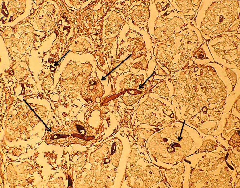
Figure 2. Invading hyphae of Aphanomyces invadans (short arrows) with development of typical mycotic granulomas (long arrows) in a histological section of muscle from of an epizootic ulcerative syndrome (EUS)-affected fish (Serranochromis robustus), north Zambia, 2014 (Grocott’s methenamine stain, x40 magnification).
Advertisement
©FAO/D. Huchzermeyer, Rhodes University

Figure 3. Epizootic ulcerative syndrome (EUS)-affected fish amongst small species (kasepa) harvested by fishermen and laid out onto drying racks in the sun, Bangweulu swamps, north Zambia, 2014.

PLATE 6 (All photos courtesy of AAHRI) PLATE 6 (All photos courtesy of AAHRI)
Histopathology of EUS-infected Thamalakane barb, Barbus thamalakanensis, Histopathology of EUS-infected Thamalakane barb, Barbus thamalakanensis,
Histopathology of EUS-infected dashtail barb Histopathology of EUS PLATE 1 Histopathology of epizootic ulcerative syndrome (EUS)-affected Thamalakane barb, Enteromius thamalakanensis, collected by scoopnet on 22 May 2007 in the shallow collected by scoopnet on 22 May 2007 in the shallow waters of Chobe-Zambezi River in Kasane, Botswana (All photos courtesy of AAHRI) collected by scoopnet on 22 May 2007 in the shallow waters of Chobe-Zambezi River in Kasane, Botswana (All photos courtesy of AAHRI) -infected dashtail showing typical mycotic granulomas showing typical mycotic granulomas waters of Chobe-Zambezi River in Kasane, Botswana
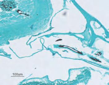
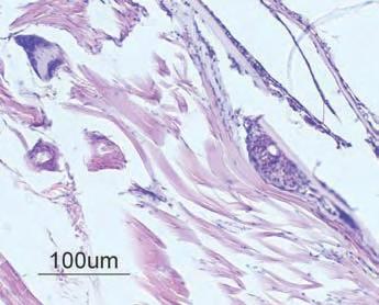
Histopathology of EUS-infected dashtail barb showing typical mycotic granulomas surrounding the invasive fungal hyphae (white arrows) in the skin layer (H&E) Histopathology of EUS-infected dashtail barb showing typical mycotic granulomas surrounding the invasive fungal hyphae (stained black, black arrows) in the skin layer (Grocott’s silver stain) Histopathology of EUS-infected dashtail barb showing typical mycotic granulomas surrounding the invasive fungal hyphae (white arrows) in the skin layer (H&E) Histopathology of EUS-infected dashtail showing typical mycotic granulomas surrounding the invasive fungal hyphae (stained black, black arrows) in the skin (Grocott’s silver stain) Histopathology of EUS-infected dashtail barb showing typical mycotic granulomas surrounding the invasive fungal hyphae (white arrows) in the skin layer (H&E) Histopathology of EUS-infected dashtail showing typical mycotic granulomas surrounding the invasive fungal hyphae (stained black, black arrows) in the skin (Grocott’s silver stain) A Histopathology of EUS-infected dashtail barb showing typical mycotic granulomas surrounding the invasive fungal hyphae (white arrows) in the skin layer (H&E) B Histopathology of EUS-infected dashtail showing typical mycotic granulomas surrounding the invasive fungal hyphae (stained black, black arrows) in the skin (Grocott’s silver stain) Histopathology of EUS-infected dashtail barb showing typical mycotic granulomas surrounding the invasive fungal hyphae (white arrows) in the skin layer (H&E) Histopathology of EUS-infected dashtail showing typical mycotic granulomas surrounding the invasive fungal hyphae (stained black, black arrows) in the skin (Grocott’s silver stain) surrounding the invasive fungal hyphae (white arrows) in the skin layer (H&E) surrounding the invasive fungal hyphae (stained black, black arrows) in the skin (Grocott’s silver stain)

AA BB CC Typical mycotic granulomas (indicated by black arrow) found in the muscle tissue of fish sample No. 1 Barbus Typical mycotic granulomas (indicated by black arrow) found in the muscle tissue of fish sample No. 1 Barbus thamalakanensis (Thamalakane barb). (A) muscle tissues with mycotic granulomas (H&E); (B) oomycete thamalakanensis (Thamalakane barb). (A) muscle tissues with mycotic granulomas (H&E); (B) oomycete hyphae penetrated into the brain of the fish; (B), (C) and (D) are stained with Grocott’s stain hyphae penetrated into the brain of the fish; (B), (C) and (D) are stained with Grocott’s stain


©FAO/AAHRI©FAO/AAHRI Results PLATE 7 Histopathology of EUS-infected dashtail barb, Barbus poechii (Steindachner, Results PLATE 7 Histopathology of EUS-infected dashtail barb, Barbus poechii (Steindachner, 21 21 A 1911), collected by scoop net on 22 May 2007 in the shallow waters of Chobe-Zambezi River in Kasane, Botswana BBA 1911), collected by scoop net on 22 May 2007 in the shallow waters of Chobe-Zambezi River in Kasane, Botswana C D PLATE 7 Histopathology of EUS-infected dashtail barb, Barbus poechii (Steindachner, 1911), collected by scoopnet on 22 May 2007 in the shallow waters of Chobe-Zambezi PLATE 7 Histopathology of EUS-infected dashtail barb, Barbus poechii (Steindachner, 1911), collected by scoop net on 22 May 2007 in the shallow waters of Chobe-Zambezi PLATE 7 Histopathology of EUS-infected dashtail barb, Barbus poechii (Steindachner, 1911), collected by scoop net on 22 May 2007 in the shallow waters of Chobe-Zambezi PLATE 7 Histopathology of EUS-infected dashtail barb, Barbus poechii (Steindachner, 1911), collected by scoop net on 22 May 2007 in the shallow waters of Chobe-Zambezi River in Kasane, Botswana PLATE 7 Histopathology of EUS-infected dashtail barb, Barbus poechii (Steindachner, 1911), collected by scoop net on 22 May 2007 in the shallow waters of Chobe-Zambezi River in Kasane, Botswana PLATE 7 Histopathology of EUS-infected dashtail barb, Barbus poechii (Steindachner, 1911), collected by scoopnet on 22 May 2007 in the shallow waters of Chobe-Zambezi PLATE 7 Histopathology of EUS-infected dashtail barb, Barbus poechii (Steindachner, 1911), collected by scoop net on 22 May 2007 in the shallow waters of Chobe-Zambezi PLATE 7 Histopathology of EUS-infected dashtail barb, Barbus poechii (Steindachner, 1911), collected by scoop net on 22 May 2007 in the shallow waters of Chobe-Zambezi PLATE 7 Histopathology of EUS-infected dashtail barb, Barbus poechii (Steindachner, 1911), collected by scoop net on 22 May 2007 in the shallow waters of Chobe-Zambezi River in Kasane, Botswana PLATE 7 Histopathology of EUS-infected dashtail barb, Barbus poechii (Steindachner, 1911), collected by scoop net on 22 May 2007 in the shallow waters of Chobe-Zambezi River in Kasane, Botswana
River in Kasane, Botswana River in Kasane, Botswana River in Kasane, Botswana River in Kasane, Botswana River in Kasane, Botswana River in Kasane, Botswana
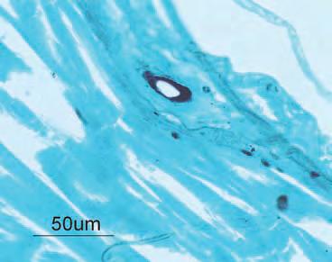
©FAO/AAHRI ©FAO/AAHRI D Histopathology of EUS-infected dashtail barb showing typical mycotic granulomas surrounding invasive fungal hyphae (white arrows) penetrating into the muscle layer (H&E) Histopathology of EUS-infected dashtail barb showing typical mycotic granulomas surrounding invasive fungal hyphae (stained black, black arrows) penetrating into the muscle layer (Grocott’s silver stain) D Histopathology of EUS-infected dashtail barb showing typical mycotic granulomas surrounding invasive fungal hyphae (white arrows) penetrating into the muscle layer (H&E). Histopathology of EUS-infected dashtail showing typical mycotic granulomas surrounding invasive fungal hyphae (stained black, black arrows) penetrating into the muscle layer (Grocott’s silver stain). D Histopathology of EUS-infected dashtail barb showing typical mycotic granulomas surrounding invasive fungal hyphae (white arrows) penetrating into the muscle layer (H&E). Histopathology of EUS-infected dashtail showing typical mycotic granulomas surrounding invasive fungal hyphae (stained black, black arrows) penetrating into the muscle layer (Grocott’s silver stain). Histopathology of EUS-infected dashtail barb showing typical mycotic granulomas surrounding invasive fungal hyphae (white arrows) penetrating into the muscle layer (H&E). Histopathology of EUS-infected dashtail showing typical mycotic granulomas surrounding invasive fungal hyphae (stained black, black arrows) penetrating into the muscle layer (Grocott’s silver stain). D Histopathology of EUS-infected dashtail barb showing typical mycotic granulomas surrounding invasive fungal hyphae (white arrows) penetrating into the muscle layer (H&E). Histopathology of EUS-infected dashtail showing typical mycotic granulomas surrounding invasive fungal hyphae (stained black, black arrows) penetrating into the muscle layer (Grocott’s silver stain). Histopathology of EUS-infected dashtail barb showing typical mycotic granulomas surrounding invasive fungal hyphae (white arrows) penetrating into the muscle layer (H&E). Histopathology of EUS-infected dashtail showing typical mycotic granulomas surrounding invasive fungal hyphae (stained black, black arrows) penetrating into the muscle layer (Grocott’s silver stain). PLATE 2 Typical mycotic granulomas (indicated by black arrow) found in the muscle tissue of fish sample No. 1 Enteromius thamalakanensis (Thamalakane barb). (A) muscle tissues with mycotic granulomas (H&E); (B) oomycete hyphae penetrated into the brain of the fish; (B), (C) and (D) are stained with Grocott’s stain. (All photos courtesy of AAHRI) CC (All photos courtesy of AAHRI) Histopathology of epizootic ulcerative syndrome (EUS)-affected dashtail barb, Typical mycotic granulomas (indicated by black arrow) found in the muscle tissue of fish sample No. 1 Barbus thamalakanensis (Thamalakane barb). (A) muscle tissues with mycotic granulomas (H&E); (B) oomycete Typical mycotic granulomas (indicated by black arrow) found in the muscle tissue of fish sample No. 1 Barbus thamalakanensis (Thamalakane barb). (A) muscle tissues with mycotic granulomas (H&E); (B) oomycete Enteromius poechii. Chobe River in Kasane, Histopathology of EUS-infected dashtail barb Histopathology of EUS-infected hyphae penetrated into the brain of the fish; (B), (C) and (D) are stained with Grocott’s stain hyphae penetrated into the brain of the fish; (B), (C) and (D) are stained with Grocott’s stainHistopathology of EUS-infected dashtail barb Histopathology of EUS-infected dashtail dashtail barb barb ©FAO/AAHRI Dashtail barb, Barbus poechii (Steindachner, 1911), exhibiting haemorrhagic dermatitis posterior to anus and towards the caudal peduncle Dashtail barb, Barbus poechii (Steindachner, 1911), exhibiting haemorrhagic dermatitis posterior to anus and towards the caudal peduncle.Dashtail barb, Barbus poechii (Steindachner, 1911), exhibiting haemorrhagic dermatitis posterior to anus and towards the caudal peduncle. Dashtail barb, Barbus poechii (Steindachner, 1911), exhibiting haemorrhagic dermatitis posterior to anus and towards the caudal peduncle.Dashtail barb, Barbus poechii (Steindachner, 1911), exhibiting haemorrhagic dermatitis posterior to anus and towards the caudal peduncle. Dashtail barb, Barbus poechii (Steindachner, 1911), exhibiting haemorrhagic dermatitis posterior to anus and towards the caudal peduncle. Botswana, 2007. Dashtail barb, Enteromius poechii, exhibiting haemorrhagic dermatitis posterior to anus and towards the caudal peduncle. Histopathology of EUS-infected dashtail barb showing typical mycotic granulomas surrounding the invasive fungal hyphae (white arrows) in the skin layer (H&E) Histopathology of EUS-infected dashtail barb showing typical mycotic granulomas surrounding the invasive fungal hyphae (stained black, black arrows) in the skin layer (Grocott’s silver stain) Histopathology of EUS-infected dashtail barb showing typical mycotic granulomas surrounding the invasive fungal hyphae (white arrows) in the skin layer (H&E) Histopathology of EUS-infected dashtail barb showing typical mycotic granulomas surrounding the invasive fungal hyphae (stained black, black arrows) in the skin layer (Grocott’s silver stain) Histopathology of EUS-infected dashtail barb showing typical mycotic granulomas surrounding the invasive fungal hyphae (white arrows) in the skin layer (H&E) Histopathology of EUS-infected dashtail barb showing typical mycotic granulomas surrounding the invasive fungal hyphae (stained black, black arrows) in the skin layer (Grocott’s silver stain) Histopathology of EUS-infected dashtail barb showing typical mycotic granulomas surrounding the invasive fungal hyphae (white arrows) in the skin layer (H&E) Histopathology of EUS-infected dashtail barb showing typical mycotic granulomas surrounding the invasive fungal hyphae (stained black, black arrows) in the skin layer (Grocott’s silver stain) Histopathology of EUS-infected dashtail barb showing typical mycotic granulomas surrounding the invasive fungal hyphae (white arrows) in the skin layer (H&E) Histopathology of EUS-infected dashtail barb showing typical mycotic granulomas surrounding the invasive fungal hyphae (stained black, black arrows) in the skin layer (Grocott’s silver stain) showing typical mycotic granulomas surrounding the invasive fungal hyphae (white arrows) in the skin layer (H&E) showing typical mycotic granulomas surrounding the invasive fungal hyphae (stained black, black arrows) in the skin layer (Grocott’s silver stain) Histopathology of EUS-infected dashtail barb showing typical mycotic granulomas surrounding the invasive fungal hyphae (white arrows) in the skin layer (H&E) Histopathology of EUS-infected dashtail barb showing typical mycotic granulomas surrounding the invasive fungal hyphae (stained black, black arrows) in the skin layer (Grocott’s silver stain) Histopathology of EUS-infected dashtail barb showing typical mycotic granulomas surrounding the invasive fungal hyphae (white arrows) in the skin layer (H&E) Histopathology of EUS-infected dashtail barb showing typical mycotic granulomas surrounding the invasive fungal hyphae (stained black, black arrows) in the skin layer (Grocott’s silver stain) Histopathology of EUS-infected dashtail barb showing typical mycotic granulomas surrounding the invasive fungal hyphae (white arrows) in the skin layer (H&E) Histopathology of EUS-infected dashtail barb showing typical mycotic granulomas surrounding the invasive fungal hyphae (stained black, black arrows) in the skin layer (Grocott’s silver stain) Histopathology of EUS-infected dashtail barb showing typical mycotic granulomas surrounding the invasive fungal hyphae (white arrows) in the skin layer (H&E) Histopathology of EUS-infected dashtail barb showing typical mycotic granulomas surrounding the invasive fungal hyphae (stained black, black arrows) in the skin layer (Grocott’s silver stain) Histopathology of EUS-infected dashtail barb showing typical mycotic granulomas surrounding the invasive fungal hyphae (white arrows) in the skin layer (H&E) Histopathology of EUS-infected dashtail barb showing typical mycotic granulomas surrounding the invasive fungal hyphae (stained black, black arrows) in the skin layer (Grocott’s silver stain) showing typical mycotic granulomas surrounding the invasive fungal hyphae (white arrows) in the skin layer (H&E) showing typical mycotic granulomas surrounding the invasive fungal hyphae (stained black, black arrows) in the skin layer (Grocott’s silver stain) Histopathology of EUS-infected dashtail barb Histopathology of EUS-infected dashtail barb Histopathology of EUS-infected dashtail barb Histopathology of EUS-infected dashtail barb showing typical mycotic granulomas surrounding showing typical mycotic granulomas surrounding showing typical mycotic granulomas surrounding showing typical mycotic granulomas surrounding invasive fungal hyphae (white arrows) penetrating invasive fungal hyphae (white arrows) penetrating invasive fungal hyphae (stained black, black arrows) invasive fungal hyphae (stained black, black arrows) into the muscle layer (H&E) into the muscle layer (H&E) penetrating into the muscle layer (Grocott’s silver penetrating into the muscle layer (Grocott’s silver stain) stain)


©FAO/AAHRI

©FAO/AAHRI Histopathology of epizootic ulcerative syndrome (EUS)-affected dashtail barb showing typical mycotic granulomas surrounding invasive fungal hyphae (white arrow) penetrating into the muscle layer (H&E). Histopathology of epizootic ulcerative syndrome (EUS)-affected dashtail barb showing typical mycotic granulomas surrounding invasive fungal hyphae (stained black, black arrow) penetrating into the muscle layer (Grocott´s silver stain). Histopathology of EUS-infected dashtail barb showing typical mycotic granulomas surrounding invasive fungal hyphae (white arrows) penetrating into the muscle layer (H&E). Histopathology of EUS-infected dashtail barb showing typical mycotic granulomas surrounding invasive fungal hyphae (stained black, black arrows) penetrating into the muscle layer (Grocott’s silver stain). Histopathology of EUS-infected dashtail barb showing typical mycotic granulomas surrounding invasive fungal hyphae (white arrows) penetrating into the muscle layer (H&E). Histopathology of EUS-infected dashtail barb showing typical mycotic granulomas surrounding invasive fungal hyphae (stained black, black arrows) penetrating into the muscle layer (Grocott’s silver stain). Histopathology of EUS-infected dashtail barb showing typical mycotic granulomas surrounding invasive fungal hyphae (white arrows) penetrating into the muscle layer (H&E). Histopathology of EUS-infected dashtail barb showing typical mycotic granulomas surrounding invasive fungal hyphae (stained black, black arrows) penetrating into the muscle layer (Grocott’s silver stain). Histopathology of EUS-infected dashtail barb showing typical mycotic granulomas surrounding invasive fungal hyphae (white arrows) penetrating into the muscle layer (H&E). Histopathology of EUS-infected dashtail barb showing typical mycotic granulomas surrounding invasive fungal hyphae (stained black, black arrows) penetrating into the muscle layer (Grocott’s silver stain). Histopathology of EUS-infected dashtail barb showing typical mycotic granulomas surrounding invasive fungal hyphae (white arrows) penetrating into the muscle layer (H&E). Histopathology of EUS-infected dashtail barb showing typical mycotic granulomas surrounding invasive fungal hyphae (stained black, black arrows) penetrating into the muscle layer (Grocott’s silver stain). Histopathology of EUS-infected dashtail barb showing typical mycotic granulomas surrounding invasive fungal hyphae (white arrows) penetrating into the muscle layer (H&E). Histopathology of EUS-infected dashtail barb showing typical mycotic granulomas surrounding invasive fungal hyphae (stained black, black arrows) penetrating into the muscle layer (Grocott’s silver stain). Histopathology of EUS-infected dashtail barb showing typical mycotic granulomas surrounding invasive fungal hyphae (white arrows) penetrating into the muscle layer (H&E). Histopathology of EUS-infected dashtail barb showing typical mycotic granulomas surrounding invasive fungal hyphae (stained black, black arrows) penetrating into the muscle layer (Grocott’s silver stain). Histopathology of EUS-infected dashtail barb showing typical mycotic granulomas surrounding invasive fungal hyphae (white arrows) penetrating into the muscle layer (H&E). Histopathology of EUS-infected dashtail barb showing typical mycotic granulomas surrounding invasive fungal hyphae (stained black, black arrows) penetrating into the muscle layer (Grocott’s silver stain). Histopathology of EUS-infected dashtail barb showing typical mycotic granulomas surrounding invasive fungal hyphae (white arrows) penetrating into the muscle layer (H&E). Histopathology of EUS-infected dashtail barb showing typical mycotic granulomas surrounding invasive fungal hyphae (stained black, black arrows) penetrating into the muscle layer (Grocott’s silver stain). Histopathology of EUS-infected dashtail barb showing typical mycotic granulomas surrounding invasive fungal hyphae (white arrows) penetrating into the muscle layer (H&E). Histopathology of EUS-infected dashtail barb showing typical mycotic granulomas surrounding invasive fungal hyphae (stained black, black arrows) penetrating into the muscle layer (Grocott’s silver stain).


