4 minute read
OpenFlexure Microscope


© B r i a n C a l l i n g h a m
Dr Richard Bowman
Advertisement
Richard is a Royal Society University Research Fellow and Proleptic Lecturer at the University of Bath. He runs a research group primarily interested in microscopy automating lab experiments using open-source hardware and software.
openflexure.org
Dr Joel Collins
Joel is a postdoctoral researcher at the University of Bath, primarily developing opensource software to control the OpenFlexure Microscope and automate collection of clinical data in Tanzania.
By focusing on the design of this open-source, 3D-printed microscope, Dr Richard Bowman’s team is helping scientists to inexpensively detect disease, as David Crookes explains
Research-grade microscopes don’t come
cheap. They cost upwards of £30,000 for a decent model with a motorised stage – putting them outside the scope of many a pocket. As such, many attempts have been made to create low-cost alternatives, and one project – OpenFlexure – could revolutionise the detection of disease in developing countries.
Dr Richard Bowman is spearheading the creation of an open-source, 3D-printed microscope that’s able to be adapted for use in labs, schools, and the home. He was inspired by his time as a Research Fellow in the NanoPhotonics Centre at the University of Cambridge, where an early attempt at such a project was falling short because most of the mechanism was based on linear bearings and metal rods.
“The bulk of the components weren’t printable and I became curious as to how much of a microscope’s mechanism you could print,” he says. “My first version had basic focus control and an extension tube for a Raspberry Pi Camera Module to turn the stock webcam lens into a basic, but functional, microscope objective lens.” The aim is to create an easily replicable scientific instrument.
Time to focus
Initially, Richard sought an ideal way of moving a sample around and picking a region to view. Rather than use sliding rails which require precise machining to be smooth, the eventual design was based on the flexibility of plastic: samples are placed on a table with bendable legs that allow for controlled focus and movement on the X and Y axes.
“It uses some fairly simple geometry to convert flexible hinges into linear motion,” explains Dr Joel Collins, who later joined the project at its home at the University of Bath. “It also means we can achieve really fine sample manipulation of tens of nanometres, for orders of magnitude cheaper than most commercial microscopes.”
Unlike traditional microscopes, the project uses an upside-down design. The camera is at the base and the viewing lens is above with the light source at the top. It makes the microscope more stable. “You can arrange things so the sample is consistently close to being in focus when you place it on the microscope’s stage, which is nice,” Richard says.
Such work has helped to keep costs down. As a consequence, a student group that became WaterScope saw its potential for cheaply identifying early-stage bacterial contamination in water. Another student project demonstrated how
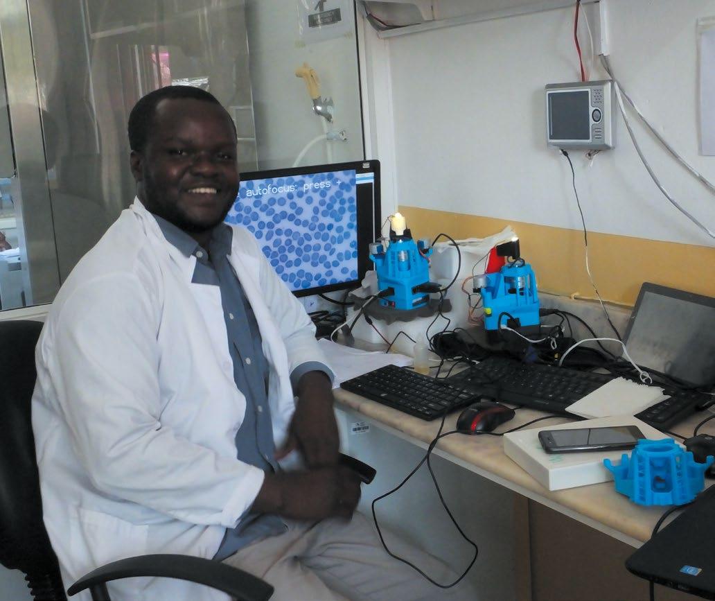
Joram Mduda is a collaborator at the Ifakara Health Institute, introducing the microscope into Tanzanian clinics for malaria diagnosis. 3D-printed components replace damaged parts
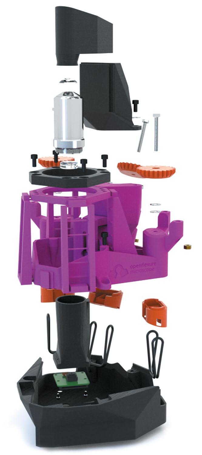
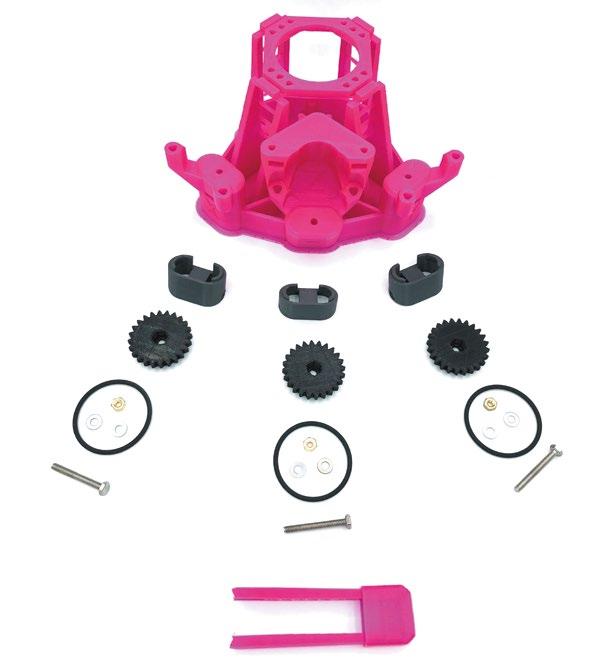
Non-printed parts have been minimised by using a ‘flexure’ mechanism to move the stage, so the whole mechanism is printable
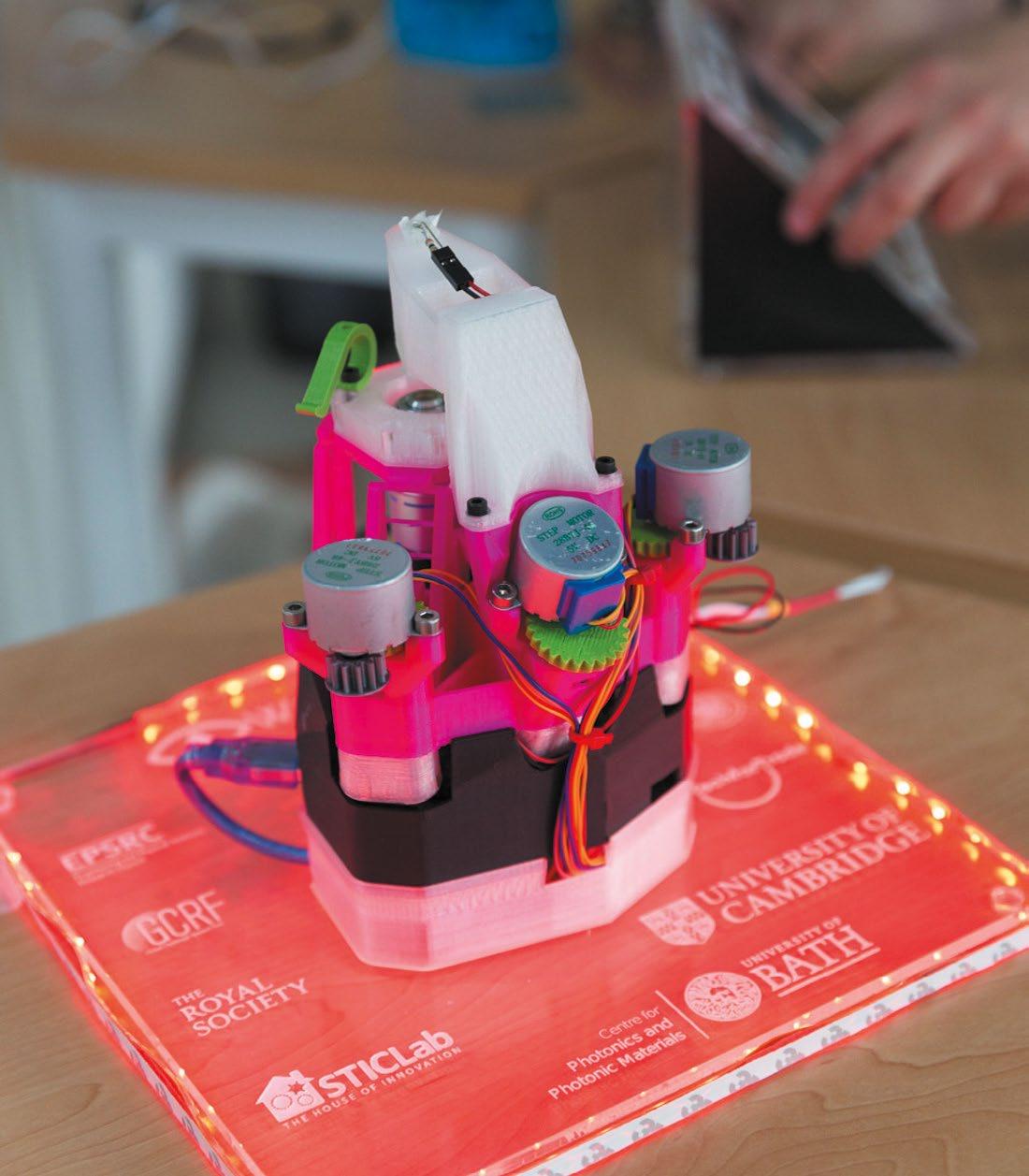
Funding from the Royal Academy of Engineering and the Royal Society is being used to develop educational materials to enable the microscope to be used in schools
it could be produced in Tanzania by STICLab. This led to a study which found the microscope could be used for malaria diagnosis.
Lighting the way
In creating the project, Richard’s team has benefited from Raspberry Pi’s bustling community. “There are great libraries for interacting with physical hardware, such as picamera and GPIO Zero,” says Joel.
“We created an HTTP API in Python and a graphical client in JavaScript that are both served by a Raspberry Pi,” he continues. “Since we have a full, powerful Linux machine in the microscope, we have many new ways to interact with the software.”
It’s possible to create an offline, standalone microscope with a fully featured interface. “If you connect it directly to your laptop via Ethernet, you can control it ‘headless’ like that, or you could have a fleet of microscopes connected to a LAN, and our software will discover all the nearby microscopes and let you control them all from a single machine,” reveals Joel.
There no doubting its impact. OpenFlexure Microscopes have been recorded on every continent, including Antarctica. The biggest challenge, however, is documentation and communication. Richard says: “Building a piece of hardware is more involved than installing prebuilt software and I have a new respect for anyone who works at IKEA – their instructions must have been so carefully tested and optimised.”
The microscope uses three screws for fine control so that the sample can be moved along the X and Y axes and focused by moving on the Z axis
The microscope uses plastic flexures to ensure motion is free from friction and vibration; the condenser mount at the top houses a condenser lens and illumination LED
The eight-megapixel Raspberry Pi Camera Module v2 is used as the microscope’s image sensor, with the electronics (including Raspberry Pi) housed in the base
Quick FACTS
> The microscope is ‘laboratory-grade’
> It’s roughly the size of a bag of sugar
> Control it remotely over Ethernet or WiFi
> It’s designed for easy replication
> More than 100 have been printed in
Tanzania and Kenya