


MEDICAL MASSAGE THERAPY TO BOOST RECOVERY AND MOBILITY
OUR PRESENTATION AND WORKSHOP OBJECTIVES


This workbook provides a holistic approach to treating stroke patients and those with hemiplegia using massage therapy, natural remedies, movement therapy, and integrative wellness strategies. It is designed for massage therapists seeking to expand their knowledge and skillset in stroke rehabilitation.

Participants will gain a comprehensive understanding of stroke as a neurological condition, including the principles of neuroplasticity, brain function, and the musculoskeletal implications of stroke recovery.
Participants will learn how to incorporate holistic approaches such as herbal medicine, anti-inflammatory nutrition, breathwork, and vagus nerve stimulation to support stroke recovery and enhance patient outcomes.
Participants will develop hands-on skills in stroke recovery massage, including lymphatic drainage, warm infused oil massage, reciprocal movement training, tapping techniques, and slow guided movement therapy.
Participants will learn to assess stroke patients' needs and develop individualized, integrative treatment plans incorporating massage, movement therapy, herbal medicine, and light therapy to support longterm recovery.
Gregory T. Lawton, D.N., D.C., D.Ac., N.D.
Dr. Lawton is national board certified in chiropractic, physiotherapy, radiology, and acupuncture.
Former vice president of large physical therapy group practice.
Founder of the Blue Heron Academy.
Founder of American Health Source.
Over 50 years in private practice at Clinical Health Maintenance.
Undergraduate and graduate studies in psychology at Arizona State University, a graduate of the National College of Naprapathic Medicine, the National University of Health Sciences, and the International Medical Acupuncture Association.
Gregory T. Lawton, D.N., D.C., D.Ac., N.D.
Dr. Lawton is national board certified in chiropractic, physiotherapy, radiology, and acupuncture.
40+ years of research in the field of brain, spinal and peripheral neurology.
Author of over 100 books and numerous health science articles.
Licensed in 3 healthcare professions in 3 US states.
50+ years as a health science educator.
Former vice president of large medical psychology practice.
Former youth probation officer and co-founder of the Berrien County Drug Rehabilitation Center


Functional chiropractic medicine is a holistic approach to healthcare that combines traditional chiropractic techniques with principles of functional medicine. This approach aims to treat the root causes of health issues rather than just addressing symptoms. Functional chiropractic medicine seeks to create a balanced and healthy state within the body by addressing the interconnected systems and promoting overall wellness. This approach can be particularly beneficial for chronic conditions, complex health issues, and patients looking for a natural and integrative approach to their health.


Holistic Assessment: Practitioners conduct a comprehensive evaluation of the patient, considering physical, biochemical, and emotional factors. This may include a detailed health history, physical examination, laboratory tests, and lifestyle assessments.
Spinal Adjustments: Traditional chiropractic adjustments are used to correct misalignments (subluxations) in the spine, which can improve nervous system function and overall health.
Functional Medicine Principles: This involves identifying and addressing underlying imbalances in the body. Functional medicine looks at aspects such as gut health, hormone levels, nutrition, and detoxification pathways to understand the root causes of disease.
Personalized Treatment Plans: Based on the holistic assessment, a customized treatment plan is developed. This plan may include dietary recommendations, nutritional supplements, exercise programs, stress management techniques, and lifestyle modifications in addition to chiropractic adjustments.


Patient Education and Empowerment: Functional chiropractors place a strong emphasis on educating patients about their health and wellness. They empower patients to take an active role in their healing process through lifestyle changes and preventive measures.
Integration with Other Healthcare Providers: Functional chiropractors often work collaboratively with other healthcare professionals, such as primary care physicians, nutritionists, and physical therapists, to provide comprehensive care.
Focus on Prevention: Preventive care is a key component, aiming to optimize health and prevent the onset of chronic diseases by maintaining proper alignment, reducing inflammation, and supporting the body’s natural healing processes.

About Your Presenter
“For hundreds of years and countless centuries herbal medicine has been an important part of the practice of traditional healthcare as well as massage and manual therapy. During the 1970's I had the great privilege of training with physicians and therapists who went to school or practiced during the late 1800's and early 1900's. A common denominator among them all was their use of herbal medicines for the treatment of their patients. Over the last 50 years of my teaching career, I have been bringing this knowledge to my students and teaching them about the value and benefits of herbal medicines and preparations, whether used internally or externally.”


The Blue Heron Academy of Healing Arts and Sciences mission is to build a healing community founded on unity, diversity, and racial harmony. Our mission is to cultivate a nurturing and inclusive community where every individual feels valued, respected, and empowered. We are dedicated to fostering unity and racial harmony by promoting understanding, compassion, and mutual support among all members of our diverse society. Through active engagement, individual and community service, education, and collaborative efforts, we strive to create an environment where healing and growth are possible for everyone. Together, we aim to break down barriers, celebrate our differences, and build a foundation of trust and solidarity, ensuring a brighter, more equitable future for all.
"Building a Healing Community" is a visionary 50-year initiative dedicated to uniting people of all backgrounds and races through the transformative power of holistic and alternative healing arts. This endeavor aims to create a space where diverse healing traditions converge, fostering understanding, compassion, and mutual respect among individuals and communities. By embracing practices such as herbal medicine, yoga, meditation, massage, and other integrative approaches, the initiative seeks to break down barriers and promote a sense of shared purpose in the pursuit of health and wellbeing. Through collective healing, education, and cultural exchange, "Building a Healing Community" aspires to cultivate a more inclusive, connected, and compassionate world, where everyone can experience the profound benefits of holistic wellness.


I recommend a system-by-system approach to patient evaluation and assessment based upon observation, palpation, and questioning. Dr. Lawton


SOAP notes provide a structured format for documenting patient progress and treatment.
SOAP stands for: S (Subjective): Patient’s reported symptoms, concerns, and goals. O (Objective): Measurable findings, including range of motion (ROM), muscle tone, swelling, and postural analysis. A (Assessment): Therapist’s professional analysis of the patient’s condition, progress, and response to treatment. P (Plan): Treatment strategy, including future sessions, exercises, and patient self-care recommendations.
S: Patient reports right arm weakness post-stroke, rates discomfort 4/10.
O: Increased spasticity noted in RUE. PROM limited to 50% shoulder abduction.
A: Spasticity and limited ROM affecting ADLs. Positive response to myofascial release.
P: Continue weekly massage focusing on reducing spasticity. Introduce reciprocal movement exercises. Patient instructed on home stretching.
ROM: Range of Motion
MMT: Manual Muscle Testing
P/AROM: Passive/Active Range of Motion
B/L: Bilateral
Tx: Treatment
F/U: Follow-Up
Dr: Doctor
Sd: Said
Dx: Diagnosis
CNS: Central Nervous System
PNS: Peripheral Nervous System
Sp: Spasticity
TTP: Tender to Palpation
Lat: Lateral
Pt: Patient
Dr sd pt HA = Doctor said patient has headaches.
Dr sd pt LBP = Doctor said patient has low back pain (L5/S1).
Pt sd dr sd LBP = Patient said doctor said they have low back pain (L5/SI)


Patient Intake


There are two main requirements for medical insurance billing:
A medical necessity for patient care (a diagnosis).
Documentation of ongoing improvement and clinical results.
If these two conditions are not met, medical insurance providers will not cover claims.


97124: Therapeutic procedure, one or more areas, each 15 minutes; massage, including effleurage, petrissage, and/or tapotement (stroking, compression, percussion).
97140: Manual therapy techniques (e.g., mobilization/manipulation, manual lymphatic drainage, manual traction), one or more regions, each 15 minutes.


Remember to cite the exact treatment procedures in your claim to ensure coverage.
Additionally, avoid citing both CPT codes 97124 and 97140 without modifier code 59, which indicates distinct and different procedures performed on the same individual within a single day or across a few days.
CPT Code 97112: Neuromuscular Reeducation
CPT Codes 97010: Hot/Cold Packs
CPT Codes 97110: Therapeutic Exercise

Remember to cite the exact treatment procedures in your claim to ensure coverage.
Additionally, avoid citing both CPT codes 97124 and 97140 without modifier code 59, which indicates distinct and different procedures performed on the same individual within a single day or across a few days.
CPT Code 97112: Neuromuscular Re-education
CPT Codes 97010: Hot/Cold Packs
CPT Codes 97110: Therapeutic Exercise

Neuroplasticity is the brain’s ability to reorganize and form new neural connections throughout life, particularly following an injury such as a stroke. This adaptive process allows the brain to compensate for lost functions by rewiring undamaged neurons to take over the roles of affected areas. Neuroplasticity plays a crucial role in stroke recovery, influencing motor function, cognitive abilities, and sensory processing. Rehabilitation strategies such as repetitive movement training, sensory stimulation, and mindfulness practices enhance neuroplasticity, helping stroke survivors regain lost abilities over time. Nutritional support, herbal medicine, and manual therapies can further optimize the neuroplastic response, promoting long-term neurological healing and functional improvement.
More important than a patient’s condition is understanding who they are. A patient’s pre-existing health, lifestyle, diet, drug use and emotional resilience significantly influence their recovery journey. An active and independent individual with strong social support may experience a different recovery process than someone managing chronic illnesses, high stress, or social isolation. Factors such as cognitive and emotional well-being, support systems, and lifestyle choices before the stroke shape the rehabilitation process. Acknowledging these aspects allows therapists to provide tailored, patient-centered care that is both effective and compassionate.
Understanding Stroke and Recovery:
Stroke is a complex neurological condition that affects both the nervous and musculoskeletal systems. There are different types of strokes, including ischemic, hemorrhagic, and transient ischemic attacks (TIAs). Recovery relies heavily on neuroplasticity, the brain's ability to reorganize and form new neural connections. Despite its importance, stroke rehabilitation faces significant challenges. Limited therapy time due to insurance constraints, therapist fatigue, and a lack of long-term treatment goals often hinder optimal recovery. Additionally, many therapists approach stroke rehabilitation purely as a musculoskeletal issue without accounting for neurological factors. Plateaus and regressions in recovery are common, yet many therapists are unprepared for these challenges. Quick discharges due to factory medicine models and a lack of trauma-informed care further complicate patient progress.
Consider this:
What were the pre-existing conditions prior to the stroke incident?
Was the patient obese?
Did the patient have diabetes, heart disease, or high blood pressure?
Was the patient an alcoholic or did they have a drug problem?
A well-rounded approach to stroke rehabilitation incorporates holistic therapies, including herbal and nutritional support. Specific herbs such as turmeric (antiinflammatory), pomegranate (vascular support), sage (cognitive enhancer), and skullcap (nervous system relaxant) can aid in stroke recovery. Bioflavonoids strengthen capillaries, while kava kava and blue vervain help with muscle relaxation and reducing anxiety. Nutrition also plays a crucial role, with an emphasis on avoiding inflammatory foods and increasing omega-3 intake from sources like flaxseed, walnuts, and salmon. Hydration and polyphenol-rich foods support neuroprotection and overall well-being.
Turmeric (Curcuma longa) is a powerful anti -inflammatory and antioxidant herb known for its neuroprotective properties. The active compound, curcumin, helps reduce neuroinflammation, protects against oxidative stress, and promotes neuroplasticity. It also improves vascular health and circulation, aiding post-stroke recovery.
Turmeric is available in capsules, tinctures, and powdered form, with a recommended dosage of 500 - 4000 mg per day of standardized curcumin powdered extract.
Pomegranate (Punica granatum) is rich in antioxidants and supports vascular health by improving endothelial function and reducing oxidative stress. Regular consumption can enhance brain circulation, helping prevent secondary strokes and cognitive decline. It can be consumed as fresh fruit, juice, or extract, with a recommended dosage of 8-12 oz of juice daily or 500 - 1000 mg of powdered extract in a capsule.
Flavonoids, a diverse group of plant compounds, provide significant vascular protection and anti -inflammatory benefits. Quercetin, found in apples, onions, and green tea, is a potent antioxidant that reduces oxidative stress and inflammation. Citrus bioflavonoids, including hesperidin and rutin, support capillary integrity and improve circulation. Green tea catechins enhance endothelial function, supporting heart and brain health. The recommended dosage of flavonoid supplements varies but typically ranges between 500 - 1000 mg per day, depending on the specific compound.
Kava Kava(Piper methysticum) is an anxiolytic and muscle relaxant that helps reduce post-stroke muscle tension and stress. It promotes relaxation and better movement coordination, making it useful in stroke recovery. It is available in tea, tincture, and capsule form, with a recommended dosage of 100 - 250 mg per day of kavalactones, the active compounds.
Blue Vervain (Verbena hastata) is a nervous system tonic that supports emotional balance, reduces muscle spasms, and promotes relaxation. It is particularly beneficial for individuals dealing with post-stroke stress or anxiety. Blue Vervain is available as a tea, tincture, or capsule, with a recommended dosage of 100 - 300 mg per day or 2040 drops of tincture.
Sage (Salvia officinalis) has been traditionally used as a cognitive enhancer and neuroprotective agent. It improves neurotransmitter function, making it beneficial for stroke survivors experiencing cognitive impairment. Sage can be taken as a tea, tincture, or capsule. The recommended dosage is 300 - 600 mg powdered extract in a capsule per day or 1 -2 teaspoons of dried herb in tea.
Skullcap (Scutellarialateriflora) is a nervous system relaxant known for its ability to reduce anxiety and neuroinflammation. It supports relaxation and promotes neural recovery, making it an excellent addition to stroke rehabilitation. It is commonly taken as a tea, tincture, or powdered extract in a capsule, with a recommended dosage of 200400 mg per day or 30 -60 drops of tincture.
A stroke recovery diet should include omega-3 fatty acids, polyphenol-rich foods, and adequate hydration. Omega-3s, found in flaxseed, walnuts, salmon, sardines, and chia seeds, reduce inflammation, improve cognitive function, and support brain cell regeneration. The recommended intake is 1-2 grams of DHA/EPA per day.
Polyphenol-rich foods, including green tea, blueberries, dark chocolate, and red grapes, support neuroprotection and vascular health. These compounds enhance brain recovery and reduce the risk of secondary strokes. Hydration is also crucial in stroke rehabilitation, ensuring proper circulation and cellular function.
It is important to avoid pro-inflammatory foods such as processed foods, refined sugar, trans fats, and excessive dairy. These dietary modifications help reduce systemic inflammation, improve overall cardiovascular health, and aid stroke recovery.
It is equally important to avoid certain oils and foods that are harmful to brain cells, neurons, and nerves. Processed vegetable oils such as soybean oil, corn oil, and canola oil are high in pro-inflammatory omega-6 fatty acids, which can contribute to neuroinflammation and oxidative stress. Hydrogenated oils and trans fats found in margarine, fast food, and commercial baked goods have been linked to neurodegeneration and cognitive decline. Refined sugars and high-fructose corn syrup cause blood sugar spikes and increase inflammation, negatively impacting brain function. Artificial sweeteners such as aspartame and saccharin may also have neurotoxic effects. Additionally, excessive dairy consumption, particularly from conventionally processed sources, can contribute to inflammation. Limiting processed meats, fried foods, and food additives such as monosodium glutamate (MSG) can further support neurological health and reduce stroke risk. By eliminating these harmful substances, individuals recovering from a stroke can create an environment that promotes brain healing and overall well-being.
Extra Virgin Olive Oil (Best Choice)
Composition: High in monounsaturated fats (~73% oleic acid), polyphenols, and vitamin E.
Benefits: Extra virgin olive oil (EVOO) is the gold standard for brain health due to its high content of monounsaturated fats, which reduce inflammation and oxidative stress. The presence of polyphenols and antioxidants supports vascular integrity, enhances circulation, and protects neurons from damage. Regular consumption has been linked to improved cognitive function and reduced risk of neurodegenerative diseases.
Recommended Use: Drizzle over salads, use in low-heat cooking, or consume raw for maximum benefits.
Flaxseed Oil
Composition: Rich in alpha-linolenic acid (ALA) (~50-60% omega-3 fatty acids).
Benefits: Flaxseed oil is an excellent plant-based source of omega-3 fatty acids, which play a key role in reducing neuroinflammation and supporting neuronal repair. ALA, the primary fatty acid in flaxseed oil, is converted into EPA and DHA, essential for brain function and stroke recovery.
Recommended Use: Best used cold in dressings, smoothies, or drizzled over cooked foods.
Walnut Oil
Composition: High in polyunsaturated fats (~63% omega-6, 10% omega-3), vitamin E, and polyphenols.
Benefits: Walnut oil contains a balanced ratio of omega-3 to omega-6 fatty acids, supporting brain function and reducing inflammation. It is also rich in polyphenols that help protect neurons from oxidative stress, making it beneficial for long-term brain. health and stroke recovery.
Recommended Use: Ideal for salad dressings, drizzling over foods, or low-heat cooking.
Composition: High in monounsaturated fats (~70% oleic acid), vitamin E, and lutein.
Benefits: Avocado oil supports cognitive function and stroke recovery by reducing inflammation and promoting healthy blood flow. It is also rich in antioxidants like lutein, which protect brain cells from oxidative stress.
Recommended Use: Suitable for high-heat cooking, salad dressings, or as a substitute for butter.
Composition: High in medium-chain triglycerides (MCTs) (~50-60% lauric acid).
Benefits: Coconut oil provides a quick source of energy for brain cells and may help support cognitive function in stroke patients. However, its high saturated fat content makes it a less preferred option for long-term cardiovascular health.
Recommended Use: Best used in moderation for occasional cooking or blended into beverages like coffee or smoothies.
Choosing the right oils can have a profound impact on stroke recovery and brain function. Extra virgin olive oil stands out as the best choice due to its high monounsaturated fat content and polyphenol-rich composition. Flaxseed and walnut oils provide essential omega-3 fatty acids, supporting neuroprotection and reducing inflammation. Avocado oil is another strong option, offering stability for cooking and antioxidant benefits. Coconut oil is beneficial for immediate brain energy. Incorporating these oils into a balanced diet while avoiding pro-inflammatory vegetable oils ensures optimal brain health and aids in stroke recovery.
A Stroke Recovery Diet should focus on whole foods, omega-3 fatty acids, polyphenolrich foods, and hydration. The Mediterranean, Ornish, and Pritikin diets are three wellresearched dietary approaches that focus on improving cardiovascular health, reducing inflammation, and promoting overall well-being. Each of these diets emphasizes whole, nutrient-dense foods while minimizing processed foods, unhealthy fats, and excessive sugars. However, they differ in their specific guidelines and dietary compositions. This document provides an overview and comparison of these three dietary plans.
If you don’t have good fuel for the cells and neurons of the body, you cannot have good recovery.
Key Features of these Diets:
High consumption of whole grains, fruits, vegetables, and legumes.
Low intake of fat (Modify this feature for stroke recovery).
Exclusion of all animal products except for non-fat dairy.
Avoidance of refined carbohydrates, oils, and processed foods
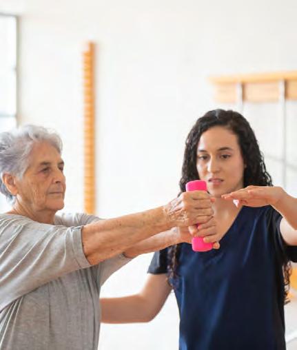
Massage therapy and manual techniques tailored for stroke patients include warm infused oil massage, lymphatic drainage, and slow guided movement therapy. Reciprocal movement training, tapping techniques, rolling ball massage, and massage hammer therapy help stimulate proprioception, improve coordination, and reestablish sensory input. Eye exercises and eye massage can further enhance neurological function and balance.
Vagus nerve stimulation through breathwork, music therapy, and cold-water immersion plays an essential role in neurological recovery. Guided diaphragmatic breathing and NADA acupuncture techniques and dry needling can help regulate the nervous system and promote relaxation. Incorporating chi kung and movement therapy into treatment enhances postural stability, core strength, and neuromuscular control, further supporting the rehabilitation process.
Warm Herbal Infused Oil Massage or Packs:
Warm infused oil massage is a highly effective technique for stroke patients as it enhances circulation, reduces spasticity, and nourishes the skin. Using herbal-infused oils such as sage, skullcap, and mugwort can help promote blood flow, relax muscles, and support nerve health. Sage is known for its anti -inflammatory and circulation-boosting properties, while skullcap helps to calm the nervous system and reduce muscle spasms. Mugwort is traditionally used to stimulate circulation and promote warmth in the affected areas. The warmth from the oil penetrates deeply into the tissues, encouraging muscle relaxation and improved sensory reintegration, making it particularly beneficial for stroke survivors working to regain mobility and function.
Lymphedema is a common post-stroke complication that results from lymphatic congestion, causing swelling in the affected areas. Manual lymphatic drainage (MLD) is a medical massage technique that encourages lymph fluid movement, reducing swelling and discomfort. Specialized strokes and light pressure are used to facilitate lymph circulation, preventing fluid buildup and improving overall limb function. This treatment is crucial for stroke patients experiencing limited mobility and circulation issues.
Stroke Recovery Massage:
Stroke recovery massage incorporates a combination of fascial-myo release, lymphatic drainage, and neuromuscular re-education. Myofascial release helps release tension in connective tissues, promoting flexibility and reducing muscle tightness. Lymphatic drainage further assists in reducing swelling, while neuromuscular re-education techniques help retrain the nervous system to establish better muscle coordination. Regular massage sessions can aid in restoring movement, improving blood flow, and enhancing overall functional recovery.
Slow (Slow) guided movements play an essential role in stroke rehabilitation by facilitating motor function retraining. By carefully guiding the affected limbs through controlled movements, therapists help patients regain muscle memory and coordination. These movements encourage neuroplasticity, the brain’s ability to form new neural connections, promoting recovery even in long-standing stroke cases. This therapy helps patients gradually regain voluntary control over their affected muscles.
Reciprocal movement training involves engaging both the affected and unaffected limbs to stimulate neurological recovery. This therapy uses repetitive, rhythmic movements to encourage balance and coordination. By working on both sides of the body simultaneously, the brain is encouraged to reestablish proper movement patterns. This technique is particularly beneficial for restoring walking ability and fine motor skills in stroke patients.
Tapping Techniques:
Tapping techniques involve rhythmic tapping along muscles and nerves to stimulate proprioception, muscle activation, and nervous system engagement. This method helps reestablish communication between the brain and the affected muscles, reducing spasticity and increasing awareness of the stroke-affected limbs. Tapping can be combined with other forms of manual therapy to maximize neuromuscular re-education and improve overall movement patterns.
Rolling Ball Massage:
Rolling ball massage utilizes small therapy balls to provide controlled pressure to the muscles and fascia. This technique helps stroke patients improve coordination, reestablish sensory input, and relieve tension in stiff muscles. Rolling movements encourage gentle mobilization, allowing patients to regain flexibility and movement awareness. It is particularly beneficial for individuals with post-stroke spasticity and reduced joint mobility.
Massage hammer therapy is a specialized technique that applies rhythmic percussive stimulation to deep tissues, activating proprioceptive pathways in the affected limbs. The repetitive motion promotes blood circulation, relaxes tight muscles, and stimulates nerve regeneration. This technique is particularly useful for patients with severe muscle tightness and impaired proprioception, helping them regain better control over their movements.
Eye exercises and eye massage support brain function, visual tracking, and balance, all of which are often affected by stroke. Techniques such as guided eye movements, focusing exercises, and gentle orbital massage improve coordination between the eyes and brain. These exercises enhance spatial awareness, reduce dizziness, and help restore visual-motor control, which is essential for overall mobility and daily activities.
Breathwork plays a fundamental role in vagus nerve stimulation, enhancing neurological recovery by promoting parasympathetic nervous system activation. Deep breathing techniques, such as diaphragmatic breathing, reverse breathing, and slow-controlled breathing, help regulate heart rate, reduce stress, and improve oxygenation to the brain. By practicing these techniques regularly, stroke patients can enhance neuroplasticity and promote relaxation, ultimately supporting their overall recovery process.
Music therapy is another effective approach to stimulating the vagus nerve due to its direct connection with the acoustic nerve. Specific sound frequencies, such as binaural beats or harmonic vibrations, can activate vagal tone, helping to regulate autonomic nervous system function. Listening to music with calming rhythms or engaging in vocal exercises, such as humming or chanting, can further enhance this connection and support brain recovery poststroke.
Cryrotherapy
Cold water immersion has been shown to improve vagal tone and brain function following a stroke. Exposing the body to cold temperatures through immersion or cold showers activates the body's natural stress-response mechanisms, which in turn strengthens vagal function. This therapy helps regulate blood pressure, reduce inflammation, and promote resilience in the autonomic nervous system, aiding in stroke rehabilitation.
NADA auricular acupuncture and dry needling involve stimulating specific neurological points to enhance vagus nerve function and promote relaxation. By targeting key acupuncture points, these techniques help reduce muscle tension, improve circulation, and enhance brain-to-body communication. Stroke patients can benefit from the relaxation response these therapies induce, which can alleviate stress, improve motor control, and support neurological healing.
Fascial Engaged Tissue Exercises (FETE) focus on strengthening muscle integrity and proprioception by engaging connective tissues and improving body awareness. These exercises use slow, deliberate movements to stimulate fascial networks, promoting better mobility and reducing muscular imbalances caused by stroke-related impairments.
Iron Shirt Chi Kung is a specialized practice designed to build core stability and neuromuscular control. By incorporating deep postural alignment techniques and focused breathing exercises, Iron Shirt Chi Kung strengthens the body's core muscles, improves balance, and enhances the body's natural resilience against external forces. This practice is particularly beneficial for stroke patients looking to regain coordination and postural stability.
Core engagement with reverse breathing is a technique that strengthens postural muscles and helps reduce muscular imbalances caused by stroke-related dysfunctions. By focusing on controlled exhalation while contracting core muscles, this technique improves spinal stability, enhances circulation, and restores balance within the musculoskeletal system. Practicing reverse breathing regularly can assist stroke patients in regaining strength and coordination in their movements.
Long sustained, slow exercise postures, such as those seen in Tai Chi Chuan, Dao Yin, or Chi Kung, are valuable in stroke rehabilitation due to their ability to integrate balance, strength, and focused breathing. These postures encourage deep muscle engagement and proprioceptive awareness while fostering relaxation and mindfulness. By holding positions for extended periods, stroke patients enhance their muscular endurance, joint stability, and neurological coordination. Additionally, these exercises promote circulation, reduce stress, and support neuroplasticity, making them an excellent complement to other rehabilitation modalities.
Red light therapy supports cellular regeneration and neuroprotection by utilizing specific wavelengths of light to penetrate deep into tissues. This therapy promotes mitochondrial function, enhances blood circulation, and reduces oxidative stress, making it an excellent tool for stroke recovery. Studies suggest that red light therapy can help accelerate neural repair and improve motor function in stroke patients.
Central neuropathy and peripheral neuropathy treatments focus on addressing nerve regeneration, pain management, and sensory deficits that often accompany stroke recovery. Therapeutic interventions such as nerve mobilization, fascial-myo release, and targeted massage techniques can help stimulate nerve regeneration and restore sensory function. These treatments play a crucial role in reducing discomfort and improving mobility in patients experiencing neurological impairments post-stroke.
Stroke recovery is a complex and individualized process that progresses through several stages, influenced by the severity of the stroke, the area of the brain affected, and the effectiveness of rehabilitation. While each patient’s recovery timeline varies, the general stages provide a structured framework for understanding how neurological function can be restored over time.
The first stage of stroke recovery is the acute phase, occurring immediately after the stroke. During this time, the primary focus is on medical stabilization, preventing further brain damage, and managing life-threatening complications such as cerebral edema, hemorrhage, or respiratory distress. Early medical interventions, including thrombolytic therapy for ischemic strokes or surgical intervention for hemorrhagic strokes, aim to restore blood flow and reduce long-term deficits. The patient may experience significant weakness, paralysis, or altered consciousness as the brain begins its initial healing process.
Following the acute phase, the subacute stage begins, typically lasting from days to weeks post-stroke. During this period, the brain initiates neuroplasticity, attempting to rewire itself and compensate for lost functions. Physical, occupational, and speech therapy are introduced to help the patient regain movement, coordination, and communication abilities. Common challenges during this stage include muscle weakness, spasticity, impaired balance, and cognitive difficulties. Rehabilitation efforts focus on restoring independence in basic activities such as sitting, standing, and eating.
Following the acute phase, the subacute stage begins, typically lasting from days to weeks post-stroke. During this period, the brain initiates neuroplasticity, attempting to rewire itself and compensate for lost functions. Physical, occupational, and speech therapy are introduced to help the patient regain movement, coordination, and communication abilities. Common challenges during this stage include muscle weakness, spasticity, impaired balance, and cognitive difficulties. Rehabilitation efforts focus on restoring independence in basic activities such as sitting, standing, and eating.
The fourth stage, chronic recovery, occurs months to years after the stroke. At this stage, recovery tends to slow, but continued improvements can still be achieved through consistent rehabilitation and lifestyle modifications. Patients may continue to experience some residual deficits, but neuroplasticity allows for gradual enhancements in mobility, cognition, and speech. Ongoing therapy, assistive devices, and community support play a crucial role in maintaining functional independence. The patient’s long-term prognosis depends on factors such as their initial severity of impairment, engagement in therapy, and overall health management.
Understanding these stages helps guide stroke survivors, caregivers, and healthcare providers in setting realistic expectations and creating tailored rehabilitation strategies. Each stage represents an opportunity for recovery, highlighting the importance of early intervention, structured therapy, and long-term support to maximize neurological healing and quality of life.
Some sources break recovery into three stages and differentiate between early and later rehabilitation, both models describe the same general process. The key takeaway is that stroke recovery is a dynamic and individualized journey, with progress depending on factors like stroke severity, treatment, and rehabilitation efforts.
Stroke recovery is a multifaceted process influenced by numerous factors beyond medical intervention and rehabilitation. A patient’s educational background, socioeconomic status, mental health, substance use history, intellectual capacity, and emotional maturity all play significant roles in determining their recovery trajectory.Education level can impact a patient’s ability to understand and follow medical advice, engage in rehabilitation exercises, and make informed decisions regarding lifestyle changes. Higher levels of education are often associated with better health literacy, enabling patients to actively participate in their own recovery process and adopt necessary modifications for long-term health improvement.
Stroke recovery is a multifaceted process influenced by numerous factors beyond medical intervention and rehabilitation. A patient’s educational background, socioeconomic status, mental health, substance use history, intellectual capacity, and emotional maturity all play significant roles in determining their recovery trajectory.
Education level can impact a patient’s ability to understand and follow medical advice, engage in rehabilitation exercises, and make informed decisions regarding lifestyle changes. Higher levels of education are often associated with better health literacy, enabling patients to actively participate in their own recovery process and adopt necessary modifications for long-term health improvement.
Socioeconomic status (SES) is another crucial determinant. Patients from lower-income backgrounds may have limited access to quality healthcare, rehabilitation services, and assistive devices. Financial constraints can also impact the ability to afford necessary medications, physical therapy, and nutritious food, all of which are vital for optimal recovery. Additionally, those with stable housing, community support, and access to transportation are more likely to attend follow-up appointments and maintain rehabilitation programs.
Mental health plays a pivotal role in stroke recovery, as conditions like depression, anxiety, and post-stroke emotional distress can hinder progress. Patients with a positive mental outlook, social support, and coping mechanisms tend to engage more in therapy and demonstrate better functional outcomes. Addressing mental health through counseling, medication, and peer support groups is essential in maintaining motivation and preventing setbacks.
Recreational drug use, including alcohol and tobacco consumption, can negatively impact stroke recovery by exacerbating vascular damage, reducing neuroplasticity, and interfering with prescribed medications. Patients with a history of substance use may face additional challenges in maintaining a healthy lifestyle and adhering to medical recommendations.
Intellectual capacity and cognitive function influence how well a patient understands, retains, and applies rehabilitation strategies. Individuals with higher cognitive function may adapt more quickly to new routines and demonstrate improved problem-solving skills when dealing with post-stroke challenges. Conversely, cognitive impairments, which are common after a stroke, may require tailored rehabilitation programs to address memory, executive function, and comprehension deficits.
Emotional maturity affects a patient’s resilience, stress management, and adaptability during the recovery journey. Individuals with strong emotional regulation skills tend to be more persistent in therapy, accept limitations with a constructive mindset, and effectively communicate their needs to caregivers and healthcare professionals.
Additional factors such as faith, social support, pre-existing health conditions, and the patient’s level of physical activity before the stroke also contribute to recovery outcomes. Family involvement, engagement in rehabilitation exercises, and adherence to medical advice all improve long-term functional independence. Stroke recovery is an ongoing process and understanding these personal and environmental influences can help healthcare providers develop holistic and patient-centered treatment plans.
What is it?


Often viewed by massage therapists as a musculoskeletal pathology or injury, stroke is first and foremost a brain, CNS, neurological, neuron, and nerve disorder resulting from disruption and destruction within the nervous system (CNS). Over treatment of the musculoskeletal system can result in harm, exacerbation of patient symptoms, delayed recovery, and at the very least interfering with and delaying patient recovery.
A stroke, also known as a cerebrovascular accident (CVA), occurs when there is a disruption of blood flow to the brain, leading to neuronal injury and potential loss of function. There are two main types of stroke: ischemic and hemorrhagic. Ischemic strokes, which account for approximately 87% of all strokes, result from an obstruction in a cerebral artery, typically caused by a thrombus (blood clot) or embolus (traveling clot or debris). The lack of oxygen and nutrients leads to energy failure in brain cells, initiating a cascade of biochemical events known as the ischemic cascade. This process involves excitotoxicity, oxidative stress, and inflammation, all of which contribute to neuronal damage and cell death.
Hemorrhagic strokes, on the other hand, occur when a weakened blood vessel ruptures, causing bleeding into the brain tissue or subarachnoid space. The accumulation of blood increases intracranial pressure and disrupts normal brain function. Common causes include hypertension, aneurysms, and arteriovenous malformations (AVMs). The presence of blood outside the vessels triggers an inflammatory response, further exacerbating neuronal injury.
Regardless of the type, stroke results in a loss of neural function in the affected area, leading to symptoms such as motor deficits, speech impairments, and cognitive dysfunction. The brain’s ability to recover depends on neuroplasticity, the capacity to reorganize and form new neural connections. Early intervention, including thrombolytic therapy for ischemic strokes or surgical intervention for hemorrhagic strokes, plays a crucial role in reducing long-term disability and improving patient outcomes.
Stroke has a profound impact on the musculoskeletal system, primarily due to the disruption of neural control over voluntary and involuntary muscle function. Muscle weakness, or paresis, often occurs on one side of the body (hemiparesis) or in more severe cases, complete paralysis (hemiplegia) of the affected limbs. Over time, muscle atrophy develops due to decreased use and neural signaling, leading to a loss of muscle mass and strength. Spasticity, characterized by increased muscle tone and involuntary contractions, is a common complication that results from damage to the motor cortex and its descending pathways. Contractures, or permanent shortening of muscles and tendons, can occur in patients with chronic spasticity, further limiting mobility and joint function. Additionally, postural imbalances and poor proprioception contribute to difficulties in movement, coordination, and weight-bearing activities, increasing the risk of falls and secondary musculoskeletal injuries.
Motor impairments following a stroke vary depending on the location and severity of brain damage. One of the most common motor deficits is hemiparesis, which affects the strength and coordination of one side of the body. Fine motor skills, such as grasping and manipulating objects, are often compromised due to decreased dexterity and impaired muscle control. Stroke survivors frequently experience impaired gait patterns, characterized by asymmetrical walking patterns, foot drop, and reduced balance, which make independent mobility challenging. Some patients also develop dysphagia, a motor impairment affecting the muscles responsible for swallowing, leading to difficulties in eating and an increased risk of aspiration pneumonia. Additionally, motor planning deficits, known as apraxia, may prevent patients from performing learned movements even when muscle strength remains intact. Rehabilitation strategies, including physical therapy, occupational therapy, and task-specific training, play a vital role in restoring motor function and enhancing quality of life post-stroke.
The sympathetic and parasympathetic nervous systems are two branches of the autonomic nervous system that help maintain homeostasis by regulating the functions of the internal organs. The sympathetic nervous system prepares the body for action by increasing heart rate, blood pressure, breathing, and metabolism, while the parasympathetic nervous system restores the body to a calm state by decreasing these functions and stimulating digestion, urination, and sexual arousal. The two systems work in opposition to each other, but also in coordination, to keep the body in a stable condition.
The controlling mechanism for this mutual coordination is the human brain and nervous system which are themselves maintained by the cerebrospinal complex composed of the cerebrospinal fluid and supporting vascular and lymphatic systems. A healthy brain and nervous system is defined by the harmonious balance between the sympathetic and parasympathetic nervous system and the achievement of homeostasis.


There is significant interest in understanding inflammatory responses within the brain and spinal cord. Inflammatory responses that are centralized within the brain and spinal cord are generally referred to as “neuroinflammatory”. Aspects of neuroinflammation vary within the context of disease, injury, infection or stress. The context, course, and duration of these inflammatory responses are all critical aspects in the understanding of these processes and their corresponding physiological, biochemical and behavioral consequences.

Neuroinflammation is inflammation of the nervous tissue in the brain, spinal cord, and throughout the nervous system.
Neuroinflammation is the root cause of many neurological disorders and the epidemic of dementia and Alzheimer’s disease.
Neuroinflammation is the activation of the immune system in the brain, which can be triggered by various factors such as diet, infection, drugs, alcohol, environmental toxins, injury, or aging. Alzheimer’s disease (AD) is a neurodegenerative disorder that causes progressive memory loss and cognitive impairment. There is growing evidence that neuroinflammation plays a key role in the development and progression of AD, as well as its symptoms.
The brain and spinal cord make up the central nervous system (CNS) which is the master control system for the entire body. It sends and receives a complicated frequency of signals with the body that dictate the function of the tissues & cells. The cerebrospinal fluid (CSF) bathes, feeds, and protects the brain and spinal cord. It also provides valuable supply of essential nutrients to neuronal and glial cells. CSF also provides a medium to transport hormones, neurotransmitters, releasing factors, and other neuropeptides.


Restoring the Power and Balance of the Nervous System: A Training Guide
The CSF maintains the electrolytic environment of the central nervous system by cleansing metabolic waste products from the brain and spinal cord. This rinsing process also plays a large role in stabilizing the critical acid-base balance throughout the CNS.

When the CSF flow becomes stagnant it is classically referred to as CSF stasis. CSF stasis has been associated with vertebral structural imbalances, mechanical tension on the spinal cord, reduced cranial rhythmic impulses & restricted respiratory function.
Reduced rates of CSF diffusion through key regions of the brain are a causative factor involved in degenerative disease.
The CSF has two major pumps that help to establish healthy flow. The pump at the top of the spine is the occiput bone which makes up the lower portion of the skull. Flexion and extension motions of the occipital bone upon the atlas help to pump CSF through the brain and spinal cord. The other pump is at the bottom of the spine in the sacrum. Flexion and extension of the sacrum is also critical to help pump the CSF.
Sedentary lifestyles and bad postural habits create an environment ripe for CSF stasis in the spinal cord. Sedentary lifestyles create poor core strength and muscle imbalances that lead to chronic vertebral displacement patterns throughout the spine. Sitting for long periods contributes to poor sacral motion and accelerated degenerative changes in the lumbo-pelvic region.
Sedentary lifestyles and poor posture contribute to the formation of forward head posture. Forward head posture is characterized by muscular tension patterns (MTPs). The majority of these MTPs have stiff cervical muscles, reduced cervical ROM, and ligamentous injury and scar formation. The MTP is also characterized by inflammation, pain, and degenerative joint disease (DJD).


Restoring the Power and Balance of the Nervous System: A Training Guide
Axoplasmic transport or axoplasmic flow, is a cellular process responsible for movement of mitochondria, lipids, synaptic vesicles, proteins, and other organelles to and from a neuron's cell body, through the cytoplasm of its axon called the axoplasm.

Since some axons are on the order of meters long, neurons cannot rely on diffusion to carry products of the nucleus and organelles to the end of their axons. Axonal transport is also responsible for moving molecules destined for degradation from the axon back to the cell body, where they are broken down by lysosomes.
Restoring the Power and Balance of the Nervous System: A Training Guide
Axoplasmic transport or axoplasmic flow, is a cellular process responsible for movement of mitochondria, lipids, synaptic vesicles, proteins, and other organelles to and from a neuron's cell body, through the cytoplasm of its axon called the axoplasm.


The sympathetic spinal ganglia, also known as paravertebral ganglia, are part of the autonomic nervous system and consist of a series of nerve cell bodies located along either side of the spinal cord. These ganglia are involved in the body’s fight-orflight response, which prepares the body to respond to perceived threats by increasing heart rate, dilating pupils, and other physiological changes.


The spinal ganglia are 20,000 to 30,000 afferent and efferent nerve cell bodies that run along on either side of the spinal cord. Afferent nerve cell bodies bring information from the body to the brain and spinal cord, while efferent nerve cell bodies bring information from the brain and spinal cord to the rest of the body. The cell bodies create long sympathetic chains that are on either side of the spinal cord. They also form para- or pre-vertebral ganglia of gross anatomy.


The bilaterally symmetric sympathetic chain ganglia, also called the paravertebral ganglia, are located just ventral and lateral to the spinal cord. The chain extends from the upper neck down to the coccyx, forming the unpaired coccygeal ganglion.
Each ganglion within this chain is either cervical, thoracic, lumbar, or sacral. Preganglionic nerves from the spinal cord synapse at one of the chain ganglia, and the postganglionic fiber extends to an effector, a visceral organ in the thoracic cavity, abdominal cavity, or pelvic cavity.


There are usually 22–23 pairs of these ganglia: 3 in the cervical region (cervical ganglia), 11 in the thoracic region (note the presence of the stellate cervicothoracic ganglia), 4 in the lumbar region and 4–5 in the sacral region. Throughout human evolution, the first thoracic and inferior cervical ganglia merged - and this resulting ganglion is called the stellate ganglion (so called because of its radiating pattern similar in appearance to a star).

There are usually 22–23 pairs of these ganglia: 3 in the cervical region (cervical ganglia), 11 in the thoracic region (note the presence of the stellate cervicothoracic ganglia), 4 in the lumbar region and 4–5 in the sacral region. Throughout human evolution, the first thoracic and inferior cervical ganglia merged - and this resulting ganglion is called the stellate ganglion (so called because of its radiating pattern similar in appearance to a star).


The celiac plexus, also known as the solar plexus because of its radiating nerve fibers,[1] is a complex network of nerves located in the abdomen, near where the celiac trunk, superior mesenteric artery, and renal arteries branch from the abdominal aorta. It is behind the stomach and the omental bursa, and in front of the crura of the diaphragm, on the level of the first lumbar vertebra. The plexus is formed in part by the greater and lesser splanchnic nerves of both sides, and fibers from the anterior and posterior vagal trunks.


The celiac ganglia are two large irregularly shaped masses of nerve tissue in the upper abdomen. Part of the sympathetic subdivision of the autonomic nervous system (ANS), the two celiac ganglia are the largest ganglia in the ANS, and they innervate most of the digestive tract. They have the appearance of lymph glands and are placed on either side of the midline in front of the crura of the diaphragm, close to the suprarenal glands (also called adrenal glands). The ganglion on the right side is placed behind the inferior vena cava.


Neurons of the collateral ganglia, also called the prevertebral ganglia, receive input from the splanchnic nerves and innervate organs of the abdominal and pelvic region. These include the celiac ganglia, superior mesenteric ganglia, and inferior mesenteric ganglia.

Neurons of the collateral ganglia, also called the prevertebral ganglia, receive input from the splanchnic nerves and innervate organs of the abdominal and pelvic region. These include the celiac ganglia, superior mesenteric ganglia, and inferior mesenteric ganglia.


Traditionally, it was believed that the brain lacked lymphatic vessels. However, recent discoveries have challenged this notion. Scientists found functional lymphatic vessels in the brain. These vessels can carry fluid and immune cells from cerebrospinal fluid (CSF) through a system called the glymphatic system. The glymphatic system, managed by glial cells, rapidly moves CSF throughout the brain, removing waste. These newly discovered lymphatic vessels may play a role in neurological disorders like Alzheimer’s disease, meningitis, and multiple sclerosis.


Nerve Peri-lymphatic System –GlympahticSystem

Nerve Peri-lymphatic System –Glympahtic System


Microcirculation of Blood and Lymph:
Peripheral nerves are surrounded by a delicate network of circulatory and lymphatic vessels. These vessels are responsible for bringing oxygenated blood to the nerve, immune defense and the removal of waste and infectious materials. Nerve-Lymphatic
Autonomic nerves (sympathetic and parasympathetic) have been detected in the walls of collecting lymphatic vessels. Differences in lymphatic innervation can affect lymphatic flow, and reductions in nerve fibers occur in aging individuals, contributing to lymphatic dysfunction.


Neuropathy, literally translated as “a sick nerve” is a condition that affects the nervous system, impairing communication between the brain, spinal cord, and peripheral nerves. It can be classified into two main types: central neuropathy and peripheral neuropathy, each with distinct causes, symptoms, and treatment approaches.
Central neuropathy arises from damage to the central nervous system (CNS), which includes the brain and spinal cord. This type of neuropathy is often associated with conditions such as multiple sclerosis, stroke, spinal cord injuries, or neurodegenerative diseases. Because the CNS controls higher-order functions such as movement, cognition, and sensation, damage to these structures can result in widespread deficits, including muscle weakness, spasticity, impaired coordination, and loss of autonomic control. Unlike peripheral neuropathy, which may involve localized nerve damage, central neuropathy can lead to broader functional impairments affecting multiple body systems.
Peripheral neuropathy, on the other hand, occurs due to damage to the peripheral nervous system (PNS), which consists of nerves outside the brain and spinal cord. This condition is often linked to diabetes, infections, toxin exposure, autoimmune disorders, or physical trauma. Peripheral neuropathy typically manifests as numbness, tingling, burning sensations, or pain, primarily in the hands and feet. It can also result in muscle weakness, loss of reflexes, and autonomic dysfunction, such as changes in blood pressure or digestion. Unlike central neuropathy, peripheral neuropathy often affects specific nerves or nerve groups, making symptoms more localized rather than widespread.
One key distinction between the two forms of neuropathy is their potential for recovery. Central neuropathy is generally more challenging to treat due to the slower regenerative capacity of the central nervous system. Treatment often focuses on managing symptoms, slowing disease progression, and promoting neuroplasticity through physical and occupational therapy. Peripheral neuropathy, however, has a greater potential for recovery, especially if the underlying cause such as diabetes or vitamin deficiency is addressed. In some cases, nerve regeneration can occur, and symptoms may improve with targeted therapies, medications, or lifestyle modifications.
Both types of neuropathy require a comprehensive approach to management, including pain control, physical rehabilitation, and addressing the underlying cause. Understanding the differences between central and peripheral neuropathy is essential for guiding appropriate treatment strategies and improving patient outcomes.
Diabetic neuropathy is a type of nerve damage that occurs in individuals with diabetes. It is a common complication of both type 1 and type 2 diabetes and can affect various parts of the body. The condition is caused by prolonged high blood sugar levels, which can damage nerves throughout the body, particularly in the legs and feet.
Types of Diabetic Neuropathy
Peripheral Neuropathy
Symptoms: Numbness, tingling, burning sensations, and pain in the extremities, especially the feet and legs.
Areas Affected: Typically affects the hands and feet.
Autonomic Neuropathy
Symptoms: Affects the autonomic nervous system, leading to problems with heart rate, blood pressure, digestion, bladder control, and sexual function.
Areas Affected: Can impact various organs and systems, including the digestive system, cardiovascular system, and urinary system.
Proximal Neuropathy (Diabetic Amyotrophy)
Symptoms: Severe pain in the hip, thigh, or buttocks, and weakness in the legs.
Areas Affected: Often affects one side of the body more than the other and can lead to significant muscle weakness.
Symptoms: Sudden weakness or pain in a specific nerve or group of nerves, causing muscle weakness or pain.
Areas Affected: Can affect any nerve in the body, leading to issues such as double vision, Bell's palsy (facial paralysis), or pain in a specific area like the front of the thigh.
High Blood Sugar (Hyperglycemia): Chronic high blood sugar levels damage the blood vessels that supply nerves with oxygen and nutrients, leading to nerve damage.
Metabolic Factors: Abnormal blood fat levels, high blood pressure, and other metabolic factors can contribute to nerve damage.
Genetic Factors: Certain genetic predispositions may make some individuals more susceptible to diabetic neuropathy.
Lifestyle Factors: Smoking, alcohol consumption, and a sedentary lifestyle can increase the risk of developing neuropathy.


The sympathetic nervous system (SNS) and the parasympathetic nervous system (PSNS) are both components of the autonomic nervous system (ANS). Together, they regulate the involuntary and reflexive functions of the human body.
The PSNS controls the ‘rest and digest’ functions of the body and maintains the body’s internal environment. It is responsible for regulating digestive and sexual function while keeping heart rate and blood pressure steady. The SNS is the driving force behind the ‘fight or flight’ response and triggers several physiological changes that prepare the body to confront or flee a perceived threat.
Restoring the Power and Balance of the Nervous System: A Training Guide
Certain forms of massage therapy, tuina, and spinal manipulation and mobilization “break” the cycle of stress and restore coordination to the CNS.
Forms of massage therapy such as classical massage, Swedish massage, neuropathic massage, medical massage, tuina, naprapathy, and low force low velocity spinal manipulation or mobilization support the parasympathetic nervous system and tonify the sympathetic nervous system resulting in nervous system and somatic coordination.
Forms of massage therapy such as classical massage, Swedish massage, neuropathic massage, medical massage, tuina, naprapathy, and low force low velocity spinal manipulation or mobilization support the parasympathetic nervous system and tonify the sympathetic nervous system resulting in nervous system and somatic coordination.
When somatic coordination is restored the impediments to healing have been removed and the body’s innate healing powers can proceed.
Innate healing is the practice of treating cause over a symptom. It is the knowledge that our health practices, from our diets to our mindset, may affect our wellness. It is the truth that our bodies, down to each and every cell, were created to heal. Innate health is your ability to positively influence your own wellbeing, inside and out.
Restoring the Power and Balance of the Nervous System: A Training Guide
A blockage in axoplasmic flow proximally may impair the function of tissues in distal regions of the nerve. For example, if a person has a proximal compression on the brachial plexus, everything distal to that site is more susceptible to pathology because there is a blockage in the axoplasmic flow and, therefore, a deprivation of nutritional supply to the distal nerve tissues. This may be the reason why patients present with comorbidity symptoms of brachial nerve entrapment and carpal tunnel syndrome.
Restoring the Power and Balance of the Nervous System: A Training Guide
“Blockage of axoplasmic flow may impair the function of tissues or organs in distal regions innervated by the nerve.”
Oakley Smith D.N., (1905) was one of the first physicians to investigate, discover, and develop treatments specifically for connective tissue compression of nerves leading to nerve dysfunction patterns and syndromes as well as somatic dysfunction.
The treatment via massage therapy should not be forced high pressure compression or employ deep tissue therapy. Compressive forces and pressures above .5 PSI would result in nerve damage and reduced axoplasmic transport. This would cause a reduction in nutrition moving through the axon and to the cell body. “Compression of a peripheral nerve can disturb the intraneural transport (axonal transport) of a large variety of substances. This may be followed by morphological and biochemical changes in the nerve cell body. These central changes may affect the axon as a whole and confer on the nerve an increased susceptibility to trauma.”
When it comes to treating nerve compression problems in the extremities, many approaches make the mistake of focusing on just one location. If someone has neuropathy in their hand due to median nerve injury or pathology, they may assume that the problem is limited to carpal tunnel compression. Effective massage treatment doesn't just target the wrist, but also addresses the entire upper extremity, shoulder, and neck regions. Massage therapists need to understand the anatomical path of the primary peripheral nerves and be familiar with common locations of nerve compression. By addressing these areas, they can reduce compression and entrapment of the nerve, reducing or eliminating symptoms and possible long-term impairment.
Dysfunctional fluid dynamics and edema, secondary to or as a part of inflammation, is a primary factor in most acute and chronic musculo-skeletal pain conditions. It is a common cause of neuritis and in chronic conditions the cause of fibrosis related to fluid and protein accumulation in the connective tissues.
Procedures and methods
Homeosomatic Neuropathic Massage: A Training Guide
When we begin the technique and hands on portion of the Neuropathic Massage Therapy (NMT) workshop we will cover the following steps in detail:
Patient Examination and Assessment
Areas and Regions of Paresthesia
Nerve Entrapment and Entanglement Regions
Specific Entrapment and Entanglement Nerve Identification
Hyperesthesia Point Location
Special Nerve Techniques
Tapping
Pincer or Pinching
Glides, Slides, and Tensioning Techniques
Spinal Osteopathic Massage Therapy – Along spinal ganglia
Scandinavian Mobilization Therapy Spinal Technique
Neuropathic Massage Therapy – Standard Swedish Massage Techniques
An examination is performed by locating the involved or pathogenic nerve and palpation of the nerve to determine its reaction to gentle palpation or challenge. When examining the nerve on the right of the body, always examine the nerve on the opposite side. When palpating examine the adjacent muscles for stiffness, hypertrophy, atrophy, or spasm.
If spasticity is present, such as in post stroke patients, then there is permanent nerve damage and/or destruction. In such cases standard massage or neuropathic massage techniques will not be effective. In cases where there is determined to be permanent nerve damage, we must utilize the Scandinavian Mobilization Therapy (SMT) protocol.


When you are performing a patient interview and assessment you are determining and assessing the physical, mental/emotion, and when appropriate, the spiritual dimension of a patient. All these assessments are made, not from a viewpoint of diagnosing disease, but rather to determine and to assess what physical or behavioral changes may need to be recommended to the patient and made by the patient to enhance the patient’s wellbeing, or to prevent illness or disease.


Peripheral nerve paresthesia is a condition characterized by abnormal sensations in the body’s extremities, such as tingling, numbness, or a “pins and needles” feeling. This occurs when peripheral nerves, which are located outside the brain and spinal cord, are damaged or irritated. Commonly experienced in the hands and feet, peripheral nerve paresthesia can also affect other areas and body functions, including digestion and urination1. While it can be transient, resulting from simple pressure on a nerve, persistent paresthesia may indicate underlying medical conditions such as neuropathies or systemic diseases.


It is recommended to assess, locate, and palpate areas of abnormal nerve activity and sensation and to document these area on a standard body mapping illustration.


Always obtain an informed consent and release form from your patient. Never treat without one.

Nerve Entrapment and Entanglement:
In cases involving nerve entrapment or entanglement due to fascial and connective tissue adhesions or scars you will need to add specific nerve gliding, sliding, tensioning and stretching to the NMT treatment protocol.


Nerve entrapment syndrome, also known as nerve compression syndrome or compression neuropathy, occurs when a peripheral nerve is compressed or pinched, often as it travels through narrow anatomical spaces. This condition can lead to symptoms such as pain, numbness, tingling, and muscle weakness in the affected area. Common types of nerve entrapment syndromes include carpal tunnel syndrome, ulnar nerve entrapment, and sciatica, among others. The causes can vary from repetitive movements, accidents, arthritis, diabetes, or anatomical abnormalities. Treatment typically involves relieving the pressure on the nerve, which may include rest, massage therapy, medications, or in some cases, surgery.


Nerve Identification:
In many cases it will be necessary to locate a specific nerve and to determine the exact location of entrapment or entanglement and upon doing so to note that location and the level of entrapment or entanglement. For example, in the case of carpal tunnel syndrome from the lower cervical vertebra at C5 to T1 along the course of the brachial plexus nerve tracts and the pathway of the median nerve.


Peripheral nerve entrapment occurs at specific anatomic locations. Familiarity with the anatomy and the magnetic resonance imaging (MRI) features of nerve entrapment syndromes is important for accurate diagnosis and early treatment of entrapment neuropathies. The purpose of this paper is to illustrate the normal anatomy of peripheral nerves in the upper and lower limbs and to review the MRI features of common disorders affecting the peripheral nerves, both compressive/entrapment and noncompressive, involving the suprascapular nerve, the axillary nerve, the radial nerve, the ulnar nerve, and the median nerve in the upper limb and the sciatic nerve, the common peroneal nerve, the tibial nerve, and the interdigital nerves in the lower limb.


The brachial plexus is a network of nerves that originates from the spinal cord in the neck and travels down the shoulder. It is responsible for the sensory and motor innervation of the upper limb, including the shoulder, arm, forearm, and hand.
The brachial plexus is formed by the anterior rami of the lower four cervical nerves (C5, C6, C7, and C8) and the first thoracic nerve (T1). These nerves converge and reorganize to form the brachial plexus, which can be divided into several parts:

Branches: The cords give rise to the major nerves of the arm. Key terminal branches include: Musculocutaneous nerve: Innervates the anterior muscles of the upper arm (biceps brachii, brachialis).
Axillary nerve: Innervates the deltoid and teres minor muscles.
Radial nerve: Innervates the posterior muscles of the arm and forearm.
Median nerve: Innervates most of the anterior forearm muscles and some muscles in the hand.
Ulnar nerve: Innervates some forearm muscles and most of the hand muscles.
Injuries or conditions affecting the brachial plexus can lead to various symptoms and disabilities, depending on which part of the plexus is involved. Common issues include:
Brachial Plexus Injury: This can occur due to trauma, stretching, compression, or i nflammation. Symptoms include weakness, numbness, and loss of function in the upper limb.
Erb's Palsy: Often results from birth trauma and affects the upper trunk (C5-C6), leading to weakness or paralysis of the shoulder and upper arm muscles.
Klumpke's Palsy: Affects the lower trunk (C8-T1), leading to weakness or paralysis of the forearm and hand muscles.
Thoracic Outlet Syndrome: Caused by compression of the brachial plexus as it passes through the thoracic outlet, leading to pain and numbness in the shoulder and arm.


The sacral plexus is a network of nerves located in the pelvis. It is responsible for providing motor and sensory innervation to the posterior thigh, most of the lower leg, the entire foot, and part of the pelvis. This plexus is formed by the lumbosacral trunk (a combination of the lumbar nerves L4 and L5) and the sacral nerves S1 to S4.


Branches:
The sacral plexus gives rise to several major nerves, each of which plays a crucial role in innervating the lower body. The primary branches include:
Sciatic Nerve: The largest and longest nerve in the body, it runs from the lower back down to the foot. It consists of two components:
Tibial Nerve: Innervates the posterior compartment of the leg and the sole of the foot.
Common Fibular (Peroneal) Nerve: Innervates the anterior and lateral compartments of the leg and the dorsum of the foot.
Superior Gluteal Nerve: Innervates the gluteus medius, gluteus minimus, and tensor fasciae latae muscles.
Inferior Gluteal Nerve: Innervates the gluteus maximus muscle.


Posterior Femoral Cutaneous Nerve: Provides sensory innervation to the skin of the posterior thigh and buttock.
Pudendal Nerve: Innervates the muscles of the perineum and provides sensory innervation to the genitalia and perineal region.
Additional Nerves:
Other smaller branches from the sacral plexus include nerves that supply the pelvic organs and muscles, such as the pelvic splanchnic nerves (which contribute to the autonomic control of the pelvic organs).


Function:
The sacral plexus serves several critical functions:
Motor Innervation: It provides motor innervation to the muscles of the posterior thigh, lower leg, and foot. This includes the muscles responsible for hip extension, knee flexion, and movements of the foot and toes.
Sensory Innervation: It provides sensory innervation to the skin of the lower limb, particularly the posterior thigh, lower leg, and most of the foot. It also supplies sensation to parts of the pelvis and perineum.
Autonomic Functions: Some branches of the sacral plexus carry autonomic fibers that regulate functions such as bladder control and sexual function.

The sacral plexus can be affected by various conditions and injuries, leading to a range of symptoms:
Sciatica: Pain that radiates along the path of the sciatic nerve, often due to compression or irritation of the nerve roots. Symptoms include pain, numbness, and weakness in the lower back, buttock, and leg.
Piriformis Syndrome: A condition where the piriformis muscle irritates the sciatic nerve, causing pain and tingling in the buttock and along the sciatic nerve.
Pelvic Fractures: Trauma to the pelvis can damage the sacral plexus, leading to motor and sensory deficits in the lower limb.
Herniated Discs: A herniated disc in the lumbar spine can compress the nerve roots that contribute to the sacral plexus, resulting in lower limb symptoms.
The lumbar plexus is a complex network of nerves that originates from the anterior rami (branches) of the lumbar spinal nerves. It is located within the posterior abdominal wall, deep to the psoas major muscle. The lumbar plexus is responsible for innervating the lower abdomen, pelvis, and parts of the lower limb, including the anterior thigh, medial thigh, and part of the lower leg.
Origin:
The lumbar plexus is formed by the anterior rami of the spinal nerves L1 to L4, with contributions from T12 and L5 in some individuals.
The nerves emerge from the intervertebral foramina (gaps between adjacent vertebrae) in the lumbar region of the spine.
Organization:
The lumbar plexus is divided into multiple branches, each with specific motor and sensory functions.
These branches innervate muscles, provide sensory input, and contribute to autonomic functions in the lower abdomen, pelvis, and lower limb.
Iliohypogastric Nerve: Innervates the skin over the lower abdomen and the muscles of the abdominal wall.
Ilioinguinal Nerve: Innervates the skin of the inguinal region, the upper part of the scrotum in males, and the labia majora in females.
Genitofemoral Nerve: Supplies the skin of the genitalia and the upper part of the anterior thigh.
Disorders affecting the lumbar plexus can lead to various symptoms and conditions:
Neuropathies: Nerve compression, trauma, inflammation, or metabolic disorders can lead to neuropathies affecting the lumbar plexus. Symptoms may include pain, weakness, numbness, or tingling in the lower abdomen, pelvis, or lower limb.
Herniated Discs: Disc herniation in the lumbar spine can compress nerve roots contributing to the lumbar plexus, leading to radiating pain, weakness, or sensory disturbances in the lower limb.
Postoperative Complications: Surgical procedures in the lumbar region or pelvis may inadvertently damage or irritate nerves of the lumbar plexus, leading to complications such as neuropathic pain or muscle weakness.
The femoral nerve is one of the largest nerves in the lumbar plexus. It provides motor and sensory innervation to the anterior compartment of the thigh and parts of the lower leg and foot. Here’s a detailed explanation of the pathway and branches of the femoral nerve:
Origin: The femoral nerve originates from the lumbar plexus, specifically from the posterior divisions of the ventral rami of the L2, L3, and L4 spinal nerves.
After forming within the psoas major muscle, the femoral nerve descends through the muscle and emerges from its lateral border. It then travels downward in the groove between the psoas major and iliacus muscles. The nerve passes deep to the inguinal ligament, entering the thigh through the femoral triangle (a region bounded by the inguinal ligament, sartorius muscle, and adductor longus muscle). Once in the thigh, it splits into several branches to innervate muscles and provide sensory input.
The femoral nerve divides into several branches after passing under the inguinal ligament. These branches can be classified into muscular, cutaneous, and articular branches:
Muscular Branches:
Iliacus: Supplies the iliacus muscle before the nerve passes under the inguinal ligament.
Pectineus: Innervates the pectineus muscle, which is involved in hip flexion and adduction.
Sartorius: Supplies the sartorius muscle, which is involved in hip flexion, abduction, and lateral rotation, and knee flexion.
Quadriceps Femoris: Consists of four parts—rectus femoris, vastus lateralis, vastus medialis, and vastus intermedius. These branches are responsible for extending the knee.
Cutaneous Branches:
Anterior Cutaneous Branches: These branches include the intermediate cutaneous nerve of the thigh and the medial cutaneous nerve of the thigh. They provide sensory innervation to the skin of the anterior and medial aspects of the thigh.
Saphenous Nerve:
The saphenous nerve is the largest and longest cutaneous branch of the femoral nerve. It travels alongside the femoral artery and vein within the adductor canal (subsartorial or Hunter’s canal) before becoming superficial at the medial side of the knee. It provides sensory innervation to the skin on the medial aspect of the leg and foot.
Articular Branches:
These branches provide sensory innervation to the hip and knee joints. They are important for proprioception and pain sensation in these joints.
Special Nerve Techniques:
Special nerve techniques are a collection of NMT techniques employed to isolate and treat specific nerves for the purpose of increasing blood and lymph activity and to promote toxin and waste removal in and around the nerve.
Tapotement (tapping) – light, mild, to moderate force applied with the fingertips.
Pinching Method – Performed with the thumb and the forefinger following along the longitudinal pathway of the nerve but perpendicular to the nerve with small massaging motions, light shaking, vibration.
When employing this method follow the nerve pathway and when adjacent to a nerve plexus treat into it.
Nerve glides, also known as nerve slides, tensioning, flossing or stretching, are exercises used in manual therapy to improve nerve movement and reduce pain. These exercises are designed to mobilize the nerves, which can become restricted due to various conditions, such as muscle strains, sciatica, herniated discs, connective tissue adhesions, and various nerve entrapment syndromes.
Objective: The main goal is to enable smooth movement of peripheral nerves. These are the nerves that extend from the brain and spinal cord to the rest of the body.
Technique: The exercises involve specific movements that stretch and mobilize the nerves without causing pain or discomfort. It’s like flossing the nerves through the tissues they pass through.
Conditions: Nerve glides can be beneficial for conditions that cause nerve tightness or compression, such as carpal tunnel syndrome, cubital tunnel syndrome, and thoracic outlet syndrome.

Glides, Slides, and Tensioning Techniques:
Upper Extremity Nerve Glides: For nerve pain in the arm or hands, exercises like the Median Nerve Glide can be prescribed. This involves movements that floss the median nerve, which helps in bending the wrist and fingers.
Lower Extremity Nerve Glides: Sciatic nerve glides, for example, can help relieve pain associated with sciatica, affecting the lower back and legs.
When employing nerve glides with a patient it is vitally important to know exactly how much tension and stretching can be safety applied.

The median, ulnar, and radial nerves are the three primary nerves responsible for providing muscle power and sensation to the forearm and hand. These nerves possess some degree of elasticity, allowing them to stretch and glide as we move our arms, elbows, wrists, and fingers. However, repetitive or acute trauma can lead to the formation of adhesions and scarring, which can restrict the normal gliding movement of the nerves. This restriction, or tethering, can cause the nerves to overstretch, resulting in pain, numbness, tingling, burning, or cold sensations in the forearm, hand, or fingers.
The following exercises aim to slide or "pump" the nerves to restore their normal gliding function. It's crucial to think of the nerve as "gliding" rather than "stretching" to avoid overstretching, which can exacerbate or cause further nerve injury. Perform these exercises slowly and with control. You may feel some tension in the surrounding muscles but avoid stretching to the point of pain or numbness. Begin by holding the glided position for about 10 seconds, gradually increasing the time to about 30 seconds as you improve. Additionally, gently "pumping" the nerve helps release adhesions.
Begin by sitting with your head straight and your palms together in a "prayer position" i n front of your face. Ensure that the entire surface of both palms is in contact.
Slowly and gently lower your hands while keeping the palms fully touching, until you feel some tension or muscle stretch. If you experience pain or numbness, stop and return to the starting position.
Hold the "prayer position" for approximately 10 seconds, then slowly raise your hands back to your face.
Repeat this exercise for 5 cycles, performing it 3-4 times a day.
The Hitchhiker
Fully extend the arm out to the side.
Extend the wrist, supinate the palm, stretch the fingers, and rotate the thumb posteriorly.
Turn the head and neck to the opposite direction and lift the chin.
If you experience pain or numbness, stop and return to the starting position.
Hold the “hitchhiker position" for approximately 10 seconds, then slowly lower your arm back to your side.
Repeat this exercise for 5 cycles, performing it 3-4 times a day.
Begin by standing with your shoulders relaxed and your hand at your side, with the back of your hand facing forward (Position 1).
Drop or push your shoulder down towards the floor, keeping your back straight and avoiding any leaning (Position 2).
Bend your wrist towards your palm so that your palm faces up and behind you in the "waiter’s tip" position, as if receiving an object from behind (Position 3).
Move your arm back, reaching behind you and slightly to the side until you feel tension in the forearm. If needed, tilt your head and neck away from your arm to increase the tension (Position 4).
If you still do not feel the tension, further bend your wrist, hand, and fingers towards the ceiling, or tilt your head and neck further away from your arm (Position 5).
Hold the position where you feel tension for about 10 seconds, then slowly return to the starting position.
Repeat this exercise for 5 cycles, performing it 3-4 times a day.

Reverse Single Whip -
Begin by sitting with your head straight and your arm extended in front of you. Slowly and gently move into Position 1 (described below), holding it for about 10 seconds before gradually moving to the next position.
Spend about 10 seconds in each position before gently transitioning to the next, completing all six positions to finish one cycle.
Repeat for 5 cycles. Perform this exercise 3-4 times a day.
Position Descriptions:
Forearm in supination (palm up), wrist and knuckles in flexion, and elbow extended.
Elbow, wrist, thumb, and fingers all extended; palm of the hand up and out.
Same as Position 2, but with the elbow flexed less than 80 degrees.
Same as Position 1, but with the arm out and to the side.
Same as Position 4, but with the thumb and wrist turned back and down.
Same as Position 5, but with your head and neck tilted away from the arm.

Phalen's test is a diagnostic procedure used to evaluate for carpal tunnel syndrome, which is a condition caused by compression of the median nerve as it passes through the carpal tunnel in the wrist.
Procedure:
Positioning:
The patient is asked to sit or stand comfortably.
Flexion:
The patient is instructed to flex both wrists maximally (bring the backs of the hands together) and maintain this position.
Alternatively, the patient can place the backs of their hands together with fingers pointing downward, keeping the wrists in full flexion.
Duration:
The patient should hold this position for 30 to 60 seconds.
Tinel's sign is a clinical test used to detect irritated nerves. It is commonly used in the diagnosis of carpal tunnel syndrome but can also be applied to other nerve compression or injury sites.
Procedure:
Positioning:
The patient is asked to sit or stand comfortably with the wrist relaxed and supported.
Tapping:
The examiner uses their fingers or a reflex hammer to tap gently over the median nerve at the carpal tunnel. The carpal tunnel is located on the palmar side of the wrist, just proximal to the wrist joint.


Osteopathic Massage Therapy (OMT):
Osteopathic massage therapy (OMT) is recommended along the spinal column from superior to inferior. When using OMT follow the route of the spinal column downward and along the spinal nerve roots and the adjacent sympathetic ganglion. This technique is named more for its treatment location along the spinal vertebral ganglion than for the uniqueness of its techniques which are essentially the same as those employed in Swedish massage therapy.


Osteopathic massage therapy has its roots in the broader field of osteopathy, a medical practice founded in the late 19th century. Osteopathy was founded by Dr. Andrew Taylor Still in 1874 in the United States. Still, a physician and surgeon, became disillusioned with the conventional medical practices of his time after the death of several family members from diseases that traditional medicine failed to cure. He sought a different approach, focusing on the body's ability to heal itself. Dr. Still developed several key principles that underpin osteopathy:
The body is a unit, and all parts are interrelated.
The body has self-regulatory mechanisms and inherent healing powers.
Structure and function are reciprocally interrelated.
Rational treatment is based on these principles.


One of the hallmarks of osteopathy is the use of manual therapy techniques. Dr. Still emphasized the manipulation of bones, muscles, and joints to improve health. This included various forms of massage, stretching, and mobilization. As osteopathy evolved, practitioners began to integrate more structured massage techniques into their practice. These techniques were aimed at enhancing circulation, relieving pain, and promoting overall health. Over time, some osteopaths specialized in the massage aspects of osteopathy, leading to the development of osteopathic massage therapy as a distinct practice. This form of therapy focused on using manual techniques to influence the musculoskeletal and nervous systems.
Scandinavian soft tissue and joint mobilization therapy is based upon Contemporary Naprapathic Manual Medicine, therapeutic passive exercise, and the American Manual Medicine Association manual medicine procedures and protocols.
Scandinavian soft tissue and joint mobilization therapy is simply referred to as Scandinavian Mobilization Therapy or SMT and it is an evidence based system of manual medicine and passive exercise that focuses on gentle and progressive physical forces directed at somatic connective tissues such as muscles, tendons, fascia and ligaments, the mobilization of joints, and the joint complex which includes the soft and hard tissue structures of joints, as well as, the vascular and nervous system components of the joint complex.
SMT is indicated when clinical and therapeutic goals include; increasing range of motion at the joint, strengthening connective tissues and connective tissue attachments at the joint complex, the reduction of joint inflammation and tissue edema, increasing nutrition to the articular surfaces of the joint, pain control via the counterirritant theory, stimulation of mechanoreceptors that inhibit pain signal transmission to the brain, endorphin release in the spinal cord and brain, and finally the stimulation of higher centers of the brain that are responsible for global control over the central and peripheral nervous systems, as well as, the humoral system.


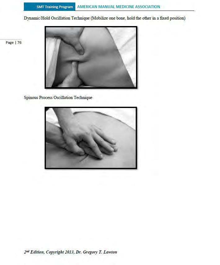


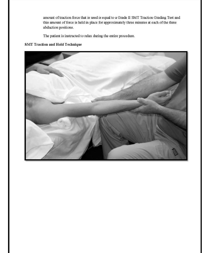


Neuropathic Massage Therapy (NMT):
When treating neurological conditions, or imbalances of the sympathetic and parasympathetic nervous system massage from superior to inferior and employ the following methods:
Light to moderate friction in a straight line along the vertebral column.
Light to moderate circular friction along the vertebral column and circling outward from the spine.
Light to moderate spiral friction along the vertebral column and circling outward from the spine.
Light to moderate elliptical friction along the vertebral column and circling outward from the spine.
Light to moderate semicircular friction along the vertebral column and circling outward from the spine.


John Harvey Kellogg (February 26, 1852 – December 14, 1943) was an American businessman, inventor, physician, surgeon, and advocate of the Progressive Movement of the early to mid 1900’s. He was the director of the Battle Creek Sanitarium in Battle Creek, Michigan, founded by members of the Seventh-day Adventist Church. It combined aspects of a European spa, a hydrotherapy institution, a hospital and highclass hotel. Kellogg treated the rich and famous, as well as the poor who could not afford other hospitals.
According to Encyclopædia Britannica, his "development of dry breakfast cereals was largely responsible for the creation of the flaked-cereal industry."


Dr. Kellogg wrote one of the foremost books on medical massage therapy, a book that is still published and studied by massage therapists today. Here is a citation for that book -Kellogg, John Harvey. The Art of Massage: A Practical Manual for the Nurse, the Student and the Practitioner. Modern Medicine Publishing Co., 1895.

John Harvey Kellogg, MD
In his book he begins by talking about the influence of the Chinese on the practice of massage. They were seen as one of the first people to use massage to help relieve illness and had particularly important contributions to the development of the field. One of the books produced from their studies was The Cong-Fou of the Tao-Tse. After the history of massage, he has a section discussing different parts of the body and how they should be manipulated. The effects of massage on the participant's body are explored, and special attention is paid to particular benefits, such as fighting or holding off certain illnesses. This is an idea Kellogg advocated for in many different contexts besides just this book. The actual practice of massage is broken down into the specific movements that a therapist could use to achieve different effects, such as kneading or stroking. Joint movements are considered, and then the piece delves into how to massage separate parts of the body. There are special massages for each distinct body part he focuses on. Kellogg believes that the techniques are important for every part of the body, whether it is necessary to perform the practice on that area or by massaging other parts of the body that are connected to it.


Edgar Cayce was a well-known American psychic who wrote on various topics, including health and healing. Among these topics, Cayce frequently discussed massage therapy. His insights emphasized the holistic nature of health and the importance of balance within the body. Cayce often mentioned the importance of good circulation and the lymphatic system in maintaining health. He suggested that massage therapy could help improve circulation, remove toxins, and stimulate the lymphatic system. Cayce pointed out the benefits of massage for the nervous system. He believed that it could help relax the nerves, reduce stress, and promote healing by restoring balance to the nervous system.


He believed in personalized treatments, tailored to the individual’s specific needs and conditions. Cayce’s readings often provided customized advice on the type, frequency, and duration of massage therapy. Edgar Cayce’s perspective on massage therapy highlights its importance as a holistic treatment that supports physical, mental, and spiritual health. His recommendations on techniques, oils, and the integration of massage with other therapies underscore his belief in the interconnectedness of bodily systems and the value of personalized, preventive care.

Neuropathic massage therapy is always indicated in conditions involving the peripheral nervous system unless specifically contraindicated.
Contraindications: Situations in which neuropathic massage therapy (NMT) should not be used). There may be exceptions, but only if ordered by a medical physician.
Open Wounds: Do not perform NMT over broken skin or recent unhealed surgical i ncisions.
Thinning Skin: Avoid performing NMT to thinning or fragile skin in the elderly or in any medical condition where the skin integrity is compromised.
Skin Ulcers: Do not use NMT on patients with skin ulceration and/or infection.
Acute Cellulitis: This condition involves inflammation of the skin caused by bacteria (usually staphylococcus or streptococcus). If a patient has acute cellulitis, characterized by redness, pain, swelling, and warmth in the infected area, NMT is not recommended.
Deep Vein Thrombosis (DVT): Do not use NMT with patients with a history or who have DVT (a blood clot in a deep vein). It can increase blood flow and dislodge the clot, potentially causing a pulmonary embolism.
Uncontrolled Diabetes: NMT can worsen pins-and-needles sensations (peripheral neuropathy) associated with uncontrolled diabetes.
Active Cancer: Do not use NMT with patients who are undergoing cancer treatment unless prescribed by a licensed physician.
Lymph Node Removal: Do not perform NMT directly over areas where lymph nodes hav e been surgically removed or damaged by trauma.
Varicose Veins: Tortuous varicose veins may be both and indication and contraindication for NMT.
Fever: Not recommended for patients with a fever.
Broken or Damaged Skin: Do not perform NMT techniques over broken or damaged skin. (Risk of infection)
Pregnancy: Care is required during pregnancy.
Certain Medications: Patients who are taking blood thinner medications may observe bruising under the skin due to the increased circulation effects.
Patients who present with signs and symptoms of nerve disorders should always be evaluated by a medical physician to determine the cause of the condition, appropriateness of manual care, and the possibility of combined medical care.
With proper screening for contraindications, cupping therapy is suitable for anyone. Therapeutic cupping helps with various issues like sports injuries, scar tissue and adhesions. muscle pain, neuropathy, chronic pain, respiratory ailments, and lymphedema.


Cupping therapy is a form of alternative medicine where glass, silicone, or plastic cups are placed on the skin to create vacuum suction to promote health and healing. Cupping therapy has been used for centuries across various cultures for muscle relaxation, detoxification, and stress relief. By creating a vacuum with cups on the skin, this alternative medicine technique promotes healing and overall well-being. Cupping therapy aids in releasing tension in soft tissue to promote circulation and lymphatic flow.


When starting cupping therapy, choose the right cup size and method for your patient. Large cups cover more area while small cups are ideal for smaller areas or contour massage. Different cupping techniques include static, flash, dynamic, shake, and rotational cupping. In our method of cupping, AcuMyoTherapy, we recommend the use of herbal infused oils to assist with the cupping process, benefits, and results.


Static cupping is the use of a cup, or several cups, with each cup left in one location on the skin and without moving or manipulating the cup. Apply a small amount of herbal infused oil before placing the cups on the skin. Gently press and release the cup to create suction or use a pump to remove the air from the cup. Start with a test site for one minute to access the patient’s response and then apply the therapy for three to five minutes per area treated. If a cup falls off, simply reposition it. Use this technique a few times a week for up to ten days before taking a break from the therapy.


Flash cupping involves placing and releasing cups on the skin to create vacuum suction, helping with blood and lymph stagnation and deep tissue release. Apply a small amount of herbal infused oil before placing the cups on the skin. Try flash cupping for up to five minutes per area for maximum benefits. The benefits include deep tissue release and nerve stimulation.


Dynamic Cupping – (Gliding or Sliding Technique)
Dynamic cupping is the gliding or sliding of cups across the skin. Apply an herbal infused oil to the skin. Compress the cup to create vacuum suction. Glide the cup in a zigzag, circular, or back and forth motion. When using dynamic cupping to provide a cupping massage limit the massage time period to twenty or thirty minutes.

Shake Cupping –
Shake cupping therapy is accomplished by shaking or vibrating cups in a static position. Apply a small amount of herbal infused oil before placing the cups on the skin. This technique is beneficial for softening scars, deep tissue work, and myofascial release.
Rotation cupping therapy is performed by placing a cup in a static position and rotating the cup over the treatment area. Apply a small amount of herbal infused oil before placing the cups on the skin. This technique is beneficial for softening scars, deep tissue work, and myofascial release.

All the techniques listed above may be combined to provide a “vacuum” massage therapy session. An expert cupping therapist will be able to demonstrate an adept flow of techniques, changing fluidly from one technique to another easily and efficiently.
To achieve this sense of proficiency first select the correct type and size of cups for the patient and your purposes. Having to switch between cup types or sizes breaks the cupping therapy routine and flow.
Also choose the correct lubricating herbal infused oil for your patient’s condition and in conformance with the patient's diagnosis.


Conclusion: The available evidence suggests that cupping therapy is effective in managing chronic pain, knee osteoarthritis, low back pain, neck pain, chronic back pain, and herpes zoster. Moreover, it represents a promising, safe, and effective non-pharmacological therapy that warrants wider application and promotion.


Background: Myofascial decompression (MFD), or cupping, and self-myofascial release (SMR) are common techniques utilized to treat soft tissue injuries and increase flexibility. MFD is a negative pressure soft tissue treatment technique using suction to manipulate the skin and underlying soft tissues. One method of SMR is a foam roller, where a patient rolls his/her bodyweight over a dense foam cylinder in a self-massaging fashion to mobilize soft tissues for the body part treated.
Conclusion: The findings suggest that both treatments are beneficial in increasing hamstring length. Patients though felt an enhanced treatment effect using MFD over SMR for perceived benefits to hamstring flexibility.


Vacuum Decompression Therapy increases blood circulation to the targeted area, which can help reduce muscle tension and promote healing. The negative pressure generated by the cups can also facilitate the removal of toxins and metabolic waste products from the tissues, thereby reducing inflammation and alleviating pain. Furthermore, cupping may stimulate the relaxation response in muscles, improving flexibility and reducing stiffness. Overall, the combined effects of enhanced circulation, detoxification, and muscle relaxation contribute to the therapeutic benefits of cupping on muscle tissue.
Lauche, R., Cramer, H., Hohmann, C., Choi, K. E., Rampp, T., Saha, F. J., ... & Dobos, G. (2012). "The Effectiveness of Cupping Therapy on Chronic Neck and Shoulder Pain: A Randomized Controlled Trial." PLoS One, 7(9), e31793.
This study examined the effectiveness of cupping therapy on chronic neck and shoulder pain, showing significant pain reduction and improved quality of life for participants.
AlBedah, A. M. N., Elsubai, I. S., Qureshi, N. A., Aboushanab, T. S., Ali, G. I. M., El-Olemy, A. T., ... & Khalil, A. A. (2019). "The medical perspective of cupping therapy: Effects and mechanisms of action." Journal of Traditional and Complementary Medicine, 9(2), 90-97.
This review article discusses the various effects and proposed mechanisms of action of cupping therapy, including increased blood flow, reduced inflammation, and enhanced tissue repair.
Huijuan Cao, Xun Li, and Jianping Liu. (2012). "An updated review of the efficacy of cupping therapy." PLoS One, 7(2), e31793.
This systematic review updated previous findings on the efficacy of cupping therapy, highlighting its benefits in treating a range of conditions, including pain and musculoskeletal disorders.
Kim, J. I., Lee, M. S., Lee, D. H., Boddy, K., & Ernst, E. (2011). "Cupping for treating pain: a systematic review." Evidence-Based Complementary and Alternative Medicine, 2011, 467014.
This systematic review evaluated the effectiveness of cupping therapy in treating various types of pain, concluding that it may be effective for pain management, especially in conditions like chronic pain.
Teut, M., Ullmann, A., Braun, V., Dornieden, K., & Irnich, D. (2012). "Cupping therapy in patients with carpal tunnel syndrome: a randomized controlled trial." The Journal of Pain, 13(3), 329-337.
This randomized controlled trial assessed the effects of cupping therapy on carpal tunnel syndrome, showing improvements in pain and functionality among participants.
The studies cited offer insights into the physiological effects of cupping therapy, such as pain reduction, improved blood flow, and potential mechanisms underlying its therapeutic benefits.


In our homesomatic method of treatment we utilize infused oils therapy along with cupping and gua sha. Later in this workbook these aspects will be covered in more detail along with the herbs and formulations that we use.

High Fever or Infection: Avoid cupping when a person has a high fever or an infection.
Convulsions or Cramps: Cupping should not be used in cases of convulsions or cramps.
Allergic Skin Conditions: If someone has allergic skin conditions, cupping is not recommended.
Ulcerated Sores: Avoid cupping over ulcerated sores.
Thin Muscle Layer: Cupping should only be done on soft muscle tissue and not where the muscle layer is thin or uneven due to bony projections or angles.
Severe Diseases: Cupping is contraindicated in severe diseases such as cardiac failure, renal failure, ascites due to hepato-cirrhosis, and severe edema.
Serious Illnesses: Individuals with serious conditions such as cancer, cardiovascular disease, or severe infections should consult with their primary healthcare provider before undergoing gua sha therapy.
Hemorrhagic Diseases: Avoid cupping in cases of allergic purpura (a form of vasculitis), hemophilia, and leukemia.
Dermatosis and Disease of the Skin: Cupping should not be used on clients with dermatosis or destruction of the skin.
Blood Disorders: Individuals with anemia or similar blood disorders should avoid cupping.
Heart Disease and Pacemakers: Cupping is not recommended for people with heart failure, kidney failure, or those with pacemakers.
Blood Thinners: If someone is on blood thinners, cupping should be avoided.
Compromised Immune System: Patients with a compromised immune system, such as those undergoing chemotherapy or with autoimmune diseases, should use caution as the therapy could potentially trigger an adverse response.
Varicose Veins: Avoid performing cupping directly over varicose veins, as the pressure may damage the veins further.
Pregnancy: While some practitioners may use gentle cupping techniques during pregnancy, it is generally advised to avoid it, especially on the abdomen and lower back, unless specifically recommended by a healthcare provider.
After a cupping therapy session, proper aftercare is essential to maximize benefits and ensure a comfortable recovery. Here are some key guidelines:
Hydrate: Drink plenty of water to help flush out toxins and stay hydrated.
Rest: Allow the body time to rest and recover after the treatment.
Avoid Strenuous Activities: Take a break from vigorous exercise immediately after the session.
Stay Warm: Cover the treated areas to maintain warmth.
Avoid Caffeine, Alcohol, and Sugary Foods: These can delay the natural healing process.
Be Mindful of Diet and Drink: Chose nourishing foods and avoid extremes like hot showers or cold environments.

Kinesio tape gently lifts the superficial layers of skin, creating a space between the skin, fascia, and underlying tissues. This lifting effect is thought to promote better circulation and lymphatic drainage, which can help reduce swelling and inflammation. In applying kinesio tape for the treatment of neurological conditions we generally follow the pathway of major nerves.
As an ancillary part of our Homeosomatic Neuropathic Massage Therapy treatment protocol, and at the end of a treatment session, we often send patients home with kinesio tape applied to an area or region that is afflicted with neuropathy.

Anchor tape from the origin of the nerve pathway and follow the pathway of the nerve


With the cross-taping technique, we identify and cross over an area of localized pain


Theraband kinesiology tape is a product that supports muscles and joints and provides pain relief. It is safe, effective, and easy to apply. The tape is latex-free, nonirritating, and allergy tested3. It lasts up to 5 days. The tape features stretch indicators for perfect application every time41. It has best in class adhesion and sticks and lasts like only the best tapes in the market.

Herb infused oils are prepared by selecting an herb or herbal blend and then cutting or grinding the dried plant material to a size and consistency that allows the oil to permeate the herbal material and to draw out and dissolve the chemical constituents into solution. In the “long” method the mixture is allowed to sit in a cool dark place for up to 8 weeks. In the “fast” method or what is called the “alcohol intermediary method”, the dried plant material is cut or ground into an acceptable size and soaked in a very small amount of alcohol for 24 hours and then it is soaked in a carrier oil for 24 to 48 hours and finally strained. During the 24-to-48-hour period the mixture is warmed to a temperature of 110-degrees Fahrenheit. When the appearance of the solution is appropriate for the herb or herbs being infused, the solution is strained. The remaining mixture of herbs may be used for other purposes.
An herb-infused oil, also known as a macerated oil, consists of a carrier oil that has been permeated (“infused” or “macerated”) with one or more herbs. The benefit of using an infused oil is that it combines the properties of both the carrier oil and the herbs. Unlike essential oils, which are highly concentrated extracts, infused oils are gentler and less concentrated. They capture the whole essence of the herb and can be used for various purposes, from skincare to cooking. For example, on average an essential oil only captures 4 or 5 percent of the chemical constituents in an herb, whereas an herbinfused oil will extract over 90 percent of the herbal constituents.
For the fast or alcohol intermediary method of extraction heat is used to speed up the process. There are several ways in which heat is used:
Warm Infusion: Heat the oil and herbs gently on the stovetop.
Solar Infusion: Place the herbs in a jar with oil and let it sit in a sunny window for 4-6 weeks.
Stovetop Infusion: Simmer the herbs and oil together.
Oven Infusion: Use low heat in the oven to infuse the oil.
Yogurt Maker: Place the mixture into jars and into a yogurt maker at 110 degrees Fahrenheit.
Double Boiler: Place the jars in a double boiler with water and set to simmer lightly.


Herbal blends and formulas include herbal infused oils using castor oil and flax seed oil as a carrier and adding essential oils such as poke root.
Castor Oil Packs
Infused Oil Treatment (IOT) (Poke Root)
Essential oils such as ginger and germanium
Mugwort i nfused oil

Mugwort is an effective ingredient for soothing even the most sensitive skin. It offers anti-inflammatory, antibacterial, and antioxidant benefits, and it could even help protect your skin against free radical damage.
Anti-Inflammatory Properties:
Mugwort soothes irritated skin and reduces redness. It’s excellent for calming inflammation caused by external factors like sun damage or pollution.
Antibacterial and Antifungal Benefits:
Mugwort has natural antibacterial and antifungal properties, making it effective against skindamaging bacteria. It helps maintain healthy skin.
Antioxidant Protection:
Rich in antioxidants, including vitamin E, mugwort protects the skin barrier from free radical damage. This contributes to preventing premature aging and other skin issues.
Softening, Moisturizing, and Toning:
Mugwort’s healing properties leave the skin soft and hydrated. Regular use can enhance overall skin health.
Ideal for Sensitive Skin:
It is well tolerated by most skin types. Its gentle nature makes it suitable for sensitive skin types.
Anti-Microbial Effects:
Mugwort’s compound called azulene has antimicrobial properties, helping maintain a healthy skin microbiome.

An herbal formulation refers to a dosage form that consists of one or more herbs or processed herbs in specific quantities. These formulations are designed to provide specific nutritional or therapeutic effects.

Arnica, Arnica montana (flowers) – Analgesic and Anti-inflammatory. Used topically or homeopathically.
Blue Vervain, Verbena hastata (all parts but mainly aerial parts) – Nervine, Relaxant, Sedative, Antispasmodic upper body.
Boswellia, Boswellia serrata - Anti-inflammatory and Analgesic, Contains boswellic acids that can treat pain and inflammation.
Calendula Flowers, Calendula officinalis – Anti-inflammatory, Anodyne, treatment of skin conditions and rashes.
California Poppy, Eschscholzia californica – Sedative, Anti-spasmodic, Anodyne, the bright orange flowers of the California poppy, leaves and other aerial parts are used.
Capsicum or Red pepper, Capsicum annuum – Vasodilatation, Anti-inflammatory, Anodyne. Use for arthritis and joint pain and inflammation.
Chamomile Flower, Matricaria chamomilla – Mild Nervine, Relaxant, Sedative.
Clove buds, Syzygium aromaticum – Anodyne, Anti-inflammatory, may be added to capsicum.
Cramp Bark, Viburnum opulus – Anti-spasmodic mainly female reproductive system and smooth muscle.
Devil’s Claw, Harpagophytum procumbens, Devil’s claw is a South African herb with medicinally active roots used to treat pain and inflammation.
Hops Flowers, Humulus lupulus – Sedative, Nervine, Relaxant, Anti-inflammatory.
Kava Root, Piper methysticum – Nervine, Relaxant, Sedative, Anti-spasmodic general and lower body.
Lavender, Lavandula spp. (aerial parts) – Nervine, Sedative, Relaxant.
Licorice Root, Glycyrrhiza glabra – Anti-inflammatory. Contains phyto sterols like hydrocortisone and estrogen.
Meadowsweet, Filipendula ulmaria (all parts but mainly flowers and leaves) – Antiinflammatory and Analgesic. Contains salicin.
Mugwort, Artemisia vulgaris (Roots, leaves, stems, and flowers) – Nervine, Sedative, Antispasmodic, Warming tonic.
Mullein Leaf and Flower, Verbascum Thapsus – Respiration, Expectorant, Anodyne, Antiinflammatory, Anti-tussive, skin irritation and inflammation.
Rosemary, Rosmarinus officinalis (all aerial parts but mainly the flowers and leaves) –Anti-inflammatory, Nervine, and Sedative.
Sage, Salvia officinalis (all aerial parts) – Anti-inflammatory, encourages new blood vessel growth and circulation, encourages nerve healing and regeneration, antispasmodic, nervine.
Skullcap, Scutellaria spp. (Aerial parts and roots) – Nervine, Sedative, Anti-inflammatory, Anti-spasmodic, Anti-convulsant, increases blood flow, prevents, and treats neuron damage in the brain.
Teasel Root, Dipsacus fullonum – Anti-inflammatory, Anodyne, Lyme disease excellent for joint pain, osteoarthritis.
Turmeric Root, Curcuma longa – Anti-inflammatory, Anti-microbial, and pain relief.
Valerian Root, Valeriana officinalis, Nervine, Sedative.
The following information provides a list of the various formulations and combinations I typically use for patient treatment:
Castor oil and flaxseed oil 50/50 blend.
Castor oil and flaxseed oil 50/50 blend plus dried skullcap infusion (nerve conditions).
Castor oil and flaxseed oil 50/50 blend plus dried sage infusion (increase circulation, edema, vascular insufficiency).
Castor oil and flaxseed oil 50/50 blend plus dried mugwort infusion (anti-spasmodic and warming tonic).
Safflower oil and dried red pepper (capsicum) infusion (increase blood flow and reduce pain).
Safflower oil and dried poke root (Phytolacca americana) infusion (for the treatment of lymphatic conditions, immune response, and lymphedema).
Castor oil and poke root infusion 50/50 blend.
The following formulation is recommended for general use for musculoskeletal conditions related to pain, inflammation, and muscle spasm.
The general formulation is blended by using flaxseed oil and adding castor oil, and mugwort infused oil.
Formulation: (Equal parts of each oil)
Castor Oil – 1 Part
Flaxseed Oil – 1 Parts
Mugwort Oil – 1 Part
To this formulation you may also add an essential oil according to correct dilution procedures.
The following formulation is used for nerve conditions and neuropathy as well as to encourage blood circulation and lymphatic activity around nerve tracks.
The nerve tonic is blended by using sweet almond oil and adding mugwort infused oil and skullcap infused oil.
Formulation: (Equal parts of each oil)
Sweet Almond Oil – 1 Parts
Mugwort Oil – 1 Parts
Skullcap Oil – 1 Parts
To this formulation you may also add an essential oil according to correct dilution procedures.
The following formulation is used for the treatment and remediation of problem scars.
The scar formulation is blended by using castor oil and flaxseed oil and adding mugwort infused oil and sage infused oil as well as rose essential oil.
Formulation:
Castor Oil – 1 Part
Flaxseed Oil – 2 Parts
Mugwort Oil – 1 Part
Sage Oil – 2 Parts
Rose Essential Oil – 18 drops per ounce
Sweet Almond Oil (Prunus amygdalus) – Mugwort Oil (Artemisia vulgaris) – Skullcap Oil
(Scutellaria lateriflora) – Castor Oil (Ricinus communis) – Flaxseed Oil (Linum usitatissimum) – Rosemary Oil (Salvia Rosmarinus) -Camphor Essential Oil (Cinnamomum camphora) – Eucalyptus Essential Oil (Eucalyptus) – Sage Oil (Salvia officinalis) - Rose
Essential Oil (Rosa/Rosa damascene/Rosa centifolia) – Frankincense Essential Oil
(Boswellia Serrata) - Valerian Root (Valeriana officinalis) - Mullein Leaf and Flower (Verbascum thapsus) - Clove Bud Essential Oil (Syzygium aromaticum) - Lavender
(Lavandula spp) Essential Oil - Arnica (Arnica montana) - Calendula (Calendula officinalis)
I recommend a system-by-system approach to patient evaluation and assessment based upon observation, palpation, and questioning. Dr. Lawton


The skin often reflects a person's overall health and well-being because it can manifest signs of internal issues, deficiencies, and systemic conditions. By observing changes in the skin, healthcare providers can often identify and diagnose underlying health issues, making the skin an important diagnostic tool for overall health and well-being.
Nutritional Status
Deficiencies: Lack of essential nutrients like vitamins (A, C, D, E), minerals (zinc, iron), and fatty acids can lead to skin problems such as dryness, rashes, and poor wound healing.
Hydration: Dehydration can cause the skin to appear dry, flaky, and less elastic.
Hydration Levels
Skin Turgor: Well-hydrated skin is firm and elastic, whereas dehydration can make it look wrinkled and saggy.
Hormonal Balance
Acne and Breakouts: Hormonal imbalances, such as those seen in puberty, pregnancy, and conditions like polycystic ovary syndrome (PCOS), can lead to acne.
Changes in Texture and Color: Conditions like thyroid disorders can cause changes in skin texture and color.
Immune System Health
Rashes and Infections: A compromised immune system can lead to frequent skin infections, rashes, and delayed healing.
Autoimmune Disorders: Conditions like lupus and psoriasis often have skin manifestations.
Stress-Related Conditions: Stress can exacerbate skin conditions such as eczema, psoriasis, and acne.
Overall Appearance: Chronic stress can lead to dull, tired-looking skin and exacerbate the signs of aging.
Jaundice: Yellowing of the skin can indicate liver problems.
Uremic Frost: In severe kidney disease, urea can crystallize on the skin.
Cyanosis: A bluish tint to the skin can indicate poor oxygenation of the blood, often related to heart or lung problems.
Redness and Swelling: Conditions like rosacea and chronic venous insufficiency can indicate underlying vascular issues.
Acanthosis Nigricans: Dark, velvety patches of skin, often in body folds, can i ndicate insulin resistance or diabetes.
Delayed Healing: High blood sugar levels can impair wound healing and increase the risk of infections.
Hives and Rashes: Allergic reactions to foods, medications, or environmental factors can manifest as skin rashes or hives.
Dermatitis: Contact with allergens or irritants can cause eczema or dermatitis.
Premature Aging: Lifestyle factors such as smoking, excessive alcohol consumption, and poor diet can accelerate skin aging, resulting in wrinkles, fine lines, and loss of elasticity.
Systemic Infections: Diseases like chickenpox, measles, and Lyme disease have characteristic skin manifestations.
Cancer: Skin cancers, such as melanoma, can indicate exposure to harmful UV radiation and overall health risks.


Central obesity, also known as abdominal obesity or visceral obesity, refers to the accumulation of fat around the abdomen and internal organs. This type of obesity is often measured by waist circumference and waist-to-hip ratio. Addressing central obesity early can significantly reduce the associated health risks and improve overall well-being and longevity.
Association with Metabolic Syndrome
Insulin Resistance: Central obesity is strongly associated with insulin resistance, a condition where cells in the body become resistant to the effects of insulin. This leads to elevated blood sugar levels and an increased risk of type 2 diabetes.
Dyslipidemia: It often coincides with abnormal levels of lipids (cholesterol and triglycerides) in the blood, specifically low HDL ("good") cholesterol and high LDL ("bad") cholesterol, which are risk factors for cardiovascular disease.
Hypertension: Central obesity is linked with hypertension (high blood pressure), which further increases the risk of heart disease and stroke.
Inflammatory and Immune Responses
Release of Inflammatory Substances: Fat cells in the abdominal area (visceral fat) release inflammatory substances and hormones that contribute to chronic inflammation. This chronic inflammation is implicated in the development of various diseases, including cardiovascular disease, type 2 diabetes, and certain cancers.
Immune System Dysfunction: Excess visceral fat can disrupt the normal functioning of the immune system, increasing susceptibility to infections and impairing wound healing.
Cardiovascular Disease
Atherosclerosis: Central obesity contributes to the development of atherosclerosis, a condition where fatty deposits (plaques) build up inside the arteries, narrowing and hardening them. This increases the risk of heart attacks, strokes, and other cardiovascular complications.
Heart Failure: Obesity, especially abdominal obesity, is a risk factor for heart failure, where the heart muscle becomes weakened and unable to pump blood efficiently.
I ncreased Cancer Risk: Central obesity is associated with an increased risk of several types of cancers, including colorectal cancer, breast cancer (in women), and prostate cancer (in men).
Non-alcoholic Fatty Liver Disease (NAFLD): Central obesity is a major risk factor for NAFLD, a condition where fat accumulates in the liver, potentially leading to liver inflammation and scarring (cirrhosis).
Kidney Disease: Obesity, particularly abdominal obesity, is linked to an increased risk of chronic kidney disease.
Psychological Well -being: Central obesity can impact self-esteem, body image, and mental health, leading to anxiety, depression, and social stigma.
Reduced Quality of Life: Health complications associated with central obesity can impair physical mobility, limit daily activities, and reduce overall quality of life.
Overall Mortality Risk: Individuals with central obesity have a higher risk of premature death compared to those with a healthy waist circumference and body composition.


Poor spinal posture can significantly impact both quality of life and overall health in various ways. Misalignment of the spine can cause conditions such as scoliosis, kyphosis, lordosis, and arthritis leading to discomfort and potential long-term health issues.
Chronic Pain:
Poor posture often leads to chronic back, neck, and shoulder pain due to increased strain on muscles and ligaments.
Reduced Flexibility and Mobility:
Poor posture can limit the range of motion and flexibility, making it difficult to perform daily activities and exercise.
Increased Risk of Injury:
Misalignment and muscle imbalances increase the risk of injuries, particularly in the back and neck.
Respiratory Issues:
Slouched posture can compress the lungs and diaphragm, leading to reduced lung capacity and breathing difficulties.
Digestive Problems:
Poor posture can compress abdominal organs, affecting digestion and potentially causing issues like acid reflux and constipation.
Decreased Energy Levels:
Chronic pain and discomfort can lead to fatigue, reducing overall energy levels and enthusiasm for daily activities.
Mood and Mental Health:
Persistent pain and physical limitations can contribute to stress, anxiety, and depression.
Self-Esteem and Confidence:
Poor posture can affect body image and self-esteem, leading to decreased confidence in social and professional settings.
Physical discomfort and pain can reduce concentration and productivity at work or school.
Reduced flexibility and pain can hinder participation in physical activities and hobbies, impacting social interactions and overall enjoyment of life.
Discomfort from poor posture can interfere with sleep quality, leading to insomnia or restless sleep.


Poor gait, which refers to an abnormal walking pattern, can have wide-ranging effects on a person's health and well-being. Identify underlying causes of poor gait, such as neurological, muscular, or skeletal issues and develop an effective treatment plan.
Musculoskeletal Issues:
Joint Pain and Damage: Abnormal gait patterns can lead to uneven weight distribution, causing stress and wear on the joints, particularly in the knees, hips, and ankles.
Muscle Strain: Compensatory movements due to poor gait can strain muscles, leading to pain and discomfort in the legs, lower back, and other areas.
Increased Risk of Falls and Injuries:
Poor gait can reduce balance and coordination, significantly increasing the risk of falls, which can result in fractures, sprains, and other injuries.
Foot Problems:
Abnormal gait can contribute to foot issues such as bunions, calluses, and plantar fasciitis, as well as exacerbate existing foot deformities.
Spinal Misalignment:
Poor gait can cause or exacerbate spinal alignment issues, leading to back pain and other spinal problems.
Reduced Physical Activity:
Difficulty walking can lead to reduced physical activity, which in turn can negatively affect cardiovascular health, contributing to conditions such as obesity, hypertension, and cardiovascular disease.
Decreased Independence:
Difficulty walking can limit a person’s ability to perform daily activities independently, leading to feelings of helplessness and decreased quality of life.
Mental Health Issues:
Chronic pain and reduced mobility associated with poor gait can contribute to mental health issues such as depression and anxiety.
Social Isolation:
Limited mobility can reduce social interactions and participation in community activities, leading to social isolation and loneliness.
Poor gait can limit a person’s ability to move around freely, affecting their ability to work, engage in hobbies, and participate in social activities.
Continuous pain and discomfort from poor gait can significantly impact daily life, reducing overall enjoyment and satisfaction.
Sleep Disturbances:
Pain and discomfort associated with poor gait can interfere with sleep, leading to i nsomnia or poor sleep quality, which in turn affects overall health and well-being.


Foot health is crucial to overall health and well-being for several reasons including all of the reasons outlined under the Gait section of this workbook.
Musculoskeletal Alignment:
The feet serve as the foundation for the body. Problems with foot health can lead to issues in the ankles, knees, hips, and lower back due to misalignment and compensation for pain or discomfort.
Prevention of Chronic Conditions:
Proper foot health can prevent chronic conditions such as neuropathy, plantar fasciitis, bunions, and arthritis, which can cause long-term pain and disability.
Circulatory Health:
The feet are farthest from the heart, and good circulation is vital for delivering oxygen and nutrients. Poor foot health can be an indicator of circulatory problems, especially in individuals with diabetes or cardiovascular disease.


Hair, skin, and nails can serve as important indicators of a person’s overall health.
Changes in these areas can sometimes be the first signs of underlying health issues.
Regular monitoring of hair, skin, and nails can help detect early signs of potential health issues.
Hair - Indicators of Health Issues:
Hair Loss: Sudden or excessive hair loss can indicate hormonal imbalances (such as thyroid disorders), nutritional deficiencies (like iron or protein deficiency), stress, or certain medical conditions like alopecia areata or lupus.
Dry, Brittle Hair: This can be a sign of nutritional deficiencies, overuse of hair styling products, or underlying health issues such as hypothyroidism.
Thinning Hair: Could be a sign of aging, hormonal changes, or nutritional deficiencies.
Skin - Indicators of Health Issues:
Rashes and Itchiness: Conditions such as eczema, psoriasis, allergies, or infections can cause these symptoms.
Discoloration: Jaundice (yellowing of the skin) can indicate liver disease; cyanosis (bluish tint) can indicate respiratory or cardiovascular issues.
Acne: While common and often related to hormonal changes, severe or sudden acne can indicate underlying hormonal disorders such as polycystic ovary syndrome (PCOS).
Dry, Flaky Skin: Can be a sign of dehydration, hypothyroidism, or vitamin deficiencies.
Wounds That Don’t Heal: Can indicate diabetes or circulatory issues.
Nails - Indicators of Health Issues:
Brittle Nails: Can indicate nutritional deficiencies (like biotin), thyroid disease, or fungal infections.
Discoloration: Yellow nails can indicate fungal infections, respiratory issues, or lymphedema; white nails can indicate liver disease.
Spoon Nails (Koilonychia): This can be a sign of iron-deficiency anemia.
Pitting or Grooving: Conditions such as psoriasis or alopecia areata can cause these changes.
Clubbing: Enlarged, curved nails can indicate respiratory or cardiovascular diseases.


Poor oral health can be an indicator of broader health issues and can also contribute to various health risks. Maintaining good oral health is crucial not just for a healthy mouth but for overall health and well-being. Regular dental check-ups, proper oral hygiene, and a balanced diet are essential for preventing oral health problems and mitigating broader health risks.
Indicators of Health Issues:
Gum Disease (Periodontal Disease):
Systemic Conditions: Severe gum disease has been linked to systemic conditions such as diabetes, cardiovascular disease, and rheumatoid arthritis.
Diabetes: People with diabetes are more prone to infections, including periodontal disease, and uncontrolled gum disease can make it harder to manage blood sugar levels.
Oral Infections:
I mmune System Issues: Frequent oral infections can indicate a compromised immune system, possibly due to conditions such as HIV/AIDS or leukemia.
Nutritional Deficiencies: Recurrent mouth sores or infections can be a sign of nutritional deficiencies, such as a lack of vitamin C (scurvy) or iron (anemia).
Heart Disease and Stroke: Inflammation and infections caused by oral bacteria can lead to cardiovascular problems. Studies have found links between periodontal disease and an increased risk of heart disease and stroke.
Respiratory Infections:
Pneumonia: Bacteria from the mouth can be aspirated into the lungs, leading to i nfections such as pneumonia, especially in vulnerable populations like the elderly or those with compromised immune systems.
Indicators of Health Issues:
Adverse Pregnancy Outcomes:
Preterm Birth and Low Birth Weight: Pregnant women with periodontal disease are at a higher risk of delivering preterm or low birth weight babies.
Diabetes Complications:
Poor Blood Sugar Control: Infections and inflammation from gum disease can make it more difficult to control blood sugar levels, worsening diabetes and its complications.
Endocarditis:
Heart Infection: Bacteria from the mouth can enter the bloodstream and infect the inner lining of the heart, especially in people with damaged heart valves or other heart conditions.


Indicators of Health Issues:
Inflammatory Conditions:
Rheumatoid Arthritis (RA): This autoimmune disease not only affects joints but can also indicate a systemic inflammatory condition that may affect organs such as the heart, lungs, and eyes. RA is associated with an increased risk of cardiovascular disease.
Psoriatic Arthritis: Often linked with psoriasis, it indicates an underlying immune system disorder and can also involve the skin and other organs.
Metabolic Disorders:
Gout: Characterized by the accumulation of uric acid crystals in the joints, gout indicates a problem with uric acid metabolism and can be associated with other conditions like kidney stones, obesity, hypertension, and cardiovascular disease.
Indicators of Health Issues:
Inflammatory Conditions:
Genetic Predispositions:
Ankylosing Spondylitis: A type of arthritis that affects the spine, it often indicates a genetic predisposition and can also lead to inflammation in other parts of the body, such as the eyes (uveitis) and the intestines (inflammatory bowel disease).
Degenerative Conditions:
Osteoarthritis: While primarily a degenerative joint disease, it can also indicate mechanical stress on the joints due to obesity, injury, or overuse. It may reflect broader issues like obesity, which is a risk factor for various other health conditions.
Cardiovascular Disease:
I nflammatory Arthritis: Conditions like RA and psoriatic arthritis are associated with an increased risk of cardiovascular diseases, including heart attack and stroke, due to chronic inflammation.
Osteoarthritis and Gout: These conditions can be exacerbated by obesity, which also increases the risk of developing other health problems such as diabetes, hypertension, and cardiovascular disease.
RA and Osteoarthritis: These conditions are often associated with an increased risk of developing diabetes. The inflammation and physical inactivity caused by joint pain can contribute to insulin resistance.
Indicators of Health Issues:
Reduced Mobility and Physical Activity:
Joint Pain and Stiffness: Arthritis can lead to decreased mobility and physical activity, which can result in muscle weakness, weight gain, and a higher risk of cardiovascular disease, diabetes, and osteoporosis.
Mental Health Issues:
Chronic Pain and Disability: Persistent pain and disability from arthritis can lead to mental health issues such as depression and anxiety, further impacting overall health and quality of life.
Respiratory Problems:
RA and Ankylosing Spondylitis: These conditions can cause lung complications, including interstitial lung disease and reduced lung capacity, leading to respiratory problems.


Eye and vision health are integral to overall health, and poor eye health can be both an indicator of and a contributor to broader health issues and risks. Eye and vision health are closely linked to overall health. Regular eye care and attention to vision changes can help detect systemic health issues early and reduce the risk of complications.
Diabetes: Diabetic retinopathy is a common complication of diabetes that affects the blood vessels in the retina. The presence of diabetic retinopathy can indicate poorly controlled blood sugar levels.
Hypertension: High blood pressure can cause damage to the blood vessels in the retina, leading to hypertensive retinopathy, which can be detected during an eye exam.
A utoimmune Diseases: Conditions such as lupus, multiple sclerosis, and rheumatoid arthritis can cause inflammation in the eyes, leading to symptoms like dry eyes, uveitis, or optic neuritis.
Cardiovascular Disease: Changes in the blood vessels in the eyes can sometimes indicate broader cardiovascular issues, such as atherosclerosis.
Impaired Vision: Poor vision increases the risk of falls and accidents, especially in older adults. This can lead to injuries, fractures, and a decrease in mobility and independence.
Reduced Independence: Vision loss can lead to difficulties in performing daily activities, reducing the quality of life and increasing dependence on others.
Mental Health Issues: Vision impairment is associated with an increased risk of depression and anxiety due to reduced independence and social isolation.
Link with Dementia: There is evidence to suggest that vision impairment is associated with an increased risk of cognitive decline and dementia. This may be due to decreased sensory input and reduced engagement in stimulating activities.
Digital Eye Strain: Prolonged use of digital devices can cause eye strain, headaches, and neck pain. This can affect productivity and overall well-being.
Uncorrected Vision Problems: Conditions such as astigmatism, hyperopia, and myopia, if uncorrected, can cause chronic headaches and fatigue.
Indicators of Health Issues:
Chronic Conditions:
Glaucoma and Age-Related Macular Degeneration (AMD): These chronic eye conditions, if not properly managed, can lead to significant vision loss and are often associated with aging and other systemic health issues.
Potential for Infections:
Contact Lens Use: Improper use and care of contact lenses can lead to eye infections, which, if severe, can result in vision loss and indicate poor hygiene practices.
Nutritional Deficiencies:
Vitamin A Deficiency: This can lead to night blindness and, in severe cases, complete blindness. It can also indicate poor overall nutritional status.
Other Nutrient Deficiencies: Deficiencies in vitamins C and E, zinc, and omega-3 fatty acids can affect eye health and may be indicative of poor dietary habits.


Reduced Blood Flow: Hearing loss can be an early indicator of cardiovascular disease. The inner ear is sensitive to blood flow, and reduced circulation from cardiovascular problems can damage the tiny hair cells in the cochlea.
Nerve Damage: Diabetes can lead to nerve damage and poor circulation, affecting the ears and leading to hearing loss. Studies show that people with diabetes are more likely to experience hearing loss than those without the condition.
Hypertension:
Blood Vessel Damage: High blood pressure can damage the blood vessels in the ear, leading to hearing loss. This can be an early sign of hypertension or poor blood pressure control.
Chronic Kidney Disease:
Shared Risk Factors: Hearing loss is more common in people with chronic kidney disease due to shared risk factors such as diabetes and hypertension. The kidneys and the ears share similar structural and functional characteristics, making them both susceptible to damage from these conditions.
Indicators of Health Issues:
Infections:
Viral and Bacterial Infections: Ear infections can be indicative of broader immune system issues. Chronic or recurrent ear infections can suggest underlying conditions that compromise the immune system.
Autoimmune Diseases:
I nflammation: Autoimmune diseases such as lupus and rheumatoid arthritis can cause inflammation in the ear, leading to hearing loss. Sudden hearing loss can sometimes be a sign of an autoimmune inner ear disease.
Ototoxic Medications:
Medication Side Effects: Certain medications, including some antibiotics, chemotherapy drugs, and diuretics, can be ototoxic (harmful to the ears) and cause hearing loss. This indicates the need for careful monitoring of medication use.
Cognitive Decline and Dementia:
Reduced Stimulation: Hearing loss has been linked to cognitive decline and an increased risk of dementia. Reduced auditory input can lead to decreased brain stimulation, contributing to cognitive deterioration.
Mental Health Issues:
Depression and Anxiety: Hearing loss can lead to social isolation, communication difficulties, and reduced quality of life, increasing the risk of depression and anxiety.
Safety Risks:
Accidents and Injuries: Hearing loss can compromise the ability to detect environmental sounds, such as alarms, traffic noises, and warnings, leading to an increased risk of accidents and injuries.
Social Isolation:
Communication Barriers: Difficulty hearing can lead to social withdrawal and i solation, impacting relationships and overall well-being.
Balance Problems:
Vestibular System: The inner ear is also responsible for balance. Hearing loss, especially when related to inner ear issues, can lead to balance problems and an increased risk of falls.


The “inconvenience” of adopting a healthy diet and way of life is nothing compared to the pain, suffering, limitations, and disabilities that result from a poor diet, bad health habits, and a lack of exercise.
Dr. Lawton’s Quote


Early indicators of dysfunction in the eleven major body systems can reveal underlying health problems and potential risks to overall health. Early indicators of dysfunction in these body systems can provide critical insights into overall health and potential risks. Recognizing and addressing these signs promptly through medical evaluation and intervention can prevent the progression of diseases and improve quality of life.


Skeletal System: Composed of bones and cartilages, the skeletal system provides mechanical support, protects internal organs, and assists in movement. There are 206 bones in an adult human body.
Muscular System: This system enables movement by contracting and relaxing muscles. It includes both voluntary (skeletal) and involuntary (smooth and cardiac) muscles.
Cardiovascular System: Responsible for circulating blood throughout the body, the cardiovascular system includes the heart, blood vessels, and blood. It transports oxygen, nutrients, and hormones while eliminating waste products.
Respiratory System: Involved in gas exchange, the respiratory system allows us to breathe. It brings oxygen into the body and removes carbon dioxide. Additionally, it helps regulate acid-base balance and is essential for phonation (speech).
Nervous System: The nervous system collects and processes information from the senses, regulates vital functions, and coordinates body movements. It includes the central nervous system (brain and spinal cord) and peripheral nerves.

Digestive System: Responsible for breaking down food mechanically and chemically, the digestive system absorbs nutrients into the body for energy. It includes organs like the mouth, esophagus, stomach, and intestines.
Urinary System (Renal System): The urinary system filters blood through the kidneys, producing urine to eliminate waste products and maintain fluid balance.
Endocrine System: This system produces hormones that regulate various bodily functions, such as the menstrual cycle, blood sugar levels, and growth.
Lymphatic System (Immune System): The lymphatic system drains excess tissue fluid, transports immune cells, and defends the body against pathogens.
Reproductive System: Responsible for producing reproductive cells (sperm and eggs), this system contributes to the process of reproduction. It differs between males and females.
Integumentary System: Comprising the skin and its appendages (hair, nails, sweat glands, and oil glands), the integumentary system provides physical protection, sensory reception, and even synthesizes vitamin D.
Cardiovascular System
I ndicators: Chest pain, shortness of breath, irregular heartbeats, high blood pressure, swelling in extremities.
Health Risks: These symptoms can indicate heart disease, hypertension, or circulatory problems, increasing the risk of heart attacks, stroke, and other cardiovascular events.
Respiratory System
I ndicators: Chronic cough, wheezing, shortness of breath, frequent respiratory infections.
Health Risks: These signs can suggest asthma, chronic obstructive pulmonary disease (COPD), infections, or lung cancer, leading to decreased oxygenation and increased risk of respiratory failure.
Nervous System
I ndicators: Persistent headaches, dizziness, numbness, tingling, changes in vision, memory problems.
Health Risks: Such symptoms can indicate neurological conditions like multiple sclerosis, migraines, stroke, or neurodegenerative diseases, affecting cognitive and motor functions.
Digestive System
I ndicators: Persistent abdominal pain, bloating, changes in bowel habits, heartburn, nausea.
Health Risks: These could be signs of gastrointestinal disorders like irritable bowel syndrome (IBS), inflammatory bowel disease (IBD), gastroesophageal reflux disease (GERD), or even cancers of the digestive tract, leading to malnutrition and systemic effects.
Endocrine System
I ndicators: Unexplained weight changes, fatigue, excessive thirst or urination, changes in skin or hair.
Health Risks: Such symptoms can indicate diabetes, thyroid disorders, adrenal dysfunction, or hormonal imbalances, which can impact metabolism, growth, and energy levels.
Immune System
I ndicators: Frequent infections, slow wound healing, fatigue, swollen lymph nodes.
Health Risks: These can suggest immunodeficiencies, autoimmune disorders, or chronic infections, leading to increased susceptibility to diseases and prolonged recovery times.
Skeletal System
I ndicators: Joint pain, stiffness, frequent fractures, back pain.
Health Risks: These signs can indicate osteoporosis, arthritis, or other bone and joint disorders, increasing the risk of immobility and chronic pain.
Muscular System
I ndicators: Muscle weakness, cramps, tremors, muscle wasting.
Health Risks: Such symptoms can be signs of muscular dystrophies, neuromuscular disorders, or chronic conditions like fibromyalgia, impacting mobility and daily functioning.
Integumentary System (Skin, Hair, Nails)
I ndicators: Rashes, persistent itching, changes in skin color or texture, hair loss, nail changes.
Health Risks: These can indicate skin conditions, infections, or underlying systemic issues like liver or kidney disease, affecting overall health and appearance.
Reproductive System
Indicators: Irregular menstrual cycles, pelvic pain, changes in libido, infertility.
Health Risks: Such symptoms can indicate hormonal imbalances, reproductive organ disorders, or sexually transmitted infections (STIs), impacting fertility and sexual health.
Urinary System
Indicators: Changes in urine color or volume, frequent urination, pain during urination, lower back pain.
Health Risks: These signs can suggest urinary tract infections, kidney stones, or chronic kidney disease, leading to impaired kidney function and systemic effects.


The “inconvenience” of adopting a healthy diet and way of life is nothing compared to the pain, suffering, limitations, and disabilities that result from a poor diet, bad health habits, and a lack of exercise.
Dr. Lawton’s Quote
In the Homeosomatic Topical Herbal Pharmacotherapy method we also use many ancillary techniques. The choice of which techniques, therapeutic modalities to use, is based upon the condition of the patient, their diagnosis, their age, and complicating factors. These ancillary techniques include:
Cupping
Red light or Bioptron Therapy
Laser
Micro Stim
High Frequency Percussion
Castor Oil Packs
Infused Oil Treatment (IOT)
Dry Needling
Dry Brushing
Gua Sha
Kinesio tape (KinesioStretch Method)
Herbal medicine and supplementation
Homeopathy


Specific nutraceuticals have been studied for their role in nerve regeneration and neuroprotection. Nutraceuticals play a vital role in supporting nerve regeneration and neuroprotection, particularly in conditions such as stroke, neuropathy, and neurodegenerative diseases. These bioactive compounds, including vitamins, minerals, and essential fatty acids, provide the necessary nutrients to facilitate neural repair, reduce oxidative stress, and promote neuroplasticity. Understanding how these nutraceuticals contribute to neurological health allows for more effective integration into rehabilitation and recovery strategies.
B-complex vitamins (especially B6, B9, and B12) – Essential for myelin repair, nerve conduction, and neuroprotection. Dosage: Vitamin B6 (Pyridoxine): 25 - 50 mg per dayVitamin B9 (Folate): 400 - 800 mcg per day - Vitamin B12 (Methylcobalamin): 1000 mcg per day (sublingual or injectable for better absorption).
Omega-3 fatty acids (EPA/DHA) – Reduce inflammation and support brain cell regeneration. Dosage: 1 - 2 grams per day of combined EPA/DHA.
Coenzyme Q10 (CoQ10) – Supports mitochondrial function and protects against oxidative damage. Dosage: 100 - 300 mg per day (ubiquinol form preferred for better absorption).
Magnesium – Helps regulate nerve signaling and reduces spasticity. Dosage: 200 - 400 mg per day (magnesium glycinate or citrate for better bioavailability).
Alpha-lipoic acid – Supports antioxidant function and may help in peripheral neuropathy associated with stroke recovery. Dosage: 300 - 600 mg per day.
Vitamin D3 and K2 – Work together to support vascular health, bone strength, and immune function. Dosage:Vitamin D3: 2000 - 5000 IU per dayVitamin K2 (MK-7 form): 100 - 200 mcg per day.
Resveratrol, found in grapes and berries, has been shown to activate sirtuins, proteins involved in neuronal longevity and protection against neurodegeneration.
Neuroprotection involves shielding neurons from injury and degeneration, ensuring the long-term health of the nervous system. Several key nutraceuticals have demonstrated strong neuroprotective properties by reducing inflammation, enhancing mitochondrial function, and preventing excitotoxicity.
Nutraceuticals provide a valuable foundation for nerve regeneration and neuroprotection, enhancing recovery in patients with neurological conditions such as stroke and neuropathy. By supplying essential vitamins, antioxidants, and fatty acids, these compounds facilitate neural repair, prevent further damage, and promote longterm brain health. When integrated with physical rehabilitation and a healthy diet, nutraceuticals can significantly improve neurological function and quality of life.
European herbal traditions have long used plant-based remedies to support stroke recovery and nerve healing. For centuries, European herbal traditions have relied on plant-based remedies to support stroke recovery, nerve regeneration, and overall neurological health. Many of these herbs possess neuroprotective, anti -inflammatory, and circulatory-enhancing properties that help mitigate post-stroke complications and promote healing.
St. John’s Wort (Hypericum perforatum)St. John’s Wort has long been recognized for its ability to support nerve healing and reduce post-stroke depression. It contains hypericin and hyperforin, which modulate neurotransmitter activity, enhancing mood and cognitive function. Additionally, it has neuroprotective properties that may aid in nerve regeneration. Recommended Dosage: 300 - 900 mg per day (standardized extract with 0.3% hypericin).
Lion’s Mane Mushroom (Hericium erinaceus)A powerful neurological tonic, Lion’s Mane mushroom stimulates the production of Nerve Growth Factor (NGF), which is crucial for nerve repair and regeneration. This fungus enhances cognitive function, reduces neuroinflammation, and promotes synaptic plasticity, making it highly beneficial for stroke recovery. Recommended Dosage: 500 - 1500 mg per day (extract containing 30% polysaccharides).
Hawthorn (Crataegus spp.)Hawthorn is widely used in European herbal medicine for cardiovascular and circulatory health. It helps improve blood flow to the brain, stabilizes blood pressure, and reduces oxidative stress. Hawthorn’s flavonoids and procyanidins strengthen blood vessels, enhancing oxygen delivery to neurons, which is vital for stroke recovery. Recommended Dosage: 300 - 600 mg per day (standardized extract with 23% flavonoids).
Skullcap (Scutellaria lateriflora)European skullcap is a well-known nerve tonic that reduces excitotoxicity, calms the nervous system, and supports nerve regeneration. Its flavonoid compounds have been shown to reduce anxiety and inflammation, which are crucial for stroke rehabilitation. Skullcap is particularly useful for stroke survivors experiencing muscle tension, spasms, and nervous system overstimulation.
Recommended Dosage: 400 - 800 mg per day (capsules) or 2-4 grams of dried herb in tea.
Ginkgo BilobaA well-documented herb for brain health, Ginkgo biloba improves cerebral circulation, enhances cognitive function, and protects against oxidative damage. Its ability to increase blood flow to the brain makes it particularly beneficial for post-stroke recovery, reducing cognitive decline and improving memory function.
Recommended Dosage: 120 - 240 mg per day (standardized extract of 24% flavone glycosides and 6% terpene lactones).
Valerian Root (Valeriana officinalis)Valerian root has been traditionally used to aid relaxation, reduce muscle spasms, and improve sleep quality. It is particularly beneficial for stroke patients who experience anxiety, restlessness, or post-stroke insomnia. Its natural sedative properties promote nervous system balance and recovery.
Recommended Dosage: 300 - 600 mg per day (capsules) or 2-5 grams of dried root in tea.
TCM views stroke as a disruption of Qi and blood flow, often caused by stagnation or internal imbalances. Herbal formulas and acupuncture have been used to improve circulation, reduce inflammation, and support nerve regeneration.
Ginkgo biloba – Improves cerebral circulation, enhances cognitive function, and protects against oxidative stress. Dosage: 120-240 mg per day (standardized extract of 24% flavone glycosides and 6% terpene lactones).
Danshen (Salvia miltiorrhiza) – Promotes blood circulation, reduces clot formation, and supports vascular health. Dosage: 200-400 mg per day (extract) or 10-15 grams of dried root in decoction.
Astragalus (Astragalus membranaceus) – Strengthens immune function, reduces fatigue, and supports nerve regeneration. Dosage: 500-1000 mg per day (standardized extract) or 9-15 grams of dried root in decoction.
Shen Tong Zhu Yu Tang – A traditional herbal formula used to clear blood stasis and promote circulation in post-stroke patients. Dosage: As prescribed by a TCM practitioner, usually taken as a decoction or granule formulation.
Homeopathy is a system of alternative medicine that was developed in the late 18th century by a German physician named Samuel Hahnemann. At its core, homeopathy is based on the principle of "like cures like," which means that a substance that causes symptoms in a healthy person can be used to treat similar symptoms in a sick person when given in a highly diluted form. The central idea behind homeopathy is that the body has an innate ability to heal itself. Homeopathic remedies, which are typically made from natural substances such as plants, minerals, or animals, are prepared through a process of serial dilution and succussion (vigorous shaking). This process is believed to transfer the healing properties of the substance into the water or alcohol base used for the remedy.
Homeopathic remedies are prescribed based on the principle of individualization, meaning that the treatment is tailored to the specific symptoms and characteristics of the individual patient rather than the disease itself. Practitioners of homeopathy conduct detailed interviews with patients to understand their physical symptoms, emotional state, and overall health in order to select the most appropriate remedy.
Homeopathic products are made from plants, minerals, or animal substances. They are often made as sugar pellets to be placed under the tongue, and can also be in other forms, such as ointments, gels, drops, creams, and tablets. Treatments in homeopathy are individualized or tailored to each person and are often based on clinical patterns of signs and symptoms that are different from those of conventional medicine.
Timing: The pellets can be taken as needed for relief of nausea and/or diarrhea.
Storage: Keep the pellets in a cool, dark, dry place.
Contraindications: There are no known contraindications for the use of Ricinus communis pellets. However, if you are allergic to castor oil, you should avoid using this medication.
Side Effects: There are no known side effects associated with the use of homeopathic Ricinus communis pellets.
Interactions: Since no solid food or liquids are required for the administration of homeopathic pellets, you can take them alongside other medications or remedies. However, it is always advisable to consult with a healthcare professional before combining medications.
Other Considerations: Homeopathic remedies, including Ricinus communis pellets, should be used responsibly and in accordance with the instructions provided by the manufacturer or a licensed homeopathic practitioner. If you experience any adverse reactions, stop using the product and consult a healthcare professional.
Arnica montana – Often used in the acute phase post-stroke to reduce bruising, swelling, and hemorrhagic effects. It is believed to support healing and minimize damage from trauma to blood vessels.
Causticum – Recommended for paralysis, muscle weakness, and speech difficulties following a stroke. It is often indicated when there is slow recovery of motor function, including difficulty swallowing or facial drooping.
Baryta carbonica – Used for post-stroke cognitive impairment, memory loss, and difficulties in processing information. It may help in cases where the patient exhibits confusion, mental fatigue, or slow comprehension.
Gelsemium – Often indicated for post-stroke muscle weakness, trembling, and lack of coordination. It may be useful for patients experiencing exhaustion and difficulty initiating movement.
Lachesis – Suggested for circulatory support and preventing further clot formation in ischemic strokes. It is often recommended for individuals with speech difficulties or difficulties with word retrieval.
Nux vomica – May help in cases where post-stroke irritability, frustration, or digestive issues arise. It is often given to patients who experience emotional stress or tension following a stroke.
Plumbum metallicum – Used for post-stroke paralysis, muscle rigidity, and severe weakness in the limbs. It may support nerve function and motor recovery.
Opium – Often recommended for stroke patients with post-stroke apathy, sluggish mental function, or loss of awareness. It may be indicated for those with a history of hemorrhagic stroke.
This workbook serves as a guide to expanding your expertise in stroke rehabilitation through a holistic and integrative approach. Continuous learning, patient-centered care, and collaboration with other healthcare providers are key to improving stroke recovery outcomes.
Next Steps:
Continue professional development in neurological massage therapy.
Implement and refine treatment strategies through case studies.
Stay updated with new research on stroke rehabilitation and holistic therapies.
Research indicates that VNS, when combined with rehabilitation exercises, can enhance motor function in stroke patients. A study published in Frontiers in Neuroscience highlighted that VNS paired with rehabilitative training significantly improved recovery of forelimb motor function compared to rehabilitation alone. This improvement is attributed to synaptic reorganization of cortical motor networks and recruitment of residual motor neurons controlling the impaired limb.
Naprapathy, Manual Therapy, SMT, and Massage:
Naprapathy: While specific studies on naprapathy a manual therapy focusing on the evaluation and treatment of neuromusculoskeletal conditions are limited, related manual therapies have shown promise in stroke rehabilitation. For instance, research indicates that manual therapy can improve motor function and reduce spasticity in stroke patients.
Tai Chi and Qigong:
Traditional Chinese practices like Tai Chi and Qigong have been shown to offer physical and psychological benefits for stroke survivors. A meta-analysis published in PLOS ONE found that these practices effectively improve mobility, balance, and activities of daily living in stroke patients. The gentle, meditative movements enhance flexibility, reduce stress, and promote overall well-being.
Red light therapy has been explored for its potential in supporting cellular regeneration and neuroprotection. While more research is needed, some studies suggest that light therapy may aid in nerve regeneration, pain management, and addressing sensory deficits associated with neuropathy.
Nutraceuticals: Incorporating certain nutraceuticals and herbal remedies may support nerve and neuron healing during stroke recovery. B-complex vitamins, particularly B12, play a crucial role in nerve regeneration. Studies have shown that high-dose vitamin B12 can promote functional recovery and peripheral nerve regeneration by reducing degeneration responses after nerve injury. Additionally, adequate intake of folate (vitamin B9) is essential for DNA synthesis and cell formation, which are vital for brain health and cognitive function.
Herbal Medicine: Regarding herbal remedies, American skullcap (Scutellaria lateriflora) has demonstrated significant antioxidant effects and may help protect against neurological disorders.
Baicalin, a flavonoid derived from Scutellaria baicalensis, has shown neuroprotective properties in various models of neuronal injury, potentially aiding in cognitive enhancement and neuroprotection.
Herbs and Rehabilitation after Stroke Study: A multi-center, double-blind, randomized, placebo-controlled trial evaluating the efficacy and safety of a traditional herbal formula in ischemic stroke rehabilitation. Results indicated a favorable trend, with 73.3% of patients in the herbal formula group achieving good outcomes at 3 months post-stroke. (PMC5066442, National Institutes of Health, 2016).
Nutritional Supplementation in Stroke Rehabilitation: A review highlighting the positive effects of proteins, amino acids, vitamins, and minerals on post-stroke recovery, showing improvements in motor function, cognition, daily activities, and mood. (PMC9833461, National Institutes of Health, 2023).
Nutritional Supplements and Neuroprotective Diets for Stroke Rehabilitation: Discusses how nutritional supplements and neuroprotective diets enhance post-stroke rehabilitation and brain recovery, emphasizing the importance of addressing malnutrition to prevent exacerbation of motor and cognitive impairments. (PMC8399756, National Institutes of Health, 2021).
Medicinal Herbs in the Treatment of Neuropathic Pain: A review suggesting that herbal medicines can serve as alternative options for relieving and managing neuropathic pain through antioxidant, anti -inflammatory, neuroprotective, and calcium inhibitory actions. (PMC5960749, National Institutes of Health, 2018).
Enhancing Brain Plasticity to Promote Stroke Recovery: Explores various therapeutic strategies aimed at enhancing brain plasticity to improve stroke recovery outcomes, highlighting that high-intensity, high-dose, and prolonged therapy can induce plastic changes that facilitate functional recovery. (Frontiers in Neurology, 2020).
Advances in Herbal Medicine for Treatment of Ischemic Brain Injury: Reviews herbal compounds such as ligustilide, tanshinone, scutellarin, and shikonin, which are under investigation for their potential benefits in treating cerebral ischemia. (Journal of Natural Products, 2014).
New Approaches to Recovery After Stroke: Discusses innovative restorative therapies, including brain-computer interfaces, robot-assisted and virtual reality therapies, brain stimulation, and cell therapies as emerging strategies in stroke rehabilitation. (Springer Neuroscience Journal, 2023).
The Role of Supplementation with Natural Compounds in Post-Stroke Rehabilitation: Presents current knowledge on the benefits of using natural compounds with antioxidant, anti -inflammatory, neuroprotective, and neuroplasticity-enhancing properties in post-stroke therapy. (International Journal of Molecular Sciences, 2021).
Add-On Effects of Chinese Herbal Medicine for Post-Stroke Spasticity: Suggests that Chinese herbal medicine may be a well-tolerated adjunct therapy for patients with post-stroke spasticity, potentially reducing spasticity and improving daily activities. (Frontiers in Pharmacology, 2019).
Effectiveness of Physiotherapy Interventions in Brain Plasticity After Stroke: Reviews how physiotherapy interventions can improve patient performance and promote brain plasticity following brain injuries such as strokes. (Neurorehabilitation and Neural Repair, 2020).
Advances in Stroke Neurorehabilitation: Discusses recent advancements in stroke neurorehabilitation, including the use of vagus nerve stimulation combined with taskoriented therapy to enhance motor recovery in chronic stroke patients. (Journal of Clinical Medicine, 2023).
Neuroprotective Phytochemicals in Experimental Ischemic Stroke: Identifies 56 terpenoids with neuroprotective activity for ischemic stroke, highlighting their mechanisms and potential therapeutic applications. (Journal of Ethnopharmacology, 2021).

NCBTMB APPROVED CONTINUING EDUCATION


No part of this proprietary material is to be used for any purpose, other than clinical therapy and treatment provided to patients, by schools or other organizations, without the express written approval of Dr. Lawton.

