

Foot Print
Bulletin of the British Orthopaedic Foot and Ankle Society


Welcome to Movilo®
The new innovative range of DARCO therapeutic comfort shoes

“The question of a good medical comfort shoe is something I come across every day. We have a lot of patients with incipient arthrosis, minor deformities, all of them far away from any surgery. Yet they have problems with off-the-shelf footwear because this type of footwear often takes little account of the limitations of these sorts of issues, and the idea was to develop a biomechanically optimised shoe that supports and stabilises the foot exactly where it requires it.”

Prof Markus Walther, MD Medical Director Schön Klinik Munich Harlaching
FEATURES:
> Forefoot relief thanks to a back-shifted ball rocker
> Continuous and reinforced rigid shoe base
> Slip-resistant rubber coated outsole
> Removable, two-part insole system
> Generous space (8 mm) for customised insoles
> Substantial and cushioned heel support
> Manufactured using high quality vegan materials
> Available in 4 colours
FOR TREATMENT OF NUMEROUS FOOT COMPLAINTS:
> Slight foot / toe deformities such as hallux valgus, hammer and claw toes
> Hallux rigidus
> Metatarsalgia
> Morton‘s neuroma
> Rheumatoid arthritis
> Polyarthritis, arthrosis, gout
> Fasciitis plantaris and heel spur
> Callus formation, panaritium prophylaxis
> Diabetes mellitus without PNP / PAOD


5 fro M the pre S identMar K d avie S
7 fro M the editorJitendra Mang W ani
8 Jone S f racture S : f ixation t echnique and t echnical p earl S
This report describes technical pearls during the surgical fixation of Jones fractures.

CONTENTS

20 evidence corner
A comprehensive list of Systematic Review and Randomised Controlled Trials published in foot and ankle conditions in 2024.
25 3d printing
Revolutionising the management of Foot and Ankle Disorders: The Hong Kong Experience.
28 f oot and a n K le r e S earch and i nnovation l ab ( faril )
Affiliated with Massachusetts General Hospital and Harvard Medical School, FARIL is advancing the field of orthopaedic foot and ankle care through innovative research and collaboration.
foot print is the bulletin of the british orthopaedic foot and ankle Society
Edited by Professor Jitendra Mangwani. Associate Editors - Mr Graham Chuter, Miss Verity Currall. Assistant EditorsMr Munier Hossain, Mr Kailash Devalia
bofaS
Company Registration Number: 01610419
Charity Number: 326114
General enquiries: administrator@bofas.org.uk
12 b riti S h o rthopaedic Medical Student S aSS ociation at bofa S
A unique feature of the BOFAS conference was the introduction of a dedicated session for medical students.
14 n ational guidance relevant to foot & an K le
16 puzzle corner
30 Salvaging the failed diabetic c harcot recon S truction Charcot Neuroarthropathy (CN) can result in instability, ulceration and osteomyelitis, which can be limb and life threatening.
34 Müller-Wei SS d i S ea S e Müller-Weiss disease is a primary affliction of the navicular bone with secondary involvement of the peri-navicular joints.
38 bofa S over S ea S educ ation Global collaboration to deliver education is embraced with great pride by BOFAS and its membership.
open box Media & communications
l Director Stuart.Walters@ob-mc.co.uk
l Director Sam.Skiller@ob-mc.co.uk
l Studio Manager Mark.Lamsdale@ob-mc.co.uk
l Production Matt.Hood@ob-mc.co.uk
l Advertising Sales Andrew.Hull@ob-mc.co.uk
18 p otential for alternative intervention S in end- S tage an K le o S teoarthriti S Claire Brockett from the University of Sheffield has spent the last decade taking a medical engineering approach to research in the ankle.
40 fello WS hip S
41 u K f oot & a n K le t hro M bo- eM boli SM ( u K- fate )
42 f oot and a n K le c ollaboration S in t rau M a: fact-1
43 the printar S tudycall to action
44 hobbie S corner
45 bofa S and partner S ocietie S event S calendar
46 top 10 u K re S earch prioritie S
Foot Print is produced on behalf of BOFAS by Open Box Media & Communications, Premier House, 13 St Pauls Square, Birmingham B3 1RB. T: 0121 200 7820.
For sales or design services, please discuss your requirements with a member of our team.
disclaimer Open box
The articles and advertisements in this publication are the responsibility of the contributor or advertiser concerned. The publishers and Open Box Media and Communications Ltd and their respective employees, officers and agents accept no liability whatsoever for the consequences of any inaccurate or misleading data, opinions or statement or of any action taken as a result of any article in this publication.
We are committed to sustainable forest management and this publication is printed by Buxton Press who are certified to ISO14001:2015 Standards (Environmental Management System). Buxton prints only with 100% vegetable based inks and uses alcohol free printing solutions, eliminating volatile organic compounds as well as ozone damaging emissions.

Open Box Media & Communications are proud to be corporate sponsors of Heart Research UK (Midlands)


WELCOME
BOFAS Pre S ident, M A rk dAvie S

Dear BOFAS members, friends and colleagues...
Firstly, I would like to congratulate, Jit Mangwani, Editor-in-chief, and his editorial team on the resounding success of the first edition of Foot Print. The broad scope of this new bulletin has received such positive feedback and interest from across the globe. Long may it continue.
Since the last edition, team BOFAS has been working exceedingly hard. In our Golden Anniversary year, it is with the greatest pleasure that I can announce some positive steps made in the engagement with the Podiatrists Practising Podiatric Surgery through the BOFAS working group, the Dean of PPPS, Ewan Kannegieter, and the CEO of the Royal College of Podiatry. A memorandum of understanding (MOU) has been drawn up and signed by both parties signalling a willingness to engage in moving forward a more collaborative working relationship

in all aspects of foot surgery, and at the same time jointly recognising the need to improve regulation of foot and ankle surgery across the UK. The common aim is to improve patient care of foot and ankle disorders in our country. This landmark document clearly lays out areas of agreement and disagreement between
“T HE COMMON AIM IS TO IM p RO v E p ATIENT CARE OF FOOT AND ANKLE DISORDERS IN OUR COUNTR y .”
the two professions and, therefore, is a foundation that can both groups can work from. I would urge all BOFAS members to read this document which is visible on the BOFAS website: www.bofas.org.uk/ clinician/home/surgical-podiatry
Two of BOFAS’ singular educational achievements are the Lectures of Distinction and the BOFAS Hyperbook.
The written content of the latter resource is exceptional but historically has had some reliance on images from web imagery. In an arrangement with Orthoracle, BOFAS will shortly be re-launching the Hyperbook with access to the high-quality and extensive Orthoracle library of images. These are significant assets to foot and ankle education to a worldwide audience. I would like to thank Mark Herron for his desire to improve the access to availability of first-rate education.
Finally with the Golden Anniversary conference in Brighton on the horizon, I would like to remind you all that registration remains open at www.bofas.org. uk/annual-meeting/home for you to attend what I hope will be a very challenging and interesting set of topics covered by a wide range of excellent speakers from our friends in Europe, the United States and South Africa. Hopefully see you in Brighton on 29-31 January 2025. n
OsteoAuger™ Bone Graft Harvesting System
Autologous bone that naturally provides bone grafts with cell, signal, and scaffold for placement on a fracture or fusion site.1
The harvester’s cutting tip accurately morselizes the bone for ideal graft handling. The AO quick connection allows for easy attachment and removal of the device.
■ Fully sterile system
■ Pilot hole creation not required


Morselized bone can be hydrated with Arthrex ACP® platelet-rich plasma, which may increase cell proliferation and support healing.2,3
The OsteoAuger™ system can be used to recover bone at common harvest sites including the iliac crest, calcaneus, distal and proximal tibia, and distal radius.

References
1. Baldwin P, Li DJ, Auston DA, Mir HS, Yoon RS, Koval KJ. Autograft, allograft, and bone graft substitutes: clinical evidence and indications for use in the setting of orthopaedic trauma surgery. J Orthop Trauma. 2019;33(4):203-213. doi:10.1097/BOT.0000000000001420
2. Arthrex, Inc. Data on file (LA0815A). Naples, FL; 2009.
3. Manini DR, Shega FD, Guo C, Wang Y. Role of platelet-rich plasma in spinal fusion surgery: systematic review and meta-analysis. Adv Orthop. 2020;2020:8361798. doi:10.1155/2020/83617988
Distal Tibia
Calcaneus
Distal Radius
AO Connection
Harvester
Cutting Tip
Plunger

Committees
Council Membership:
President: Mark Davies
President Elect: Robert Clayton
Immediate Past President: Rick Brown
Honorary Secretary: Jitendra Mangwani
Treasurer: Hiro Tanaka
Media & Comms Committee Chair: Karan Malhotra
Education Committee Chair: Callum Clark
Clinical Practice Committee Chair: Lyndon Mason
Scientific Committee Chair: Dave Townshend
Chief Operating Officer: Jo Millard
Director: Paul Halliwell
Trustee: Steve Hepple
Trustee: Steve Bendall
EFAS Representative: James Ritchie
EDI Lead: Anna Chapman
Clinical Practice Committee Membership:
Chair: Lyndon Mason
Secretary: Nilesh Makwana
Caldicott Guardian: Jitendra Mangwani
Registry Lead: Ed Wood
Data Protection Officer: Jo Millard
Member: Tim Clough
Member: Ben Hickey
Member: Joel Humphrey
Member: Shilpa Jha
Member: Robbie Ray
Co-opted: Andy Goldberg
Co-opted: Karan Malhotra
Education Committee Membership:
Chair: Callum Clark
Secretary: Howard Davies
Member: Maneesh Bhatia
Member: Jim Carmichael
Member: Graham Chuter
Member: Verity Currall
Member: Vivek Dhukaram
Member: Rajesh Kakwani
Member: Krishna Vemulapalli
Member: Matthew Welck
Overseas Comm: Tim Williams
Media & Communications Committee Membership: Chair: Karan Malhotra
Secretary: Togay Koç
Member: May Labidi
Co-opted: Charline Roslee
Scientific Committee Membership:
Chair: David Townshend
Member: Toby Jennison
Member: Sarah Johnson-Lynn
Member: Dev Mahadevan
Member: Madhu Tiruveedhula
Member: Nijil Vasukutty
Co-opted: James Ritchie
FROM THE EDITOR
As
we enter the Golden Jubilee celebration year of the British Orthopaedic Foot and Ankle Society, I would like to wish you all a happy, healthy, and peaceful 2025.
Thank you for your overwhelmingly positive feedback to the inaugural issue of Foot Print. It has certainly encouraged us to put together this edition with contributions from many parts of the world. The vision of Foot Print as a global platform of communication amongst healthcare professionals delivering foot and ankle care to the patients worldwide is being realised in the fact that the last edition was downloaded in more than 15 countries.
This has only been possible because of the engagement and support of the partner societies: American Orthopaedic Foot & Ankle Society, European Foot & Ankle Society, Canadian Foot & Ankle Society, Indian Foot & Ankle Society, Association of Foot & Ankle Physiotherapists and the British Indian Orthopaedic Society. Support for this global platform continues to grow as we welcome another partner society, Hong Kong Foot & Ankle Society.
The authors and editorial board have worked hard to create great educational content for this issue. The feature articles have been written by key opinion leaders and experts in the field of foot and ankle surgery, and focus on several important topics such as Salvaging Failed Charcot Reconstruction, Role of 3D Printing, Müller-Weiss Disease, and Technical Tips for Fixing Jones Fractures.
The importance of research and innovation in the field of foot and ankle cannot be overemphasised. In this issue, we have focussed on both basic science and clinical research; featuring ‘Top 10’ research priorities in the UK, Foot and Ankle Research and Innovation Lab, and Improving understanding of alternative treatment options in Ankle arthritis.
In addition, our Chief Operating Officer shares her experience of BOFAS overseas education, a medical student shares his experience of attending the BOFAS annual meeting and being inspired to pursue a career in orthopaedics. There are new features such as Evidence, Puzzle, and Hobbies corners.
We remain very grateful to the industry for continuing to support this endeavour. I very much hope that you like this issue. Your feedback is extremely important to make sure we keep the content relevant to the readership. Please feel free to drop me a line at honorarysecretary@bofas.org.uk if you have any suggestions or wish to contribute to future issues.
Jit Mangwani - Editor - Foot Print


Verity Currall Munier Hossain Kailash Devalia
Graham Chuter
Foot Print
J O neS Fr Acture S
Fix Ati O n t echnique
A nd t echnic A l Pe A rl S
Fifth metatarsal fractures are the most commonly seen fractures of the foot.1 Proximal metadiaphyseal fractures occurring at the 4th and 5th tarsometatarsal articulation, also known as Jones fracture, have a high predilection for delayed union or nonunion due to the poor, retrograde blood supply to this region.2 Surgical fixation of such fractures is often preferred for these injuries in certain populations where more rapid healing is desirable, such as collegiate and professional athletes and active-duty military personnel.3–5 Operative fixation with an intramedullary (IM) screw is often recommended in such cases; however, the unique anatomy of the 5th metatarsal can make these cases challenging at times to achieve the optimal starting point, maximise screw fixation and minimise complications. This report describes technical pearls during the surgical fixation of Jones fractures.


Selene G. Parekh, MD, MBA Professor of Orthopaedic Surgery, Rothman Orthopaedic Institute
The patient is placed supine on a radiolucent table. Following standard sterile prep and draping, the bony landmarks of the 5th metatarsal are marked out. A 1 cm incision is made 1 cm proximal to the base of the 5th metatarsal in line with the peroneal tendons (Figure 1). The sural nerve is protected and the underlying fascia is incised. A curved hemostat is placed deep to the peroneal tendons down to the 5th metatarsal base, then spread while removing to create a soft tissue channel. The mini C-arm is then brought in from the end of the bed and the foot is placed flat on the C-arm base while holding the hip in internal rotation. The forefoot is adducted to allow access to the “high and inside” starting point by using your right hand for a left foot or your left hand for a right foot (Figure 2). The guidewire is placed on the AP view in this position and a lateral view can be easily checked by externally rotating at the hip without moving the position of the mini C-arm. Once an adequate starting point is confirmed, the forefoot is abducted to reduce the fracture, and the guidewire can be advanced into the mid-shaft.


Joseph
A.S. McCahon, DO Orthopaedic Surgery Resident, PGY-4, Jefferson Health New Jersey


Figure 1: Bony landmarks of the 5th metatarsal are marked out and an incision is made 1 cm proximal to the base of the 5th metatarsal parallel to the peroneal tendons.


3: The solid partially threaded screw is placed under fluoroscopy on the mini C-arm. Just before the screw head goes below the level of the skin, lateral X-ray is again checked by external rotation at the hip to ensure that the screw is in bone on all views.
2: Starting point of the IM screw fixation can be obtained by placing the operative extremity on the base of the mini C-arm and abducting the forefoot.
The protection sleeve is then placed over the guidewire and drilled. A 4.5 mm cannulated tap is used. Screw size is determined by the ability of the tap to engage the metatarsal shaft and cause the metatarsal head to visibly torque. A 5.5 mm tap is used if the 4.5 mm tap does not provide enough engagement. Once the appropriate size tap has been determined, the length of the screw must be finalised.
Ideally, only the threads of the screw should pass the fracture site, and advance no further than the isthmus of the bone, or where the curvature of the bone becomes severe enough that a straight screw would disrupt the cortical integrity. The guidewire and tap are removed, and a solid screw is placed under fluoroscopy (Figure 3). Just before the screw head goes below the level of the skin, lateral X-ray is again checked by external rotation >>


4: AP and lateral views of the final IM screw construct. In addition to ensuring anatomic reduction of the fracture and adequate screw placement, final views are used to ensure that the screw head is not impinging on the cuboid.
Figure
Figure
Figure
at the hip to ensure that the screw is in bone on all views. The screw can then be seated down to the bone. An AP view is again checked to ensure that the screw head is not impinging on the cuboid (Figure 4).
Following screw placement, the wound is irrigated and closed with 2-0 Vicryl and staples. The patient is placed in a bulky Jones splint for 2 weeks and later transitioned to the CAM walker boot and permitted to start weight bearing at 4-6 weeks.
Conclusion
Jones fracture fixation can occasionally be technically difficult due to several patient and surgical factors. The senior author describes a novel surgical technique that allows adequate surgical exposure and ease of getting appropriate radiographs. By placing the foot on the base of the mini C-arm as it comes in from the end of the bed, the position of the foot can be manoeuvred to allow for a high and inside starting point then subsequent
anatomic fracture reduction during drilling and screw placement. Orthogonal radiographs can be easily obtained by maintaining this position and externally rotating at the operative hip. Finally, utilising the tap to aid in screw diameter determination ensures optimal screw purchase to increase the strength of fixation while minimising complications of inserting too large of a screw. We hope that surgeons can take the above surgical pearls into their practice in the hope of improving patient outcomes while minimising operative time and complications during the Jones fracture fixation. n
References
1. Rasmussen CG, Jørgensen SB, Larsen P, Horodyskyy M, Kjær IL, Elsoe R. Population-based incidence and epidemiology of 5912 foot fractures. Foot Ankle Surg. 2021;27(2):181-185. doi:10.1016/j.fas.2020.03.009
2. Smith JW, Arnoczky SP, Hersh A. The intraosseous blood supply of the fifth metatarsal: implications for proximal fracture healing. Foot Ankle. 1992;13(3):143-152. doi:10.1177/107110079201300306
3. Lareau CR, Hsu AR, Anderson RB. Return to play in National Football League players after operative Jones fracture treatment. Foot Ankle Int 2016;37(1):8-16 doi:10.1177/1071100715603983
4. Metzl JA, Bowers MW, Anderson RB. Fifth metatarsal Jones fractures: Diagnosis and treatment. J Am Acad Orthop Surg. 2022;30(4):e470-e479. doi:10.5435/JAAOS-D-21-00542
5. Ruta DJ, Parker D. Jones fracture management in athletes. Orthop Clin North Am. 2020;51(4):541-553. doi:10.1016/j.ocl.2020.06.010


EXPERTISE IN FOOT & ANKLE
Developed and Engineered with Surgeons


BritiSh OrthOPAedic MedicAl Student S ASSOciAtiOn At BOFAS
Aunique feature of this year's BOFAS conference was the introduction of a dedicated session for medical students. This session provided an invaluable opportunity to explore the intricacies of an orthopaedic career, understand the training pathway, and discuss common foot and ankle trauma cases.
A highlight of the session was a practical sawbones workshop on scarf osteotomies. This hands-on experience not only helped consolidate our understanding of the surgical techniques but also fostered a sense of confidence in handling orthopaedic and surgical instruments, which is often lacking at the undergraduate level.


Peter Coughlin
Final Year Medical Student, Queen's University, Belfast
I also enjoyed the compelling keynote address on "The Burden of Foot and Ankle Trauma in Africa" by Dr James Munthali. This presentation shed light on the global challenges faced in orthopaedic care, particularly in resource-constrained settings.
There was an opportunity for the students to present, with the prize for the best presentation being a week-long observership at the Royal National Orthopaedic Hospital (RNOH). Each presentation was followed by a Q&A session, providing an opportunity for critical engagement and reflection. I was honoured to receive the award for the best presentation with my casebased discussion titled, "The Ilizarov Frame: Revolutionary or a Relic?"
Observership at RNOH
In September 2024, I undertook my observership at the Royal National Orthopaedic Hospital. I spent the week with the Stanmore Foot and Ankle Team, led by Mr Nick Cullen, Mr Matthew Welck, Mr Shelain Patel and Mr Karan Malhotra. The observership


included a variety of clinical and theatrebased experiences, complemented by teaching sessions, and multidisciplinary team meetings. This exposure allowed me to witness a diverse range of procedures, from hallux valgus corrections to complex Achilles tendon repairs and flat foot surgery. One particularly interesting case involved a 70-year-old gentleman undergoing a total ankle replacement for severe arthritis. Before this, I had predominantly seen ankle fusions performed for this condition, so it was

“ THIS ENTIRE E xp ERIENCE WAS INCREDIBL y v ALUABLE AND GA v E ME A GREAT
INSIGHT INTO THE WORLD OF ORTHO
enlightening to learn about the emerging evidence supporting ankle replacements for select patients. It broadened my understanding of the evolving landscape of orthopaedic treatments and how innovative approaches can offer improved outcomes for patients.
p AEDICS .”
During my observership, I was most struck by the seamless collaboration among the multidisciplinary team. The respect and open communication between surgeons, nurses, physiotherapists, and anaesthetists exemplified the importance of teamwork in delivering highquality patient care. I came to appreciate that surgery is not an isolated act but rather a coordinated effort that requires the collective expertise of various healthcare professionals. This holistic approach to patient management is something I aim to carry forward into my future practice.


allowed me to gain hands-on experience and understand the nuances of surgical procedures that textbooks simply cannot convey. This experience has certainly enriched my appreciation for the complexities of orthopaedic surgery and has reinforced my ambition to pursue a career in this field.
I would also like to extend my gratitude to the Stanmore consultants who generously shared their expertise and provided exemplary teaching throughout my time at the RNOH. Their willingness to engage in teaching, both in the clinic and theatre, was a testament to their dedication to fostering the next generation of orthopaedic surgeons. Their encouragement and mentorship
Conclusion
In conclusion, this entire experience was incredibly valuable and gave me a great insight into the world of orthopaedics. For medical students, opportunities to attend conferences, present cases, and participate in observerships are often scarce due to the competitive nature and associated costs. Therefore, initiatives such as the BOFAS medical student sessions are vital. They not only demystify the pathway to a career in orthopaedics, but also provide a platform for students to develop the essential skills required for academic success. I sincerely hope that these sessions will continue in the years to come, encouraging more medical students to explore a career in orthopaedics. n
national guidance relevant to foot and ankle published over the past three years
Synthetic cartilage implant insertion for first metatarsophalangeal joint osteoarthritis (hallux rigidus)
NICE Guidance
Interventional procedures guidance [IPG727] Published: 01 June 2022 https://www.nice.org.uk/guidance/ipg727
Recommendations
1.1 For people with advanced disease for whom arthrodesis is indicated, evidence on the safety of synthetic cartilage implant insertion for first metatarsophalangeal joint osteoarthritis (hallux rigidus) shows no major safety concerns in the short term. But evidence on efficacy is limited in quantity and quality. Therefore, for these people, this procedure should only be used with special arrangements for clinical governance, consent, and audit or research. Find out what special arrangements mean on the NICE interventional procedures guidance page https://www.nice.org.uk/about/ what-we-do/our-programmes/niceguidance/interventional-proceduresguidance/recommendations.
1.2 For all other people with hallux rigidus, evidence on the safety of synthetic cartilage implant insertion for hallux rigidus shows no major safety concerns in the short term. But evidence on efficacy is inadequate in quantity and quality. Therefore, for these people, this procedure should only be used in the context of research. Find out what only in research means on the NICE
interventional procedures guidance page https://www.nice.org.uk/about/ what-we-do/our-programmes/niceguidance/interventional-proceduresguidance/recommendations
1.3 Clinicians wanting to do synthetic cartilage implant insertion for hallux rigidus for people with advanced disease for whom arthrodesis is otherwise indicated should:
• Inform the clinical governance leads in their healthcare organisation.
• Give people (and their families and carers as appropriate) clear written information to support shared decision making (https://www. nice.org.uk/about/what-we-do/ourprogrammes/nice-guidance/niceguidelines/shared-decision-making), including NICE's information for the public (https://www. nice.org.uk/guidance/IPG727/ InformationForPublic).
• Make sure that people (and their families and carers as appropriate) understand the procedure's safety and efficacy, and any uncertainties about these.
• Enter details about all people having synthetic cartilage implant insertion for first metatarsophalangeal joint osteoarthritis (hallux rigidus) onto the British Orthopaedic Foot & Ankle Society (BOFAS) Registry (https://www.bofas.org.uk/patient/ bofas-registry) and review local clinical outcomes.
• Discuss the outcomes of the procedure during their annual appraisal to reflect, learn and improve.
1.4 Healthcare organisations should:
• Ensure systems are in place that support clinicians to collect and report data on outcomes and safety for every person having this procedure.
• Regularly review data on outcomes and safety for this procedure.
1.5 Further research should include suitably powered randomised controlled trials. These should report details of patient selection, including stage of osteoarthritis, and patient-reported outcomes such as pain, mobility and quality of life, and long-term outcomes related to the implant.
Percutaneous image-guided cryoablation of peripheral neuroma for chronic pain
NICE Guidance
Interventional procedures guidance [IPG747] Published: 04 January 2023 https://www.nice.org.uk/guidance/ ipg747/chapter/1-Recommendations
Recommendations
1.1 Evidence on the safety and efficacy of percutaneous image-guided cryoablation of peripheral neuroma for chronic pain is inadequate in quality and quantity. Therefore,
this procedure should only be used in the context of research. Find out what only in research means on the NICE interventional procedures guidance page https://www.nice.org.uk/about/ what-we-do/our-programmes/niceguidance/interventional-proceduresguidance/recommendations
1.2 For Morton's neuroma, further research should preferably be in the form of randomised controlled trials and should report details of patient selection and the procedure, quality of life and pain reduction in the short and long term. For stump and other traumatic neuromas, further research could be in the form of case series.
Minimally invasive percutaneous surgical techniques with internal fixation for correcting hallux valgus
NICE guidance
Interventional procedures guidance [IPG789] Published: 20 June 2024 https://www.nice.org.uk/guidance/ipg789
Recommendations
1.1 Use minimally invasive percutaneous surgical techniques with internal fixation as an option for correcting hallux valgus with standard arrangements in place for clinical governance, consent and audit.
1.2 These challenging techniques should only be done by a clinician with specific training and specialist experience in the procedure techniques.
1.3 Clinicians should enter details about everyone having these procedures onto a registry, which could include
the BOFAS registry (https://www. bofas.org.uk/clinician/bofas-registry). This should include:
• details of patient selection
• the technique used
• the type of implant used
• short and longer-term patient-reported outcomes
Mobilisation and weightbearing after orthopaedic surgery / musculoskeletal injury
BOA Standard, August 2024 https://www.boa.ac.uk/asset/C735F86 B%2D6172%2D43EF%2DAA90005C8E 3F0706/?_gl=1*15qomh7*_up*MQ..*_ga* MjQ4MTE2MDk1LjE3Mjk2ODU5NjM.*_ ga_GZM76H2EF6*MTcyOTY4NTk2Mi4xLj AuMTcyOTY4NTk2Mi4wLjAuMA
Includes standards on documentation, instructions using specific weightbearing terms, walking aids, in-patient review. n

P uzzle cOrner


Wordsearch
Can you find all fifteen words related to the foot and ankle listed below, in the grid?
• achilles
• cub O id
• cuneifO rm
• d O rsiflexi O n
• fibula
• fOOt
• heel
• inversi O n
• metatarsal
• navicular
• phalanges
• subtalar
• talO crural
• talus
• tibia
Wordsearch solution can be found on page 43
crossword
Across
3. Solid copper globe injected into two hearts, for example (6)
4. Trendy translation making change to the usual order (9)
5. Measure brought by Labour leader of yesteryear (4)
8. Doctor treats a small skinless bone in the foot (10)
10. Flu jab initially gone wrong, with first of injections in part of wrong limb! (6)
11. Reversing large deer not applicable (5)
12. Letters from Crete about large pile of rocks (5)
Down
1. Part of the skeleton uniform in grotesque carnival (9)
2. Ancient writing inside nice forum (9)
6. Hound, or a command he’s given (4)
7. I chewed up a bone (5)
9. Pains besetting ailing hero at Troy (8)
Crossword solution can be found on page 43

P OtentiA l FO r A ltern Ative interventi O n S in end- StAge
A nkle OSteOA rthriti S
Claire Brockett, Professor of Biomechanics at the University of Sheffield has spent the last decade taking a medical engineering approach to research in the ankle. Noting the relatively poor outcomes in total ankle replacement, when compared to other lower limb joints, she was keen to investigate whether more conservative, or regenerative treatment approaches might be possible by undertaking some basic science research during her time at the University of Leeds.
Using donor tissue retrieved from ankle fusion, in collaboration with Mr Mark Farndon and Mr David Lavalette (Harrogate and District NHS Foundation Trust), studies examined the structure of the trabecular bone and the presence and potency of multipotential stem cells (MSCs).
Extremely high detail images of the small tissue specimens were captured using micro-CT scanning. The morphology of the bone was assessed using specialist software to compare properties between arthritic and non-diseased tissues. Key findings showed increased bone volume fraction and trabecular thickness in the osteoarthritic tissues. The shape and surface area of the trabeculae also differed in the arthritic specimens, demonstrating changes in the microarchitecture of
the bone associated with remodeling. Further work is needed to characterize the significance of this with regards to the mechanical properties of the tissues.
MSC isolation and culture – to investigate whether stem cells were present in the arthritic tissues and


Claire Brockett Professor of Biomechanics, University of Sheffield
whether they were able to differentiate into useful musculoskeletal cells – was performed on arthritic tibial and talar tissue taken at ankle fusion procedures. Additionally, histology was performed to investigate the tissue morphology in the arthritic tissue and examine if MSCs were contributing to chondral regeneration. MSCs were found at a 30-fold higher frequency than for control tissue and were shown to be capable of both cartilage-cell and bonecell formation. Histology showed thickening of the subchondral bone plate with osteoarthritis, in regions of cartilage loss. MSCs were more concentrated in regions of higher damage and were likely to be contributing new bone formation to the thickened bone plate. The study suggests changing the biomechanical environment could enhance chondrogenic difference of the MSCs
Figure 1: Images of non-diseased (ND) and arthritic tibial bone specimens (OA), numbers denote depth from subchondral bone plate1

Figure 2: Histological characterisation of osteoarthritic ankle tissues retrieved at ankle fusion A, Safranin O histology of talus or distal tibial osteochondral tissue sections obtained from ankle fusion patients. Red staining shows glycosaminoglycans, and blue shows bone. (i—Examples of grade 3 distal tibia/talus. ii—Example of grade 5 distal tibia/talus. iii—Glycosaminoglycan loss in cartilage. iv—Fibrillation of cartilage. v— Fibrillation and tidemark duplication. vi—Denuded cartilage. vii—Chondrocyte clustering. viii—A subchondral bone cyst. ix—example of grade 6 tissue with osseous repair extending above the previous surface. x—and hypertrophy/clustering of chondrocytes (i scale bar 500 µm, ii-ix scale bar of 200 µm)2
for use in joint preserving regenerative treatments.
A review (https://doi.org/10.2217/rme2017-0104) of existing regenerative interventions with a focus on stem cell therapies whilst exploring interventions that you may be more familiar with –such as osteochondral grafting, ACI and joint distraction – different approaches that use tissue/cellular replacement or aim to activate intrinsic MSCs to enable self-regeneration3. Each of these approaches demonstrated some success but are not without complications. MSC-based clinical studies in earlier-stage ankle lesions have shown promising outcomes, and we conclude that MSC-based therapies have potential for use as a regenerative intervention when combined with other biological and mechanical factors, such as scaffolds and growth factors. More research is needed to identify the optimal mechanical and biological conditions to promote MSC differentiation and appropriate tissue remodeling.
To bring these studies together, changes in the morphology of bone in the arthritic ankle tissues were identified and linked to cartilage damage and the presence of MSCs. It seems that the remodeling may be related to changes in the local mechanical environment associated with the cartilage damage, and so suggests that changing the mechanical environment in the joint could improve the likelihood of cartilage formation instead of bone, and support joint regeneration.
However, there is much further research to be done in this space. If you’d be interested in getting involved with future projects, please get in touch –C.Brockett@sheffield.ac.uk n
This research has been conducted through the PhD projects of Dr Lekha Koria and Dr William Jones in the EPSRC Centre for Doctoral Training in Tissue Engineering and Regenerative Medicine. We are immensely grateful to the patients and donors of the tissue specimens used in these studies.
References
1. Koria L, Farndon M, Jones E, Mengoni M, Brockett C (2024) Changes in subchondral bone morphology with osteoarthritis in the ankle. PLoS ONE 19(6): e0290914. https://doi.org/10.1371/journal. pone.0290914
2. Jones, W. G., El-Jawhari, J. J., Brockett, C. L., Koria, L., Ktistakis, I., & Jones, E. (2021). Multipotential stromal cells in the talus and distal tibia in ankle osteoarthritis–Presence, potency and relationships to subchondral bone changes. Journal of Cellular and Molecular Medicine, 25(1), 259-271. https://doi.org/10.1111/jcmm.15993
3. El-Jawhari, J. J., Brockett, C. L., Ktistakis, I., Jones, E., & Giannoudis, P. V. (2018). The Regenerative Therapies of the Ankle Degeneration: A Focus on Multipotential Mesenchymal Stromal Cells. Regenerative Medicine, 13(2), 175–188. https://doi.org/10.2217/ rme-2017-0104
evidence cOrner
High-quality and reliable evidence is paramount to make informed healthcare decisions and provide the best possible treatment to our patients. Unfortunately, due to the abundance of orthopaedic literature, busy practitioners often struggle to keep in touch with the latest developments in research.
Ms Pip Divall, Clinical Librarian Service Manager at the University Hospitals of Leicester has put together a comprehensive list of Systematic Review and Randomised Controlled Trials published in foot and ankle conditions in the year 2024.
The editorial team has highlighted five articles in each of the sections to assist your quest for evidence-informed practice. For the sake of brevity, only the title of the articles is cited. You can access the more detailed search and abstracts by scanning the QR codes at the end of this article.
Systematic Review
1. Robot-assisted versus traditional fixation for the treatment of calcaneal fractures: a meta-analysis
Authors: Shi, Jiaxiao; Shen, Jiaxin; Guo, Wei and Zhang, Chaochao
Publication Date: Jul 27, 2024
Journal: BMC Musculoskeletal Disorders 25(1), pp. 591–1
DOI: https://libkey.io/10.1186/s12891024-07726-1
2. Current Lack of Evidence on Treatment Strategies and Clinical Outcomes for Osteochondral Lesions of the Subtalar, Talonavicular, and Calcaneocuboid Joints: A Systematic Review
Authors: Shimozono, Yoshiharu; Rammelt, Stefan and Takao, Masato
Publication Date: Ma, 2024
Journal: Cartilage 15(1), pp. 7–15
DOI: https://libkey. io/10.1177/19476035231216182
3. Posterior tibialis tendon entrapment as a complication of posterior malleolar fractures in complex ankle fractures
Authors: Syziu, Anxhela; Aamir, Junaid and Mason, Lyndon William
Publication Date: Mar 28, 2024
Journal: Bone & Joint Open 5(3), pp. 252–259
DOI: https://libkey.io/10.1302/26331462.53.BJO-2023-0139
4. Weightbearing Imaging Assessment of Midfoot Instability in Patients with Confirmed Hallux Valgus Deformity: A Systematic Review of the Literature
Authors: Talaski, Grayson M.; Baumann, Anthony N.; Sleem, Bshara; Anastasio, Albert T.; Walley, Kempland C.; O'Neill, Conor N. and Adams, Samuel B.
Publication Date: Jan 16, 2024
Journal: Diagnostics (Basel, Switzerland) 14(2), pp. 193. doi: 10.3390/diagnostics14020193
DOI: https://libkey.io/10.3390/ diagnostics14020193
5. Comparative analyses of arthroscopic and open repairs of lateral ligament complex injuries of the ankle: a systematic review and meta-analysis of the medium-term outcomes
Authors: Tonsuthanluck, Sora; Handoyo, Henry Ricardo; Tharincharoen, Ramita and Angthong, Chayanin
Publication Date: Ap, 2024
Journal: European Journal of Orthopaedic Surgery & Traumatology: Orthopedie Traumatologie 34(3), pp. 1487–1495


Pip Divall
Clinical Librarian Service Manager, University Hospitals of Leicester
DOI: https://libkey.io/10.1007/ s00590-023-03825-2
Randomised Controlled Trial
1. Use of removable support boot versus cast for early mobilisation after ankle fracture surgery: cost-effectiveness analysis and qualitative findings of the Ankle Recovery Trial (ART)
Authors: Baji, Petra; Barbosa, Estela C.; Heaslip, Vanessa; Sangar, Bob; Tbaily, Lee; Martin, Rachel; Docherty, Sharon; Allen, Helen; Hayward, Christopher and Marques, Elsa M. R.
Publication Date: Jan 11, 2024
Journal: BMJ Open 14(1), pp. e073542–073542
DOI: https://libkey.io/10.1136/ bmjopen-2023-073542
2. Early versus delayed weightbearing following operatively treated ankle fracture (WAX): a non-inferiority, multicentre, randomised controlled trial
Authors: Bretherton, Christopher Patrick; Achten, Juul; Jogarah, Vidoushee; Petrou, Stavros; Peckham, Nicholas; Achana, Felix; Appelbe, Duncan; Kearney, Rebecca; Claireux, Harry; Bell, Philip; Griffin, Xavier L. and WAX Investigators
Publication Date: Jun 29, 2024
Journal: Lancet (London, England) 403(10446), pp. 2787–2797
DOI: https://libkey.io/10.1016/ S0140-6736(24)00710-4
3. Operative vs Nonoperative Management of Unstable Medial Malleolus Fractures: A Randomized Clinical Trial
Authors: Carter, Thomas H.; Oliver, William M.; Bell, Katrina R.; Graham, Catriona; Duckworth, Andrew D. and White, Timothy O.
Publication Date: Jan 2, 2024
Journal: JAMA Network Open 7(1), pp. e2351308
DOI: https://libkey.io/10.1001/ jamanetworkopen.2023.51308
4. Is radial extracorporeal shock wave therapy (rESWT), shamrESWT or a standardised exercise programme in combination with advice plus customised foot orthoses more effective than advice plus customised foot orthoses alone in the treatment of plantar fasciopathy? A double-blind, randomised, sham-controlled trial
Authors: Heide, Marte; Røe, Cecilie;
Mørk, Marianne; Myhre, Kjersti; Brunborg, Cathrine; Brox, Jens Ivar and Hoksrud, Aasne Fenne
Publication Date: Jul 31, 2024
Journal: British Journal of Sports Medicine 58(16), pp. 910–918
DOI: https://libkey.io/10.1136/ bjsports-2024-108139
5. Tendon Elongation and Function After Delayed or Standard Loading of Surgically Repaired Achilles Tendon Ruptures: A Randomized Controlled Trial
Authors: Hoeffner, Rikke; Agergaard, Anne-Sofie; Svensson, Rene B.; Cullum, Camilla; Mikkelsen, Rasmus Kramer; Konradsen, Lars; Krogsgaard, Michael; Boesen, Mikael; Kjaer, Michael and Magnusson, S. Peter
Publication Date: Feb, 2024
Journal: The American Journal of Sports Medicine 52(4), pp. 1022–1031
DOI: https://libkey. io/10.1177/03635465241227178
For recently published systematic reviews and meta-analyses in PubMed:
For recently published clinical trials:
If you have any feedback, please contact: pip.divall@uhl-tr.nhs.uk n


3d Printing Revolutionising the management of Foot and Ankle Disorders: The Hong Kong Experience
The application of 3D printing is transforming the management of foot and ankle disorders, offering unprecedented opportunities for personalised care, surgical precision, and improved outcomes. This article outlines the various applications of 3D printing in orthotics, prosthetics, surgical planning, and bone reconstruction, highlighting its role in reducing costs, improving accessibility, and enhancing the efficiency of surgical procedures. Challenges such as cost, training requirements, and regulatory considerations are also discussed, along with future directions for integrating this cutting-edge technology into orthopaedic practice.
Technological advancements in medicine continually redefine patient management strategies. One such innovation is 3D printing, which is revolutionising the treatment of foot and ankle disorders. From customised orthotics to patientspecific surgical tools, this “cutting edge” technology has enabled a new level of precision and personalisation, improving patient care outcomes.
Applications of 3D Printing
1. Orthotics and Prosthetics: Orthosis is an important aspect in management of foot and ankle disorders. 3D printing enables the production of highly customised orthoses and prostheses, such as UCBL devices and lower limb prosthetic sockets for amputees, tailored to match the patient’s anatomy. Advanced scanning techniques allow for precise measurements, improving comfort, functionality, and aesthetics.


Total talus replacement using a patient-specific 3D-printed prosthesis
reconstructions. Identifying potential challenges prior to actual procedure, helps surgeons to reduce the operating time, minimise complications, and improve surgical precision.
This technology also facilitates rapid prototyping, significantly reducing production time and cost. Lightweight yet durable materials enable the creation of devices tailored to specific activities including daily use & sports.
2. Surgical Planning and Simulation: The use of 3D-printed anatomical models enhances preoperative surgical planning and trial simulations. It allows surgeons to visualise complex anatomy in three dimensions, perform trial procedures, and practice complex
Patient specific cutting guide for Lapidus procedure in hallux valgus reconstruction

3. Personalised Implants: Traditional implants often fail to accommodate unique anatomical variations due to limited sizes and shapes. 3D printing allows for the fabrication of patientspecific implants based on preoperative scans, ensuring precise fit and reducing the risk of complications such as loosening or discomfort.
4. Bone Defect Reconstruction:
Custom-made 3D-printed bone grafts and scaffolds facilitate the treatment of significant bone loss or deformities. These personalised solutions may promote faster integration, healing and improved long term outcomes for complex injuries.
5. Patient-Specific Instrumentation:
Customised cutting guides and drill templates can streamline surgeries, reduce errors, and enhance the accuracy of implant placement and bone cuts. >>
Dr. Man Shui-wah
Consultant orthopaedic surgeon, Head of Foot & Ankle Service, Queen Elizabeth Hospital, Hong Kong Council member, Hong Kong Foot & Ankle Society
3D printed universal cutting guide for

distal chevron osteotomy in hallux valgus reconstruction


Challenges and Future Directions
Despite its potential, challenges such as high costs, specialised training requirements, and regulatory barriers limit the widespread adoption of 3D printing. Continued research and technological advancements are essential for overcoming these obstacles. As 3D printing evolves, it promises to further enhance conservative and surgical approaches to foot and ankle care, ushering in an era of patientcentred orthopaedic management.
Conclusion
3D printing is poised to reshape the field of orthopaedic surgery, particularly in the management of foot and ankle disorders. By enabling personalised care and surgical precision, this technology represents a paradigm shift with significant implications for improving patient outcomes. Ongoing collaboration among researchers, surgeons, and technology developers will further solidify its role in advancing orthopaedic medicine. n
Acknowledgments
The author acknowledges the contributions of the 3D Printing Office, Queen Elizabeth Hospital; Prosthetic & Orthotic Department, Queen Elizabeth Hospital; Department of Orthopaedics and Traumatology, Prince of Wales Hospital and The Chinese University of Hong Kong; Stryker (Hong Kong); and KOLN 3D Technology (Medical) Limited.

References
• Clinical applications of custom 3D printed implants in complex lower extremity reconstruction, Kadakia et al. 3D Printing in Medicine (2020) 6:29
• Preoperative Planning Using 3D Printing Technology in Orthopedic Surgery, BioMed Research International, Volume 2021, Article ID 7940242
• Efficacy of 3D-printed customized titanium implants and its clinical validation in foot and ankle surgery, International Journal of Bioprinting, Volume X Issue X (2023)
• The use of 3D printed cutting guide improves accuracy of distal chevron osteotomy in

hallux valgus surgery – A comparative technical study, Denis HF Yee / SW Man, poster presentation, Asian Federation of Foot and Ankle Surgeons, 9th AFFAS Meeting, November,2023, Kaohsiung, Taiwan
• Clinical outcomes of joint preserving procedure for hammer toe repair in conjunction with HV reconstruction, SW Man, free paper presentation, Asian Federation of Foot and Ankle Surgeons, 9th AFFAS Meeting, November,2023, Kaohsiung, Taiwan
• Conversion total ankle replacement - Application of pre-operative navigation technique in a case of symptomatic ankle fusion, SW Man, poster presentation, International Federation of Foot and Ankle Societies, IFFAS 2024, Seol, Korea
• Randomised Control Trial Comparing the Clinical Outcomes of PatientSpecific-Instrumentation Assisted Vs Conventional Modified Lapidus Arthrodesis in Hallux Valgus Reconstruction: Preliminary Radiological and Functional Results during Early Follow-up, Ester MW Chow, Free paper presentation, Hong Kong Orthopaedics Association Annual Congress, November,2024


Innovations for Foot and Ankle Trauma
and Elective Procedures
Nearly 50 specific Foot and Ankle plates. Wide range of Nitinol implants for Continuous Compression. The most comprehensive portfolio of cannulated compression headless screw offerings on the market.*1


Foot and Ankle research and innovation l ab (FAril)
Affiliated with Massachusetts General Hospital and Harvard Medical School, the Foot and Ankle Research and Innovation Lab (FARIL) is advancing the field of orthopaedic foot and ankle care through innovative research and collaboration.
Founded and chaired by Professor Christopher W. DiGiovanni, MD, and directed by Soheil AshkaniEsfahani, MD, FARIL represents a cornerstone of orthopaedic research at Harvard Medical School. With a focus on combining clinical expertise with rigorous research, FARIL is pushing boundaries in diagnosing, treating, and preventing complex foot and ankle pathologies. At its core, FARIL is a collaborative space where surgeons, engineers, and scientists work together to create new solutions that impact patient outcome and enhance quality of life.
FARIL International: Advancing Global Collaboration in Orthopaedic Foot and Ankle Research
The FARIL International Initiative exemplifies the belief that geographically diverse and collaborative teams drive optimal patient outcomes. Since its inception in 2017, FARIL has hosted more than 300 students, researchers, and trainees from 18 U.S. states and 25 countries. The team includes individuals at all career stages, ranging from undergraduate students, MD graduates, engineers, residents and fellows to accomplished orthopaedic surgeons. It collaborates with academic institutions, industry leaders, and entrepreneurs to translate research and bright ideas into practical innovations that improve patient care. By bridging the gap between laboratory discoveries and clinical applications, FARIL continues to accelerate advancements in foot and ankle surgery.


Soheil Ashkani-Esfahani, MD, MPH Director, Foot & Ankle Research and Innovation Lab, Massachusetts General Hospital
Assistant Professor, Orthopaedic Surgery, Harvard Medical School


Christopher W. DiGiovanni, MD, FAOA, FAAOS Chair, Foot & Ankle
Research and Innovation Lab, Massachusetts General Hospital
Professor, Orthopaedic Surgery, Harvard Medical School
Vice Chair, Academic affairs, Massachusetts General Hospital

This global network supports multicenter clinical trials, extensive database studies, and biomechanical research, fostering novel solutions. FARIL International also promotes the development of unbiased global AI solutions, facilitates international workshops and educational programs, and provides resources to orthopaedic surgeons in limited-resource settings. Additionally, FARIL’s Minimally Invasive Surgery (MIS) Initiative invites orthopaedic professionals to learn from leading MIS experts, promoting hands-on education and skill development.


Biomechanical and Clinical Research Excellence
FARIL is a leader in biomechanical and clinical research, with an annual average of more than 40 peer-reviewed publications, 60 presentations, 10 intellectual property applications, and numerous contributions to textbooks. Its presence at prominent conferences, such as AOFAS, EFAS, IFAS, AAOS, OTA, ORS, MIFAS, AOSSM, and NEOS, underscores its commitment to advancing knowledge in foot and ankle care.
Innovations in Imaging Technology
FARIL is revolutionising foot and ankle diagnostics by integrating advanced imaging technologies, including weightbearing CT (WBCT) devices that provide precise imaging under natural load conditions. The lab is also exploring portable ultrasound and X-ray devices, which enhance diagnostic accessibility in remote settings and reduce costs, particularly in resource-limited settings. FARIL employs minimally invasive arthroscopy devices that enable precise, targeted imaging and intervention, setting new standards in orthopaedic diagnostics and care.
FARIL is at the forefront of applying AI to orthopaedic foot and ankle surgery. The lab has developed predictive and diagnostic tools such as FIXUS AI, an automated image interpretation system, and Ankle Insight 3D, which analyses WBCT scans. Other AI-driven innovations include FallAware, a predictive tool for fall risk assessment,
and Zaloo, a venous thromboembolism prediction tool. These AI applications enhance clinical decision-making, enabling personalized and data-driven patient care.
Looking Ahead
FARIL remains committed to expanding its global impact. Supported by the MGH Division of Foot and Ankle, led by John Kwon, MD, and prominent faculty members such as Daniel Guss, MD, MBA; Gregory Waryasz, MD; Lorena Bejarano-Pineda, MD; and Amgad Haleem, MD, PhD; FARIL aims to connect researchers, clinicians, and scientists worldwide.
The lab’s efforts are further supported by the MGH Department of Orthopaedic Surgery, chaired by Professor Mitchel B. Harris, MD, and Vice Chair Professor Christopher Bono, MD. FARIL's diverse team of scientists, surgeons, engineers, business advisors, and international ambassadors, including George Alex, Ofer Nemirovsky, Atta Taseh, MD; Siddhartha Sharma, MD, PhD; Bedri Karaismailoglu, MD; Matthias Peiffer, MD; Arne Burssens, MD, PhD; Omer Subasi, PhD; and Lercan Aslan, MD; exemplifies its commitment to innovation and knowledge sharing.
By fostering collaboration among academic institutions, industry leaders, and global researchers, FARIL continues to transform orthopaedic care and enhance patient outcomes worldwide. n
For further information, visit: https://faril.Mgh.Harvard.Edu
printing and prototyping
WBCT, Xray, Ultrasound
Researchers' area
Conference and leisure room
SA lvAging the FA iled diAB etic c h A rcOt recO n Structi O n
Charcot Neuroarthropathy (CN) can result in instability, ulceration and osteomyelitis, which can be limb and life threatening. The broad aims of Charcot foot reconstruction surgery are outlined below (1):
1. A foot with no ulcers
2. A foot which fits into specialised footwear
3. A foot which permits ambulation
4. No collapse or recurrent instability
5. A plantigrade foot with no bony prominences and ideally with a medial longitudinal arch
6. A limb which is not painful or a burden to the patient
This is an evolving field and has been shown to be a successful limb preserving procedure for the treatment of CN. However, it is well recognised that 5-34% of patients report complications in the form of infection, metal work failure, non-union, re-ulceration, and a minority will lead to amputation or revision (2).
CN Reconstruction Failure
A recent systematic review showed a high overall complication rate of 36%, with an ulceration rate (non-healing or recurrent) of 6.25%; amputation rates are low at 5.5% (2). Survivorship is currently defined as no amputation, ulceration requiring surgical debridement or revision for any cause, but may not capture all episodes of failure. A patient may ambulate with the help of a CROW-walker for a fibrous non-union or may accept repeated outpatient visits for care of a recurrent or chronic ulceration. In this patient group, there is a spectrum of outcome, ranging from a stable ulcer-free weight-bearing shoeable foot, through a “contained” foot (allowing weightbearing, but with ongoing risk of ulceration), to a life- or limb-threatening failure requiring revision or amputation.
Establishing the cause of CN reconstruction failure is of paramount importance and will help in defining a treatment strategy; patient factors, implant factors and surgical techniques should be assessed. One study identified peripheral arterial disease, renal disease, delayed postoperative healing, post- operative wound infection, postoperative osteomyelitis, transfer ulceration, new site of CN in the same foot and post-reconstruction non-union as predictors of post-reconstruction amputation (3). In addition, our unit has found predictors of failure for hindfoot nails, including not achieving an isthmic fit of the nail and not using supplementary hindfoot screws. An intact medial malleolus is protective against non-union and subsequent metalwork failure (5).
Patient factors include impaired healing due to poorly controlled diabetes, a high BMI, adverse biology, poor soft tissues, wound healing problems (can be as high as 10%), impaired bone stock, and ongoing neurological dysfunction. Patients with diabetic Charcot frequently have significant peripheral arterial disease (PAD), and, if untreated, this can lead to failure (6). After reconstruction, patients have greater activity levels and consequently higher biomechanical requirements and loadbearing stress on their metalwork, especially in the presence of bone loss and delayed bone healing.
Implant factors include adverse local tissue reactions, fatigue or implant fracture, and implant loosening at bone implant interfaces. Long segment fixation is championed with rigid fixation and creation of fusion masses that are stable. However, variable surgical techniques are used in Charcot reconstruction,


Anil Haldar Consultant T&O Surgeon, Kings College Hospital NHS Foundation Trust


Ines Reichert BORS President (Elect) and Consultant T&O Surgeon, King's College Hospital NHS Foundation Trust


Raju Ahluwalia Hunterian Professor, RCS Eng and Consultant T&O Surgeon, King's College Hospital NHS Foundation Trust
including internal, external and hybrid strategies (2). Therefore, surgical factors such as metalwork position, implanting technique, bone cuts and intra-operative stability are very important. Most implants are not designed to be used with bone loss in mind, hence anatomical positioning and careful creation of stable constructs are recommended.
Standards in CN Reconstruction Failure
1. Patients must have access to a Multidisciplinary Foot Team (MDT) outpatient review to allow timely investigation, particularly when infection is suspected.

Second opinions are encouraged, as are regional referrals to specialist units in more complex cases and in cases of diagnostic uncertainty. The MDFT should comprise diabetologists; orthopaedic, vascular and plastic surgeons; radiologists; microbiologists; and podiatrists.
2. Initial investigation should include clinical examination (including a detailed vascular examination), blood tests for infection (e.g. CRP, ESR) +/- bone biopsy or tissue sampling. If infection is present or suspected, then further investigation according to BOAST ‘Investigation and Treatment of Infection’ guidelines should be undertaken. Infection should be diagnosed using specific validated criteria (4) and discussed at a regional MDT.
3. Investigation is aimed at identifying a cause for the patient’s problem. Several specific surgically correctable causes of a problematic Charcot reconstruction are recognised:
• Soft tissue problems (wound breakdown, haemarthrosis, dehiscence and recurrent ulceration)
• Hardware malposition/failure
• Infection
• Periprosthetic fracture
• Functional instability.
4. In Charcot reconstruction, component loosening, failure or loss of osseous reduction should be assessed with plain weightbearing radiographs. CT scans may offer additional information and SPECT CT may have an additional
negative predictive value in cases of diagnostic uncertainty. Early discussion with a regional MDT is recommended to guide further investigation.
5. The decision to proceed to revision surgery should be based on the identification of a specific surgically correctable problem. In the absence of a firm diagnosis, MDT discussion is essential, to optimise surgical outcome for the patient.
6. It is recognised that in a proportion of patients, no specific cause for their problem will be found. Each unit should have an early pathway formulated by the MDT for their subsequent management. This may involve palliative pathways with >>

Case 1: Neurological pattern of CN and hindfoot midfoot CN. Reconstruction through ankle and subtalar joint and medial column stabilisation, in 2015. In 2016, non-union and nail failure noted, heel ulcer developed. Auto dynamisation and prolonged TCC. Hindfoot stability and union, implant rotational stability was preserved, so healing occurred. Ambulatory at last visit in 2023.
Case 2: Acute CN treated with hindfoot nail, secondary infection at 6 months – CRP >200 and WCC >32. Metalwork failure, instability and ulceration. Multi-stage wound debridement, initial stabilisation with cement nail and then K- wires and hindfoot docked through tibiotalar fusion. At two years, ambulatory in AFO, as treated by senior author.
Problem Diagnostics Management
Ulceration & Infection
• Blood tests - FBC, renal and liver function tests, CRP, ESR & HbA1c
• At least 5 intra-operative samples for microbiological assessment & 2 for histological evaluation
• MRI can help assess the proximal extent of infection & identify soft tissue collections which need to be drained as part of infection eradication debridement surgery
Instability
Vascular insufficiency
Medical Optimisation
• Stability of the ankle and foot including hindfoot alignment, Achilles tightness and the development of midfoot and forefoot deformities should be documented, and clinical photographs should be obtained in the clinic.
• Weightbearing foot & ankle radiographs
• CT assesses deformity and bone stock for surgical planning
• Production of a 3D printed model
• Assessment should initially involve doppler studies to assess peak systolic velocity (PSV)
• Any abnormalities should be discussed with the vascular surgeon in the MDT
• Blood tests - markers of nutritional status (e.g. albumin)
regular visits to the diabetic foot clinic for ulcer care and adjustment of orthosis; pain services as required; physiotherapy or other specialist teams.
7. Units undertaking the surgical management of patients with problematic Charcot reconstruction:
1. Must be part of a network with access to regional MDT meetings
2. Should be achieving (or projected to achieve) suitable volumes of these procedures and meet quality metrics for these procedures
3. Should have standardised, recorded and auditable MDT discussions.
8. Complex cases should be routinely discussed at a regional network MDT. A complexity-grading tool may be used to stratify patients for network discussion and timing of treatment.
9. Each unit should submit data to national databases such as the Bone & Joint Infection Registry (BAJIR) to monitor performance against national benchmarks and standards, or used to create standards. Clear signposting of ‘Charcot neuropathy’ is useful for identification of this important group of patients.
• Comprehensive debridement to healthy tissue
• Local antibiotic carrying bone void fillers can be used
• Systemic antibiotics as per microbiology guidance, adjusted with intra-operative microbiology results
• Temporarily stabilising the ankle & subtalar joints and medial & lateral columns after debridement improves the chances of successful infection eradication
• Use 2.5mm threaded K-wires or external fixation
• Vascular surgeon-led optimisation of vascular supply (ideally before revision)
• Optimise blood glucose control & renal function
• Address nutritional deficiencies before revision surgery
10. The benefit to the patient of treating the failed Charcot reconstruction, irrespective of cause, should not be underestimated, and treatment should not be delayed. Timely and appropriate treatment may be life- and limb-saving, and the cost to the provider and patient increases with the consequences of untreated failure.
Salvage Pathways and EndStage Treatment in Failed Reconstruction
Salvage pathways are defined by complexity of the clinical situation. The key objectives are infection control, stability and ensuring vascular sufficiency. Early emergent treatment for the great pathologies in diabetes (infection and vascular disease) with local stabilisation to promote healing is at the core of the treatment pathway. The aim for each patient needs to be discussed and outlined for a personalised treatment plan, considering patient wishes, compliance, anatomy, vascularity and comorbidities.
There is a hierarchy of adverse factors to be controlled, with infection needing to be controlled first, whereas instability may be controlled sufficiently with orthotics. Adequate vascular supply is a pre-requisite for wound healing and infection control.
If infection is present, then the following treatment principles according to different clinical scenarios should be followed:
1) Acute Infection Control SurgeryIn our experience, in the presence of acute and florid infection, emergent surgery is required for symptomatic treatment and infection control with removal of any infected metalwork and radical debridement to healthy tissue This is summarised in Table 1.
2) Infection Control with a Stable Construct – Infection may be present in patients who have mal-union, non-union or metalwork failure, but nonetheless stable constructs. If this leads to recurrent pressure areas/non-infected ulceration, an exostectomy may be a simple surgical option to avoid large scale revision surgery and reconstruction salvage (see case 2). If infection control is achieved, but instability ensues, then reconstruction is necessary.
3) First Stage Charcot Reconstruction –Infection control surgery may be accompanied by first stage Charcot reconstruction, correcting any deformity and creating a temporarily stable, plantigrade foot. Alternatively, first stage
Table 1: Early management of the 3 major issues (ulceration, stability, and vascular insufficiency).
“S AL v AGE p ATHWA y S ARE DEFINED B y COM p LE x IT y OF THE CLINICAL SITUATION . T HE KE y OBJECTI v ES ARE
INFECTION CONTROL , STABILIT y AND ENSURING v ASCULAR SUFFICIENC y.”
reconstruction can be performed at a separate time if the expertise is not available at the time of acute surgical debridement. Thought must be given to docking of the hind-foot and midfoot, ideally with bony apposition, though a spacer loaded with antibiotic may be used. Patients are managed post-surgery in a total contact cast (NWB), or a circular frame, until they are ready for definitive surgical fixation (see case 1). The first stage may be repeated for infection control or adjustment of position if required. In some patients, it may even be the final stage, provided the foot can be managed in a CROW walker.
4) Second Stage or Definitive Charcot Reconstruction – After satisfactory infection eradication, definitive second stage reconstruction surgery may be considered. This definite step of salvage reconstruction may be taken in patients who have adequate bone stock and viable fixation options and can be medically optimised for surgery.
The principles of long segment, rigid internal fixation with good bony apposition can be employed (6) in order to obtain the desired outcome of a stable, plantigrade, ulcer free, shoeable foot. Increased rigidity compared to the original surgery should be achieved using hindfoot nails of greater length and diameter; neutralisation plates as adjuncts to beaming and threepoint fixation; near far concepts and triangulation of implants (3). Application of local antibiotics and bone stimulating products (e.g.
antibiotic coated bone graft) can help to reduce infection risk (7).
Patients must be compliant and avoid weightbearing in total contact casts until satisfactory signs of union on radiographs (at least 3 months). If compliance is an issue, serious thought and must be given to proceeding with definitive reconstruction.
5) Amputation – In some patients, amputation may be the best, or even the only, option. This may be due to technical surgical reasons (inadequate bone stock, poor blood supply which cannot be improved) or after discussion with the MDT (outcome would not be functional, patient too high risk for anaesthesia). In addition, the patient may not wish to undergo further surgery or extended rehabilitation. The patient should have a full MDT assessment and appropriate counselling before this decision is taken.
Conclusion
The failed diabetic foot reconstruction is a challenging issue and must be managed within an MDT. There is an emerging need for a reliable registry or independent data collection to benchmark outcomes and standardise processes, and ensure all implants and techniques are robust. Systematic assessment and treatment may lead to successful salvage, resulting in the eradication of infection and a stable construct on which patients can safely ambulate. n
Bibliography
1. Ahluwalia R, Maffulli N, LázaroMartínez JL, et al. Diabetic foot off loading and ulcer remission: Exploring surgical off-loading. Surgeon. 2021 Dec;19(6):e526-e535. doi: 10.1016/j.surge.2021.01.005. Epub 2021 Feb 26. PMID: 33642205.
2. Ha J, Hester T, Foley R, Reichert ILH, et al. Charcot foot reconstruction outcomes: A systematic review. J Clin Orthop Trauma. 2020 May 1;11(3):357–68.
3. Capobianco CM, Ramanujam CL, Zgonis T. Charcot foot reconstruction with combined internal and external fixation: case report. J Orthop Surg Res. 2010 Feb;5:7.
4. Najefi AA, Zaidi R, Chan O, et al. Predictors of metalwork failure and nonunion after hindfoot Charcot reconstruction. Bone Jt J. 2022;104 B(6):703–8.
5. Parvizi J, Gehrke T, Chen AF. Proceedings of the International Consensus on Periprosthetic Joint Infection. Bone Joint J. 2013 Nov;95-B(11):1450–2.
6. Meloni M, Ahluwalia R, Bellia A, et al. The Neuro-Ischaemic Charcot Foot: Prevalence, Characteristics and Severity of Peripheral Arterial Disease in Acute Charcot NeuroArthropathy. Journal of Clinical Medicine. 2022; 11(21):6230. https://doi.org/10.3390/jcm11216230
7. Ahluwalia R, Lázaro-Martínez JL, Reichert I, et al. Advances in pharmacotherapy for diabetic foot osteomyelitis. Expert Opin Pharmacother. 2021 Nov;22(16):22812291. doi:10.1080/14656566.2021. 1954159. Epub 2021 Jul 29. PMID: 34323622.
8. Kavarthapu V, Budair B. Two-stage reconstruction of infected Charcot foot using internal fixation : a promising functional limb salvage technique. Bone Joint J. 2021 Oct; 103-B(10):1611–8.X
Müller-Weiss
Müller-Wei SS di S e AS e

Müller-Weiss disease (MWD) is a primary affliction of the navicular bone with secondary involvement of the peri-navicular joints. Although it is registered in the National Organization for Rare Disorders (NORD), its incidence is now rising exponentially due to increasing awareness and recognition worldwide.1
Its aetiology remains unclear. Spanish researchers detected a relationship between the birth year of MWD sufferers and the famine caused by the Spanish Civil War. They concluded that nutritional insufficiency and environmental stress caused delayed ossification of the immature navicular, predisposing a weakened navicular to deformation under eccentric loading.2 However, other centres throughout the world do not share the same experience.3 Even in Spain nowadays, except for the occasional case born during the era of the Spanish famine, the same researchers report no new cases with a history of nutritional insufficiency.4
Current consensus is that an overlong second metatarsal or first ray brachymetatarsia leads to eccentric compression of the lateral navicular. The medial pole of the comma-shaped navicular subluxes medially, while the talar head shifts laterally and plantarwards towards the lateral side of the foot, bringing the calcaneum medially along with it. The net result is a medial talonavicular subluxation accompanied

John Wong-Chung
Trauma and Orthopaedic Research Centre (TORC), Musgrave Park Hospital, Belfast Ulster Independent Clinic, Belfast Ulster University, School of Medicine, Londonderry
by a decrease in Kite’s AP talocalcaneal angle. Another as yet unidentified factor must be responsible, since not all index minus feet develop MWD.
Maceira and Rochera hypothesized that lateral shift of the talar head automatically implies medial displacement of the calcaneum, since both form part of the ‘acetabulum pedis’ articulation. They coined the term “paradoxical pes planus varus” to describe the flatfoot deformity encountered in MWD.2 This catchy latinised phrase has unwittingly helped perpetuate the myth of universal heel varus in MWD, so much so that certain authorities adamantly insist that, if the hindfoot is not in varus, it is not MWD.
To begin with, the medial arch is neutral in stage I, apex dorsal in stage II and returns to neutral in stage III, according to Maceira’s own classification. It only sags in stages IV and V disease. Evidence from different centres worldwide indicates that the heel is not always in varus. In a major study, only 33% of 64 feet with MWD had confirmed heel varus on hindfoot alignment radiographs. The combination of heel varus and flatfoot was present in just 26%, the commonest combination being of heel valgus and flatfoot at 56%.5

Figure 1: Early-onset Müller-Weiss disease in a 12-year-old girl. Note compression of the lateral navicular more so on the cuneiform side, fracture at the lateral corner of the navicular, severe degree of medial subluxation of the navicular, accompanying lateral shift of the talar head, decreased Kite’s angle, negative dorsoplantar talo-first metatarsal angle indicative of midfoot abduction and a hypertrophied long second metatarsal.
The misconception of obligatory heel varus in MWD may have arisen for a number of reasons. The true presence or absence of a peekaboo sign should only be elicited with the patient standing with both feet at shoulder width, the medial border of both feet parallel, the toes pointing straight towards the examiner, and by looking down the corridor so formed at ankle level, and not at knee level. Looking from an elevated height at knee level, the medial border of most heels will appear readily visible, creating a false positive peekaboo sign. It is also incorrect to say that the heel is in varus by palpating its position during heel rise, since heels normally varise when standing on tiptoes.2,4 Furthermore, Maceira and Rochera inferred that, since the calcaneum displaces into valgus when the talar head shifts medially in progressive collapsing deformity (PCFD), it follows that the calcaneum must move into varus when the talar head shifts laterally in MWD. While it is not known why the heel does not always move into varus as the talar head shifts laterally within the acetabulum pedis, the term ‘paradoxical pes planus varus’ should be

Figure 2: (A) Wong Group 3A Müller-Weiss disease in the right foot of a 53-year-old lady. Established degenerative changes at the talonavicular joint with mild medial talonavicular subluxation. Lateral shift of the talar head. Long second metatarsal. (B) Sagittal CT slice demonstrating intact middle and lateral naviculocuneiform joints. Osteophyte formation at the anterior facet of the subtalar joint and anterosuperior aspect of the calcaneocuboid joint. (C, D) Dorsoplantar and lateral radiographs following triple arthrodesis.
restricted to instances when both are present. This becomes especially relevant when advocating lateral displacement calcaneal osteotomy for all cases.
Any classification should ideally guide treatment, give an indication of prognosis and where possible cause of the disease. To date, the only classification in use has been that proposed by Maceira and Rochera in 2004.2 It relies solely on the lateral Méary’s angle. Even Maceira and Monteagudo later admitted that, while it is useful for descriptive purposes, it bears little prognostic value and does not guide treatment.4 However, its stage V is a reliable endpoint because it marks end-stage disease, whereby the navicular has subluxed medially, the dorsolateral navicular fragment extruded dorsally and the talar head articulates directly with the lateral cuneiform, forming the so-called talo-cuneiform pseudoarticulation.
From a detailed radiological analysis of 95 feet with MWD, Wong-Chung et al. suggested a comprehensive classification based on (a) age of onset (b) previous evidence of a radiologically normal navicular (c) the peri-navicular joints involved (d) lateral Méary’s angle and (e) the dorsoplantar talo-first metatarsal angle.
It contains 3 groups and 3 subgroups.6 Combined with the Maceira stages, the Wong groups provide an all-encompassing universal platform for reporting the condition and treatment outcomes (Fig. 3).
• Group 1 early-onset disease comprised the 11 youngest feet. These feet had the greatest degree of navicular compression, the greatest extrusion of the medial pole and the lowest Kite’s angle. Fracture was present at the lateral corner of the navicular in 91% (Fig. 1).
• Group 2 late-onset Müller-Weissoid feet had previous radiologically normal navicular in early adulthood and developed disease in late adulthood. They had the lowest degree of navicular compression, lowest degree of medial pole extrusion, the highest Kite’s angle and no fracture of the lateral navicular.
• Group 3A late-onset disease affected the talonavicular joint only and also had no associated lateral navicular

Figure 3: Wong Group 3B Maceira Stage IV Müller-Weiss disease, affecting the talonavicular more than the naviculocuneiform joint, in the right foot of a 55-year-old man. (A) Dorsoplantar standing and (B) medial oblique radiographs show severe compression of the lateral navicular, marked medial talonavicular subluxation and lateral shift of the talar head. Dorsal extrusion of the lateral navicular fragment on (C) lateral standing radiograph. The medial arch sags, corresponding to Maceira Stage IV. (D-F) Talonaviculocuneiform arthrodesis with interpositional tricortical iliac crest bone graft. Fixation with a medial column locking plate. Two dorsal bridging plates hold the bone graft in place, across the second and third rays. Note the medial talonavicular subluxation has been well reduced.

Figure 4: Wong Group 3C reverse MüllerWeiss disease, affecting the naviculocuneiform more than the talonavicular joint, in the left foot of a 70-year-old man. (A, B) Dorsoplantar and medial oblique radiographs show greater compression on the cuneiform than the talar side, marked midfoot abduction and markedly overlong second metatarsal. Degeneration also involves the tarsometatarsal (TMT) joints. (C, D) Talonaviculocuneiform arthrodesis extended across the TMT articulations. Fixation with a medial column plate, dorsal locking plate across the second ray and a compression staple across TMT3. Midfoot abduction has been corrected.
fracture (Fig. 2). Group 3B and 3C lateonset disease affects both sides of the navicular. In group 3B, the talonavicular joint (TNJ) is more affected than the naviculocuneiform (NCJ). It has the highest incidence of Maceira stage V with a fracture rate of 65% (Fig. 3). In group 3C, also termed “reverse MüllerWeiss disease”, compression is more marked on the cuneiform side and the NCJ more affected than the TNJ. It has the greatest degree of midfoot abduction, the greatest overlength of the second metatarsal and a fracture rate of 32% (Fig. 4). >>
Currently, treatment recommendations and outcomes are based on Maceira stages. This accounts for the lack of gold standard in the management of MWD, since Maceira’s classification guides neither treatment nor prognosis. On the other hand, the Wong grouping system provides a guide to surgical treatment, should nonoperative treatment fail. Thus, none of the Group 1 early-onset feet (oldest 33 years of age) has required surgery to date. Surgery for Group 2 and Group 3A late-onset MWD consists of either isolated TNJ arthrodesis, double medial or triple arthrodesis depending upon whether the subtalar joint is also symptomatic (Fig. 2).
Surgery for Groups 3B and 3C consists of talonaviculocuneiform (TNC) arthrodesis ± interpositional tricortical iliac crest bone graft, in case of fracture or inadequate bone stock at the lateral navicular (Figs. 3,4). If the medial talonavicular subluxation is adequately reduced and arthritis is limited to the lateral naviculocuneiform (NC3) joint is asymptomatic, a case may be made for isolated TNJ arthrodesis in Group 3B disease.
Recent studies have demonstrated better radiological and clinical outcomes, whether an isolated TNJ or a TNC arthrodesis is performed, when the medial talonavicular subluxation is reduced than left unreduced (Fig. 3).7
The role of lateral displacement calcaneal osteotomy requires to be defined according to the Wong groups.8,9 It may work for certain groups and not all of them. Similarly, whatever nonoperative treatment is employed, its outcomes need to be defined according to a combination of the Wong groups and Maceira stages, the type of orthosis used, and heel position as verified radiologically on hindfoot alignment views. Future research should also utilise weightbearing CT to study the relationships of the subtalar facets and the peri-navicular joints, as with Progessive Collapsing Foot Deformity. n
This article is an aperçu of the contents of a textbook entitled “Comprehensive Guide to Müller-Weiss Disease”. It is written by BOFAS Emeritus Member Professor John Wong-Chung, who is based in Northern
Group 1
Early-onset
Group 2
Late-onset
Müller-Weissoid
Group 3A
Late-onset
Group 3B
Late-onset
Group 3C
Late-onset
(Reverse MüllerWeiss disease)
• Youngest
• Greatest compression & extrusion
• Lowest Kite's angle
• Fracture rate 91%
• Previous radiologically normal navicular
• Highest Kite's angle
• No fracture
• Only TNJ affected
• No fracture
• TNJ more affected than NCJ
• Highest incidence of Maceira stage V
• Fracture rate 65%
• Blood tests - markers of nutritional status (e.g. albumin)
No surgery required so far
Talonavicular / Double Medial / Triple Arthrodesis
Talonavicular / Double Medial / Triple Arthrodesis
Talonaviculocuneiform arthrodesis
± TICBG if fracture of lateral navicular A,B (Consider talonavicular arthrodesis only)
C Talonaviculocuneiform arthrodesis
± TICBG if fracture of lateral navicular
Abbreviations: TNJ, talonavicular joint; NCJ, naviculocuneiform joint; Double Medial, arthrodesis of talonavicular and subtalar joints; TICBG, tricortical iliac crest bone graft.
A Consider reducing medial talonavicular subluxation and perform talonavicular arthrodesis only
B Consider talonavicular arthrodesis only if NC joints are asymptomatic
C Naviculocuneiform joints need to be included in fusion as they are, by definition, more involved than the talonavicular joint.
Table 1: Wong's Classification of Müller-Weiss disease.
Ireland where the incidence of MWD appears prevalent, especially in the Western regions.
The book is aimed at foot and ankle orthopaedic surgeons, podiatrists, physiotherapists and other health professionals involved in the care of the foot and ankle worldwide.
The book presents the Author’s own experience and opinions, disease Classification and surgical treatment algorithm. It discusses at length the numerous controversies regarding its management and explores avenues for future areas of Research, with the aim of defining Gold Standards of treatment.

References
The book is published by Springer-Nature and is available in Kindle form and Hardcover.
1. Mueller-Weiss disease. NORD. September 2022. http://www.rarediseases.org
2. Maceira E, Rochera R. Müller-Weiss disease: clinical and biomechanical features. Foot Ankle Clin. 2004;9(1):105-25. https://doi.org/10.1016/S10837515(03)00153-0
3. Doyle T, Napier RJ, Wong-Chung J. Recognition and management of
Müller-Weiss disease. Foot Ankle Int. 2012;33(4):275-81. http://doi.org/10.3113/FAI.2012.0275
4. Monteagudo M, Maceira E. Management of Müller-Weiss disease. Foot Ankle Clin 2019;24(1):89-105. http://doi.org/10.1016/j.fcl.2018.09.006
5. Wong-Chung J, McKenna R, Tucker A, Gibson D, Datta P. Radiographic analysis of Müller-Weiss disease. Foot Ankle Surg. 2021;27(5):501-509. https://doi.org/10.1016/j.fas.2020.06.009
6. Wong-Chung J, Walls A, Lynch-Wong M, Cassidy R, McKenna R, Wilson A, Stephens M. Towards understanding Müller-Weiss disease from an analysis of 95 cases. Foot Ankle Surg. 2023;29(5):401-411. http://doi.org/10.1016/j.fas.2023.05.004
7. Bai W, Xu J, Zhang H, Li X, Zou Y, Shen G, Zhu Y. Müller-Weiss disease: Midfoot arthrodesis in reduction vs malreduction. Foot Ankle Int. 2024;45(3):225-235. https://doi.org/10.1177/10711007231220911
8. Hetsroni I, Nyska M, Ayalon M. Plantar pressure distribution in patients with Müller-Weiss disease. Foot Ankle Int 2007;28(2):237-241.
https://doi.org/10.3113/FAI.2007.0237
9. Li S-Y, Myerson MS, Monteagudo M, Maceira E. Efficacy of calcaneus osteotomy for treatment of symptomatic Müller-Weiss disease. Foot Ankle Int. 2017;38(3):261-69.
https://doi.org/10.1177/1071100716677741


BOFAS Overseas educatiOn
Humanitarian Origins

Jo Millard
BOFAS Chief Operating Officer

Tim Williams
BOFAS Global Committee Chair

Mark B Davies
BOFAS President

Rick Brown Vice-Chair,
EFAS Humanitarian Committee

Global collaboration to deliver education is embraced with great pride by BOFAS and its membership. We all recognise the importance of disseminating knowledge and expertise beyond local borders to promote a truly international exchange of ideas and best practices.

When we married this to the strengths of our established ‘BOFAS Principles course’ a humanitarian educational programme was born.
‘Principles of Foot & Ankle Surgery’
Our first foray was to Delhi with the Indian Foot & Ankle Society (IFAS) in 2017. UK and local faculty joined together on a course involving patients and cadaveric workshops to teach basic approaches and procedures. Having been so well received, it has continued annually in India ever since. It is now almost completely run by our Indian colleagues, such that demand has driven a request for an ‘Advanced’ course.
It has been recognised that the core principles of F&A surgery apply in all healthcare settings and the emphasis on faculty interaction applies globally. The opportunity to teach examination techniques on real patients is greatly received by the delegates.
We began to develop the syllabus for our African Principles Courses in 2018. That year the inaugural Principles course in Zambia started the development of close relationships with local surgeons and COSECSA (College of Surgeons of East, Central and Southern Africa)
such as James Munthali head of the COSECSA Orthopaedic examination, who quickly became a personal friend and a great part of BOFAS.
Six courses have been run across Eastern and Southern Africa so far and well over 200 surgeons have attended the Principles course, even attracting young West African surgeons to attend. This has led to a recent BOFAS Instructional Session at the Nigerian Orthopaedic Association Congress in November 2024. BOFAS faculty have also given instructional lectures on trauma topics at the West African College of Surgeons (WACS) in Cameroon and Burkina Faso. There is planning to commence Principles Courses in West Africa in the near future.
We have recently appointed a local ‘BOFAS Director of African Education’ in Dr JC Allen Ingabire from Rwanda whose role will be to build both face to face and digital education and training. We are seeking to attract more faculty from a wider part of the BOFAS membership.
Out of Africa
In October 2024, a team travelled to Bangladesh which had been a target nation for several years. What we experienced was a country with minimal expertise in delivering formal foot and ankle care with an enormous foot and ankle trauma burden. The keen interest in our course
BOFAS


was reflected by the >120 applications we received to attend. Brilliantly hosted at the National Institute of Traumatology and Orthopaedic Rehabilitation (NITOR) Dhaka, we witnessed the surgical teams operating on 40+ acute cases using two trauma theatres 24 hours a day. With 1000 beds, 27 more elective operating theatres and often over 1500 inpatients NITOR was a remarkable specialist hospital to see.
Bangladesh gave us time to reflect on our future objectives. We concluded that, in the most under-resourced countries, BOFAS needs to commit to teaching and training on the ground for at least three consecutive years to leave a sustainable teaching and training programme. This sort of commitment creates a skilled network of local surgeons who can subsequently further disseminate the knowledge and skills to their peers in the future.
Prioritising our Resources – LMIC
BOFAS-funded humanitarian work is focused on Lower-Middle-Income Countries (LMIC). The World Health Organisation publishes the list of LMIC countries each year based on a ceiling of Gross National Income (GNI) per capita. With over 50 LMIC countries, there is the exciting prospect of much more BOFAS education and training work to come.
The enduring essential core of our programme remains teaching a high standard of foot & ankle trauma care while the management of common non-traumatic conditions such as bone infection and deformity correction are also taught. Furthermore, with the global prevalence of diabetes and its attendant foot complications increasing, this is another key area to focus in education.
Often the care of F&A conditions requires a comprehensive, multidisciplinary approach which is not yet in place in many LMICs. BOFAS is committed to adding to its current Principles programme to include Allied Health Professionals (AHP) skills such as total contact casting techniques and encouraging the development of an MDT approach to diabetic foot management that has transformed care in the UK.
The Future & Its Funding
We have many more invitations from LMIC, such as Nepal, Pakistan and Sri Lanka, but this all comes at a cost, both financially and of time away from the hospital for the team involved. The cost of running courses is increasingly being paid by the host country as well as some support from the BOFAS finances. If you, or anyone you know, has philanthropic inclinations, please do get in touch via email, coo@bofas.org.uk.
The advent of online platform education has been a game-changer globally. Already LMIC attendees join the BOFAS conference for free through MedAll
BOFAS
COSECSA Principles Courses
2018 Zambia 2019 Ethiopia 2020 Tanzania 2022 Kenya 2023 Zambia 2024 Rwanda
(our digital host) and our COVID-era Lectures of Distinction series has proven popular across the globe. A huge thanks go to those who contributed to these lectures. Our very own Rick Brown has become vice Chairperson for the brand new EFAS Humanitarian Committee. This has already borne fruits and BOFAS welcomed Dr Manuel Sousa from Portugal to experience the recent Rwanda BOFAS Principles course, before running a version of this in Mozambique in Portuguese!
We would love to hear from BOFAS members who have the desire and energy to help with this programme. The first step is to teach in a UK Principles course. We also welcome anyone who has connections in countries that would wish to join and host a collaboration. It is an amazing and fruitful experience for all involved.
What it means to me
I have been personally enriched by these experiences over the years I have worked overseas with BOFAS. Travelling with teams of surgeons and watching all those present grow in their knowledge and friendship is indeed humbling. In my many guises I have been labelled as the Iron Lady in Africa, but most recently, The Lady with the Lamp in Bangladesh – choose what you may, I am not averse to either!
There is so much more to come, and you can make a meaningful difference whilst meeting some wonderful new friends. n

t he kA r t eO h
vi SitAtiO n FellOWShi P
Honouring a life dedicated to family, patients, and surgical excellence
This Fellowship, created in memory of Dr. Kar Hao Teoh, aims to inspire future foot and ankle surgeons by reflecting Kar's deep dedication to his family, patients, and the pursuit of surgical excellence.
Known for his meticulous approach and ability to connect with people, Kar was a beloved consultant at Princess Alexandra Hospital, where he led initiatives that demonstrably enhanced patient outcomes. He trained in Scotland, England and Wales and personified turning challenges into triumphs. Always seeking to elevate his knowledge and skills, he authored 66
Dr. Kar Hao Teoh

research papers by the age of 40y and earned accolades for his leadership in clinical trials.
Kar Hao trained under internationally respected specialists and won prestigious fellowships in Europe and the USA. His passion for exploring diverse cuisines and traveling the world, enriched his life and broadened his horizons. This Fellowship award upholds Kar's legacy by encouraging future surgeons to embrace his dynamic approach to achieve surgical excellence for the benefit of patients and the medical community. n
BOFAS FellOWShiPS - uk And AB rOAd
Hospitals in the UK host some of the best Foot & Ankle Fellowships in the world. BOFAS aims to continually improve the quality of Foot & Ankle Surgical training for senior trainees after the completion of their specialist training, by providing co-ordination and support.
BOFAS follows stringent criteria before recognising each Fellowship. We expect the Fellows receive a broad experience across the different areas of Foot & Ankle Surgery. A BOFAS recognised Fellowship should provide a balance between operative experience and experience of decision making in outpatients with a balance of at least two clinics and three theatre sessions
per week. There should be a regular multi-disciplinary team meeting and a session for Research & Audit.
BOFAS Education Committee monitors the standards of these Fellowships by receiving annual feedback from the Fellows. On commencing their Fellowship, all the Fellows are encouraged to register on the BOFAS website so that they are included into the 18-month BOFAS Fellows Programme, which involves an Advanced Fellows Forum, Research Training, a Diabetic Foot Course, and a course designed for New Foot & Ankle Consultants.
There are a number of fellowships on offer from the society. Please visit BOFAS website www.bofas.org.uk to learn more about them. n
Travelling Fellowship Grants: https://www.bofas.org.uk/clinician/ fellowships/travelling-fellowships
Malawi mini-Fellowship 2014: https://www.bofas.org.uk/clinician/ fellowships/bofas-malawi-minifellowship
Overseas Consultant Bursaries: https://www.bofas.org.uk/clinician/ fellowships/overseas-consultantbursaries
Allied Heath Professionals Bursary: https://www.bofas.org.uk/clinician/ fellowships/ahp-bursaries-forannual-meeting
uK Foot & ankle thromboembolism (uK-Fate)
Rationale: There is wide variation in the threshold and provision of thromboprophylaxis in the treatment of foot and ankle conditions and units may have varying protocols. One of the difficulties in affecting change in practice in this area is the low incidence of postoperative, symptomatic VTE. Therefore, a large number of patients need to be included in any series for meaningful conclusions to be drawn.
The primary aim was to analyse the 90day incidence of symptomatic VTE related to elective foot and ankle surgery, trauma foot and ankle surgery and following the treatment of Achilles tendon ruptures (operative and non-operative).
Secondary aims were to assess the chemical anticoagulation variability in the UK and assimilate risk factors associated with development of symptomatic VTE in foot and ankle surgery in the UK.
Methods: UK-wide Multicentre prospective national audit. Looked at data from 11,363 foot and ankle patients who underwent surgery in an operating theatre (or treatment for Achilles tendon ruptures) between the 1st of June to 30th of November 2022.
Key findings: 11,363 patients were included. In total, 0.87% of patients were diagnosed with a symptomatic VTE (n=99). The mean time to VTE was 36.61 days from admission (range 0 to 88 days). There were 3 cases of VTE related mortality (0.03% of
all cases, 3% of VTE cases). All of these were following surgery for ankle trauma. 11 different types of anticoagulants were recorded. On multivariable analysis, the strongest predictors for 90-day VTE were found to be the type of foot and ankle procedure (Achilles tendon rupture, operative or non-operative, OR 9.84 – 12.83) and the ASA grade (ASA grade 3/4, OR 2.70).
Conclusions: The incidence of 90-day post procedure VTE and VTE-related mortality foot and ankle surgery in this UK audit is low. There was significant variability of chemical anticoagulants reported in the study, with 11 different anticoagulants used. The significant risk factors reported in the study of development of 90-day symptomatic VTE on multivariable analysis were Achilles tendon rupture management and high ASA grade.

Publications:

• 13,569 patients from 68 sites
• 11,363 patients included
Details can be found in our outputs below.
BOFAS report
Abstracts
BOA Annual Congress 2023
AOFAS Annual Meeting
BOFAS 2024 Scientific Papers
• Nominated for the IFFAS award in the American Orthopaedic Foot and Ankle Society (AOFAS) meeting.
• Won the best podium prize at the British Indian Orthopaedic Society meeting held on 6th and 7th July 2023.
• EFAS Presidential prize, December 2023 (pictured left).
• Best Podium Prize winner, BOFAS conference, 2024 (pictured right).
• UK Foot and Ankle Thromboembolism (UK-FATE). Bone Joint J. 2024 Nov 1;106B(11):1249-1256. https://pubmed.ncbi.nlm. nih.gov/39481430/
• The variation of anticoagulation prescribed in foot and ankle surgery in the UK - UK foot and ankle thrombo-embolism audit (UKFATE). Foot Ankle Surg. 2024 Jun 26:S12687731(24)00146-2. https://pubmed.ncbi.nlm. nih.gov/38944567/
• Characteristics of Patients Not Receiving Chemical Thromboprophylaxis Following Foot and Ankle Surgery: Data From the Multicenter, Prospective UK Foot and Ankle Thrombo-Embolism Audit (UK-FATE). Foot Ankle Int. 2024 Sep;45(9):943-949. https:// pubmed.ncbi.nlm.nih.gov/38872313/ n
Acknowledgments
Ms Joanne Millard, BOFAS Chief Operating Officer; Dr Linzy Houchen-Wolloff, Senior Research Associate, University Hospitals of Leicester NHS Trust; Mr Dan Lawday, Statistician, NIHR, Leicester Biomedical Research Centre; Mr Carl Walker and Ms Vicki Snow, Audit & Clinical Governance, University Hospitals of Leicester NHS Trust; Ms Lucy Teece, Ms Sarah Booth and Mr Aiden Smith, Statisticians, University of Leicester.

Thank you to all UKFATE Collaborative Contributors.

BOFAS S u PPORTED n ATIO n A l CO llABORATIVES
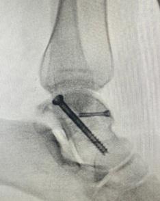
FACT-1 is the first in a series of collaborative studies to explore complex foot and ankle injuries in greater detail to help us answer the questions that may help guide our clinical management. The FACT Network aims to have at least one PI in every trauma unit and trauma centre. Each FACT study will cover a different injury with a panel designing the study to provide well-powered and robust data to better our understanding of complex injuries, risk factors for poor outcomes and the results of more novel treatment strategies with FACT-1 focussing specifically on fractures of the talus.
Talar fractures carry high rates of complications and morbidity, namely non-union, avascular necrosis, malreduction and early arthritis. These injuries often occur in a young, active population with significant impact on quality of life and morbidity. Treatment strategies vary greatly with different views on best practice. Complications pose a challenging clinical situation, and rates vary greatly between papers. Evidence is often underpowered due to this being a relatively rare injury. To combat this power is often increased by performing meta-analyses, systematic reviews or studying a cohort over a long time period, during which significant changes in management, techniques and implants may render previous management strategies obsolete.
FOOt A nd Ankle cO ll ABO r AtiO n S in t r Au MA : FAct-1
This multi-centre, retrospective review of talus fractures treated within the past 10 years aims to produce a well powered study with a variety of managements and complications. This will identify specific risk factors for complications and compare results of different treatments to guide best management.
Data will be retrospectively collected using each hospital’s electronic notes system and adult patients treated between 1st January 2013 and 1st January 2023 with a minimum of 18 month follow up will be included. Patient factors,
injury factors, fracture factors, treatment factors, outcomes and complications will be recorded electronically using a standardised form and data will be collated and assessed centrally.
FACT trials will be run via the umbrella collaboration platform DARFI (Database of Atypical and Rare Fractures and Injuries). Please make sure your hospital is signed up to the DARFI platform (https://bit.ly/DARFI-EoI) and express interest in FACT-1 (fact-network.co.uk). More information on the FACT-1 protocol at (www.darfi.net/studies/fact-1/) n
BOFAS S u PPORTED n ATIO n A l CO llABORATIVES
the P rintAr Study – cAll tO ActiOn!
Prosthetic Joint Infection (PJI) is a rare but potentially devastating complication in total ankle arthroplasty (TAR). With a growing number of ankle joint replacements being performed, the incidence of PJI is expected to increase.
The evidence base for managing PJI in TAR is limited and therefore the current practice guidelines have relied on the comparatively extensive evidence available from hip and knee arthroplasty colleagues. Current treatment recommendations for infected ankle prostheses remain ambiguous. A systematic review suggested that either one-stage or two-stage revision surgery may be the most effective approach. However, the study also reported that Debridement, Antibiotics, and Implant Retention (DAIR) was associated with a high re-infection rate of 39.8%, alongside a 5.6–22.2% risk of major amputation (Kunutsor et al., 2020). These statistics underscore the urgent need for more detailed investigations into effective treatments for PJI in TAR.
Where numbers are small, a collaborative approach is needed. The PRINTAR (PRosthetic Infection in Total Ankle Replacement) Study is a multicentre, retrospective service evaluation designed to address this evidence gap. The aims of the PRINTAR study are to: Identify common pathogens associated with prosthetic joint infections in total ankle replacements; Characterise pathogen sensitivity and resistance profiles to inform antibiotic therapy; Identify sampling methodologies utilised during revision surgeries to ensure optimal pathogen detection; Examine management variability in PJI treatment across different sites; Assess 12-month clinical outcomes to provide insight into the long-term efficacy of current management strategies.
The PRINTAR Study is already underway in a number of BOFAS affiliated sites. Each site is contributing data from cases of revision ankle joint replacement surgery conducted between 1st January 2014, and 1st January 2024. This multi-centre retrospective service
evaluation is limited to eligible patients who underwent revision surgeries due to suspected or confirmed PJI at one of the participating sites.
The PRINTAR Study is nearing the completion of its registration phase within the United Kingdom but if you have cases you can contribute, we would love to hear from you. PRINTAR soon plans to extend to include additional sites across Europe through collaborations with EFAS (European Foot and Ankle Society) colleagues.
The findings of the PRINTAR Study are expected to contribute valuable insights into the management of PJIs in TAR, a field with limited existing data and no standardised treatment guidelines. By establishing a clearer understanding of pathogen profiles, resistance patterns, and effective sampling techniques, the study aims to improve patient outcomes and inform evidence-based clinical practice. n
If you would like to get involved, please email research@bofas.org.uk
h OBB ie S cO rner
Hobbies are a great way for a person to reduce stress. Evidence suggests that there is a good association of enjoyable leisure activities leading to better work life balance, psychological, and physical well-being.
In this issue, we feature hobbies of two of our colleagues, Munier Hossain and Rajiv Limaye.
Munier was able to capture the snowy scenery of the Peak District with his camera as the unexpected cold snap brought with it a taste of arctic weather very recently and turned many parts of the country into a winter wonderland.
Rajiv has found painting as the biggest stress buster throughout his career in medicine. n

Above and right: Photographs by
If you have a hobby you would like to share with us, please email the editor at honorarysecretary @bofas.org.uk
Below: Artworks by Rajiv Limaye


Munier Hossain
Foot Print
BOFAS and partner societies events calendar 2025
16-19 January 2025 AOFAS Winter Meeting Lake Louise, Alberta, Canada
29-31 January 2025 BOFAS Annual Scientific Meeting / BOFAS 50th Year Anniversary Doubletree by Hilton Hotel, Brighton, UK
4 February 2025 AOFAS Foot & Ankle Focus Webinar: Social Media as a Tool for Success Online
24-25 February 2025 Principles of Foot and Ankle Surgery Course
London Chigwell Prince Regent Hotel Woodford Green, London, UK
14 March 2025 AOFAS/AOSSM Specialty Day San Diego Convention Center San Diego, California, USA
3-4 April 2025 MIS Course (1st) Keele University, UK
28 April 2025 Diabetic Foot Course (Basic) Kents Hill Park, Milton Keynes, UK
29 April 2025 Diabetic Foot Course (Advanced) Kents Hill Park, Milton Keynes, UK
11-13 June 2025 Advanced Fellows Foot & Ankle Forum (AFAF) Jesmond Dene House / NSTC Newcastle-upon-Tyne, UK
20-21 June 2025 British Indian Orthopaedic Association Conference Grantham, UK
28 June 2025 4th FOOT-HK cadaveric course Chinese University, Hong Kong
3 July 2025 Basic Arthroscopy Course Arthrolab Solihull, UK
4 July 2025 Advanced Arthroscopy Course Arthrolab Solihull, UK
29-31 August 2025 Indian Foot and Ankle Society Conference Patna, India
TBC September 2025 AFAP / BOFAS (AHP Course) Newcastle-upon-Tyne, UK
10-13 September 2025 AOFAS Annual Meeting Savannah, Georgia, USA
10-11 October 2025 EFAS Congress Vilnius, Lithuania
20-21 October 2025 Principles of Foot and Ankle Surgery Course Taunton, UK
26-28 November 2025 BOFAS Annual Scientific Meeting SEC Glasgow, UK
• For details of all events listed above, please visit the individual website of the relevant society.
Congratulations from...

On behalf of the Hong Kong Foot and Ankle Society, we extend our warmest congratulations on the successful launch of your new bulletin, Foot Print. This initiative is a testament to your dedication to advancing knowledge and sharing expertise in the field of foot and ankle orthopaedics.
We applaud your commitment to providing a platform for education, collaboration, and innovation within the foot and ankle community. The Foot Print bulletin will undoubtedly serve as a valuable resource for surgeons and researchers alike.
We look forward to continued partnership and collaboration between our societies.
Samuel Ling, President of the Hong Kong Foot and Ankle Society (HKFAS)


Foot and Ankle JlA PSP Steering Group
Project lead: Jitendra Mangwani
Patient and carer representatives: Amanda Roberts, Steve Ingram
Clinical representatives: Brigitte Scammell, Patricia Allen, Mark B Davies, Noelene Davey, Rebecca Kearney, Lyndon Mason, Stephen Lines, Nick Gallogly, Veena Patel
Information specialist team: Melinda Hau, Lauren Thomson, Douglas Grindlay
James lind Alliance Adviser: Katherine Cowan
Project administrator: Jo Millard
t OP 10 uk re S e A rch
P ri O ritie S
British orthopaedic Foot and Ankle Society (BOFAS) and James Lind Alliance Priority Setting partnerships joined forces to establish ‘Top 10’ Research Priorities in the UK.
Our partnership brought together those affected by foot and ankle problems including patients, carers, and clinicians to decide what research to focus on next. BOFAS and the James Lind Alliance worked together to establish a Priority Setting Partnership (PSP) following the methods set out by the James Lind Alliance at www.jla.nihr.ac.uk.
The importance of involving those directly affected by a condition in identifying research priorities, is increasingly being recognised. This ensures that the research being done is relevant and timely.
The scope of the Foot & Ankle Surgery PSP included:
• Surgical treatments and injections for the treatment of foot & ankle conditions and injuries in adults (over 18 years).
• Post-surgical rehabilitation or injection aftercare. n
The full protocol for the BOFAS James Lind Alliance can be found at: www.bofas.org.uk/Portals/0/BOFAS%20Documents/JLA%20 PSP%20Documents/JLA-BOFAS-PSP-Protocol-Final.pdf
Publications
https://bmjopen.bmj.com/content/bmjopen/13/5/e070641.full.pdf


Backed by data and designed with patients’ mobility in mind, the Vantage® Total Ankle Mobile Bearing System was engineered to be both anatomic and bone preserving.1 With its lateral curvature on the tibia and dual radius of curvature on the talus, the Vantage Ankle is designed to create stability in all the right places.1
TO LEARN MORE: enquiries@exac.com 01527 591555