26 minute read
ChaPTer 6 emergency Care


Chapter
6Emergency Care
Advertisement
Objectives Upon completion of this chapter, the reader should be able to identify and understand terms related to the following: 1. Emergency-prevention techniques. Discuss the importance of prevention, the procedures taken to prepare for emergencies, and taking vital signs. 2. Emergency-prevention equipment and materials. List and identify the major equipment and materials needed in emergency prevention and treatment. 3. Airway obstruction and resuscitation protocol. Discuss the methods to clear the airway, and define the terms related to resuscitation. 4. Classification of shock. List and discuss the various types of shock. 5. Common medical emergencies. List and describe the most frequent medical emergencies and conditions affecting dental care. 6. Common dental emergencies. Describe the most common emergencies occurring in the dental facility.
Emergency-Prevention Techniques
The best treatment for emergencies is to prevent them from happening. With careful training, observation, and preparation, many medical and dental emergencies can be averted. Two of the fundamental methods employed in facility readiness are: = Patient health history: written and oral communication regarding the patient’s present and past health status, including medication, treatment, allergies, and health concerns. = Vital signs: body indications of the patient’s present health status, including blood pressure, pulse, respiration, temperature, and the patient’s concept of pain.
Blood Pressure
Pulse
Blood pressure (BP) is an indication of the pulsating force of blood circulating through the blood vessels at rest diastolic (dye-ah-STAHL-ick) and while under the highest pressure of the circulating blood, the systolic (sis-TAHL-ick) pressure. BP is recorded in even numbers, with systolic pressure numbers placed before diastolic pressure numbers, for example, 120/80 (systolic/diastolic). Relevant terms are: stethoscope (STETH-oh-scope): device employed to intensify body sounds.
It has a set of earpieces inserted into rubber tubing that combines the two ear tubes into one and extends to a metal bell-shaped or flat disc diaphragm. Stethoscopes used in training may have two earpieces combined to one diaphragm for instructional purposes. diaphragm (DYE-ah-fram = thin covering): a thin layer over the disc end of the stethoscope that helps to enlarge or amplify pulse and body sounds. sphygmomanometer (sfig-moh-man-AHM-eh-ter): an instrument employed to measure the arterial blood pressure. This instrument is available in portable, wall mounted, or mobile floor units and consists of a squeeze bulb on rubber tubing, an arm cuff, and a pressure or aneroid (AN-er-oyd = air pressure) dial or a graduated marked mercury column. Digital sphygmomanometers use only a wrap around cuff with machine read-out gauges; no stethoscope is required. The mercury column unit is considered the most reliable recorder and may be used to calibrate the aneroid system. antecubital fossa (an-tee-CUE-bee-tal FAH-sah): interior depression or bend of the elbow; the approximate area for the placement of the stethoscope diaphragm to determine blood-pressure sound. brachial artery: situated at the inside, upper arm area; selected site of bloodpressure cuff placement.
Pulse is the beating force for blood circulating through arteries, which is classified according to rate, rhythm, and condition. Pulse counts may be taken at various body areas. Figure 6-1 shows the sites for pulse and blood-pressure readings. Abnormal pulse rates can be: accelerated: faster pulse rate than normal or expected, also called “rapid.” alternating: changing back and forth of weak and strong pulsations. Other terms related to pulse are: arrhythmia (ah-RITH-mee-ah): irregular heartbeat or pulsations. bradycardia (bray-dee-KAR-dee-ah): pulse rate under 60 beats per minute (bpm). tachycardia (tack-ee-KAR-dee-ah): an abnormal condition of pulse rates over 100 bpm (except in children). deficit (DEF-ah-sit = lacking): lower pulse rate at the wrist than at the heart site; “heart flutter.”
Carotid pulse
Cuff placement area
Radial pulse Brachial pulse
Figure 6-1
Pulse sites used in blood-pressure readings and pulse counts. Note: The pulse point at the wrist is the radial pulse; in the elbow, the brachial pulse; and in the neck, the carotid pulse.
© Cengage Learning 2013
febrile (FEEB-ril): normal pulse rate becoming weak and feeble with prostration or illness. frequency: pulse count; number of pulsations, which differs with age, sex, body position, health of patient. Frequency can be: = intermittent: occasional skipping of heartbeats. = irregular: variation of force or frequency in pulse rate. = regular: uniform pulse force, frequency, and duration. = thready: a fine, hard-to-locate, barely perceivable pulse.
Respiration
Respiration is the inhaling or breathing in of oxygen and the exhaling or expelling of carbon dioxide. One respiration count requires an inspiration (in-spurAY-shun = breathing in) and an expiration (ecks-purr-AY-shun = breathing out). Respirations are described according to rate, character, and rhythm as: absent: suppresses respiratory sounds. apnea (AP-nee-ah): cessation of breathing, usually temporary. Cheyne-Stokes: respirations gradually increasing in volume until climax, and then subsiding and ceasing for a short period of time before starting again; may be noted in dying.
deep: strong inhalation of air with exhalation. dyspnea (DISP-nee-ah): out of breath; difficult or labored breathing. frequent: rapid breathing that may be noted in children, those with disease, those in hysteria, or those in a drug-induced condition. rale (RAHL): noisy, bubbling sounds from lung mucous, heard on inhalation. shallow: short inhalation with small rise in chest. slow: fewer than 12 respirations per minute. stertorous (STARE-toe-rus): rattling, bubbling, or snoring sounds that obscure normal breaths.
Temperature
Temperature is the balance of heat loss and production in a body and may be taken at various sites, such as oral, rectal, axillary (ACK-sih-lair-ee = armpit), and aural (ORE-ahl = pertaining to the ear). Terms relating to temperature are: fever: elevated body temperature, usually considered over 38.3°C (100–103°F). hyperthermia (high-per-THER-mee-ah): body temperature exceeding 40°C (104°F). hypothermia (high-poh-THER-mee-ah): body temperature, below 35°C (95°F). tympanic (tim-PAN-ick = pertaining to eardrum): measurement of body heat registered by an ear thermometer. The normal ranges of four vital signs are listed in Table 6-1.
Table 6-1 Vital Signs Ranges
Ages Blood Pressure Pulse
Respiration Temperature
Infants 70–100/50–70 80 to 160 bpm 30 to 70 bpm 99.2–99.8 2–5 years 82–110/50–75 80 to 120 bpm 22 to 35 bpm 98.5–99 6–12 years 84–120/54–80 75 to 110 bpm 18 to 25 bpm 98–98.5 13–18 years 90–140/62–88 60 to 90 bpm 16 to 20 bpm 97–99 Adults 90–140/60–90 60 to 100 bpm 15 to 20 bpm 97–99 Geriatric +70 90–140/60–90 60 to 100 bpm 15 to 20 bpm 96–99
Patient’s Concept of Pain
A sixth vital sign, the patient’s concept of pain, is added to the patient’s assessment. Recording the patient’s concept of pain endured can be used as a measurement in the determination of the patient’s condition. The patient rates the level of pain on a scale of 1–10 in intensity. Any increase or decrease in this pain concept may indicate the course of the disease. This vital sign is subjective because it is received from the patient while the other five listed are objective and can be seen by others and recorded.
Emergency-Prevention Equipment and Materials
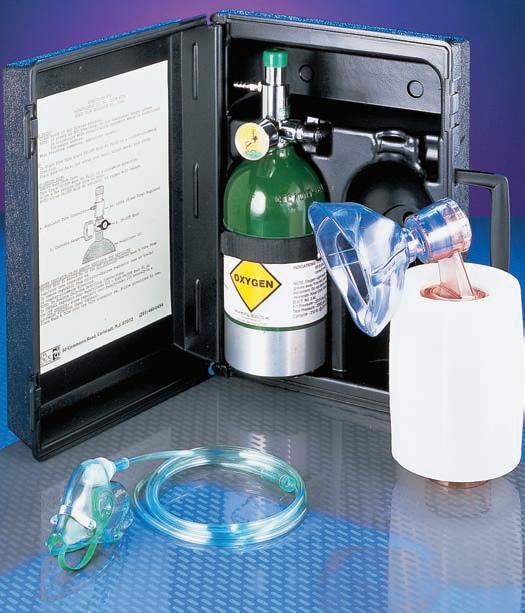
All facilities should maintain the basic equipment and materials necessary to deal with emergencies. Knowledge of the location and use of the following emergency items is essential: emergency call list: important phone numbers necessary in an emergency, which are located in a prominent position near every available phone. oxygen source: container with oxygen gas tank, colored green; obtained in various sizes and may be centrally supplied to each work station (see
Figure 6-2). oxygen regulator: device used to control the flow of oxygen. oxygen flowmeter: gauge used to adjust the flow amount of oxygen. oxygen mask: device placed over a patient’s nose and mouth to administer gas; may be clear or tinted plastic or rubber material. demand-valve resuscitator: device attached to an oxygen mask to apply pressure to the oxygen flow and thereby inflate the lungs. AMBU-bag: handheld squeeze device with a mask that is placed over the patient’s nose and mouth and used to force atmospheric air into the
Figure 6-2
Oxygen supply unit. All practice personnel should know how to operate the oxygen unit in preparation for an emergency.
Photo supplied by Mada Medical Products, Inc.
Figure 6-3
Emergency tray, which is an essential need for most dental-practice emergencies

patient’s lungs; may also be attached to the oxygen supply to force oxygen to lungs. emergency tray: a tray assembled with materials and items necessary for emergencies; often supplied in kit form with medicines, administration items, and chemicals to be used for various emergency events. Emergency trays must be updated frequently and close at hand. All dental personnel should know how to use each item (see Figure 6-3).
Airway Obstruction and Resuscitation Protocol
One of the most-feared emergency situations is an airway obstruction, which occurs when a blockage prevents the patient from receiving air into the lungs. Symptoms include an inability to speak or make a noise, fearfulness, opened eyes, clutching of the throat, and cyanosis (sigh-ah-NO-sis = blue condition), which is a bluish discoloration of the skin caused by a lack of oxygen. The following are terms related to airway obstruction and resuscitation: abdominal thrust: quick, jabbing pressure and force at belt line to force air up the windpipe. asphyxiation (ass-fick-see-AY-shun): not breathing; a result of oxygen imbalance. chest thrusts: applying quick pressure on the chest to force air upward in the windpipe to dislodge the obstruction; may be used on pregnant women as a substitute for abdominal thrusts.
cricothyrotomy (kry-koh-thigh-ROT-oh-mee; crico = ring, thyreo = shield, tomy = cut): an insert or cut into the thyroid and cricoid cartilage to introduce an emergency air supply. gastric distension: a condition resulting from air having been forced into the abdomen instead of the lungs. Heimlich maneuver: procedure in which abdominal thrusts are applied to a choking patient, which forces air from the diaphragm upward to expel a blockage in the airway. hypoxia (high-POCK-see-ah = lack of oxygen): a lack of inspired oxygen. stoma (STOH-mah = mouth): an artificial opening into the windpipe that is placed between the mouth and lung; the opening is at the frontal base of neck into the windpipe for air intake. tracheotomy (tray-kee-AH-toh-mee = cutting into the trachea): a cut and an insertion of a tube into the trachea for an emergency air supply.
Cardiopulmonary Resuscitation
Occasionally, an emergency requires cardiopulmonary resuscitation, commonly known as CPR, a life-saving measure that combines artificial respiration with external cardiac massage. In 2010, the American Heart Association (AHA) and the International Liaison Committee on Resuscitation, believing that air intake is less important than external compression, has changed their recommended guidelines. The following CAB (Compression, Airway, Breathing) regimen is now preferred for anyone who is unresponsive and not breathing normally: 1. Perform immediate compression to distribute the oxygen already present in the lungs. The purpose of compressions is not to “restart the heart” but to circulate the victim’s blood supply. 2. After 30 compressions, establish an open airway, and give two breaths. 3. Maintain the breathing cycle with 2 breaths and 30 chest compressions at 100 compressions per minute rate.
Chest compression should be at a depth of at least 2 inches using one hand for adults and children and using two-finger pressure of 1.5 inches for babies. Rescuers should avoid leaning on the chest so that it may return to its starting position after compression. Note: Unresponsive victims of cardiac arrest such as drowning or drug-overdosed persons may receive the original resuscitation technique of ABC.
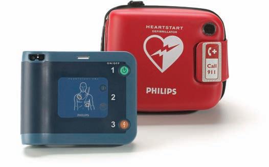
If present at an emergency involving an unresponsive victim, it is wise to survey the immediate surroundings for the presence of life-saving devices that could be used such as an AED (automated external defibrillator), which is a mechanical/electrical device used to revive and stimulate the heart of a patient in cardiac arrest. Dated electrode pads are placed on the patient’s chest to determine if pulseless ventricular tachycardia or a ventricular fibrillation is occurring. After diagnosis, the unit will signal the need for a shock and administer it, analyze the patient, and indicate the need for CPR or administer another shock. Figure 6-4 shows the AED machine and pad placement.
Figure 6-4
Automated external defibrillator unit; showing pad placement
Some common terms used in connection with artificial resuscitation are: airway device: tube inserted into the mouth and down the throat to provide air to the windpipe. compression: force applied to the chest, providing pressure on the heart to imitate a heartbeat or pulsation. finger sweep: using a finger in the mouth of an unconscious person to locate and wipe out any airway obstruction. sternum (STIR-num): “breastbone,” which is the flat bone between the ribs. xiphoid (ZIF-oyd) process: lowest portion of the sternum (breastbone) with no ribs attached.
Classification of Shock
When a patient incurs a condition that alters the intake of oxygen and its passage and use, the body may react by shutting down any or all of its systems. The most common symptom of shock is syncope (SIN-koh-pee = fainting). The pulse may become weak, fast, or irregular, and blood pressure may drop. Breathing may increase, accompanied by pale skin, sweating, and possibly vomiting. Treatment includes finding the source of shock, providing suitable treatment, maintaining body functions, and obtaining assistance. The nine basic types of shock are: anaphylactic (an-ah-fih-LACK-tick): shock arising from a reaction to a body allergen. cardiogenic (kar-dee-oh-JEN-ick): shock arising from improper heart action. hemorrhage: shock arising from excessive blood loss. metabolic (met-ah-BAHL-ick): shock arising from endocrine diseases and disorders, such as diabetes.
neurogenic (new-row-JEN-ick): shock arising from nervous impulses. postural (PAHS-tour-al): shock arising from a sudden change in body positions. psychogenic (sigh-koh-JEN-ick): shock arising from mental origins. respiratory (res-per-uh-TORE-ee): shock arising from insufficient breathing. septic (SEP-tick): shock arising from a microbial infection.
Common Medical Emergencies
During the course of treatment in the dental facility, an occasional medical emergency will arise that complicates the handling and care of the patient. These include allergies, asthma, reactions related to diabetes, epilepsy, hyperventilation, and heart conditions.
Allergies
An allergic reaction is caused by a person’s sensitivity to a specific antigen that can result in a variety of symptoms. Some are as mild as a slight rash, and others are quite involved, including death from severe anaphylactic shock. anaphylaxis (an-ah-fill-ACK-sis): an allergic reaction of the body resulting in lowered blood pressure, swelling of the throat, shock, and even death. itching: a condition of irritation to the skin, scalp, or mucous membranes. erythema (air-ih-THEE-mah = skin redness): a red rash or blotching of the skin. edema (eh-DEE-mah): a tissue swelling, enlargement of a body area. vesicle (VES-ih-kuhl = small blister) formation: small, watery blisters. urticaria (yur-tih-CARE-ee-ah = vascular skin reaction): commonly called hives or wheals.
Asthma
Asthma (AZ-mah = panting) is a chronic disorder characterized by shortness of breath, wheezing, and coughing caused by spasms of the bronchial tubes or swollen mucous membranes. Asthmatic conditions are classified as: extrinsic: resulting from allergens (animal, dust, foods) entering the body (usually affecting children). intrinsic: resulting from bronchial infection allergens (usually affecting older patients). status asthmaticus (AS-matic-us): severe asthma attack that may be fatal.
Allergies, asthma, diabetes, and other emergencies occurring in the dental practice need immediate attention and sometimes medication that is found in a prepared emergency tray as shown earlier in Figure 6-3.
Diabetes Mellitus
Epilepsy
Diabetes mellitus (dye-ah-BEE-tus = passing through, mel-EYE-tus = sugar in the urine) is a disorder of the metabolism of carbohydrates. This disease has been divided into three types. = Type I, insulin-dependent diabetes (once termed juvenile diabetes), has an early onset and is more severe in course. Treatment consists of insulin intake. = Type II, noninsulin-dependent diabetes, usually develops later in life and may be regulated by diet control and/or taking oral medication. = Type III gestational diabetes mellitus may occur in pregnant women who have never had or been tested for diabetes before pregnancy.
Various terms related to diabetic emergencies are: = diabetic coma: loss of consciousness because of severe untreated or unregulated hyperglycemia, a condition termed diabetic acidosis. = glucose (GLUE-kose): sugar, an important carbohydrate in body metabolism. = hyperglycemia (high-purr-gly-SEE-mee-ah; hyper = over, glyco = sweet, emia = blood): a condition characterized by an increase in blood sugar. = hypoglycemia (high-poh-gly-SEE-me-ah; hypo = under, glyco = sweet, emia = blood): a condition in which the blood sugar is abnormally low. = insulin (IN-sahlin): a hormone released by the pancreas that is essential for the proper metabolism of sugar (glucose). = insulin shock: a condition produced by an overdose of insulin resulting in a lowered blood sugar level (hypoglycemia). = juvenile diabetes: onset of diabetes in a person under 15 years of age. = ketone (KEY-tone): acidic substance resulting from metabolism; sometimes produces an acetone mouth odor similar to the odor of nail polish remover.
Epilepsy (EP-ih-lep-see = a seizure) is a disease characterized by recurrent seizures resulting from disturbed brain functioning. Symptoms can range from mild twitching to periods of unconsciousness accompanied with body movements and actions. Caregivers maintain safety by assuring that the patient has an open airway and does not harm himself or herself during this period. Terms related to epilepsy are: = petit mal (PEH-teet-mahl) seizure: small seizures consisting of momentary unconsciousness with mild body movements or actions of staring off into space; sometimes called absence seizures (ab-SAH-ence; French pronunciation). = grand mal seizure: significant epileptic attack that may include an aura, unconsciousness, spasms, mouth frothing, incontinence, and coma.
Hyperventilation
= status epilepticus: a rapid succession of epileptic attacks without the person’s regaining consciousness between the occurrences. = aura (AW-rah = breeze): a subtle sensation of oncoming physical or mental disorder. = clonic (KLON-ick = turmoil): seizure marked by alternating contraction and relaxation of muscles, producing jerking movements. = partial epilepsy: form of epilepsy consisting of convulsions, without loss of consciousness, that are restricted to certain areas, such as one side of the body. = tonic (tahn-ick) seizure: seizure marked by continuous muscular tension, producing rigidity or violent spasms. = incontinence (in-KAHN-tin-ense): loss of bladder control, which may occur during a seizure.
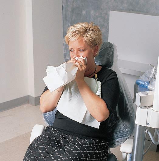
Hyperventilation (high-per-ven-tih-LAY-shun) is a condition of increased inspiration resulting in a carbon dioxide decrease (acapnia) in the body. This may cause tingling of fingers and/or toes, a drop in blood pressure, dizziness, and possible syncope. Treatment consists of calming the patient’s fears, remaining reassuring, and asking the patient to breathe into a paper bag, or to cup the hands over the mouth and take deep breaths. This helps the body regain a carbon dioxide/oxygen balance (see Figure 6-5).
Figure 6-5
Hyperventilation—patient breathing into a paper bag
© Cengage Learning 2013
Heart Conditions
Heart problems are cardiac diseases and conditions that are related to the heart, the muscular organ that powers the circulatory system. The heart, as seen in Figure 6-6, is encased in a sac called the pericardium (pear-ih-KARdee-um = sac around the heart), which has three layers: epicardium (epp-ih-KAR-dee-um): outer serous layer. myocardium (my-oh-KAR-dee-um): middle cardiac muscular layer. endocardium (en-doh-KAR-dee-um): inner layer lining the four heart chambers. Various terms are used to describe heart structure, conditions, and diseases: atrium (AY-tree-um = corridor; plural is atria): two upper chambers of the heart, right and left. ventricle (VEN-trih-kul = little belly): two lower chambers of the heart, one on each side, beneath the atrium. valve (tiny fold ): a structure for temporary closing of the blood vessels; valves also control the flow of blood through the heart. atrioventricular (ay-tree-oh-ven-TRICK-you-lar) orifice: an opening between the atrium and ventricle where the valves are situated. semilunar valves: heart valves. The aortic valve is found at the entrance of the aorta to the heart, and the pulmonary valve or tricuspid valve is situated between the right atrium and the right ventricle of the heart. The mitral, or bicuspid valve, is on the left side between the atrium and the ventricle.
Aorta
Right atrium
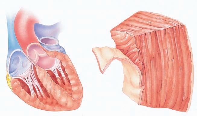
Tricuspid valve
Right ventricle Myocardium
Endocardium
Figure 6-6
Myocardium
Epicardium (visceral layer of the serous pericardium)
aorta (ay-ORE-tah): main artery that exits from the heart. murmur: abnormal sound heard over the heart or blood vessels, an indication of improper blood flow or valve action. arteriosclerosis (ar-teer-ee-oh-skleh-ROH-siss): thickening and hardening of small arteries. atherosclerosis (ath-er-oh-skleh-ROH-sis): blocking of larger artery, often from plaque buildup. angina pectoris (an-JINE-ah PECK tor-is): a pain in the chest caused by a heart malfunction. myocardial infarction (my-oh-KAR-dee-ahl in-FARK-shun): necrosis or death of the myocardium muscle tissue, a heart attack (MI). bacterial endocarditis: sometimes termed infective endocarditis, an inflammation of the heart lining of patients who have had rheumatic fever, open-heart surgery, body part replacements, or implants. Although this is not an immediate emergency chairside threat, these patients require pretreatment with antibiotic therapy to ward off future infections. nitroglycerin: medication for the immediate relief of heart pains, particularly angina pectoris.
Stroke
A common illness related to the blood supply and circulation is stroke. A stroke is technically termed a cerebrovascular (sare-ee-broh-VASS-kyou-lar = pertaining to the blood vessels of the brain) accident (abbreviated as CVA). A stroke is the result of insufficient blood supply to the brain because of a rupture or blockage. Recognition of symptoms and immediate care is important because medical attention given within three hours can be of great benefit. There are two types of strokes: hemorrhagic stroke: occurs when a blood vessel in some part of the brain weakens and bursts open causing damage to the brain cells; occurs in 15% of strokes. ischemic (is-KEY-mick) stroke: caused when the blood supply to the brain is blocked by either a blood clot traveling to the brain (embolic shock) or a blood clot forming in an artery (thrombotic stroke); occurs in 85% of strokes. Other terms related to stroke are: transient ischemic attack (TIA): localized, temporary anemia resulting from an obstruction of the blood circulation; also called a mini-stroke. Ischemia (iss-KEE-me-ah) means a holding back of blood. TIA attacks resolve in less than 24 hours and may be precursors of a stroke (CVA). embolism (EM-boh-lizm): a floating clot or air bubble that may lodge in a blood vessel; if lodged in the brain, it is called cerebral embolism. hemorrhage (HEM-or-rij = blood burst): a rupture in a brain artery. infarction (in-FARK-shun): a decreased blood supply causing necrosis or tissue death. thrombosis (throm-BOE-siss): a clot forming in a blood vessel; atherosclerosis.
hemiplegia (hem-ih-PLEE-jee-ah = a paralysis on one side of the body): may result from a brain lesion, thrombosis, hemorrhage, or tumor of the cerebrum. aneurysm (AN-you-rizm = dilation or bulging of a blood vessel because of wall weakness): a balloon-like enlargement of a cerebral artery or another vessel.
Signs or symptoms of a possible stroke vary but can be detectable by observers: walking and balance off, speech slurred, face droopy, coordination not working, confusion, and so on.
If you see these symptoms, act FAST: = Face: Ask the person to smile. Does one side of the face droop? = Arms: Ask the person to raise both arms. Does one arm drift down? = Speech: Ask the person to repeat a simple sentence. Are the words slurred? Was it repeated correctly? = Time: If the person shows any of these symptoms. Time is important. Call 911. Go to the hospital.
Common Dental Emergencies
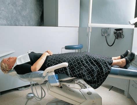
Some emergencies are specifically dental related, occurring as a result of recent dental treatment or the need for dental treatment: alveolitis (al-vee-oh-LIE-tis): inflammation of the alveolar area, commonly called “dry socket.” avulsed (uh-VULST): a tooth or body part that has been knocked out, forced, or torn away. epistaxis (ep-ih-STACK-sis): nosebleed. hemorrhage: excessive bleeding. Treatments for hemorrhage are: = astringent (ah-strin-JENT): agent that has a binding effect; constricts. = coagulant (koh-AG-you-lant): an agent that causes blood to coagulate or congeal. = hemostatic (hee-moe-STAT-ick): agent that stops bleeding, such as vitamin K. trismus (TRIZ-mus): tonic contraction of muscle, perhaps muscles of mastication (jaw). postural hypotension: a decrease in blood pressure resulting from quickly raising the body after having been in a lowered position for a period of time; rapid sit-up resulting in dizziness. sequestra (see-KWESS-trah): small bone pieces or spicules working to the surface after surgery, causing bleeding and soreness. syncope (fainting): the most common emergency in a dental facility. The
Trendelenburg position refers to the patient being placed in a subsupine position with the feet higher than the head; used in office syncope emergencies (see Figure 6-7).
Figure 6-7
Trendelenburg position; emergency position to restore blood to the brain
© Cengage Learning 2013
Review Exercises
Matching
Match the following word elements with their meaning. 1. ____ vesicle A. pulse rate under 60 bpm 2. ____ apnea B. artificial neck opening for inspiration of air 3. ____ dyspnea C. breastbone 4. ____ ketone D. continuous muscular tension 5. ____ axillary E. cessation of breathing 6. ____ tonic F. nosebleed 7. ____ insulin G. small, watery blister
8. ____ sternum H. pulse rate over 100 bpm 9. ____ thrombosis I. loss of bladder control 10. ____ bradycardia J. clot formed in a blood vessel 11. ____ epistaxis K. acid substance resulting from body metabolism 12. ____ atrium L. difficult, labored breathing 13. ____ stoma M. upper chamber half of heart 14. ____ tachycardia N. pertaining to the armpit 15. ____ incontinence O. hormone released from pancreas for metabolism
Definitions
Using the selection given in each sentence, choose the best term to complete the definition. 1. The machine that is exclusively used to determine arterial blood pressure is the: a. sphygmomanometer b. arteriogram c. diaphragm d. thermometer 2. Which type of shock is caused by a rupture of blood vessel in the head? a. atherosclerosis b. cardiac c. hemorrhagic d. ischemic
3. A small epileptic seizure of momentary unconsciousness and mild body involvement is a/an: a. grand mal seizure b. clonic seizure c. aura seizure d. petit mal seizure 4. Thickening and hardening of the wall of an artery is termed: a. angina pectoris b. arterosclerosis c. atherosclerosis d. arythrocardia 5. What color designates that the gas content is oxygen? a. green b. blue c. red d. yellow 6. A body temperature below 35°C (95°F) is termed: a. hypothermia b. hyperthermia c. aurathermia d. diathermia 7. Chest thrusts given in cardiac resuscitation on an adult should be how deep? a. 2 inches b. 1 inch c. 3 inch d. undetermined 8. The automatic external defibrillator (AED) is used for which type of emergency? a. pregnancy b. trauma c. cardiac d. bleeding 9. Which condition exists when air is forced into the abdomen instead of the lungs during CPR? a. hypoxia b. gastric distension c. tracheotomy d. stoma 10. What type of shock arises from the body’s reaction to an allergen? a. anaphylactic b. cardiogenic c. metabolic d. psychogenic 11. The sac covering that encases the heart is the: a. pericardium b. endocardium c. myocardium d. epicardium 12. A bluish cast to the skin due to a lack of oxygen is called: a. cyanamide b. cyanosis c. aura d. cryptitis 13. An astringent would be prescribed for which of the following? a. diabetes mellitus b. epilepsy c. asthma d. hemorrhage 14. Which type of asthmatic attacks results from an allergen and is usually seen in children? a. intrinsic b. extrinsic c. spastic d. status asthmaticus 15. The normal rate of respirations per minute for an adult is: a. 8–10 b. 10–15 c. 15–20 d. 20–25 16. The outer serous layer of the heart muscle is the: a. epicardium b. myocardium c. endocardium d. mitral 17. The condition caused by increased inhalation resulting in lower body carbon dioxide is: a. erythema b. gastric distension c. hypertension d. fever 18. The inner layer of the heart, lining the four chambers is the: a. epicardium b. myocardium c. endocardium d. mitral 19. Another term for the common nosebleed is: a. apnea b. xiphoid c. trismus d. epistaxis 20. A balloon-like enlargement of a cerebral artery or another blood vessel is a/an a. infarction b. aneurysm c. embolism d. hemorrhage Building Skills
Locate and define the prefix, root/combining form, and suffix (if present) in the following words: 1. arteriosclerosis prefix ___________________ root/combining form ___________________ suffix ___________________ 2. defibrillation prefix ___________________ root/combining form ___________________ suffix ___________________ 3. psychogenic prefix ___________________ root/combining form ___________________ suffix ___________________
4. tracheotomy prefix ___________________ root/combining form ___________________ suffix ___________________ 5. hyperventilation prefix ___________________ root/combining form ___________________ suffix ___________________
Fill-Ins
Write the correct word in the blank space to complete each statement. 1. An artificial opening into the windpipe is called a/an ____________________________. 2. The tympanic temperature reading is obtained by using a/an ___________________ thermometer 3. A pulse indicating an occasional skipped beat is termed _________________________. 4. A/an ______________________________ is an instrument used to intensify body sounds. 5. In a blood-pressure reading, the force of the blow flow at rest is called the _________________________ reading. 6. One method of removing airway obstructions is to use the ___________________ maneuver. 7. Another word for the breastbone used in giving
CPR is _________________________. 8. Sensitivity to an allergen present in the body is a/an ____________________________. 9. The ______________________________ bone is situated at the tip of the breastbone. 10. A severe microbial infection may cause ___________________________ shock. 11. The metabolic hormone released by the pancreas is ___________________________. 12. An awareness of an oncoming physical or mental disorder is a/an ___________________. 13. A quick application of pressure to simulate a heartbeat in CPR is called a/an _______________. 14. The _______________________ are the upper halves of the heart, left and right. 15. The _____________________ is the main trunk artery exiting the heart. 16. In a health history, body temp, blood pressure, pulse, and respiration are considered the patient’s ___________________________ signs 17. A/an _______________________ is a tonic concentration of the mastication muscles. 18. ____________________ or fainting is the most common emergency in the dental facility. 19. Another term for redness or blotching of the skin is _________________________. 20. The ________________________________ fossa used in BP readings is situated at the inner elbow space.
Word Use
Read the following sentences, and define the meaning of the word written in bolded letters. 1. The fearful or excited patient can present an elevated systolic count in the blood pressure reading.
________________________________________ 2. Since her cerebrovascular accident (CVA),
Mrs. Brown’s speech and motor skills have been limited.
________________________________________ 3. A patient’s complaint of throat swelling and breathing difficulty indicate an oncoming
anaphylactic reaction.
________________________________________ 4. The receptionist noted that Mr. Harris included hyperglycemia as a medical condition on his health history.
________________________________________ 5. The stethoscope’s diaphragm is placed on the antecubital fossa during the blood pressure procedure.
Audio List
This list contains selected new, important, or difficult terms from this chapter. You may use the list to review these terms and to practice pronouncing them correctly. When you work with the audio for this chapter, listen to the word, repeat it, and then place a checkmark in the box. Proceed to the next boxed word, and repeat the process.
anaphylactic (an-ah-fih-LACK-tick) aneroid (AN-er-oyd) aneurysm (AN-you-rizm) antecubital fossa (an-tee-CUE-bee-tal FAH-sah) aorta (ay-ORE-tah) apnea (AP-nee-ah) arrhythmia (ah-RITH-mee-ah) arteriosclerosis (ar-teer-ee-oh-skleh-ROH-siss) asphyxiation (ass-fick-see-AY-shun) atherosclerosis (ath-er-oh-skleh-ROH-sis) atrioventricular (ay-tree-oh-ven-TRICK-you-lar) atrium (AY-tree-um) aural (ORE-ahl) axillary (ACK-sih-lair-ee) bradycardia (bray-dee-KAR-dee-ah) cardiogenic (kar-dee-oh-JEN-ick) cerebrovascular (sare-ee-broh-VAS-kyou-lar) clonic (KLAHN-ick) cricothyrotomy (kry-koh-thigh-ROT-oh-mee) cyanosis (sigh-ah-NO-sis) defibrillation (dee-fib-rih-LAY-shun) deficit (DEF-ih-sit) diaphragm (DYE-ah-fram) diastolic (dye-ah-STAHL-ick) dyspnea (DISP-nee-ah) embolism (EM-boh-lizm) endocardium (en-doh-KAR-dee-um) epicardium (epp-ih-KAR-dee-um) epilepsy (EP-ih-lep-see) epistaxis (ep-ih-STACK-sis) erythema (air-ith-EE-mah) expiration (ecks-purr-AY-shun) febrile (FEEB-ril) hemiplegia (hem-ih-PLEE-jee-ah) hyperthermia (high-per-THER-mee-ah) hyperventilation (high-per-ven-tih-LAY-shun) hypoglycemia (high-poh-gly-SEE-me-ah) hypothermia (high-poh-THER-mee-ah) hypoxia (high-POCK-see-ah) incontinence (in-KAHN-tin-ense) inspiration (in-spur-AY-shun) ischemia (iss-KEE-me-ah) metabolic (met-ah-BAHL-ick) myocardial infarction (my-oh-KAR-dee-ahl in-FARK-shun) myocardium (my-oh-KAR-dee-um) neurogenic (new-roh-JEN-ick) pericardium (pair-ih-KAR-dee-um) postural (PAHS-tour-al) psychogenic (sigh-koh-JEN-ick) respiratory (RESS-purr-ah-tore-ee) resuscitation (ree-SUSS-ih-tay-shun) septic (SEP-tick) sequestra (see-KWESS-trah)
sphygmomanometer
(sfig-moh-man-AHM-eh-ter) sternum (STIR-num) stethoscope (STETH-oh-scope) stertorous (STARE-toe-rus) stoma (STOW-mah) systolic (sis-TAH-lick) tachycardia (tack-ee-KAR-dee-ah) tracheotomy (tray-kee-AH-toh-mee) trismus (TRIZ-mus) tympanic (tim-PAN-ick) urticaria (yur-tih-CARE-ee-ah) ventricle (VEN-trih-kul) vesicle (VES-ih-kuhl) xiphoid (ZIF-oyd)


