26 minute read
ChaPTer 16 Periodontics
Chapter
16 Periodontics
Advertisement
Objectives Upon completion of this chapter, the reader should be able to identify and understand terms related to the following: 1. Anatomy of the periodontium. Identify and describe the major tissues comprising the periodontium. 2. Etiology and symptoms of periodontal diseases. Describe the cause of periodontal diseases and the symptoms involved. 3. Classifications of periodontal diseases. List the various classes and types of gingivitis and periodontitis. 4. Periodontal examination and evaluation. Describe the examination measures required to evaluate periodontal diseases. 5. Measurement and recording of periodontal conditions. Discuss the methods used to measure and record various periodontal conditions, and identify and explain the index ratings for tooth and periodontal conditions. 6. Periodontal treatment methods. Identify the nonsurgical and surgical methods used to care for periodontal problems.
7. Periodontal involvement with dental implants and cosmetic dentistry.
Identify the various types of bone implants and cosmetic procedures performed in periodontic dentistry. 8. Instrumentation for periodontics. Explain the use of instruments commonly used in periodontal treatment and care.
Anatomy of the Periodontium
All dentists deal with the periodontal tissues in the course of their treatment, but complicated or complex cases can be referred to a periodontist—a dentist who has completed two to three years of additional education in the treatment of periodontal diseases. Periodontology (pear-ee-oh-dahn-TAH-loh-jee) is one of the ADA-recognized specialties and is the field of dentistry that deals with the treatment of diseases of the tissues around the teeth, commonly called
the periodontium. The periodontium serves as an attachment apparatus and is composed of four major tissues (see Figure 16-1): A. gingival: fibrous, epithelial tissue surrounding a tooth; may be divided into three types: attached: the portion that is firm, dense, stippled, and bound to the underlying periosteum, tooth, and bone. The keratinized (KARE-ah-tin-ized = hard or horny) tissue, also called masticatory mucosa, where the gingiva and mucous membrane unite, is indicated by the color changes from pink gingiva to red mucosa, and is called the mucogingival junction. marginal: the portion that is unattached to underlying tissues and helps to form the sides of the gingival crevice; also called the free margin gingiva and forms the gingival sulcus (SUL-kus = groove), approximately 1 to 3 mm in depth. papillary: the part of the marginal gingiva that occupies the interproximal spaces. Normally this tissue is triangular and fills the tooth embrasure area, and also is called the interdental papilla.
Marginal gingiva (free gingiva)
Gingival groove (free gingival groove) Gingival crest Enamal
Dentin
Gingival sulcus Epithelial attachment
Attached gingiva
Mucogingival junction
Alveolar bone Lamina dura
Periodontal ligaments
Cementum
Figure 16-1
The tissues of the periodontium
© Cengage Learning 2013
B. periodontal ligaments: bundles of fibers that support and retain the tooth in the alveolar socket. The five principal types of periodontal membrane fibers are: = alveolar crest fibers: located at the cementoenamel junction; assists with the retention of the tooth in its socket and protects the deeper fibers. = horizontal fibers: attached along the upper side of the root; assists in the control of lateral movement. = oblique fibers: connects the majority of the root in the alveolar socket; assists with the tooth’s resistance to axial forces. = apical fibers: arranged in bundles and attaches the apex of the tooth to the alveolar bone; assists with prevention of tipping and dislocation, and also protects the nerve and blood supply to the tooth. = interradicular fibers: also arranged in bundles and located in the furcations of multiple rooted teeth; assists the tipping, turning, and dislocation of the tooth. An illustration of these fibers is presented in Figure 3-5 in Chapter 3, Tooth Origin and Formation. C. cementum: outer hard, rough surface covering of the root section of the tooth that permits the fiber attachment for tooth retention. D. alveolar bone process: compact bone that forms the tooth socket; supported by stronger bone tissue of the mandible and maxilla and accepts periodontal fiber attachment. The alveolar process makes up the cribriform (KRIB-rih-form = sieve-like) plate to form and line the tooth socket. This outline is called lamina dura (LAM-ih-nah = lining, thin layer) and is easily viewed on radiographs.
Etiology and Symptoms of Periodontal Diseases
In the etiology of problems affecting the periodontium, the major contributing factors are plaque (PLACK = plate or buildup) and calculus (KAL-kyou-lus). Teeth acquire an adhering biofilm or pellicle (PELL-ih-kul), which harbors an assortment of bacterial pathogens and enables plaque to build up. With the addition of calcium and phosphorus salts found in saliva and mouth fluids, a hard substance called calculus (also know as tartar) forms. These gingival irritants are classified as either supragingival (found on the tooth crowns) or subgingival (found on root surfaces below the gingival margin). When this collection of calculus and plaque with pathogens extends into periodontal pockets, it causes irritation and disease. Many health organizations are finding a correlation between periodontal health and major body diseases, such as stroke and circulatory illnesses. Tobacco use and genetic make up play a part in the chances of periodontal disease.
Indications of periodontal disease are: erythema: The gingiva is red and appears inflamed. edema: The tissue is overgrown from hyperplasia (high-per-PLAY-zeeah = excessive number of tissue cells) and hypertrophy (high-per-TROEfee = excessive cellular growth). The gingiva looks swollen and irritated.
Hyperplastic gum tissue may be caused by drug reactions, allergies, and hormonal changes, as well as response to local irritants and disease. loss of stippling (STIP-ling = spotting): Tone or tissue attachment loosens, and puffy gums become smooth and shiny. pocket formation: Gingiva is unattached, recession occurs, and the root may be observed. alveolar bone loss with exudate (EX-you-dayt = passing out of pus): A foul odor is present as supporting bone resorbs; retention is lessening. mobility: The tooth seems loose and moves under pressure because of loss of attachment. The tooth eventually is lost from lack of support or from extraction.
Classification of Periodontal Diseases
Periodontal diseases can be divided into two main divisions: = Gingivitis, an inflammation of gingival tissue with no supporting tissue loss = Periodontitis, inflammation of gingival tissue with involvement of other tissues of the periodontium
Destruction of these tissues varies in degree, intensity, and overall action. To identify each stage and type of periodontal involvement, the American Academy of Periodotology (AAP) devised the following classification system for periodontal diseases: A. Gingival disease: = Dental plaque involvement: (most common) Tissues react to irritants. = Dental plaque with systemic factors included: Pregnancy, hormone, medication, or malnutrition may modify and intensify the disease course of action; sometimes called induced gingivitis. = Nondental plaque tensions: These are of specific bacterial, viral, fungal, or genetic origin, such as gonorrhea, herpes, HIV, and candida infections. = Allergies: The patient may be allergic to dental-restorative materials and have reactions to food, additives, and so forth. = Traumatic lesions, injury: The patient my have been subjected to an external force or have been injured in some way. B. Periodontal disease: = Chronic periodontitis: Previously termed adult periodontitis, this is the most common type of slowly progressive periodontal disease. May be subdivided according to extent and severity into localized with <30% involvement and generalized with >30% involvement. Severity is measured based on the amount of clinical attachment loss (CAL) as slight, moderate, or severe.
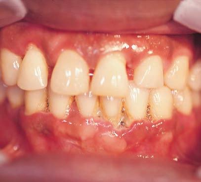
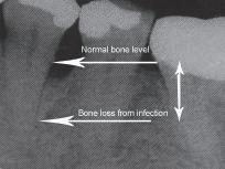
= Aggressive periodontitis: Previously termed early-onset periodontitis, this is a rapidly progressive disease. Subclassifications are: Localized aggressive periodontitis, formerly termed localized juvenile periodontitis (LPJ) affecting young adults Generalized aggressive periodontitis, formerly termed prepubertal or rapid-progressing periodontitis (RPP) = Refractory periodontitis: The periodontitis progresses in spite of excellent patient compliance and provision of periodontal therapy; may be applied to all types of periodontitis. Tissues that are painful, red, and sloughing are said to be desquamative (des-KWAM-ah-tiv = shedding, or scaling off ). = Periodontitis as manifested of systemic disease: Periodontal inflammatory reactions occur as a result of diseases and genetic disorders, such as leukemia, HIV, malnutrition, and hormones. = Necrotizing periodontal diseases: Rapid gingival tissue destruction with bacterial invasion of connective tissue may be a manifestation of systemic disease, such as HIV infection. This category is subdivided into two divisions: Necrotizing ulcerative gingivitis (NUG): With foul odor and a loss of interdental papilla, sometimes called “trench mouth” (see Figure 16-2A). Necrotizing ulcerative periodontitis (NUP): With bone pain and rapid bone loss (see Figure 16-2B).
(A)
Courtesy of Joseph L. Konzelman, Jr., DDS
(B)
Figure 16-2
(A) Necrotizing ulcerative gingivitis; (B) X-ray evidence of significant bone loss around molar tooth.
C. Abscesses of the periodontium: Abscesses are classified according to location, such as gingival, periodontal, and pericoronal. D. Periodontitis associated with endodontic lesions: This simple classification was added to distinguish between periodontitis and periodontitis with endodontic inflammation involvement. E. Developmental or acquired deformities and condition: Deformities appear around teeth, edentulous ridges, and from trauma.
Periodontal Examination and Evaluation
The patient must receive a thorough exam and evaluation before treatment can be established. This procedure involves: medical history: questions regarding diabetes, pregnancy, smoking, hypertension, dedication, substance abuse, and so forth. dental history: chief complaint, past dental records and radiographs, complete assessment of restoration condition, tooth position, mobility. extraoral structure assessment: exam of oral mucosa, muscles of mastication, lips, floor of mouth, tongue, palate, salivary glands, and the oropharynx area. periodontal probing depth: charting and recording findings of probe depths, assessing plaque and calculus presence, soft tissue, and implant conditions. assessing intraoral findings: exam for tori palatinus or tori mandibularis growths abnormal frenum placement and size, and furcation involvement.
The periodontal examination involves charting and recording tooth conditions and the status of the oral mucosa, particularly the periodontium. Prior to the perio exam, the teeth receive a prophylaxis (proh-fil-LACK-sis = scaling, root planing, and polishing of teeth). The professional cleaning of teeth involves the scaling or scraping off of calculus. This is usually accomplished by hand instruments and ultrasonic scaling tips that vibrate and remove the hard deposits from above the gingival crest (supragingival) and below the gingival crest (subgingival). Deposits in the periodontal pockets around the root area are planed by curettage or ultrasonic tips. The combination of the two processes is termed scaling and root planing (SRP) and is followed with a polishing by a slow handpiece with rubber cups or air polishers that spray a force of abrasive powder to cleanse the surfaces. Completion of this process leads to the periodontal survey.
Clinical examination requires obtaining and recording an index (IN-decks = measurement of conditions to a standard), which rates the status of a particular subject and provides a method to measure the progress of the tested item, such as bleeding or plaque.
To be effective, the index procedure must be followed for each patient, and the same method must be used from one dental source to another. Different
The Periodontal Probe
indices apply to different conditions. The common indexes used in periodontics cover plaque, oral hygiene, calculus, debris, periodontal pocket depth, bleeding, grade mobility, root furcation, gingival margin, periodontal disease, and suppurative (pus) involvement, among others. Each practice will choose those measurements, which are considered most indicative, such as calculus, bleeding, and tissue attachment. To obtain measurements for indices, the dentist or hygienist will use a periodontal probe to insert into the gingival sulcus and record the findings.
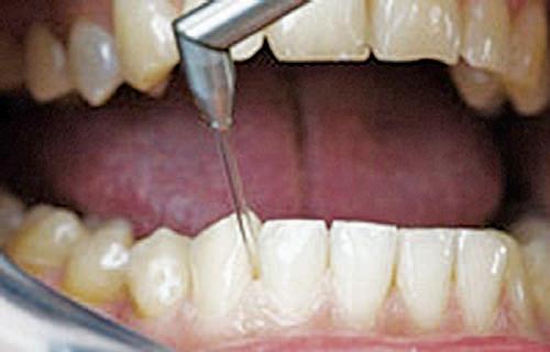
The most common instrument used in measuring is the periodontal probe, a round- or flat-bladed hand instrument marked in millimeter increments. The probe tip is inserted into six specific areas with the deepest pocket marking measurement recorded for that spot. The six areas are facial (F), mesiofacial (MF), distofacial (DF), lingual (L), mesiolingual (ML), and distolingual (DL). Also available for measurement is an electronic probe such as the DiagnoPen with a perio probe that indicates the pocket condition after the teeth have been scaled and planed (Figure 16-3): = <5 indicates clean gingival pockets. = 4–40 indicates little calculus or residual calculus left. = >40 indicates calculus in gingival pockets.
Measurements may be handwritten by the operator, dictated to a recording assistant, or spoken to a voice recorder that transmits the findings to a perio chart. When pocket markings are finished and recorded, the depths are connected by a continuous line giving a visual indication of the gingival crest, indicating the heights of the interdental papilla, and the depths of gingival pockets, including furcation involvement. Dental radiographs are examined to determine bone resorption and loss. The tooth is palpated and tested for mobility, and the patient’s health history is reviewed and discussed.
Figure 16-3
Perio point on DiagnoPen used to determine pocket condition after scaling and root planning
Periodontal Treatment Methods
Treatments for periodontal conditions vary with the severity of the disease. Treatment may be of a nonsurgical nature, conducted in the dental office with a program of home care, or it may involve extensive surgical care.
Nonsurgical Treatment of the Periodontium
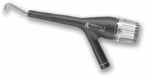
The nonsurgical treatment of periodontal irregularities and diseases involves a variety of procedures and therapies. prophylaxis debridement: removing supragingival and subgingival plaque, calculus, stain, and irritants through tooth-crown and root-surface scaling and root planing (SRP). This treatment usually involves ultrasonic tip scaling and hand instrumentation. tooth and surface polishing: polishing surfaces to remove accumulated extrinsic (ex-TRIN-sick = outer) stains on the tooth surface and endotoxins (en-doh-TOCKS-inz = absorbed pathogens) on the accessible surfaces. This treatment can be completed using air polishers, abrasive cleansing, and handpiece application of nonabrasive rubber cups and points with polishing paste or powder (see Figure 16-4). selective polishing: term applied to the polishing of chosen tooth sites or areas. prophylaxis: term applied to the combination of debridement and tooth polishing; used for purposes of insurance and scheduling. Tooth aids, such as flossing and bridge cleaners, perio brushes, and the like are applied and used in necessary attention areas to complete a thorough cleansing. patient education: customized instruction in oral hygiene; the care of teeth and gingival tissue. antimicrobial (an-tie-my-KROH-bee-al = against small life) therapy: includes prescription mouthwashes (chlorhexidine digulconate); over-the-counter mouthwashes; systemic antibiotics such as tetracycline, penicillin, clindamycin, erythromycin, and metronidazole medicines; and local delivery systems to the site using a gel pack, syringe insertion, or gelatin chip containing antimicrobal products. A new method called Perio Protect offers an antimicrobial medicated application in a custom-made patient tray for patient application. occlusal adjustment: selective grinding of occlusal cusps to eliminate premature contact. tooth stabilization: splinting, wire ligation, or bonding of teeth to lessen tooth mobility. occlusal guards: custom-formed acrylic nightguard to protect from tooth grinding.
Figure 16-4
Air polisher used to polish teeth after the scaling and root planing
Courtesy of KaVo Dental
Periodontal Surgery Techniques
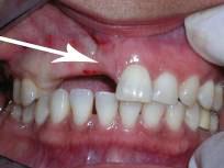
Various specialized surgical treatments are applied in cases of extensive disease of the periodontium. Surgery may involve periodontal knives, instruments, and scalpels. Lately, laser incision with controlled power levels and selected wavelengths to lessen bleeding, swelling, and discomfort is becoming more popular. Some procedures are: mucogingival excision: used to correct defects in shape, position, or amount of gingiva around the tooth; eliminates the pocket formation and pericoronitis typically found on erupting third molars. gingivectomy: excision of pocket tissue areas. One end of a pocket marker is inserted the entire depth of pocket and squeezed until the opposite pointed tip penetrates the gingiva, thereby marking a setting for a surgical template pattern. Necrotic tissue is excised and removed. gingivoplasty: instrumental or laser surgical contour of gingival tissue to remove excessive tissue or pellical edges. periodontal flap surgery: a loosened section of tissue is separated from the adjacent tissues to enable elimination of deposits and contouring of alveolar bone. The several types of flap surgery are: envelope flap: no vertical incision with the mucoperiosteal flap retracted from a horizontal incision line. mucoperiosteal: mucosal tissue flap, including the periosteum, reflected from the bone; also called full thickness flap. partial thickness flap: surgical flap, including mucosa and connective tissue but no periosteum. pedicle flap: tissue flap with lateral incisions. positioned flap: flap that is moved to a new position apically, laterally, or coronally. repositioned flap: surgical flap replaced into its original position. sliding flap: pedicle flap resituated to a new position. osseous surgery: tissue surgery with alteration in bony support of the teeth. re-entry: second-stage surgical procedure to enhance or improve conditions from a previous surgical procedure. vestibuloplasty (ves-TIB-you-loh-plas-tee): surgical alteration of the gingival mucous membrane in the vestibule of the mouth, including frenum reposition or frenectomy and change in muscle attachment. ENAP (excisional new attachment procedure): removal of chronically inflamed soft tissue to permit formation of new tissue attachment. guided tissue regeneration: placement of a semipermeable membrane (Gore-Tex) beneath the flap to prevent ingrowth of epithelium between the flap and the defect; encourages the growth of new periodontal attachment. See an example of tissue surgery in Figure 16-5.
Periodontal dressing packs are placed over the surgical site to assist with protection and healing. These packs are mixed, prepared, and expressed on the surgical site. They are supplied in four ways: = Zinc oxide and eugenol powder and liquid that are mixed to a paste, rolled, and pressed over the site
Figure 16-5
One type of periodontal surgery is restoration and adjustment of marginal ridge.
Bone Grafting
Courtesy of Dr. Louis Poulos (www.drpoulosperio.com)
= Zinc oxide paste and a non-eugenol base paste that are mixed together and applied to the area. = Syringe-dispensed periodontal paste that is expressed on the site and light cured = Gelatin packs that dissolve and do not need to be removed
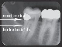
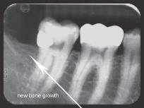
Some periodontal surgical techniques require bone tissues. Bone grafts involve transplants to restore bone from periodontal disease (Figure 16-6). Related terms are: allograft (AL-oh-graft): human bone graft from someone other than the patient.
Courtesy of Dr. Louis Poulos (www.drpoulosperio.com)
autograft (AW-toh-graft): bone graft from another site in the same patient. xenograft (ZEE-no-graft): graft taken from another species, such as cow or pig bone (experimental). allogenic (al-loh-JEN-ick): addition of synthetic material to repair or build up bone.
Periodontal Involvement with Dental Implants and Cosmetic Dentistry
In addition to treatment of the gingiva and periodontium of the mouth, the periodontist performs a variety of tissue and bone augmentations or reductions needed for a functional or cosmetic improvement. Some of these procedures used to provide optimal dental care are listed in the following subsections.
Dental Implants
Dental implants are titanium or ceramic devices that are surgically placed into the alveolar bone to provide firm, fixed anchors for dental appliances or dentures. The bone and the implant complete a process of uniting called osseointegration (oss-ee-oh-in-teh-GRAY-shun = union of bone and device). Implants are explained and illustrated in Chapter 14, Oral and Maxillofacial Surgery, and terminology relevant to periodontics is defined here: endosteal: implants of various designs placed within the bone. subperiosteal: implant placement beneath the periosteum and onto the bone. transosteal: implant placement through the bone. endodontic: the implant is set within the apex of the root.
Periodontal cleaning and scaling procedures performed on implants must be completed with plastic, sonic instruments, or the newer titanium scalers to protect the implants from surface scratching.
Cosmetic Dentistry
Some treatments performed in a periodontist’s practice are used for cosmetic purposes to complete the treatment plan or to improve the esthetic appearance of the patient. Some of these procedures are: crown lengthening: removal of excessive gingival covering tooth enamel in the sulcus area. May be done to gain access to areas of decay for restoration work or to remove a “gummy smile” appearance for esthestic reasons (see Figure 16-7). soft tissue graft: periodontal flap coverage of exposed root areas or repair of pocket damage. pocket reduction: eliminate collection area from pocket position; may include bone grafts. ridge augmentation: bone graft inserts to reshape to the natural contour of gingival and alveolar bone. sinus augmentation: raising the floor of the sinus cavity and building bone replacement may be necessary for placement of maxillary implants in cosmetic surgery. combination procedures: union of more than one procedure to achieve cosmetic effect.
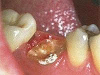
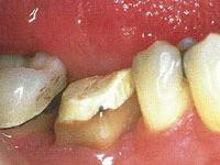
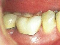
Figure 16-7
An example of a functional crown lengthening treatment. (A) View of decayed tooth (B) Tooth prepared for crown with gingiva shortened (C) completed crown placement on prepared tooth with crown lengthening.
Courtesy of Dr. Louis Poulos (www.drpoulosperio.com)
Instrumentation for Periodontics
Periodontal treatment requires specialized instruments, most of which are hand instruments, although ultrasonic, sonic, laser, and power-driven tools are also part of the necessary setup.
Periodontal Hand Instruments
Hand instruments employed in periodontal treatment, illustrated in Figure 16-8, are used in a push or pull method to accomplish a desired outcome. Push instruments use a push stroke direction with a blade-to-tooth angle of less than 45 degrees perpendicular to the instrument’s shaft. An example is the chisel. Pull instruments use a pull stroke direction with a blade-to-tooth angle of between 45 and 90 degrees perpendicular to the instrument’s shaft. The most effective pull angle is approximately 75 degrees. Examples include scalers, curettes, hoes, and files. Hand instruments are:
Anterior scaler
Posterior scaler
Scalette
Universal curette
Hoe
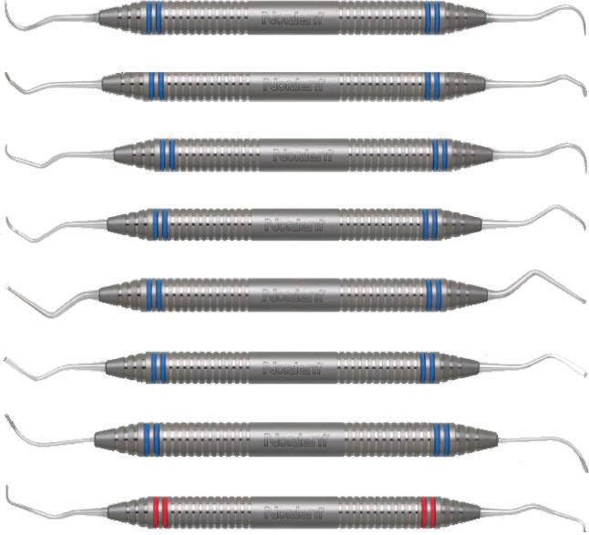
Figure 16-8
An assortment of periodontal hand instruments.
Debridement curette
Diamond furcation file
Implant curette
periodontal probe: used to measure the depth of the periodontal pocket by determining the amount of gingival tissue attachment. A probe may be flat or round bladed and is marked in measured increments. Automatic periodontal probes are available and are used by inserting the probe wire into the sulcus area to determine and record the measurement on a computer. explorer: instrument with a longer, tapered, thin wire tip to determine calculus formation, restoration overhangs, and any root furcation involvement. scaler: instrument with a sharpened blade to remove supragingival calculus deposits and stains. Scalers are available in various shapes such as sickle for universal use, straight for anterior areas, and contra-angled for posterior areas. hoe: instrument with a long shank and a hoe-like tip; used to remove heavy or thick supragingival calculus in posterior areas. chisel: instrument with a longer shaft and a chisel-bladed tip; used to break off and remove heavy calculus in the anterior region. curette: instrument with longer shaft and working end with a rounded toe and back edge to access and remove subgingival deposits. A universal curette has a cutting edge on each side of the blade to enable use in all areas.
Other curettes are designed for specific areas, such as the Gracey curettes with a cutting edge on one side of the blade. file: hand instrument with multiple cutting edges; used to smooth off rough and uneven tissues and remove stubborn calculus deposits. ultrasonic and sonic instrument tip: inserted into the ultrasonic handle; sonic forces move the tip in rapid, short (0.001 inch) waves at speed frequencies of 20,000 to 35,000 vibrations per second to break apart and dislodge deposits from the tooth surface. The tips or inserts are designed for specific
areas and designated use. Machine tips are cooled with a water spray to lessen friction heat. Some handpieces have light sources for better viewing.
Polishing Instruments
Instruments used to polish the tooth surfaces include straight handpieces, prophylaxis contra-angles, and rubber cups as well as polishing agents, such as pumice, cleaners, and chemically impregnated rubber points. Polishing and stain removal is completed by using an air-power-driven calcium carbonate powder spray unit or a slow handpiece rotary rubber cup with pumice.
Surgical Periodontal Instruments
The following are instruments used in surgery: periodontal pocket marker: set of instruments similar to tweezers with a sharp point on one tip for insertion into the depth of the pocket and then compressed to make puncture marks indicating pocket depth. There is one marker for each side of the pocket. periodontal knives: used to make incisions for removal of tissue or to obtain flap design. Blade shape may be round or pointed and long edged. electrosurgery tips/unit: apparatus using electrical current to incise tissue and coagulate blood at the same time; useful in periodontal flap, tissue grafting, crown lengthening, and other tissue surgeries. laser tip/unit: apparatus delivering energy in light form at different wavelengths; can be used in soft or hard tissue curettage surgery when regulated to the specific bacterial target.
Review Exercises
Matching
Match the following word elements with their meaning. 1. ____ papillary gingiva A. free marginal gingiva 2. ____ gingivectomy B. used to measure depth of attached gingiva 3. ____ file C. professional cleaning of teeth 4. ____ allograft D. removal of subgingival and supragingival calculus 5. ____ debridement E. implant placed within apex of tooth root 6. ____ transosteal implant F. hard gingival tissue called masticatory mucosa 7. ____ sulcus G. hand instrument with rasp edges 8. ____ endodontic implant H. bone graft from another site on the same patient 9. ____ furcation I. used to mark depth of tissue pocket for excision 10. ____ periodontal probe J. marginal gingiva occupying interproximal space 11. ____ periodontal pocket marker K. branching or forking of tooth roots 12. ____ prophylaxis L. implant placed through the mandibular bone 13. ____ etiology M. excision and removal of gingival tissue 14. ____ autograft N. bone graft from person other than patient 15. ____ keratinized gingiva O. study of the cause of a disease
Definitions
Using the selection given for each sentence, choose the best term to complete the definition. 1. Transplanting bone to restore bone lost from periodontal disease is termed: a. osteoplasty b. bone graft c. bone therapy d. osseotectomy 2. Splinting, wire ligating, or bonding of teeth together are forms of tooth: a. stabilization b. mobility c. transplantation d. adjustment 3. Increase in the size of affected gingiva is termed: a. hypertrophy b. hypotrophy c. hypoextension d. hyperextension 4. Scraping or flaking off of any hard deposits or accumulated stain on tooth surfaces is: a. prophylaxis b. scaling c. root planing d. tooth polishing 5. Cleansing and shining of tooth surfaces and exposed tooth tissue is termed: a. prophylaxis b. scaling c. root planing d. tooth polishing 6. Which is not considered a periodontal pull hand instrument? a. periodontal probe b. scaler c. curette d. hoe 7. A bone graft insertion to provide a more natural ridge for dentures is termed: a. soft-tissue graft b. crown exposure c. ridge augmentation d. pocket reduction 8. A surgical flap replaced into its original position is what type of flap surgery? a. sliding b. pellicle c. partial d. repositioned flap 9. Which record is not considered an indexing recording of conditions? a. calculus b. bleeding c. radiograph d. stain 10. Another term for the common name “trench mouth” is: a. halitosis b. gingivitis c. ANUG d. periodontia 11. Hard substances formed from calcium and phosphorus salts and deposited on teeth are called: a. stains b. calculus c. plaque d. pellicle 12. Interradicular periodontal fibers may be found around which teeth? a. incisors b. cuspids c. bicuspids d. molars 13. Which is not considered a tissue of the periodontium? a. gingiva b. alveolar bone c. dentin d. cementum 14. Which of the following is not considered a periodontal index factor? a. bleeding b. color c. root furcation d. pocket depth 15. What is considered the proper amount of probe sites per tooth for pocket data? a. 1 b. 2 c. 4 d. 6 16. Painful, red, sloughing gingival epithelium is called: a. irritated b. desquamative c. gingivitis d. refractory 17. Which of the following is not a symptom of gingivitis? a. redness b. mobility c. foul odor d. coral stippling 18. Calculus deposits found on the exposed coronal surfaces of the teeth are termed: a. coronal b. subgingival c. subgingival d. supragingival 19. Selective grinding of occlusal edges of cuspids to eliminate premature contact is: a. occlusal guarding b. occlusal adjustment c. tooth stabilization d. cuspid alignment 20. The periodontium tissue that makes up the tooth socket is: a. alveolar bone b. gingiva c. periodontal fibers d. gums
Building Skills
Locate and define the prefix, root/combining form, and suffix (if present) in the following words. 1. periodontology prefix ___________________ root/combining form ___________________ suffix ___________________ 2. osseointegration prefix ___________________ root/combining form ___________________ suffix ___________________ 3. subperiosteal prefix ___________________ root/combining form ___________________ suffix ___________________ 4. hypertrophy prefix ___________________ root/combining form ___________________ suffix ___________________ 5. gingivitis prefix ___________________ root/combining form ___________________ suffix ___________________
Fill-Ins
Write the correct word in the blank space to complete each statement. 1. The ________________ is a periodontal hand instrument with a longer shaft and neck to access subgingival areas. 2. The chisel is used to break off and remove heavy calculus deposits in the ______________________________ region. 3. The major contributing factor to diseases of the periodontium is ______________________ formation. 4. Pus formation in a pocket area is called pocket ________________________________________. 5. An adhering tooth film that lacks the sticking quality of plaque is called _________________ film.
6. Hard-tissue gingiva, also known as masticatory mucosa, is ____________________________ gingiva. 7. The _________________________ is comprised of the tissue surrounding the teeth. 8. _____________________________ gingiva is unattached to the underlying tissues and forms the gingival crevice. 9. Scalpels, lasers, and _______________________ are used to incise gingival tissue. 10. Periodontal instruments that vibrate rapidly using little strokes to break away deposits are called _________________________________ instruments.
11. The uniting of the implant and bone tissue is a process called ______________________. 12. Using a bone graft taken from another species is termed a/an ___________________________. 13. A/an _________________________________ implant is placed under the periosteum and onto the bone.
14. A periodontal instrument used in a pushing motion should have less than a _______ degree angle on the blade. 15. The use of prescription mouthwashes and treated fibers to destroy microbes is termed ______________________________ therapy. 16. The selective grinding of occlusal cusps to prevent premature contacts is _________________. 17. Acrylic nightguards worn to protect from tooth grinding are called _______________________. 18. Polishing selected teeth during a prophylaxis is termed _________________________________. 19. ______________________________ refers to the procedure of contouring gingival tissue. 20. The measure of a condition to determine standards is called a/an ____________________.
Word Use
Read the following sentences, and define the word written in boldface letters.
1. The dentist explained to Mrs. Bailey that her induced gingivitis is only temporary and will cease after the delivery of her baby. ________________________________________ 2. Dr. Goldenberg’s office called to arrange an appointment for a gingivoplasty procedure for her patient. ________________________________________ 3. One of the treatment plan procedures for the ANUG patient involves antimicrobial
therapy. ________________________________________
4. The dental hygienist uses a periodontal probe to establish the extent of the marginal
gingiva.
________________________________________ 5. The laboratory returned the study cast and the occlusal guard they have prepared for
Mr. Mason.
Audio List
This list contains selected new, important, or difficult terms from this chapter. You may use the list to review these terms and to practice pronouncing them correctly. When you work with the audio for this chapter, listen to the word, repeat it, and then place a checkmark in the box. Proceed to the next boxed word, and repeat the process.
allograft (AL-oh-graft) antimicrobial (an-tie-my-KROH-bee-al) augmentation (awg-men-TAY-shun) autograft (AW-toh-graft) cribriform (KRIB-rih-form) desquamative (des-KWAM-ah-tiv) endotoxins (en-doh-TOCKS-inz) exudate (EX-you-dayt) furcation (fur-KAY-shun) hyperplasia (high-per-PLAY-zee-ah) hypertrophy (high-per-TROH-fee) keratinized (KARE-ah-tin-ized) mucogingival (myou-koh-JIN-ih-vahl)
osseointegration
(oss-ee-oh-in-teh-GRAY-shun) pellicle (PELL-ih-kul)
periodontology
(pear-ee-oh-dahn-TAH-loh-jee) refractory (ree-FRACK-tore-ee) somatic (soh-MAT-ick) stippling (STIP-ling) suppurative (SUP-you-rah-tive) vestibuloplasty (ves-TIB-you-loh-plas-tee)


