28 minute read
ChaPTer 14 Oral and Maxillofacial Surgery


Chapter
14 Oral and Maxillofacial Surgery
Advertisement
Objectives Upon completion of this chapter, the reader should be able to identify and understand terms related to the following: 1. Duties and functions of an oral and maxillofacial surgeon. Describe the functions and roles of an oral/maxillofacial surgeon. 2. Instrumentation related to oral surgery. List and identify the instruments commonly used in the practice of oral surgery. 3. Surgical procedures involved in exodontia. Discuss the various types of tooth extractions and the assorted types of tooth impactions. 4. Procedures involved in soft-tissue surgery. Identify the common tissue surgeries, biopsy methods, and diseases encountered in oral/maxillofacial surgery. 5. Procedures involved in minor bone surgery. Discuss the various minor bone surgeries performed with or without tooth extraction. 6. Surgical procedures involved in fracture repair. Identify and determine the differences between open and closed reduction of bone fractures. 7. Procedures involved in maxillofacial surgery. List and describe the various types of complicated bone surgery, including arthrotomy and genioplasty alterations. 8. Oral surgery procedures involved with TMJ dysfunction. Discuss the classification of TMJ disorders, testing for diagnosis, and possible treatments. 9. Surgical procedures involved in implantology. Identify the various types of implants and their composition materials. 10. Oral surgery role in esthetic dentistry. Discuss the practices of oral and maxillofacial surgery in cosmetic and esthetic completion of treatment and procedures.
A dentist who completes training and board certification in dentistry is able to perform extractions and dental surgery. Prescription writing and hospital privileges are afforded to members of the dental profession, but in many cases, a dentist may not want to perform some surgical procedures. Because of complex measures, patients’ health problems, or lack of specific training, a dentist may refer a patient to an oral and maxillofacial surgeon for treatment. An oral maxillofacial surgeon is a dentist who has completed additional oral surgical and anesthesia studies of two to three years, as well as a hospital internship and residency program. Studies may also be extended two to four years to receive a fellowship in a specific area or a double degree of DDS and MD.
The procedures performed by the specialist include exodontia (ecksoh-DAHN-shah = extraction of a tooth); repair of a fractured maxilla and/or mandible; reconstruction of irregular facial bones and tissues; TMJ (temporomandibular joint) disorders; biopsies and surgical treatment of cysts, tumors, cancers, and other diseases of the oral cavity; placement of implant prostheses, cosmetic adjustments and corrections; and other miscellaneous surgery in the oral cavity.
Oral surgeons may work in private practice or hospital settings and perform solo or as a member of a reconstruction team with various specialists. They mainly work with referred patients who return to their family dentist after the surgical treatment is completed.
Instrumentation Related to Oral Surgery
Each specialization requires instruments designed, adapted, or modified to complete certain desired effects. In addition to regular dental implements, there are numerous specialized oral surgical instruments. See Figure 14-1 for illustrations of common surgical instruments. Common instruments are: forceps (FOUR-seps = pincers for seizing, holding, or extracting): forceps are made for maxillary or mandibular use. Tooth forceps have a handle, a neck, and nib or beaks, which are angled and designed to grasp, hold, and provide leverage to a specific tooth for extraction. Many forceps come in right-sided and left-sided pairs and are numbered with an R or L, such as 88R or 88L molar forceps. Forceps that can be used on both the right and left sides are called universal (see Figure 14-1A). scalpel: a small surgical knife used to cut open or excise tissue from a surgical area. Made of metal or disposable plastic; may be one-piece style or composed of a detachable blade and handles of varying lengths. Blades are designed to work in a certain area and are numbered according to this design and shape. Much soft-tissue surgical work is completed using lasermachine tips that complete the tissue separation and cauterization of the area without the blood flow connected with surgery (see Figure 14-1B). bone file: heavier and thicker than the file used on tooth and restoration surfaces. Bone files may be single ended, but most are double ended with
serrated file edges and different head sizes on opposite sides. They are used to smooth off irregular bone edges remaining from extracted teeth or bone restructure (see Figure 14-1C). elevators: devices used to raise the tooth; three types of elevators are used in oral surgery (see Figure 14-1J): periosteal (pear-ee-OSS-tee-al = concerning the periosteum): used to loosen the periosteum tissue from bone, or detach the tissue around the cervix of the tooth and retract tissue in the surgical site, also called the periosteotome (pear-ee-OSS-tee-oh-tome = cutting tissue around bone). exolever (ECKS-oh-lee-ver = device to raise or elevate): used to elevate or luxate a tooth from its natural socket; also called root elevators. Tips are designed to be used in the mesial or distal, and maxillary or mandibular area. Handles may be the grasp type or T-handed for extra leverage. apical (AY-pih-kal = pertaining to apex or tip): used to elevate or pick out remains of a fractured root tip; also called root tip elevators/picks. These elevators have thinner handles and longer shanked tips than other tooth elevators. hemostat (HE-mow-stat = device or drug used to arrest blood flow): scissorsstyle device with a locking joint and serrated beaks; used to clamp off or hold onto and transfer. Hemostats come in various lengths and may have straight or curved beaks (see Figure 14-1G). needle holder: similar to a hemostat except that the nose of the instrument is rounded and blunted with serrated criss-crossed edges inside its beaks to assist with holding a needle. Suture needles are curved and triangular in shape to avoid tissue trauma during puncturing. Needles are numbered according to their sizes. scissors: various specialized scissors used in oral surgery (see Figure 14-1H). tissue: longer handled scissors with a serrated blade edge that is used to grasp and hold the tissue during cutting. suture: smaller scissors with one curved, half-moon blade that is inserted under the suture thread during cutting. bandage: scissors used to cut materials and dressings during surgery; usually have one longer, blunted blade tip to insert under material. rongeurs (RON-jeers = bone cutting): grasp-handled instrument similar to forceps but with a spring in the handle to provide a “nipping” action.
Beaks may be sharp cutting points (ends) or round sided (blades); used to snip off bony edges and rough areas (see Figure 14-1I). aspirating tips: disposable or metal suction tips with longer handles and narrower tip openings; used to aspirate sockets, deeper throat areas, and surgical sites. chisel: device that is longer, thicker, and heavier than tooth chisels. Available in small, medium, and large blade-width tips. Chisels are used to chip away bone and to apply force enough to break impacted molar teeth that will be removed in sections (see Figure 14-1E). mallet (MAL-ett = surgical hammer): hammer-like device used to apply pressure to chisels. A mallet may have a plain metal face or removable nylonpadded facing (see Figure 14-1F).
curette: hand instrument with a spoon-shaped face that is inserted in the socket or surgical site to scrape out infection and debris. A surgical curette is larger than a dental operative curette. Curettes may be single ended or double ended (see Figure 14-1D). retractor (ree-TRACK-tore = draw back): a hand device used to draw back cheeks and/or tissue to provide more access or light to the surgical area.
Three types are used in surgery: tissue: may be a hemostat-type device with notched tips to hold tissue. Assistants use these claw-like blades with holding tips to retract and hold tissue during surgical procedures. cheek retractor: may be bent wire-shaped device or flat, curved handles used to scoop and hold cheek tissue; may be metal or plastic. After position is obtained, the retractor is hand stabilized against the bone to avoid cheeks and tissues from moving around. Excessive movement may be the cause of swelling and bruising after surgery. tongue retractor: scissor type of instrument with longer shaft and padded or serrated edges; used to grasp and hold the tongue. Occasionally, the operator will use a sterile piece of gauze to grasp, hold, and extend the tongue for examination. mouth prop: small, medium, or large pieces of hard rubber; also called a bite block. Another style of prop, or gag, is a scissors-like instrument with padded ends instead of blades. The padded ends are placed into the tooth occlusion while in a “bite” and later used to spread the jaws apart while the patient is asleep during surgery. suture (SOO-chur = closure): used to close up a wound or incision. To remove the suture, suture thread of silk or nylon material is required. Resorbable suture material of gut or collagen substances does not require removal. surgical bur: similar to dental burs but larger in size; used to remove bone, to expose root tips, or to score and divide teeth in preparation for forced sectioning and removal.
Surgical Procedures Involved in Exodontia
Tooth removal (exodontia) can be a simple or a complex procedure, depending on the tooth or teeth involved, the condition or disease of the site, and the patient’s general health.
Single Extraction
A single extraction is a removal of one tooth during the procedure. The tooth may require a routine extraction, as in a single extraction of an impacted tooth. A single extraction may also entail additional surgical intervention, as when a tooth needs to be sectioned or severed in half.
Impacted Teeth
FULL SIZE
FULL SIZE FULL SIZE FULL SIZE
(A)
KA I KA I
10 11 KA I KA I KA I KA I
12 12B 15 15C
Figure 14-1
Assorted oral surgery instruments (A) assorted dental forceps (B) scalpel handle/surgical blades (C) bone file (D) double-ended curettes (E) bone chisel (F) surgical mallet (G) hemostat (H) surgical scissors, tissue scissors, suture scissors (I) rongeur forceps (J) root elevators (K) root tip picks (L) periosteal elevators
3
(B)
85
75
2
(E)
(F)
FULL SIZE FULL SIZE
(G)
SURGICAL SCISSORS SUTURE SCISSORS
TISSUE SCISSORS (H)
(I) (J)
78 79 80
(K) (L)
(A) Horizontal Tooth Impaction
(C) Distoangular Tooth Impaction (B) Vertical Tooth Impaction
(D) Mesioangular Tooth Impaction
Figure 14-2
Illustrated examples of impaction classifications
© Cengage Learning 2013
for extraction. A bone and tissue impacted tooth is covered with tissue and bone. Bone impacted teeth are typed and named for the tilt angle of the impaction. Examples of the impact classifications are given in Figure 14-2 and described here: horizontal impaction: the tooth is horizontally tilted (see Figure 14-2A); may be leaning parallel to the floor at various angles; crown may be perpendicular to an adjacent tooth crown. vertical impaction: tooth is in upright position but in close proximity to or under the crown of a nearby tooth (see Figure 14-2B). distoangular impaction: crown of the tooth is slanted toward the distal surface and is covered by tissue and/or bone (see Figure 14-2C). mesioangular impaction: the crown of the tooth is mesially tilted and covered by tissue and/or bone (see Figure 14-2D). transverse impaction: tooth is situated sideways to the adjacent teeth and occlusal plane, and it is covered by tissue and/or bone.
Multiple Extraction
A multiple extraction involves the removal of two or more teeth during one procedure. When multiple teeth are extracted, the alveolar bone crests have to be removed and smoothed to prepare the ridges for denture or appliance wear. This reduction procedure is termed an alveolectomy (al-vee-oh-LECK-toh me).
Full Mouth Extraction
In a full mouth extraction, all remaining teeth in the oral cavity are removed. Immediate dentures may be inserted over the sutured site at the time of surgery. A surgical template (TEM-plate = pattern) is used as a guide for the alveolectomy and resection of the area before the placement of the immediate denture.
One potential complication resulting from extraction of teeth is alveolitis (al-vee-oh-LIGH-tiss = infection or inflammation of the alveolar process). This loss of the natural clotting is commonly called a dry socket.
Procedures Involved in Soft-Tissue Surgery
Many procedures performed by the oral surgeon are limited to, or involve, soft tissue of the oral cavity. Some of these soft-tissue surgeries are commonly completed by the general dentist and other specialists, particularly the periodontist, as well: gingivectomy (jin-jih-VECK-toh-me): surgical excision of unattached gingival tissue. gingivoplasty (jin-jih-voh-PLAS-tee): surgical recountour of the gingival tissues. periodontal flap surgery (surgical excision and removal of pocket or tissue extensions): laser or scalpel sectioning and tissue removal that may be necessary for extensive singular pocket involvement or when during tooth eruption, tissue flap coverage of incoming teeth, particularly third molars, obstructs or impacts food around the crown, causing gingival irritation and an infection termed pericoronitis. frenectomy (freh-NECK-toh-me = surgical removal or resectioning of a frenum): surgery that may be performed on the maxillary labial frenum to correct diastema (die-ah-STEE-mah = a space between two teeth), or on the mandibular lingual frenum to correct ankyloglossia (ang-kill-oh-GLOSSee-ah = shortness of the tongue frenum, tongue tied). incision and drainage (I &D): procedure performed for a periodontal abscess.
An incision is made into the affected area, and an opening is obtained to remove and drain infected matter. In some cases, a small piece of rubber dam, or Iodoform gauze, is inserted into the incision to maintain the opening for drainage.
Other tissue surgery may involve removal of the salivary glands, cysts, or any assorted malady (MAL-ah-dee = disease or disorder) of the mucous membranes and oral structures.
Tissue Biopsy
Another tissue surgical procedure performed by an oral surgeon is a biopsy (BYE-op-see = small tissue incision). The common types of dental biopsies are: incision biopsy: removing a wedge-shaped section of affected tissue along with some normal adjacent tissue.
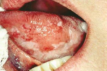
Figure 14-3
Leukoplakia of the tongue
Tissue Diseases
Courtesy of DoctorSpiller.com
excision biopsy: removal of the entire lesion of affected tissue with some underlying normal tissue. exfoliative (ecks-FOH-lee-ah-tiv) biopsy: scraping with glass slide or tongue depressor to collect tissue cells for microscopic study. brush biopsy: much like the exfoliative test, a pipe stem brush is drawn across the mouth tissues, scraped against a glass slide, fixed with a solution, and sent to the lab for a computer-assisted reading.
The term applied to cancerous tumors is malignant (mah-LIG-nant= harmful or growing worse). By contrast, benign (bee-NINE = nonmalignant) tumors are not considered life-threatening or deadly. Some of the tissue diseases occurring in the oral cavity are: leukoplakia (loo-koh-PLAY-key-ah): formation of white patches on the mucous membrane of the oral cavity that cannot be scraped off and have the potential for malignancy (Figure 14-3). fibroma: benign, fibrous, encapsulated connective tissue tumor. papilloma (pap-ih-LOH-mah): benign, epithelial tumor of the skin or mucous membrane. hemangioma (he-man-jee-OH-mah): benign tumor of dilated blood vessels. granuloma (gran-you-LOH-mah): grandular tumor usually occurring with other diseases. melanoma (mel-ah-NO-mah): malignant, pigmented mole or tumor. basal or squamous cell carcinoma: malignant growth of epithelial cells.
Procedures Involved in Minor Bone Surgery
Some tissue surgeries involve treatment of the alveolar bone (osteoplasty = OSS-tee-oh-plas-tee = forming or recontouring bones). Related terms are: alveolectomy (al-vee-oh-LECK-toe-mee): usually performed to remove alveolar bone crests remaining after tooth extraction in preparation for a smooth bone ridge for denture wear.
(A) (B)
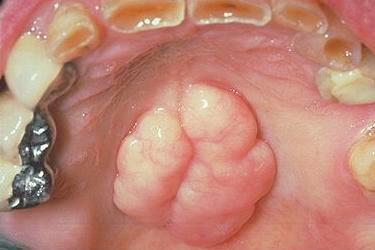
Figure 14-4
(A) Torus mandibularis, (B) torus palatinus
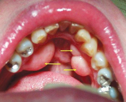
apicoectomy (ay-pih-koh-ECK-toh-me): usually requires opening of the periodontium, including some alveolar bone, and exposure with removal of the root apex. Many times this surgery is followed with a retrofill root canal treatment (RCT). exostosis (ecks-ahs-TOH-sis = bony outgrowth): removing overgrowths and smoothing off of bone edges in preparation for dentures. torus (TORE-us = rounded elevation): an excessive bone growth; a torus on the lingual side of the mandible is termed a torus mandibularis (man-dib-u-
LAIR-iss = concerning the mandible). In the roof of the mouth, it is termed torus palatinus (pal-ah-TEEN-us = in the palate). See Figure 14-4 for an example of each. cysts (SISTs = abnormal, closed-walled sac present in or around tissue): usually
X-ray detected and removed before they enlarge and destroy bone tissue.
Some types are: dentigerous (den-TIJ-er-us): cystic sac containing a tooth or tooth bud particle. radicular (rah-DICK-you-lar): cyst located alongside or at the apex of a tooth root; also called periapical cyst. ranula (RAN-you-lah): cystic tumor found on the underside of the tongue or in the sublingual or submaxillary ducts; usually the result of a blocked duct.
Surgical Procedures Involved in Fracture Repair
Repair of fractured maxillae and mandible bones are reserved for treatment by an oral maxillofacial specialist. Fracture reduction can be completed in two fashions: closed or open reduction.
= Closed fracture reduction: repair with interoral fixation, tooth wiring, or ligation methods in which the teeth are “wired together” in proper alignment while awaiting bone healing. = Open fracture reduction: more complicated procedure involving osteotomy and rigid fixation, perhaps bone plate, mesh, pins, grafts, and other fixation devices. Open reduction requires not only alignment by fixation of the teeth but also repositioning and correction of fractures after surgical access through the periosteum.
Procedures Involved in Maxillofacial Surgery
More complicated or involved surgical intervention with tissue and bone elements is called maxillofacial reconstruction and beautification. Some procedures come as a result of cogenital deformities, some from trauma occurrences, and others from disease damage. The oral maxillofacial surgeon may work alone or with other professionals in a team effort. Examples of reconstruction surgeries are: genioplasty (JEE-nee-oh-plas-tee): plastic surgery of the chin or cheek. Chin size is classified in one of six ways: macrogenia (mack-roh-JEE-nee-ah): large or excessive chin. microgenia (my-kroh-JEE-nee-ah): undersized chin. lateral excessive/deficient: excessive bone in one direction and deficient bone in another. asymmetrical (ay-sim-EH-trih-kal): lack of balance of size and shape on opposite sides. pseudomacrogenia (soo-doh-mack-roh-JEE-nee-ah): excess of soft tissue presenting a chin with the look of abnormal size.
“witch’s chin”: soft tissue ptosis (TOH-sis = dropping or sagging of an organ).
Chin size may be altered by chin augmentation, which could involve tissue liposuction or implanting bone cartilage, grafts, or alloplastic materials.
Figure 14-5
An example of an alloplastic implant graft used in chin augmentation
© Cengage Learning 2013
Osteotomy (oss-tee-AUGH-toh-me = bone incision), surgical movement of bone, or osteoplasty (OSS-tee-oh-plas-tee = to form bones) removal of bone, usually completed with surgical burs. chin augmentation: may be termed sliding genioplasty because it is the option of moving the chin forward by making an incision inside the lower lip and inserting an artificial chin implant or moving the severed bone tip segment forward. In cases where the chin is too posterior, the cut segment may be advanced and secured to enlarge the chin. ridge augmentation: use of bone grafts to build or correct an underdeveloped or missing ridge possibly needed for tooth or denture.implant or preparation for denture wear. surgical correction: in conjunction with the orthodontist and/or the prosthodontist, the oral surgeon may expose and band or peg erupting teeth to prepare the mouth for orthodontic treatment or may remove hidden or retained root tips, cysts, or foreign bodies before taking denture impressions. arthrotomy (ar-THRAH-toh-me = cutting into a joint): reconstruction and alignment of the mandible for TMJ disorders. The mandible may be altered to obtain one of these three movements: retrusive (ree-TRUE-sive): position with mandible backward. protrusive (proh-TRUE-sive): position with mandible forward. lateral (LAT-er-al): position to the side; mesiolateral is toward the center of the face, and distolateral is toward the outside of the face. cleft lip repair: tissue fissure or incomplete juncture of maxillary lip tissues; congenital effect. cleft palate repair: congenital fissure in roof of mouth with an opening into the nasal cavity; may be unilateral (one sided) or bilateral (two sided); also may be complete or incomplete. orthognathic (ore-theg-NATH-ick) surgery: surgical manipulation of the facial skeleton to restore facial esthetics and proper function to a congenital, developmental, or traumatic-affected patient; performed in cooperation with orthodontic involvement in planning and treatment.
Osteotomy and osteoplasty techniques are used on orthodontic prebraced teeth and jaws. This technique is further discussed in Chapter 15,
Orthodontics.
Oral Surgery Procedures Involved with TMJ Dysfunction
The temporomandibular joint (TMJ), composed of condyle of the mandible and the fossa eminence of the temporal bone, is responsible for vertical and lateral movement of the lower jaw. Any malposition or derangement of these parts of the TMJ may cause pain and dysfunction. Repair of a dysfunctional TMJ depends on the severity of the malady. One classification of internal derangement is listed in Table 14-1.
Table 14-1 Wilkes Classification of TMJ Internal Derangement
Stages Symptoms
Stage I Early
Painless clicking Stage II Early/Intermediate Occasional painful clicking, headaches
Stage III Intermediate Frequent pain, joint tenderness, headaches
Stage IV Intermediate/Late Chronic pain, headaches
Stage V Late Variable pain, joint crepitus (KreP-ah-tus): grinding
Motion Function
No restrictive motion
Intermittent locking
Painful chewing, locking, restricted motion
Restricted motion <35 mm
Painful function
Medical tests used to determine malposition of the TMJ are: computerized mandibular scan (CMS): 3D tracking device to record functional movement of the jaw during opening, closing, chewing, and swallowing. electyromyograph (EMG): surface electrodes instrument to determine muscle activity during function; healthy muscles have low levels of electrical activity, and disarranged muscles register high activity. electrosonograph (ESG): recording of sounds during opening and closing of the jaw; also observed by use of a stethoscope. CT (computed tomography, also known as CAT scan): uses X-ray images taken at different angles and computerized into a cross-section of anatomical features. It is used for diagnosis as well as for the preparation of Co-
Cr-Mo (Cobalt-Chromium-Molybdenum) prostheses. See Figure 14-6 for an example of a CAT scan view involving the temporomandibular joint disorder (TMD).
Figure 14-6

Courtesy of Imagining Services, Inc.
Treatment of TMJ Dysfunction
Although some minor cases of TMJ dysfunction can be treated by selective grinding and aligning of tooth surfaces, night sleep guards, or temporary stabilization of the bite process, more severe cases of TMJ dysfunction require oral surgical services. Surgical intervention in TMJ treatment includes a variety of techniques, such as: hemiarthroplasty (HEM-ee-are-throw-plas-tee): surgical repair of a joint with a partial joint implant reconstruction. This may be completed by: autogenous reconstruction: rebuilding of the joint using organic material supplied by the patient, such as toe or rib bone grafts. alloplastic reconstruction: rebuilding of the joint using inert, synthetic man-made materials; can be manufactured to be resorbable or nonresorbable. allograft reconstruction: graft material taken from human donors, which can be tested, sterilized, and accepted by patient’s body to rebuild the jawbone. xenograft: harvested from animals, most commonly the cow; specially processed to become biocompatible. total joint reconstruction: surgical intervention and use of artificial prostheses for the condyle, disc, and fossa of the temporal bone. revision surgery: surgical correction of an area that has been operated on previously, occurring when further degeneration happens, when previous implants have failed, or when going from a partial joint implant to a total implant.
Surgical Procedures Involved in Implantology
The oral maxillofacial surgeon may work in association with a prosthodontist or dentist in the construction and completion of a dental appliance involving a single implant or multiple dental bone implants. After extensive X-ray, CT scans, measurements, examinations, and planning (Figure 14-7), the surgeon may perform one of the following types of dental implants: endosteal (en-DOSS-tee-ahl = placement within the bone): also known as osseointegrated implants; can be used as an anchor for a single tooth or multiple areas, depending on the style of implant. subperiosteal (sub-pear-ee-OSS-tee-ahl = beneath the periosteum and placed onto the bone): usually a cast framework implant with protruding pegs that is placed over the bone and covered by the periosteum; used to hold a base plate for tooth-replacing device, similar to a denture base. transosteal (trans-OSS-tee-ahl = through the mandibular bone): anchor implants that are placed all the way through the mandible. These are also called staple implants. endodontic (en-doh-DON-tick = within the tooth): titanium post placed in the apex of endodontically treated tooth to improve the crown–root length ratio (Figure 14-8). intramucosal insert: indentations in the palate used to provide anchorage for special mushroom-shaped pads built into the gum side of a removable denture.
Figure 14-7
An example of an i-CAT radiograph scan used for the planning of an implant, abutment, and restoration placement

Courtesy of Imaging Sciences, Inc.
(A) (B)
Figure 14-8
Illustrations of dental implants: (A) endosteal implant (B) subperiosteal implant (C) transosteal implant (D) endodontic implant
© Cengage Learning 2013
Implant Material
When the jawbone is too thin or insufficient to accommodate an implant device, a bone graft or ridge augmentation may be completed beforehand. With the use of bone-grafting materials and methods mentioned previously in the paragraph on TMJ arthroplasty, the jawbone may be repaired and later used for dental implants. Maxillary sinus openings may cause a lack of bone structure for implant placement. The sinus may be elevated, and bone-grafting materials may be placed to encourage bone growth thick enough for implant devices.
Implant pins or frameworks may be fabricated using any of several materials: titanium: biocompatible with high strength; oxidizes readily on contact with tissue fluid and has a minimum amount of corrosion. zirconia (zircon oxide ceramic): biocompatible, light colored; may be used in soft-tissue areas; for patients with allergies to other materials and patients who do not want metal framework in the mouth. polymers and composites: in the research stage; may be used as abutments in partially edentulous mouth. stainless steel and cobalt-chromium alloys: older but less used metal materials. cobalt-chromium-molybdenum: implant material used in prosthesis construction for TMJ replacement.
Oral Surgery Role in Esthetic Dentistry
As part of a combined project consisting of maxillofacial surgeons, prosthodontists, orthodontists, dentists, speech therapists, and others, the following procedures may be performed to alter, repair, give proper function, and present an improved cosmetic appearance: cleft tongue repair: bifid or split tongue; usually split at the tip. cleft palate repair: closing of lip and palatine tissues combined with orthodontic treatment. cleft lip repair: tissue closing and repair of opening tissue gap. cosmetic alterations: some cosmetic repair or improvement work is necessary with extensive bone and tissue replacement or movement. Examples that may be performed are: rhytidectomy (rit-ah-DECK-toe-mee): excision of wrinkles by plastic surgery. blepharoplasty (BLEF-er-oh-plas-tee): plastic surgery of the eyelid. rhinoplasty (RINE-no-plas-tee): plastic surgery of the nose. septoplasty (SEPT-toe-plas-tee): plastic surgery of the nasal septum. neck liposuction: suction of fat tissue of the neck. lip, cheek, or chin augmentation: improvement of tissue. injectible botox and chemical peel: skin (dermal) adjustment.
Review Exercises
Matching
Match the following word elements with their meaning. 1. _____ fibroma A. extraction of a tooth 2. _____ malady B. surgical movement of a bone 3. _____ diastema C. undersized chin 4. _____ microgenia D. surgical removal of unattached gingival tissue 5. _____ exodontia E. infection of tissue surrounding crown of tooth 6. _____ gingivectomy F. white, precancerous patch on mucous membrane 7. _____ osteotomy G. witch’s chin 8. _____ crepitus H. open space between two teeth 9. _____ template I. disease or disorder 10. _____ ptosis J. bony overgrowth or rounded elevation 11. _____ macrogenia K. large or excessive chin 12. _____ torus L. fibrous, encapsulated connective tissue tumor 13. _____ ranula M. cyst sac under the tongue 14. _____ leukoplakia N. pattern for surgical adaptation 15. _____ pericornitis O. grinding
Definition
Using the selection given for each sentence, choose the best term to complete the definition. 1. Which instrument is used to clamp off the blood supply in an artery or vein? a. needle holder b. curette c. retractor d. hemostat 2. Anklyloglossia is an immobility or fixation of which tissue? a. uvula b. tongue c. tooth d. cheek 3. Which instrument is used to nip or cut off bony tissue ridges? a. rongeurs b. scissors c. forceps d. hemostats 4. A biopsy technique taking some normal tissue and the entire removal of the lesion is: a. excision b. general c. incision d. exfoliative 5. The building up of an undeveloped or missing mandible area is a/an: a. excision graft b. ridge augmentation c. reduction d. osteotomy
6. A blephoplasty is a surgical correction of which body part? a. lip b. eyelid c. chin d. brow 7. A surgical hammer is a/an: a. retractor b. mallet c. chisel d. periosteal 8. The surgical contouring of the gingival tissue is termed: a. gingivoplastomy b. osteoplastomy c. gingivectomy d. osteoectomy 9. A surgical correction of a previously operated area is termed a/an: a. excision b. osteoplasty c. revision d. exodontia 10. An impaction of a tilted third molar with crown perpendicular to an adjacent tooth is which type of impaction? a. transverse b. distoangular c. vertical d. horizontal
11. The infection of a tooth socket following an extraction may result in the condition called: a. alveolectomy b. alveolitis c. apicoectomy d. apicalalgia 12. A surgical overlay pattern used to prepare for immediate dentures is called a/an: a. template b. implant c. mouth prop d. pattern 13. A torus of the mandibular bone may be termed: a. torus palatinus b. torus lingualis c. torus mandibularis d. ranula 14. In the Wilkes classification of TMJ internal derangement, variable pain, crepitus, and limited motion is considered which stage? a. Stage II b. State III c. Stage IV d. Stage V 15. When the mandible is situated in a drawn backwards position, it is said to be: a. anterior b. prognastic c. retrusive d. posterior 16. A cystic sac containing a tooth or tooth bud is termed: a. radicular b. cystic c. fibromic d. dentigerous 17. The rebuilding of a joint using man-made materials is which type of reconstruction? a. autogenous b. homogenous c. alloplastic d. arthogenous 18. An implant appliance that is placed within the bone is termed: a. ossifous b. exosteal c. subperiosteal d. endosteal 19. The rebuilding of a joint using organic material, such as a rib, is which type of reconstruction? a. autogenous b. homogeneus c. alloplastic d. arthogenous 20. A surgical forcep designed to remove a maxillary or mandibular tooth is called: a. multipurpose b. general c. omnipotent d. universal
Building Skills
Locate and define the prefix, root/combining form, and suffix (if present) in the following words.
1. pericoronitis prefix ___________________ root/combining form ___________________ suffix ___________________ 2. ankyloglossia prefix ___________________ root/combining form ___________________ suffix ___________________ 3. endosteal prefix ___________________ root/combining form ___________________ suffix ___________________ 4. apicoectomy prefix ___________________ root/combining form ___________________ suffix ___________________ 5. exodontia prefix ___________________ root/combining form ___________________ suffix ___________________
Fill-Ins
Write the correct word in the blank space to complete each statement. 1. Another word for a tongue that has an unusual split at the tip is a/an _____________________ tongue. 2. A surgical implant appliance that is placed beneath the periosteum and onto the bone is a/ an ______________________________ implant. 3. ___________________________________ is the termed given to a malignant, pigmented mole.
4. A/an ___________________________ is a cystic tumor found on the underside of the tongue or in a sublingual or submaxillary gland. 5. A __________________ is an abnormal, closedwalled tissue sac. 6. A protrusive mandible is positioned in a _________________________ position during a bite registration. 7. Another term given to a disease or ill condition is a/an __________________________________. 8. A cystic sac containing a tooth or tooth part is a/an _______________________________ cyst. 9. ________________________________ is the term for surgical contouring of gingival tissue. 10. Rhinoplasty is plastic surgery of the ________________________________________. 11. The loss of a clot shortly after extraction is commonly called a/an _____________________. 12. The removing of a wedge piece and some nearby tissue for study is termed a/an _________________________________ biopsy. 13. The surgical removal or resectioning of a frenum is a/an _____________________________. 14. A/an _____________________________ is a tumor of dilated blood vessels. 15. A torus mandibularis elevation is found on the ___________________________________ bone. 16. A rongeurs instrument is used to _____________________________ bony edges. 17. A cyst located adjacent to or at the side of a tooth is a ____________________ cyst. 18. An implant device that is placed within the mandible bone is a/an __________________ implant. 19. I&D is the abbreviation for the ______________________________ procedure. 20. Another word for a surgical hammer is a _________________.
Word Use
Read the following sentences, and define the boldfaced word. 1. To avoid pericoronitis, the dental hygienist instructed the teenager to carefully maintain a clean area round the erupting third molar. ________________________________________ 2. The office manager is sure to list the correct insurance code for the alveolectomy procedure. ________________________________________ 3. The needle holder has a blunt-ended nose while the hemostat’s nose tip is pointed and long. ________________________________________ 4. When setting up the surgical tray for a full mouth extraction and immediate denture insert, the assistant makes sure the template is included.
________________________________________ 5. The surgeon advised Mr. Towers that there must be a long waiting period between the implant appointments to allow for
osseointegration.
_________________________________
Audio List
This list contains selected new, important, or difficult terms from this chapter. You may use the list to review these terms and to practice pronouncing them correctly. When you work with the audio for this chapter, listen to the word, repeat it, and then place a checkmark in the box. Proceed to the next boxed word, and repeat the process. allograft (AL-oh-graft) alveolectomy (al-vee-oh-LECK-toh-me) alveolitis (al-vee-oh-LIGH-tiss) ankyloglossia (ang-kill-loh-GLOSS-ia) arthrotomy (ar-THRAH-toh-me) asymmetrical (ay-sim-EH-trih-kal)
benign (bee-NINE) blepharoplasty (BLEF-er-oh-plas-tee) crepitus (KREP-ih-tus) cyst (SIST) dentigerous (den-TIJ-er-us) diastema (die-ah-STEE-mah) endodontic (en-doh-DAHN-tick) endosteal (en-DOSS-tee-ahl) exfoliative (ecks-FOH-lee-ah-tiv) exodontia (ecks-oh-DAHN-shah) exolever (ECKS-oh-lee-ver) exostosis (ecks-ahs-TOH-sis) forceps (FOUR-seps) frenectomy (freh-NECK-toh-me) genioplasty (JEE-nee-oh-plas-tee) gingivectomy (jin-jih-VECK-toh-me) gingivoplastomy (jin-jih-voh-PLAS-toh-me) hemangioma (heh-man-jee-OH-mah)
hemiarthroplasty
(HEM-ee-are-throw-plas-tee) hemostat (HE-moh-stat) macrogenia (mack-roh-JEE-nee-ah) malady (MAL-ah-dee) malignant (mah-LIG-nant) mallet (MAL-ett) melanoma (mel-ah-NO-mah) microgenia (my-kroh-JEE-nee-ah) orthognathic (ore-theg-NATH-ick) osteoplasty (OSS-tee-oh-plas-tee) osteotomy (oss-tee-OT-oh-me) papilloma (pap-ih-LOH-mah) periosteal (pear-ee-OSS-tee-al) periosteotome (pear-ee-OSS-tee-oh-tome)
pseudomacrogenia
(sue-doh-mack-roh-JEE-nee-ah) ptosis (TOH-sis) radicular (rah-DICK-you-lar) ranula (RAN-you-lah) retractor (ree-TRACK-tore) rhinoplasty (RINE-oh-plas-tee) rhytidectomy (rit-oh-DECK-toe-me) rongeurs (RON-jeers) septoplasty (SEPT-oh-plas-tee) subperiosteal (sub-pear-ee-OSS-tee-ahl) suture (SOO-chur) torus (TORE-us) transosteal (trans-OSS-tee-ahl)


