Better Health
Peace. Love. Cure.


Every October, a “sea of pink” leaves Western Massachusetts awash with hope.
By T iera N W right Special to the Republican
“The Rays of Hope Walk & Run Toward the Cure of Breast Cancer is a powerful and inspiring event that brings together over 10,000 participants, all united by the cause of raising awareness and funds for breast cancer,” said Michelle Graci, manager of events for the Bay-
“The
morning’s activities at 8:30am; followed by the Walk Towards the Cure at 10:30am.
“We are excited to see so many participants gearing up to walk, run, fundraise, and show their support,” said Sandy and John Maybury, 2024-2025 Rays of Hope Co-Chairs, Survivor and Co-Survivor. “The enthusiasm and commitment from our community is truly inspiring.
Whether you are a survivor, a co-survivor, a supporter, or a corporate sponsor, your involvement is what makes this event so powerful.”
This year’s Walk & Run can be summed up by three words, Peace, Love, Cure.
“We strive to create a peaceful and loving environment for everyone Rays of Hope touches on our way to a cure,” said Gra-
survivors.”
In 2023, Rays of Hope raised over $557,000; and to date, the annual charity event has amassed over $17 million. Since its inception, all the money donated to Rays of Hope has remained local and been utilized for patient care, services and research throughout Western Massachusetts.
“Our fundraising goal is to raise more than last year,” said Graci. “With that being said our main focus is to raise awareness for early detection by getting mammograms, supporting survivors through treatment and beyond with special programs and complimentary therapies, and finding a cure through the Rays of Hope Center for Breast Cancer Research. If we are able to do this, our goal has been
enthusiasm and commitment from our community is truly inspiring. Whether you are a survivor, a co-survivor, a supporter, or a corporate sponsor, your involvement is what makes this event so powerful.”
- Sandy and John Maybury, 2024-2025 Rays of Hope Co-Chairs, Survivor and Co-Survivor
state Health Foundation.
Participants converge at Temple Beth El in Springfield and journey through Forest Park, decked out in their finest shades of pink symbolizing their solidarity and support for those impacted by breast cancer. “It’s truly a moving experience,” said Graci. “As you walk, you’ll see happy smiles mixed with tears of joy and remembrance.”
Founded in 1994, the annual Walk & Run will be held Sunday, October 27. The Run Toward a Cure will kick off the
ci. “Our ultimate goal is a world without breast cancer.”
Additionally, dragonflies will play a big role at this year’s Walk & Run.
“A dragonfly is a symbol of change, transformation and self-realization,” said Graci. “It teaches us to love life, to rejoice and have faith even amidst difficulties. Dragonflies symbolize joy and happiness. They serve as a testament to the importance of living in the moment and enjoying each day as it comes; something that resonates deeply with our
reached!”
Participants can choose to register as an individual or a team.
“Individual and group fundraising are the cornerstones of Rays of Hope,” said Kate Weir, Rays of Hope event coordinator. “Our fundraisers help to ensure that we can continue funding breast cancer programs, services, and community initiatives that provide essential support for breast cancer patients, survivors, and co-survivors.”


While the best way to contribute to the walk and run is to fundraise through the Rays of Hope website and participate as a walker or runner at the event on October 27th, community members can also organize their own fundraising events.
“Leading up to the Walk & Run, we encourage supporters and businesses to host their own fundraising event, online or in person, or become an event sponsor,” said Weir. “Popular fundraisers include hosting a Pink & Denim dress down day at your office, pink themed drinks or meal discounts at restaurants and breast cancer awareness sporting events.” Corporate sponsors are very important to the success of Rays of Hope. This year’s major sponsors are Hyundai Hope on Wheels, Gilead,
Kinsley Group, Zasco Productions, Baystate Health Breast & Wellness Center, Baystate Health Breast Specialists, City Tire, Gary Rome Hyundai, Golden Years Home Care Services, Pfizer, Radiology & Imaging, and USA Waste & Recycling.
In addition to the Rays of Hope Shop, there will also be a number of local business and healthcare exhibitors on hand, as well as the Pink Hope Survivor’s Lounge, a special space dedicated to honoring and celebrating the strength and resilience of breast cancer survivors.
“The Pink Hope Lounge is a big pink hug from Rays of Hope and serves as a gathering place for survivors to connect, share their stories, and feel the love and support of the community,” said Graci. “Survivors are celebrated with pride, while those we’ve lost are honored with heartfelt tributes. It’s a day filled with emotions, but also a sense of
hope and strength.”
Meticulously decorated by McClelland’s Florist & Gifts, and curated in collaboration with Gary Rome Hyundai, Graci added that the Pink Hope Survivor’s Lounge “is beautifully decorated with crystals and vibrant flowers, creating a serene and uplifting atmosphere that radiates hope and beauty. It’s a space filled with warmth, appreciation, and a reminder that they are not alone.”
“It’s more than just a walk,” concluded Graci. “It’s a celebration, a chance to support ongoing research, and a meaningful way to connect with a compassionate community.”
For more information or to register for the Rays of Hope Walk & Run Toward the Cure, visit www.baystatehealth. org/raysofhope.





When Cathy Englehardt, 60, was diagnosed with breast cancer, she felt a whirlwind of emotions and fears—central to it all was how her battle with the fatal disease would affect her grandchildren.
The Hatfield woman cares for a 10-year-old boy and 13-year-old girl.
“My husband and I are raising our grandchildren. We’ve had them for nine years,” she said.
Englehardt was diagnosed with breast cancer in the spring of 2023, had a lump and lymph node removed two months later and infusions of chemotherapy from September through last December.
Her grandchildren are special needs, and worry about her, so she tried to limit what they knew about her cancer treatments. But there would be no hiding her loss of hair if it began falling out.
“I was very cognizant of how they would be affected by me having a serious illness. They have anxiety issues, and I didn’t want to contribute more to that, if I could help it,” she said.
Doctors and nurses at the Mass General Cancer Center at Cooley Dickinson Hospital
in Northampton successfully treated Englehardt. They are now monitoring her condition, watching out for a possible recurrence of the cancer.
Cool caps
During treatments, hospital staff introduced the Hatfield woman to a new procedure designed to limit the loss of hair because of chemotherapy. Before, during and after her rounds of chemo, Englehardt wore a gel cap, laced to her head by a canvas covering that secured everything in place.
A cooling unit, situated on the floor next to where she sat, circulated freezing cold water up a tube and through the cap. The process is called scalp cooling and is designed to protect hair follicles.
“It cools the scalp, which means it lowers the temperature to the hair follicles as you are getting your chemotherapy,” said Lisa Martensson, the nurse manager who worked with Englehardt. “It makes your veins tighten up so there’s less [chemotherapy] drug getting to your hair follicles, thus preventing it from damaging them and making the hair fall out. It lessens the chance of losing


your hair,” she explained.
Scalp cooling is being used widely in the United States and Europe. CDH is leasing two units manufactured by Paxman, an English company. Martensson said not all chemotherapy drugs cause hair loss, and scalp cooling doesn’t always help with those that do. But in the last year, the hospital has successfully used the device to help more than a dozen patients keep most of their hair.
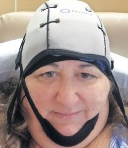
“It doesn’t guarantee no hair loss, but it does minimize loss during the treatment. It also speeds up regrowth after you’re done with your chemo,” said the nurse manager. “It depends on the drug you’re
getting, what chemotherapy regimen you’re on. It works better for some chemotherapy treatments versus others, so you might have more or less loss, depending upon the drug.”
Western Mass pioneer
Englehardt was the first patient to undergo scalp cooling at Cooley Dickinson Hospital. The hospital is part of the Mass General Brigham

system, where the hair-saving procedure is also being used. Englehardt refused treatment in eastern Massachusetts because it would have taken her away from her grandchildren for days at a time.
“Because of the kids at home I didn’t want to be away. I wanted to get things done close to home, so it’s important to have that healthcare locally,” she said.
Scalp cooling can cost patients up to $2,500, depending on how many times they receive the procedure. CDH has a grant program for patients that do not have insurance to cover the hair-saving measure, or can’t afford it.
Englehardt received a grant and did not have to pay for the scalp cooling she received.
“I don’t know if it’s vain or not, but my hair has always been very important to me, and I felt losing it would be a very obvious and significant visual for the kids that I was sick. I was interested in trying the cool cap to see if it could help minimize the visual impact to the kids,” she said.
Englehardt described feeling a cold spike when the icy, 24 degree water ran over her head. The feeling lasted for about ten minutes, until she said her scalp went numb. When the gel caps are removed after sitting somewhat heavily on a patient’s head for several hours, there is a visible, frosty aftermath.
“You get cold pretty quickly,

so a lot of people bring in a blanket. You do have a little bit of frost on your hair when you take your cap off because your hair is damp when you put it on. That helps to conduct the coldness,” said Martensson.
Englehardt’s long, blonde hair flows over her shoulders down to her lower back. Keeping it for the sake of her grandchildren was at the root of her decision to choose scalp cooling. The Hatfield woman did lose some hair volume, but not enough for anyone to notice, she said.
“I was extremely relieved. It was important to retain that part of myself, even if I felt tired or weak from my treatments,” she said, “I didn’t have to appear like I was ill and sickly, and that helped me keep positive.”
When asked if she is looking forward to regaining all of her energy and caring for the kids full-time, she quipped, “I never really stopped!”
Baystate Health does not offer scalp cooling, but a spokesperson said some patients bring in and use their own apparatus. The Sister Caritas Cancer Center at Mercy Medical Center does not offer scalp cooling but is looking into it, according to a spokesperson.


The term ‘breast
does not describe a single type of cancer, but rather several forms of a disease that can develop in areas of the breast. The American Cancer Society says breast cancer type is determined by the specific cells in the breast that become cancerous. There are many different types of breast cancer, and the medical community’s understanding of the disease is based on decades of research and millions of patients treated.
In 2001, Dr. Charles Perou first classified breast cancer into subtypes based on genomic patterns. The Breast Cancer Research Foundation says breast cancer is broadly divided into two types: non-invasive breast cancers and invasive breast cancers. Non-invasive breast cancers are called Stage 0 breast cancers or carcinomas in situ. These are thought to be the precursors to breast cancer, says the BCRF. While non-invasive breast cancers
are not initially life-threatening, if left untreated, they can develop into invasive breast cancers, which can be fatal. Here is a look at some of the different types of breast cancer.
Invasive ductal carcinoma
This is the most common type of breast cancer, advises the National Breast Cancer Foundation, Inc. Invasive ductal carcinoma accounts for 70 to 80 percent of all breast cancer diagnoses in women and men. This cancer forms in the milk ducts and spreads beyond.
Invasive lobular carcinoma
This is the second most common type of breast cancer, accounting for 10 to 15 percent of diagnoses, says the BCRF. Invasive lobular carcinoma originates in the milk-producing glands of the breast known as lobules. Tumors that form due to inva-
sive lobular carcinoma more commonly grow in lines in the breast rather than in lumps, so they present differently on a mammogram.
Inflammatory breast cancer
Inflammatory breast cancer is a rare, fast-growing type of breast cancer. The inflammatory name comes from the appearance of the skin of the breast. It looks red and inflamed, which is caused by breast cancer cells blocking lymph channels in the breast and skin, says Breast Cancer Now, a research and support charity.
Tripe-negative breast cancer


Upon being diagnosed with breast cancer, women and their families are presented with a wealth of information regarding the disease. Some of that information is unique to each patient, but much of it is based on decades of research and millions of successful treatments.


are still contained in a small area. Stage I breast cancer may be characterized as stage IA, which indicates a tumor is about as large as a grape and cancer has not spread to the lymph nodes, or stage IB, which indicates the tumor may be slightly smaller but is accompanied by small clusters of cancer cells in the lymph nodes or there is no tumor and only the small clusters in the lymph nodes. The ACS also reports a 99 percent five-year survival rate for patients diagnosed with stage I breast cancer.




The NBCF says a diagnosis of triple-negative breast cancer means the three most common types of receptors known to cause most breast cancer growths are not present in the cancer tumor. These receptors are estrogen, progesterone and the HER2/ neu gene. Since the tumor cells lack necessary receptors, certain treatments like hormone therapy and drugs that target these receptors are ineffective. Chemotherapy is still an option.
Metastatic
breast cancer
This type of breast cancer is also known as Stage IV breast cancer. Metastatic breast cancer originates in an area of the breast, but spreads (metastasizes) to another part of the body, most commonly the bones, lungs, brain, or liver, indicates BreastCancer.org.
Individuals hoping to learn more about breast cancer should be aware that there are various types of the disease. Which type an individual has is an important variable doctors consider as they plan a course of treatment.
The American Cancer Society reports that cancer staging is a process during which doctors will attempt to determine if a cancer has spread and, if so, how far. Breast cancer stages range from stage 0 to stage IV. Each stage signifies something different, and recognition of what each stage indicates can make it easier for women to understand their disease.
The Memorial Sloan Kettering Cancer Center notes that when a woman is diagnosed with stage 0 breast cancer, that means abnormal cells are present but have not spread to nearby tissue. The National Breast Cancer Foundation, Inc.¨ indicates stage 0 breast cancer is the earliest stage of the disease and is highly treatable when detected early. Indeed, the American Cancer Society reports a five-year survival rate of 99 percent among individuals diagnosed with stage 0 breast cancer.
Stage I is still considered early stage breast cancer.
The MSKCC notes a stage I diagnosis indicates tumor cells have spread to normal
A stage II breast cancer diagnosis indicates the tumor is at least 20 millimeters (about the size of a stage IA tumor) and potentially as large as 50 millimeters. The tumor also can be larger than 50 millimeters if no lymph nodes are affected (stage IIB). The ACS notes the size of the tumor may indicate if the cancer is stage IIA or stage II B. The MSKCC notes that a stage IIA diagnosis could indicate there is no tumor or there is a tumor up to 20 millimeters and the cancer has spread to the lymph nodes under the arm.
A tumor determined to be between 20 and 50 millimeters that has not spread to the lymph nodes also indicates a stage IIA diagnosis. A stage IIB diagnosis indicates the tumor in the breast is between 20 and 50 millimeters and has spread to between one and three nearby lymph nodes. According to Cancer Research UK, the five-year survival rate for stage II breast cancer is around 90 percent.
Stage III breast cancer is considered regional, which
roughly 86 percent survival rate between 2013 and 2019. The MSKCC notes that a stage III diagnosis indicates the tumor is larger than 50 millimeters and has affected lymph nodes across a wider region than in less developed stages of the disease. Cancers that have reached stage III may be categorized as stage IIIA, stage IIIB or stage IIIC. The American College of Surgeons reports that stage IIIA indicates a tumor of any size that has spread to between four and nine lymph nodes or a tumor larger than five centimeters that has spread to between one and three lymph nodes. Stage IIIB indicates any size tumor and that the cancer has spready to the chest wall. A stage IIIC diagnosis indicates the tumor can be any size and has spread to 10 or more lymph nodes.
Stage IV is the most advanced form of breast cancer. If the cancer has reached stage IV, that indicates the tumor can be any size and has spread beyond the breast to other parts of the body, potentially including organs and tissues. The ACS reports that survival rate for this stage, which is considered distant, is 31 percent. However, the breast cancer advocacy organization Susan G. Komen notes that only around 6 percent of breast cancer diagnoses in women diagnosed for the first time have reached stage IV at the time of diagnosis.
Staging makes it easier to understand a breast cancer diagnosis. More information about breast cancer staging is available at mskcc.org and cancer.org.


By D ebbie Gardner
Special to The Republican Mercy’s breast care program is the only one in Springfield accredited by both the National Accreditation Program for Breast Centers (NAPBC) and the American College of Surgeons Commission on Cancer, assuring that Mercy meets the highest standards of care for patients with diseases of the breast. More information can be found at www.trinityhealthofne.org/ services/cancer-care
The Republican reached out to Dr. Sarah J. McPartland, MD, MS, FACS, medical director of the Center for Breast Health and Gynecologic Oncology at Mercy Medical Center with questions about
the importance of breast cancer detection and current treatment options. McPartland offered the following answers and insights.
Early detection is always touted as the best defense against breast cancer. How have the recommendations for screenings changed in recent years? Are they suggested at a younger age?
Detecting breast cancer at its earliest stages – when the cancer cannot be felt in the breast but can be seen as a change on a mammogram – is very important because it offers the best chance for cure. The age at which wom-


en should start mammogram screening has received a lot of recent media attention as national societies have provided sometimes conflicting recommendations. As a breast surgeon, I follow the guidelines from the American Society of Breast Surgeons (ASBrS) - women who are at average risk of breast cancer should start yearly screening exams at the age of 40. There are situations where breast cancer screening is recommended before age 40, sometimes in women with a strong family history of breast cancer diagnosed at a young age or in those who have genetic testing results that indicate a higher risk of breast cancer.

How much emphasis is now put on digital and/or MRI mammography, and when are these screenings appropriate?
A mammogram in the simplest terms is an x-ray of the breast. Similar to a camera, x-rays can be captured on film or digitally. Both film and digital mammograms are considered adequate for screening; however, digital mammography has higher detection rates of breast cancer in young women and those with dense breast tissue. The use of tomosynthesis, also known as 3D mammography, reduces the likelihood of getting “called back” for additional pictures and having a “false positive” result (i.e. the mammogram suggests a possible cancer when one does not actually exist; this situation can lead to unnecessary biopsies). The use of digital mammography combined with tomosynthesis makes it easier to find very small breast cancers, particularly in women who have dense breasts.
There are certain genesBRCA1 and BRCA2 –that make women more susceptible to breast cancer. When is it appropriate to add gene testing to screenings?
high risk of breast cancer, can decrease the chance of breast cancer by achieving a healthy weight, performing moderate daily exercise, and avoiding smoking and excess alcohol.
If a woman’s mammogram suggests the presence of breast cancer, what are the next steps in diagnosis?
When a screening exam reveals a change or new finding, the woman will be called back for a more sensitive test called a diagnostic mammogram. This type of exam takes additional magnified pictures of any areas of concern. Sometimes a breast ultrasound is also performed. If a mass or change persists, a biopsy may be recommended. Breast biopsies are performed in several different ways using mammogram, ultrasound, or MRI to guide a needle into the area of concern. The needle allows for a small amount of tissue to be removed. This tissue is then sent to the lab, where a pathology physician will test the tissue and determine if cancer is present. Staff at the Center for Breast Health at Mercy understand a breast biopsy can be a very stressful process, and our team are often able to provide biopsy results within 1 to 2 days.

breast will be recommended after surgery. This helps to reduce the risk that the cancer will return.
Medications that prevent hormones from stimulating breast tissue are commonly used in breast cancer treatment. Because these medications are usually well tolerated with fewer side effects, they can also sometimes be an option for additional treatment in women who may not be healthy enough to receive chemotherapy.
Are there new treatments or approaches to breast cancer on the horizon?
Are there any in use now?


Early detection of breast cancer is important. Mammograms are safe, fast, and easier than you may think. If you or someone you love is concerned about breast disease, Mercy Medical Center offers comprehensive resources for screening, diagnosis and treatment for cysts, lumps, breast pain and breast cancer.
For scheduling information, please scan the QR code or visit TrinityHealthOfNE.org/Mammo.
We have now identified multiple genes associated with breast cancer. The presence of these mutated genes can increase a woman’s lifetime risk of breast cancer. These genes can be passed down through generations within families. All women should discuss their personal and family history with their physician to determine if genetic testing is recommended. In my practice, we offer genetic testing to all women diagnosed with breast cancer as recent studies show that relying on family history alone misses a sizeable population of women with gene mutations that may impact their breast cancer risk.
Are there environmental factors that can increase a woman’s chance of developing breast cancer? There has been recent talk about an association between alcohol use and breast cancer. How strong is the relationship? What should women do?
There is much we still do not fully understand about how breast cancer begins. Known risk factors include older age, family history of breast cancer, a personal history of radiation treatments to the chest, and exposure to hormones/ estrogen. Being overweight or obese increases the risk of breast cancer, as do smoking and excessive alcohol consumption. The more you drink or smoke, the higher the risk of breast cancer developing. Women, even those at
Are both radiation and chemotherapy always follow-up treatments after tumor removal? Are there instances where one or both of these treatments are not necessary?
How is that determined?
What might be used as alternatives if these standard treatments are not employed?
The best cancer care happens when a team of cancer physicians work together to develop and implement a treatment plan. Breast cancer team members typically include medical oncologists, breast surgeons, radiologists, pathologists, and radiation oncologists, along with nurses, genetic counselors, social workers, experts in rehabilitation, and nutritionists. Teams meet to discuss a patient’s case, then utilize national guidelines to determine if additional treatment is needed after (and sometime before) surgery. Cancer care has also become more individualized with the use of sophisticated tests on the breast tumor itself. These tests provide specific information regarding the aggressiveness of a patient’s tumor. When used in conjunction with other lab results, these tests can help determine whether chemotherapy would be helpful.
For many women who undergo a lumpectomy (procedure that removes the tumor and a small amount of adjacent healthy breast tissue), radiation treatments to the
Breast cancer is the subject of significant research and thankfully there have been many recent advances. Many of the newer treatments utilize components of the immune system to fight cancer. Our program is pleased to be able to offer women the option of enrolling in a clinical research trial as part of their treatment plan.
There are also many new technologies and procedures in the surgical treatment of breast cancer. Our center employs wireless and radiation free localization of tumors for surgery with tiny magnetic clips. We also utilize intra-operative specimen mammography. These technologies allow for more precise surgical procedures which have a higher likelihood of complete removal of the cancer at the time of initial surgery.
What about men and breast cancer? Though it is rare, it does happen. What are the symptoms? Are treatment approaches different?
Breast cancer can occur in men, although it is rare. Male breast cancer cases account for less than 1% of all breast cancers diagnosed in the United States each year. In general, the treatment of breast cancer and prognosis for men and women is similar. However, in part because there is an absence of routine screening for breast cancer in men, they are more commonly diagnosed at a later stage of disease once the mass becomes palpable. This
impact

By L indsey B ever
Washington Post
The
Generation X and millennials are at an increased risk of developing certain cancers compared with older generations, a shift that is probably due to generational changes in diet, lifestyle and environmental exposures, a large new study suggests.
In a new study published in the Lancet Public Health journal, researchers from the American Cancer Society reported that cancer rates for 17 of the 34 most common cancers are increasing in progressively younger generations. The findings included:
Cancers with the most significant increased risk are kidney, pancreatic and small intestine, which are two to three times as high for millennial men and women as baby boomers.
Millennial women also are at higher risk of liver and bile duct cancers compared with baby boomers.
Although the risk of getting cancer is rising, for most cancers, the risk of dying of the disease stabilized or declined among younger people. But mortality rates increased for gallbladder, colorectal, testicular and uterine cancers, as well as for liver cancer among younger women.
“It is a concern,” said Ahmedin Jemal, senior vice president of the American Cancer Society’s surveillance and health equity science department, who was the senior author of the study.
If the current trend continues, the increased cancer and mortality rates among younger people may “halt or even reverse the progress that we have made in reducing cancer mortality over the past several decades,” he added.
While there is no clear explanation for the increased cancer rates among younger people, the researchers suggest that there may be several contributing factors, including rising obesity rates;
altered microbiomes from unhealthy diets high in saturated fats, red meat and ultra-processed foods or antibiotic use; poor sleep; sedentary lifestyles; and environmental factors, including exposure to pollutants and carcinogenic chemicals.
Two decades of cancer data
Researchers analyzed data from more than 23.5 million patients who had been diagnosed with 34 types of cancer from 2000 to 2019. They also studied mortality data that included 7 million deaths from 25 types of cancer among people ages 25 to 84 in the United States.
Expanding on their previous research, which had identified eight types of cancer in which incidence rates increased with each successive generation,
and population sciences at the University of Texas MD Anderson Cancer Center.
At the same time, the researchers also noted that there has been a drop in cervical cancer rates among younger women, which they attribute to vaccination against the human papillomavirus (HPV). Smoking-related cancers such as lung, larynx and esophageal squamous cell carcinoma also declined, though progress has slowed among the youngest age groups, the researchers said.
Difficulties with detection
Routine screening tests are recommended for only four cancers - colon, cervical, breast and, for some people, lung - and a number of younger people who are at average risk do not meet the age re-
gallbladder and other biliary cancers, and uterine cancer rates increased across almost all age groups, rising faster among younger generations. While breast cancer rates among women younger than 40 remain low, in a separate study, breast cancer still accounted for the highest number of early-onset cancer cases.
Lowering screening ages
Recent and growing evidence showing that more women in their 40s are getting breast cancer prompted the U.S. Preventive Services Task Force last year to change its previous guidance, lowering the age for routine screening mammograms from age 50 to 40.
Routine mammograms are not as effective for women
that increases in stomach and colorectal cancer rates were confined to younger age groups, meaning that while colorectal cancer rates are declining overall, there is a rise in incidence in younger populations.
But many people who are eligible are not getting colorectal cancer screenings. A 2021 study reported that fewer than 4 million of the eligible 19 million adults ages 45 to 49 years were up-to-date on screenings, which can include a newly approved blood test, a stool test or a visual test such as a colonoscopy or CT colonography.
Even when symptoms arise, “I think many younger folks ignore them, thinking they cannot get cancer because they’re young,” said Rashmi Verma, an oncologist who specializes in gastrointestinal
Cancers with the most significant increased risk are kidney, pancreatic and small intestine, which are two to three times as high for millennial men and women as baby boomers.
the researchers have found an additional nine, including some that had previously declined among older birth cohorts before rising in younger populations.
The study did not examine factors including household income, insurance status, race or ethnicity.
Younger people, or those under 50, represent a minority of the overall population of those who develop cancer, “but the concern is that cancer is occurring at younger and younger ages, so this increased incidence raises very real concerns as that population continues to age,” said Ernest Hawk, vice president and head of the division of cancer prevention
quirements or, for various reasons, are not getting screened. Some experts have pointed to potential harms from widespread screening, including false positives that may take a psychological toll and lead to unnecessary follow-up tests and procedures.
“The problem becomes that patients are getting younger and younger, we don’t always have good screening to begin with, and then we can’t really screen such large populations,” said Andrea Cercek, a gastrointestinal oncologist and co-director of the Center for Young Onset Colorectal and Gastrointestinal Cancers at Memorial Sloan Kettering Cancer Center. In the new study, breast,
with dense breasts, however, which is more common among those who are younger, said Elizabeth Comen, a breast cancer oncologist and associate professor at New York University Langone Health’s Perlmutter Cancer Center.
“Understanding how we can better screen and detect cancers in younger patients is a massive unmet need,” said Comen, who added that many of her younger patients find their own breast cancer.
In recent years, the recommended age for colorectal cancer screening was also lowered, from age 50 to 45, as research has shown a trend toward diagnoses at younger ages. The new study found
cancers at the Comprehensive Cancer Center at the University of California at Davis, adding that she has treated patients as young as 20.
Others may be uninsured or not aware that screening tests are recommended for them, experts said.
Missed diagnoses
When some younger patients seek care for gastrointestinal symptoms, they are misdiagnosed with other conditions such as hemorrhoids or irritable bowel syndrome, so it is important to consult a gastroenterologist, Verma said.
But for most cancers, including pancreatic cancer, there are no screening tests - at any age - which can lead to late diagnoses and more limited treatment options, experts said.
While there have been advances in diagnostics and treatment, oftentimes, malignant pancreatic tumors (and some others) are discovered incidentally during imaging for other issues, said Charles J. Yeo, a professor and chair of the department of surgery at Sidney Kimmel Medical College at Thomas Jefferson University.
The increasing cancer rates among younger generations highlights the need for further study to both pinpoint a cause for planning prevention as well as to develop better - and, in many cases, any - screening tests to help detect cancers in younger people earlier in the course of disease when treatment is often more effective, experts said.
The rising rates also raise questions about what happens to younger patients in survivorship.
“There are going to be young cancer survivors who are deeply impacted biologically, physically and psychologically from these diagnoses,” Comen said. “And that’s going to have a ripple effect in our society that our medical community needs to be equipped to address.”



By L indsey B ever
The Washington Post
A national health panel has recommended lowering the age for routine screening mammograms by 10 years, now advising women ages 40 to 74 at average risk of breast cancer to get screened every two years.
Previously, the guidance from the U.S. Preventive Services Task Force was for women to make individual screening decisions in their 40s but start no later than 50.
The change has left many women with questions about how the new guidance affects their personal health. We spoke with experts to get answers.
Why were the guidlines changed?
Cancer rates among younger Americans are on the rise. More women in their 40s are getting breast cancer, with the number of newly diagnosed women increasing about 2 percent each year, said John Wong, an internist and professor of medicine at Tufts University School of Medicine, and vice chair on the task force.
Black women are more likely than White women to be diagnosed with breast cancer at a younger age and more likely to be diagnosed with an aggressive form called triple-negative breast cancer. They are also about 40 percent more likely to die
of breast cancer than White women, research shows.
Overall, more than 42,000 women die of breast cancer each year in the United States, data shows.
The task force proposed the new guidelines last year to address the rising breast cancer rates among younger women and mitigate racial disparities, and has now formally approved the advice.
“It could potentially save as many as 1 in 5 women, or 20 percent, from dying from breast cancer,” Wong said. Why aren’t older women advised to get screened?
The task force concluded there wasn’t enough evidence
to assess the benefits vs. harms for screening mammograms for women older than 74. Potential harms include false positives that may take a psychological toll and lead to unnecessary follow-up tests and procedures, as well as the added - yet minimal - radiation exposure, the task force noted.
What are the screening recommendations for women with dense breasts?
Guidance for women with dense breasts was also inconclusive in some ways. While all women are encouraged to start screening mammograms at age 4o, mammograms may not be as effective for women with dense breasts, who make up nearly half of women 40 and older who get screened, research shows.
The task force did not find enough evidence for supplemental screenings such as ultrasounds or MRIs for women with dense breasts.
Women at high risk of developing breast cancerincluding those who have a genetic marker or syndrome associated with a high risk of breast cancer such as BRCA1 or BRCA2 genetic variation - are not covered by the new task force recommendations. In 2019, the task force advised that primary care providers assess women who are considered high risk and, when indicated, prescribe genetic counseling and then, if needed, genetic testing. In these cases, private insurers and state Medicaid expansion programs are required to cover the cost for the counseling and testing.
Will the new guidelines change insurance coverage for mammograms?
Most insured women in the United States are already covered for annual screening mammograms without cost-sharing starting at age 40 based on existing guidelines from independent medical
and scientific recommending bodies.
“The task force recommendation is not going to change what insurance plans are required to cover,” as far as mammograms are concerned, said Alina Salganicoff, a senior vice president and director of the Women’s Health Policy Program at KFF, formerly the Kaiser Family Foundation.
Women with employer-based insurance and private insurance, which include nearly 70 percent of women ages 19 to 64, are covered starting at age 40 at least biennially but as frequently as annually through at least age 74, though “age alone should not be the basis to discontinue screening,” according to guidelines from the Health Resources and Services Administration.
Women who qualify for Medicaid expansion are subject to the same coverage rules as the private insurance plans. For women who are not part of the expansion program, the scope of coverage is up to the states.
Medicare, which has its own rules but considers recommendations from the task force, covers screening mammograms once a year for women 40 and older, with a one-time baseline screening between ages 35 and 39. “We think of our recommendations as more of a floor than a ceiling,” Wong said, noting that lawmakers, regulators and insurers can make their own coverage decisions. “We do seek to inform clinicians, but we also seek to inform the public and people who are thinking about decisions to help themselves live longer and better lives.”
Why do different medical groups give different advice?
William Dahut, chief scientific officer of the American Cancer Society (ACS), said he thinks the task force is “moving in the right direction” by lowering the age for screening
mammograms to 40. But the task force guidelines still differ from other recommendations.
The ACS, for instance, recommends that all women at average risk of breast cancer start annual screening mammograms, not biennial screenings, by age 45 and continue annual screenings at least to age 54. Starting at age 40, women can consider speaking with their medical provider about starting annual screenings, and those 55 or older can consider switching to biennial screenings. Unlike the task force guidelines, the ACS does not put an age limit on screenings, stating that women should continue as long as they are healthy and expected to live at least 10 more years.
The American College of Radiology and Society of Breast Imaging recommend that women at average risk start at age 40, but by 25, all women should talk to their doctors about their individual risk factors to determine whether earlier screening may be needed for them.
The American College of Obstetricians and Gynecologists (ACOG) recommends mammograms every one to two years beginning at age 40 for patients at average risk of breast cancer. After age 55, it is “reasonable” to reduce screening to every two years “to reduce the frequency of harms, as long as patient counseling includes a discussion that with decreased screening comes some reduction in benefits,” according to ACOG.
“The good news is, every major national guideline in the United States is now recommending that women at average risk of breast cancer should be offered screening or recommended to have screening starting at age 40,” said Mark Pearlman, professor emeritus at University of Michigan Health System and senior author of ACOG’s most recent practice bulletin on breast cancer screening.





Events like National Breast Cancer Awareness Month and the Susan G. Komen “MORE THAN PINK Walk” have been integral to raising awareness about the most commonly diagnosed cancer in women across the globe. Though such campaigns and events have helped many women better understand breast cancer and their own risk for the disease, certain myths surrounding breast cancer persist. Such myths are not harmless and can, in fact, lead to unsafe outcomes that jeopardize women’s health. Questions about breast cancer should always be directed to a physician. Though physicians may not have all the answers, they remain use-


ful allies in the fight against a disease that the World Cancer Research Fund International reports will be discovered in roughly three million women this year. As women seek more knowledge of breast cancer, it can be just as important to recognize some common myths surrounding the disease.
Myth: MRIs are more effective than mammograms.
The National Breast Cancer Coalition notes that no evidence exists to support the assertion that a magnetic resonance imaging exam is a more effective screening test for breast cancer than a mammogram. The NBCC acknowledges that an MRI

can be an effective diagnostic tool when doctors suspect something is wrong. However, the NBCC advises against using MRI to screen for breast cancer since it is more likely to yield a false-positive result than a mammogram. Indeed, the National Breast Cancer Foundation identifies mammography as the gold standard for the early detection of breast cancer.
Myth:
Breast size and breast cancer risk are connected are connected. This myth typically suggests breast cancer is more common in women with large breasts. The NBCF notes there is no connection between breast size and breast


cancer risk. Breast density, not size, may be associated with a greater risk for breast cancer. The Mayo Clinic notes dense breast tissue refers to the ways breast tissue appears on a mammogram. Women with dense breasts, which the National Cancer Institute notes affects roughly half of all women over age 40, are at higher risk for breast cancer because the dense tissue makes screening for the disease more difficult. But breast size and breast density are not one and the same.
Myth:
Most breast cancer patients have a family history of the disease.
The NBCC notes that roughly 15 to 20 percent of


women diagnosed with breast cancer report a family history of the disease. Assuming only those with a family history are vulnerable to breast cancer gives women with no such background a false sense of security, which may discourage them from taking measures to lower their risk.
Myth:
All breast lumps are cancerous. The NBCF indicates only a small percentage of breast lumps end up being cancerous. Lumps should never be ignored, and should be reported to a physician immediately. But it’s important to avoid jumping to

conclusions after finding a breast lump. A clinical breast exam can determine what’s behind the lump, and women who discover a lump should remain calm until such an exam is conducted.
These are just some of the many myths circulating about breast cancer. More information about the disease can be found at nationalbreastcancer.org.


Please donate to Big Y’s Partners of Hope program to benefit MA and CT organizations dedicated to Breast Cancer research and treatment.


Purchase a Partners of Hope Ribbon at any register for only $1 now through Oct. 23rd or scan here to donate.


