IBI - International Biopharmaceutical Industry Journal
mRNA and AAV as Vectors for Novel Cell and Gene Therapies
Breaking Down Barriers for Postgraduate Researchers
Sponsor Company:
Enhancing Therapeutic Antibody Development via Synthetic Phage Display Technology Bench to Bedside: A Roadmap for Developing Novel Gene Therapies www.international-biopharma.com
DIRECTOR: Mark A. Barker
INTERNATIONAL MEDIA DIRECTOR: Anthony Stewart anthony@senglobalcoms.com
All rights reserved. No part of this publication may be reproduced, duplicated, stored in any retrieval system or transmitted in any form by any means without prior written permission of the Publishers.
The next issue of IBI will be published in Winter 2024.
ISSN No.International Biopharmaceutical Industry ISSN 1755-4578.
The opinions and views expressed by the authors in this journal are not necessarily those of the Editor or the Publisher. Please note that although care is taken in the preparation of this publication, the Editor and the Publisher are not responsible for opinions, views, and inaccuracies in the articles. Great care is taken concerning artwork supplied, but the Publisher cannot be held responsible for any loss or damage incurred. This publication is protected by copyright.
06 Integrating the X-Tube Processor® Flex 2.0: Trust in Excellence
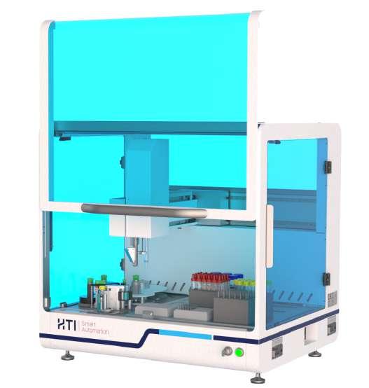
Technologies are at the forefront of modern manufacturing and production, not only offering an advantage but rather being a necessity to the world of science. Torun of Mabtech shares their collaborative works with HTI Automation in developing and enhancing HTI FLEX into the FLEX 2.0.
WATCH PAGE
08 Bridging the Care Gap for Children with Medical Complexity
Children with medical complexity (CMC) often face a daily reality that is incomprehensible to most of us: the inability to walk, talk, eat, or take part in typical childhood activities. The Roald Dahl Charity are asking for your help to develop their Roald Dahl Nurses scheme and allowing them to provide better direct care to children with CMC.
10 Breaking Down Barriers
One of the hardest things about starting a research project is the literature review that forms the basis of the entire programme, and with limited availability and reduced access to these, this can make these projects even harder. DeSci Labs looks at ways to dismantle obstacles such as paywalls and create a space where it is easier for scientists to retrieve and distribute research and data.
REGULATORY AND COMPLIANCE
14 Leveraging Simplicity to Enhance Efficiency for First-in-human Clinical Trials
Drug developers are under ever-increasing pressure to deliver effective therapies to patients faster, without compromising both drug safety or quality. Martin Wing-King of Quotient Sciences addresses the increasing demand for first-in-human clinical trials and how to best tackle this with a revised, simpler formulation approach and the means of collaboration.
RESEARCH/ INNOVATION/ DEVELOPMENT
18 CellShip: An Ambient Temperature Transport and Shortterm Storage Medium for Mammalian Cell Cultures
Cell culture is fundamental to many research and industrial processes, but traditional transport methods are fraught with challenges. Jenny Murray of Life Science Group, introduces the concept of CellShip, an ambient temperature transport medium that combats the complexities and logistical obstacles that typically make cell culture transportation difficult.
TECHNOLOGY
22 AI in Drug Discovery: High-Throughput SPR Boosts Breakthroughs
Until now, the main obstacle for researchers has been the ability to quickly screen and characterise large libraries of candidates for an efficacious therapeutic, as well as the inability to spot failed candidates earlier in the process. Josh Eckman of Carterra, David Eavarone and Jens Plassmeier of Absci, give us an insight in the how
Artificial Intelligence (AI) is beginning to revolutionise drug discovery through its ability to improve efficiency and save time.
30 Small Molecule Microarrays: An established Hit-identification Technique with New Potential for Challenging Targets
Small molecule microarray (SMM) is a well-established method for identifying small molecule (SM) binding interactions with a diverse range of target types. Adam Buckle , Iain McWilliam and Julia Unsicker of Arrayjet explores what sets SMM apart from other target-based drug discovery approaches, highlighting its success in identifying novel RNA-binding small molecules and workflow efficiencies.
36 Enhancing Therapeutic Antibody Development via Synthetic Phage Display Technology
Phage display technology has played a key role in the discovery and optimisation of antibodies for a wide range of clinical or research applications, with its greatest impact seen in the development of antibody-based drugs. John Cardone of Bio-Rad discusses the revolutionary impacts of combining advanced bioengineering and optimised selection strategies for phage display to be able to enhance therapeutic antibody development.
MANUFACTURING AND PROCESSING
42 Understanding the Corporate Sustainability Reporting Directive in Europe and Its Implications for Medium-sized Companies
The issue of sustainability has garnered increasing attention across the globe, with particular emphasis on small and medium-sized enterprises (SME). Michael Gorek of Richter-Biologics urges that medium-sized companies dedicate the time and want to improve their sustainability, regularly reviewing and assessing their practices and where they can improve as it is essential regardless of its obstacles.
CELL AND GENE THERAPY
50 Considerations on In Vitro Disease Models with focus on Fibrosis
The evolution of disease modelling has seen a paradigm shift from the traditional use of animal models to more advanced in vitro human-based systems. This has been driven by the need for more accurate predictions of human disease mechanisms and drug responses in humans. Tanmay Gharat and Emanuela Costigliola of Newcells dive into the specific ways In Vitro models are used in the treatment of Fibrosis.
58 mRNA and AAV as Vectors for Novel Cell and Gene Therapies
Technological advancements are at the forefront of cell and gene therapy developments as we try to confront and overcome the major challenges of safety, efficacy and production. Dr. Sandy Tretbar and Dr. Jacqueline Breuer of Fraunhofer highlight the benefits and drawbacks of two emerging technologies: mRNA technology and adeno-associated virus technology (AAV) in improving cell and gene research.
62 Bench to Bedside: A Roadmap for Developing Novel Gene Therapies
Over the past few decades, advancements in molecular biology and genetic engineering have significantly propelled
the development and application of gene therapy. Here, Dr. Anandkumar Nandakumar, Dr. Stefania Fedele and Dr. James Dixon of Theragenix provide insights into progressing a novel gene therapy into clinical trials and the considerations taken to do so.
APPLICATION NOTES
26 Next-generation Proximity-based Luminescence Assays & CRISPR/Cas9Genome Engineering and nanoBRET
Bioluminescence resonance energy transfer (BRET) is a technology often used to probe the proximity of molecules within live cells, monitoring interactions and screening for drug candidates. Barry Whyte of BMG LabTech describes the study of G protein-coupled receptors and discusses how the strategy used can be used more widely for assays.
40 Meeting Drug Development Challenges: An Insider’s View on CDMO Services for Biologics
Maider Parikh of Thermo Fisher gives an insight into the latest trends and challenges in drug development and manufacturing, and just how Thermo Fisher’s CDMO services a positioned to address and manage these obstacles.
45 Unlocking Early Drug Development Potential with CDMO Expertise
Early-phase drug product development and manufacturing are crucial steps in transforming promising drug candidates into viable, life-changing therapies and Jeff Clement of PCI delves into the key role CDMOs play in accelerating the rate in which the process of developing is carried out, the specific and specialised approach it provides and the improved cost efficiency.
56 NK Cell Therapy – An IP Update
Advances in CAR-T cell therapy have carved a path for improved treatments in field of oncology, in particular haematological cancers, however, the same success is yet to be realised for solid tumours. And so, Amy Dawson of Harrison Goddard Foote informs us of the latest NK cell therapeutic developments and how it targets the elements that are not met by CAR-T cell therapy.
QC -Analysis of Inhaled Antisense Oligonuclotides and mRNA
Analytical
support for ASO and mRNA based therapies from early development to batch release
Antisense oligonucleotides (ASOs) and full-length mRNA -based therapeutics promise diseasespecific treatment options . One of the challenges of these therapies is the tissue- or cellspecific delivery. Inhalation has been an effective means to deliver pharmaceutically active substances to the respiratory tract for centuries. Today there is a growing interest in the industry to leverage on the experience with different inhalation technologies to deliver ASO and mRNAbased treatments directly to diseased respiratory tissues.
A&M STABTEST has over 15 years of experience in supporting drug development and QCanalysis of nasal and orally inhaled products Specially trained staff and dedicated laboratory facilities are the key to analyze inhaled products throughout their lifecycle with the same constantly high quality In the past 10 years A&M STABTEST has gained in-depth experience in stability and release testing of nucleic acid -based therapies ranging from ASO to mRNA -LNP therapies Very recently we have embarked on a project to combine our know-how of inhalable drug and nucleic acid testing to support our clients with inhaled ASO and mRNA therapies
For QC-analysis of mRNAs formulated as lipid -nanoparticles
Figure 1: A) displays the different charge states of a 40 mer oligo in a negative mode full MS B) Deconvoluted spectrum resulting from the full MS spectrum The mass difference between the calculated mass and the observed mass is 8ppm C) Celibatarian curves from a parallel detection by UV (above) and MS (below)
A&M STABTEST has established the panel of methods described in the USP draft guidance on “Analytical Procedures for mRNA Vaccine Quality” Especially the determination of polydispersity by DLS is a valuable indicator to evaluate how well different LNP formulations respond to aerosolization Combined with our generic cell -based bioassay, we can help to predict the transfection efficiency of different LNP formulations. A&M STABTEST has also developed a generic LC-MS/MS method to support early ASO development projects, offering data on quantity, purity and impurities in one analytical run.
A&M STABTEST can support your early development and more advanced ASO and mRNALNP projects whether inhaled or not With our experience spanning well over two decades of providing the pharmaceutical industry with high quality analytical services, we are well suited to find analytical solutions for the industries most innovative drug development programs
Before you start reading the great lineup of informative articles in this autumn issue of IBI, I would like you to turn your attention to the feature about the role of the Roald Dahl Nurses supporting families with children with medical complexity. As professionals in the pharmaceutical industry, we are all aware of the breathtaking advances made to treat and cure different diseases in the past decades and the great promise that novel therapies hold for the not-so-distant future. However, most of us are completely detached from the realities of patients receiving these life-changing treatments. In the case of children with medical complexity we are talking about extremely poor children which are often unable to walk, talk, eat, and generally cannot partake in normal childhood activities. The parents of those children, apart from the grief of having a severely ill child, also must take on the responsibility for their complex care. This includes educating about the medical condition, treatment options, talking to specialists, learning about the operation and maintenance of medical equipment, the day-to-day care, while simultaneously being a loving parent. The Roald Dahl nurse specialists give families long term support by taking charge of many aspects of the child’s care. There are already about 150 trained Roald Dahl nurses supporting over 30000 children throughout the UK. With sufficient funding many more children and their families could benefit for their support. Now with the end of the year approaching, many companies and individuals are looking at different organizations and charities to donate to. I think, the Roald Dahl´s Marvelous Children´s Charity and their specialist nurses’ program is a cause worthy of our attention.
The identification of new drug modalities often starts with the cumbersome task of screening many candidatemolecules for specific chemical or biologic properties. These high-throughput screening (HTS) approaches need to be designed in a way that they are relevant for the respective disease model and sufficiently specific to eliminate unsuitable candidates. The emergence of in vitro human-based disease models has significantly improved the predictability for testing novel therapeutic compounds. Tanmay Gharat and Emanuela
IBI – Editorial Advisory Board
• Ashok K. Ghone, PhD, VP, Global Services MakroCare, USA
• Bakhyt Sarymsakova – Head of Department of International Cooperation, National Research Center of MCH, Astana, Kazakhstan
• Catherine Lund, Vice Chairman, OnQ Consulting
• Cellia K. Habita, President & CEO, Arianne Corporation
• Chris Tait, Life Science Account Manager, CHUBB Insurance Company of Europe
• Deborah A. Komlos, Senior Medical & Regulatory Writer, Clarivate Analytics
• Elizabeth Moench, President and CEO of Bioclinica – Patient Recruitment & Retention
• Francis Crawley, Executive Director of the Good Clinical Practice Alliance – Europe (GCPA) and a World Health Organisation (WHO) Expert in ethics
• Hermann Schulz, MD, Founder, PresseKontext
• Jim James DeSantihas, Chief Executive Officer, PharmaVigilant
Costigliola of Newcells dive into the specific ways in vitro models are used in the treatment of Fibrosis. The Phage-display technique has been at the center of therapeutic antibody development. John Cardone of Bio-Rad discusses the impacts of combining advanced bioengineering and optimized selection strategies for phage display to improve antibody development. What phage display is for the development of specific antibodies, small molecule microarrays (SMM) are for the identification of small molecule binding partners. Adam Buckle, Iain McWilliam and Julia Unsicker of Arrayjet explore what sets SMM apart from other target-based drug discovery approaches, highlighting its success in identifying novel RNA-binding small molecules. HTS produces vast amounts of data that need to be carefully analyzed to identify suitable and eliminate unsuitable candidates. Especially when evaluating complex data sets like those from SPR measurements, suitable AI models are ideally suited to look for the needle in the haystack. Josh Eckman of Carterra, David Eavarone and Jens Plassmeier of Absci, give us an insight in the how Artificial Intelligence (AI) is beginning to revolutionize drug discovery through its ability to improve efficiency and save time.
Lastly, I want to highlight the discussion about the exciting field of CAR-T cell therapies and how our understanding of the different T-cell species and their phenotypes will enable the development of cancer and autoimmune related therapies. Amy Dawson of Harrison Goddard Foote informs us of the latest NK cell therapeutic developments and how it targets the elements that are not met by CAR-T cell therapy.
I also want to congratulate Polypure on their 20th Anniversary. a research-intensive production company specialized in R&D and manufacturing of uniform polyethylene glycol derivatives with a wide range of applications.
I hope you enjoy this edition of IBI, and I look forward to meeting many of you at up coming events.
Dr. Steven A. Watt, CBDO (Chief Business Development Officer) at A&M STABTEST GmbH
• Jeffrey W. Sherman, Chief Medical Officer and Senior Vice President, IDM Pharma.
• Mark Goldberg, Chief Operating Officer, PAREXEL International Corporation
• Maha Al-Farhan, Chair of the GCC Chapter of the ACRP
• Rick Turner, Senior Scientific Director, Quintiles Cardiac Safety Services & Affiliate Clinical Associate Professor, University of Florida College of Pharmacy
• Robert Reekie, Snr. Executive Vice President Operations, Europe, Asia-Pacific at PharmaNet Development Group
• Stanley Tam, General Manager, Eurofins MEDINET (Singapore, Shanghai)
• Stefan Astrom, Founder and CEO of Astrom Research International HB
• Steve Heath, Head of EMEA – Medidata Solutions, Inc
Single-length polyethylene glycol derivatives for demanding applications in pharma
Integrating the X-Tube Processor®_Flex 2.0:
Trust in Excellence
In the world of modern manufacturing and production, staying at the forefront of technology is not just an advantage; it's a necessity. Mabtech understands this well. We had the opportunity to sit down with Torun Ekblad, COO of Mabtech, to gain insights into their journey with the HTI FLEX and their exciting transition to the enhanced model “FLEX 2.0”.
Could You Briefly Explain What Mabtech is Doing?
We are a Swedish life science tool provider. Our primary focus lies in the development of immunoassays. Our customers primarily consist of scientists who are involved in research related to the immune system. At the heart of our company, we specialise in two main aspects. Firstly, we excel in the production of monoclonal antibodies, which serve as essential binding molecules in various scientific applications. Secondly, we are dedicated to creating assays specifically designed for measuring these analytes, thereby enabling scientists to conduct precise and meaningful research in the field of immunology.
Could You Give Us an Insight into Your Current Production Processes and How the Current FLEX is Integrated into Them?
Currently, the FLEX is integrated at the end of our production process. After we've produced a batch of the desired quality, we need to package it in portions suitable for our customers. These portions must fit our kit format, which is relatively small. To maintain the high quality and sterility of the batch, we must fill many vials in a controlled environment. The FLEX plays a crucial role in filling the samples with precision and accuracy. We've been using the FLEX for six years now. We were early adopters, and while we did face some initial challenges, we always received excellent support and assistance to overcome them. We've also made adaptations as our needs evolved over time.
What Are the Key New Features You Expect from the FLEX 2.0 Compared to the Previous Version?
The previous FLEX is still used for some products with high demand, mainly bulk filling and capping. However, the FLEX 2.0 is designed for more complex tasks. It can manage the entire kit assembly process, cover storage, and the entire workflow, including storing various reagents, filling, capping, decapping, labelling, and sorting finished tubes. Its functionality has significantly increased, and an operator can simply request a job, and the machine will handle the rest, allowing our team to focus on other valuable tasks. Additionally, the compact footprint of the FLEX 2.0 is a game-changer for us. It allows us to reduce our physical footprint, which was a limiting factor in our growth.
How has the Collaboration with the HTI Team Been so far in the Development of the FLEX 2.0? Are There Specific Aspects of the Collaboration That You Would like to Highlight?
Our collaboration with the HTI team has been a rewarding journey. We share a similar thought process, and solving complex problems together has built great trust between our teams. While the process required patience, there was no hesitation. Both sides have shown bravery and inspiration throughout the collaboration. We appreciate HTI’s deep understanding of our needs and their willingness to customise solutions to meet our requirements.
We are committed to excellence in everything we do. Our goal is to be best in class and deliver superior products. Finding a partner like HTI who shares this vision is crucial. When excellence meets excellence, great things happen. We trust that our long-term discussions and collaboration will result in something exceptional.
Torun
Ekblad, PhD. COO, Mabtech
What Specific Requirements or Challenges has Your Company Identified that have Led to the Need for an Enhanced Version of the FLEX, i.e., a FLEX 2.0?
Several needs have driven our decision to upgrade to FLEX 2.0. First, our company is growing and we need to increase our production capacities. The current system limits our growth potential. Additionally, automation is a crucial strategy for our business growth without significantly increasing manual work. It's essential to address these challenges in a smart way and automate processes wherever possible to ensure efficiency and utilise the unique qualities of our team members effectively.
Can You Tell Us Some of the Key Benefits You Expect From Integrating the Enhanced FLEX 2.0 into Your Production Process?
Integrating FLEX 2.0 into our production process will enable us to allocate our resources more effectively. Automation will handle tasks that can be automated, allowing our team to focus on higher-value activities. An automated system can work round the clock, 24/7, without human supervision, enhancing our production capacities and enabling our growth.
Are There Specific Technical Challenges or Adjustments Required to Seamlessly Integrate the New Machine into Your Existing Infrastructure?
We have a well-thought-out strategy to address any technical challenges step by step, both on the software and hardware fronts. We trust in the expertise of the HTI team to make the integration seamless.
What is Your Timeframe for Implementing the FLEX 2.0 in Your Operation, and What Milestones Must be Met to Achieve This Goal?
Implementing FLEX 2.0 is a critical step for our operations. It's at the heart of our production process. To minimise downtime,
we need to retire the old production line to make space for the new one. Our project timeline includes installation and factory acceptance tests at HTI's facilities. We aim to ensure that everything works at our site according to specifications, and we're working together with HTI to shorten the installation time as much as possible.
What Considerations Have You Made Regarding Training Your Team to Operate the FLEX 2.0 Safely and Efficiently?
Our collaboration has been strong, with teams working closely together from the design phase onwards. This ensures that our team is well-prepared to operate FLEX 2.0 safely and efficiently. Can you give us some insight into your long-term vision for using the FLEX 2.0?
The adoption of FLEX 2.0 is a significant investment, both financially and in terms of time. We see it as a strategic decision to automate our processes and expect to rely on this system for many years to come. We look forward to continuing our collaboration and anticipate further growth in our business.
Thank you, Torun, for your time and the exciting insights you provided!
About Mabtech
Mabtech is a Swedish life science tool provider specialising in immunoassay development. The company caters to scientists
The collaboration with Mabtech is a mutually rewarding experience. Both, Mabtech and HTI strive to be among the best in the industry. We value learning from each other, engaging on an eye level. Our relationship is built on trust and mutual success. We are looking forward to continuing our journey with Mabtech.
as its primary customer base, offering a range of products designed for the study of immune system reactions in various contexts, such as vaccine development, infectious diseases, and allergies. At its core, Mabtech focuses on the production of monoclonal antibodies and their utilisation as binding molecules. These meticulously crafted antibodies are pivotal tools that enable scientists to conduct precise investigations into immune responses. In addition to producing antibodies, Mabtech also develops assays for the accurate measurement of these analytes. These cutting-edge assays equip researchers with the means to quantify immune system activity with high precision.
About HTI Automation
HTI is a family-owned, global provider of laboratory and production automation solutions with a strong focus on high quality standards and flexible solution capabilities. With many years of expertise, HTI supports customers from the pharmaceutical and life science industries, among others, in optimising their processes and increasing their efficiency. Highly qualified specialists are available to assist customers at every stage of the project, from planning and design to implementation and maintenance. HTI understands the individual requirements of the customers and provides tailored solutions that meet their specific needs.
Dr. Wolfgang Heimberg, Owner & Managing Director, HTI Automation
Bridging the Care Gap for Children with Medical Complexity
Children with medical complexity (CMC) live with congenital or acquired multi-system diseases, severe neurologic conditions with marked functional impairment, and/or are technology dependent for activities of daily living.1
These children often face a daily reality that is incomprehensible to most of us: the inability to walk, talk, eat, or take part in typical childhood activities, coupled with the frightening prospect of life-limiting diagnoses.
The CMC Care Gap
Medical advances mean that these children are living longer; presenting the complex challenge, for their families and healthcare providers, of how best to achieve a quality of life that goes beyond mere survival.
The NHS is built on an acute care system, which results in fragmented care. This is wholly unsuitable for children with medical complexity who require coordinated, multifaceted care that unites diverse medical specialisms and therapies.
Navigating the healthcare system as it stands is a complicated and exhausting endeavour for these families. As one parent told us, “I’m not just a mum looking after a child, I have to be a doctor, a nurse, a physiotherapist, every person… It’s a constant battle for the right care.”
The reality for these families is a constant cycle of reexplaining their child’s complex medical history to an everchanging roster of consultants and healthcare professionals, alongside frequent frustrating trips to emergency departments and conflicting appointments.
What Would ‘Best Practice’ Look Like?
Parents of children with medical complexity need stable, long-lasting relationships with healthcare professionals, providing consistency, reassurance and a joined-up holistic approach. Done well, care should be coordinated by a specialist with expert knowledge of the child and their condition, as well
as advanced communication skills to liaise with the many teams involved. They should also have an awareness of the family, including aspects of their social situation, be it financial, housing or knowledge of their educational needs.
In 2023, Roald Dahl’s Marvellous Children’s Charity took the pioneering step of launching a programme to establish dedicated Roald Dahl CMC Nurses to improve the care for children with medical complexity and their families.
Roald Dahl Nurse Specialists are ideally positioned to meet the care gap, as their knowledge of child and family goes hand-in-hand with professional relationships, multi-agency working, sourcing equipment, and managing medication, all of which facilitates consistent and effective continuity of care for the patient.
Introducing Roald Dahl Nurses
Founded in 1991, Roald Dahl’s Marvellous Children’s Charity establishes specialist nurses to care for seriously ill children. There are now over 150 nurses caring for more than 36,000 children across the UK, but thousands more are still living without this support.
Roald Dahl Nurses are trained and dedicated specialists whose expert care reduces A&E visits, hospital admissions and consultant appointments, uniquely positioning them to provide exactly the style of care to best-support CMC.
Roald Dahl CMC Nurses extend their care beyond the conventional medical framework, building a comprehensive network of support that addresses the medical, emotional, and social needs of each child and their family's lives.
Their deep understanding of the unique challenges these families face – from managing intricate care regimens to interfacing with social services and education – allows them to provide support tailored to the specific needs of the conditions they help to manage.
As one parent explained, “You get support for more than just the illness and it’s invaluable for a child with complex needs, as there’s no clear process and you need help to navigate that. It’s a huge benefit, I cannot say how useful and worthwhile this role is; every hospital should have a Roald Dahl Nurse.”
How Can You Help?
Investment in Roald Dahl CMC Nurses is a necessary step towards a comprehensive model of care for these inadequately supported children. Acknowledging the critical gaps in current healthcare provision for CMC is paramount and Roald Dahl’s Marvellous Children’s Charity is striving for more structured support to ensure exceptional care standards and a holistic approach.
Roald Dahl Nurses are uniquely positioned to meet the complex care needs of children with medical complexity and ease the pressure on these families during an overwhelming and stressful time.
We are urgently seeking corporate partners to support our fundraising efforts. Your business could help ensure that more
children with medical complexity have access to the vital specialist care of a Roald Dahl CMC Nurse. Children like Max, who lives with epilepsy and a GRIN2D-related disorder.
To donate or learn more about how your business could help, please contact partnerships@roalddahlcharity.org, including the reference IBI-2024, or visit https://www.roalddahlcharity. org/get-involved/corporate-partnerships/.
If your business runs a charity scheme, please nominate us and let our team know via the email address above.
REFERENCE
1. Cohen E, Kuo DZ, Agrawal R, et al. Children with medical complexity: an emerging population for clinical and research initiatives. Pediatrics 2011;127:529–38.
Roald Dahl’s Marvellous Children’s Charity
Roald Dahl’s Marvellous Children’s Charity provides specialist nurses and support for seriously ill children, with over 150 Roald Dahl Nurses caring for over 36,000 children across the UK. Roald Dahl Nurses are specialists in their field and their dedication and expertise and reduce A&E visits, hospital admissions and consultant appointments.The charity’s Marvellous Family Support Services also provide financial and emotional support for families under the care of a Roald Dahl Nurse.
Breaking Down Barriers for Postgraduate Researchers
How truly open access publishing models improve project outcomes for postgraduate research students
One of the hardest things about starting a research project is the literature review that forms the basis of the entire programme. This allows the student and their academic supervisor to understand existing literature and ensure essential criteria for passing the qualification is met –novelty. However, with huge swathes of research locked behind paywalls, this is often difficult to do. Here, Philipp Koellinger the co-founder of open access publishing start-up DeSci Labs, explains why, if we are to solve the world’s scientific challenges, the future of scientific publishing must truly be open access and not concentrated into the hands of a select few publishers.
The journey into postgraduate research is often heralded as an exciting adventure into the unknown. But for many aspiring scientists, the reality can be far less exhilarating as their adventures hit an immediate obstacle: the literature review.
Challenges Accessing Literature
With Google Scholar at our fingertips, it’s easy to be overwhelmed by the sheer volume of available research online. However, simply combing through the search results to find journal articles that are relevant to your chosen research area is only half the battle. A significant portion of the existing literature is locked behind paywalls, creating a frustrating obstacle for students trying to grasp the nuances of their chosen field.
Furthermore, the vast majority of published articles do not provide access to the code and data on which their claims are based, and publishers lack the technology to evaluate and publish anything other than manuscripts. Building on this fragmented, incomplete, and partly paywalled content is like trying to build a house with only half the blueprints.
This is far more than just an inconvenience. It’s a systemic issue that hinders scientific progress. By restricting access to published scientific research based on paid subscriptions, we create an uneven playing field that favours academic institutions, or individual researchers, with the resources to afford expensive journal subscriptions.
And by publishing only the final manuscript, with the accompanying data and code being inaccessible or lost, we have to rely on the author’s statements without the ability to check for ourselves. The lack of data and code in the scientific record also means that many researchers inadvertently end up reinventing the wheel over and over again, making unnecessary mistakes along the way.
Also, it leads to many false research findings being published because referees lack the incentives and the means to check how the authors arrived at their results. These flaws
not only limit the potential pool of future scientists but also slow down the pace of scientific discovery. Ultimately, the collective flaws in the publishing landscape harm scientists’ ability to solve the world’s key scientific challenges, from drugs to treat diseases like cancer to green technology to tackle climate change.
Bring Down the Walls
At its finest, science is a collaborative effort. After all, you don’t improve research outcomes by hiding scientific data or code. Sharing knowledge freely is essential for accelerating innovation as, by making experimental methods and their results more accessible, it allows for greater scrutiny, replication, and an enhanced capacity to building on existing work. It also fosters a culture of transparency and accountability, which are fundamental to the ongoing integrity of science.
An open access publishing landscape also makes it easier to address global challenges. From climate change to disease outbreaks, the world needs science to provide solutions to the biggest scientific challenges of the day.
By making all components of research freely available, including the data and code that underpin published findings, we empower scientists everywhere to contribute to finding answers. We break down geographical and economic barriers, ensuring that the best minds can work together to tackle humanity’s most pressing problems.
Furthermore, when the world faces major scientific challenges that impact the public in a profound way, being able to freely access scientific research can limit the spread of misinformation and dramatically speed up scientific discovery. For example, during COVID19, many scientists shared their research results on the virus immediately on preprint servers such as BioRxiv, instead of waiting months or years before a publishing in a scientific journal and having their findings locked behind publisher’s paywalls.
The appearance of COVID19 papers on BioRxiv reflected the global spread of the disease: The first results were posted by Chinese scientists, followed by Italians, and so forth. The immediate sharing of results was instrumental for the fast development of vaccines, treatments, and disease management.
Look to the Future
It’s time to recognise that the current publishing model is outdated and counterproductive. The future of science depends on new researchers being able to enter the field, assess the current literature landscape, and get to work trying to build on it with minimal disruption.
By dismantling paywalls and making research freely available, we improve the experiences of postgraduate students globally,
Discovery Park is one of the largest science and technology parks in the UK, providing all the space, support and facilities you need to unlock your company’s potential.
• World-class laboratory, manufacturing and office spaces.
• Excellent connectivity to London & European markets.
• Collaborative ecosystem fostering innovation and partnerships.
• Comprehensive business support and access to investment.
• Ideal setting offering the optimum quality of life for employees.
Get in touch today for a tour and find out how we can help your business succeed.
make their research projects run smoother, and deliver better outcomes. This will, ultimately, encourage more would-be scientists to undertake postgraduate research programmes.
By including data and code in the publishing process and making them freely available with truly open access publishing, we will increase visibility for scientists’ research and make it easier to credit the authors for all their work. Also, publications that include data and code tend to get more citations, which benefits the authors’ careers while opening up new learning possibilities for young researchers as they get more chances to look under the hood and grow their own understanding.
Creating more opportunities for recognition in this way will make research programmes more personally rewarding for individual researchers, making it more likely that people pursue careers in research.
This will only benefit scientific research in the long-term as there will be more active researchers globally and they will be happier and more motivated in their work.
DeSci Labs is helping to make this more open future possible by building a traceable digital version of the scientific record that treats data and code as “equal citizens”, following the company raising $6.5 million in seed funding. DeSci Publish is an open-source peer-to-peer platform for scientific publishing, and the first publishing solution based on versionable research objects that can contain any file type.
By transitioning to an open access publishing landscape, postgraduate scientists can benefit from a more equitable, efficient, and impactful research environment. It will also help address the replication crisis and ensure that all of scientists’ work can be seen and recognised, regardless of where the audience is based or how much funding they have available.
The current version of the platform allows researchers to share up to 100 GB of content for free, with persistent identifiers for each file uploaded. There are no publication charges or paywalls on DeSci Publish – it’s built by scientists for scientists. If this sounds interesting, visit publish.desci.com to try the platform for free.
Philipp Koellinger
Prof. Philipp Koellinger is the CEO and cofounder of DeSci Labs, which aims to make science more reliable, transparent, independent and openly accessible. He is published in leading journals such as Nature and Science, has more than 17,000 citations for his work in social science genetics, economics and neuroscience, and has worked as a full professor at the University of Wisconsin Madison and Vrije Universiteit Amsterda.
DeSci Labs platform for open sharing of research data
talkfuture@pci.com pci.com
•
•
•
•
•
Building
Regulatory and Compliance
Leveraging Simplicity to Enhance Efficiency for
First-in-human Clinical Trials
It is well-known that going from drug discovery to commercialisation takes, on average, 10–15 years and poses significant costs – approximately $2.6 billion when R&D, materials, manufacturing, and related expenses are added up.1 In this challenging landscape, drug developers are under ever-increasing pressure to deliver effective therapies to patients faster and without compromising drug safety or quality. This has increased the industry's need to find ways to improve first-in-human (FiH) clinical trial pathways for nearly all indications.
The US Food & Drug Administration (FDA) has defined four ways a pharma or biotech may accelerate through development and approval to meet the goal of getting to patients earlier. The FDA’s designations for priority review, breakthrough therapy, accelerated approval, and fast track status each have their own set of requirements.2 A drug that has been designated as fast track, for example, must fill an unmet need and provide therapy where none exists or that has a significant advantage when compared with the available therapy.2 Similarly, the European Medicines Agency (EMA) may recognise a drug with a PRIME status, which grants similar benefits to the FDA fast-track designation. Conducting FiH trials more efficiently can benefit both developers and patients but, this can be challenging and costly particularly for emerging pharmaceutical companies or biotechs with limited resources.
Adding to the pressure, development challenges faced at the clinical stage can ultimately lead to a bleak future for a molecule. Only 12% of the new molecular entities (NMEs) entering clinical trials gain U.S. Food and Drug Administration (FDA) approval.1 Lack of efficacy and drug toxicity remain the leading reasons why an NME may fail at the clinical stage.3 One potential way to progress forward is to leverage simplicity in formulation by directly filling capsules with the active pharmaceutical ingredient (API). This approach allows for a faster path to the clinic, where drug efficacy, safety, and toxicity data can be obtained early in development.
In this article, Martin Wing-King, Vice President and General Manager at Quotient Sciences -Reading explores the demand for accelerated FiH trials and outlines how simple formulations can be leveraged for a faster way to progress through early clinical studies.
What is Driving the Demand for Accelerated FiH Trials? FiH clinical trials are the first instance where an investigational drug is introduced to human subjects. A primary focus of these studies is a safety assessment, which monitors adverse events and dose-limiting toxicities. Data about a drug’s absorption, distribution, metabolism, and elimination (ADME) are essential for determining optimal dosing regimens and administration schedules. Additionally, FIH trials provide crucial
pharmacokinetic (PK) and pharmacodynamics (PD) data that support a better understanding of how the body interacts with the drug and its impacts on the body, mechanism of action, and potential efficacy.
FiH trials often involve dose escalation to identify the maximum tolerated dose (MTD) and the recommended starting dose for subsequent trials. This information is vital in determining safe dosage ranges for the drug as it moves forward in clinical investigation. In all instances, early PK/PD data can influence decisions about the drug’s further development.
Increased efficiency in FiH trials is in demand across nearly all indication areas but in rare and orphan diseases in particular, the benefits are clear. In these cases, developers are faced with small patient populations that are both hard to recruit for clinical testing and equally as challenging when it comes to deciding the commercial viability of a drug downstream. Removing costs and time from traditional drug development is imperative to do as early as possible.
The data generated may support continuing clinical development and provide insight that de-risks the process. Using efficacy, safety, and toxicity data can allow developers to decide whether or not to continue developing a drug, especially when it comes to addressing challenges with the increasing complexity of APIs.
Enhancing Efficiency in Clinical Trials with a Simple Formulation Approach
Simplifying formulation development can streamline FiH trials and enable developers to obtain crucial clinical data. A “drug in capsule” approach, also known as “blend in capsule” or filling a capsule directly with API and limited (if any) excipients, is a dosage form that requires minimal development and can be used to simplify formulation and accelerate FiH trials. This approach enables developers to get their API into the clinic and obtain FiH data before investing in more complex dosage forms, such as dry powder inhalers.
API-filled capsules can reduce the need for complex formulation development while still enabling the drug to be manufactured in a GMP setting and administered to patients easily. This approach improves the time to reach the clinic while significantly reducing the costs associated with API synthesis and the potential waste of manufacturing excess material.
Specialised equipment that can help with API drug-incapsule filling includes semi-automated and fully-automated capsule filling systems that microdose a precise amount of API into capsules. With choices in equipment that can handle doses ranging from 0.1 mg to 100 mg, drug developers have flexibility in dosing with rapid encapsulation of APIs with good flow and solubility properties for their FIH trials. Additionally, fill-toweight capsule filling machines that can accommodate a wide
CONTRACT DEVELOPMENT AND MANUFACTURING OF BIOPHARMACEUTICALS
Richter BioLogics is a Germany-based GMP manufacturer specialized in products derived from bacteria and yeasts, with a proven 35-year track record.
Count on us to flexibly provide a comprehensive range of services and customized solutions. Clients worldwide have already benefited from our commitment to good manufacturing practice and total transparency. Our work focuses on recombinant proteins, plasmid DNA, antibody-like sca olds (VHH/Nanobody), cell-free expression and vaccines.
Your product – our competence and dedication. Contact us +49 40 55290-801 www.richterbiologics.eu
LEARN MORE ABOUT OUR SERVICES AND CAPABILITIES
Regulatory and Compliance
range of APIs make it possible to handle complex formulations with poor properties and spray-dried powders. Using a robotic capsule filling system that integrates check weighing and containment can also be beneficial for the safe handling of high-potency APIs (HPAPI).
Case Study: A Simple Formulation Approach in Oncology Drug Development
In one program, an EU-based biotech focused on the development of oncology therapies required the development, manufacture, and supply of a solid dose formulation for first-in-patient (FIP) trials. The first-in-class molecule was designated as FDA “fast track” for various oncology indications but had limitations in the availability of the API that could have compromised the ability to perform the study and progress into the potential treatment phase if patients responded to the drug.
To start patient trials with a simpler product that conserved API, Quotient Sciences supported the program design that entailed the development of a blend-in-capsule (BiC) formulation at three strengths with a flexible manufacturing scale of up to 3 kg batches. Over the course of three years, more than 30 small batches of product were manufactured using a semi-automated capsule filling system and over 100 product shipments were packed and labelled. The BiC formulation was scaled up to 10 kg batches to continue larger patient trials seamlessly while still conserving API and providing bright stock-labelled product to patients on demand. In parallel, Quotient Sciences supported an ICH stability program to be able to continue to extend product shelf-life.
Thinking about the end goal in mind from the start, once the simple BiC formulation was deemed effective in-patient trials, the goal was to continue to support the biotech in the transition to a patient-friendly, commercial-ready tablet drug product. The dry blend composition for the BiC remained the same for the tablets and included excipients that would support a dry granulation process. A film coat for taste masking was also applied to the tablet. The tablet was used in continuing Phase II and III patient trials and is currently being scaled up for registration batches and commercial supply.
Specialised capsule-filling technology and a simpler approach to formulation have the potential to shave months
off the drug development process by significantly reducing development timelines and eliminating the need for repeated stability studies. In this case, the program design by Quotient Sciences ensured optimal use of the available API inventory while meeting clinical study needs. More generally, the ability to configure an “on-demand” GMP manufacturing solution, with flexibility in batch size can enable developers to either proactively defer API scale-up investment until post-POC, or reactively manage programs where API availability is otherwise limited.
Fast-tracking FiH trials: The power of Collaboration Partners that understand the urgency of reaching clinical milestones under tight timelines by leveraging their extensive experience and specialised equipment are critical to the success of FiH trials. Ultimately, using simple formulation methods to generate data early in development helps prevent unnecessary formulation development expenditures on compounds that may ultimately prove ineffective or unsafe.
The complexity of tech transfer is often underestimated, and development and manufacturing can be significantly streamlined when conducted within the same provider network, but especially within the same facility. Additionally, a company that offers dedicated project management ensures efficient coordination, timely communication, and adherence to timelines and budgets – ultimately benefiting drug developers and patients alike.
By leveraging simplified formulation approaches such as capsules filled with API, drug developers can significantly expedite their path to the clinic. Strategic collaborations with experienced partners offering integrated drug development and manufacturing services can also streamline the process, ensuring efficient tech transfers and project management. Ultimately, these strategies help with collecting FiH data early, paving the way for successful drug development and benefiting patients.
REFERENCES
1. Research and Development, accessed May 28, 2024, from: https:// phrma.org/policy-issues/Research-and-Development-PolicyFramework
2. U.S. Food and Drug Administration Fast Track, accessed August 21, 2024, from: https://www.fda.gov/patients/fast-track-breakthroughtherapy-accelerated-approval-priority-review/fast-track
3. Sun, D., Gao, W., Hu, H., & Zhou, S. (2022). Why 90% of clinical drug development fails and how to improve it? Acta pharmaceutica Sinica. B, 12(7), 3049–3062. https://doi.org/10.1016/j.apsb. 2022.02.002
Martin Wing-King
Martin Wing-King is Vice President & General Manager, Quotient Sciences - Reading. Martin joined Pharmaterials in 2008, a company later acquired by Quotient Sciences in 2017, and has held various business development, project management leadership, and operations leadership roles with the company for over 16 years. Martin holds a Bachelor of Science in Chemistry and Management from Brunel University London.
• Central Lab: Kitting, Logistics, and Biostorage With Virtual Sample Inventory Management
• Biospecimens: Curated Inventory and Analysis With Advanced Biobanking
• Preclinical: Advanced Cell-Based Assays and Target Validation
• Genomics: Single-Cell to Multiplex Studies
• Bioanalytics: Immunogenicity and PK Testing
• Immune Monitoring: Comprehensive Cell Phenotyping and Profiling
• Tissue and Liquid Biopsy: Rare Cell and CTC Isolation and Analysis
• Clinical Trials: Design, Strategy, and Full-Spectrum Support
• Diagnostics and CDx: Regulatory Consulting, Companion Dx, and NGS
• Data Sciences: Biometrics and Biostatistics via QuartzBio® precisionformedicine.com
CellShip: An Ambient Temperature Transport and Short-term Storage Medium for Mammalian Cell Cultures
Cell culture is fundamental to many research and industrial processes, but traditional transport methods are fraught with challenges. Cryopreservation, the most common technique, requires potentially cytotoxic cryoprotective and can lead to poor cell recovery. Additionally, the logistics of cryopreservation are complex, requiring specialised equipment and careful handling. An ambient temperature transport medium like CellShip could significantly ease these issues.
This study introduces CellShip, a novel transportation medium for mammalian cells. Five commonly used cell lines (HEK293, CHO, HepG2, K562, and Jurkat) were successfully transported and stored at ambient temperature for up to 96 hours, maintaining high cell viability.
Methods of Cell Transportation
Current cell transportation methods largely rely on cryopreservation, where cells are frozen using cryoprotective agents (CPAs) such as dimethyl sulfoxide (DMSO). This process, though effective, has several drawbacks:
• Logistical Challenges: Requires slow freezing at -1°C per minute to -80°C, storage in liquid nitrogen, and transport on dry ice.
• Cytotoxicity: Prolonged exposure to DMSO can lead to cellular changes and toxicity.
• Recovery Issues: Cells must be thawed quickly to avoid ice crystal formation, followed by careful removal or dilution of DMSO to prevent osmotic shock.
Alternatively, cells can be shipped as growing cultures in sealed vessels, which is feasible for short durations but presents risks of contamination and cell detachment, leading to cell death (anoikis).
Development of CellShip
CellShip was developed to address these issues by providing a medium that allows for ambient temperature transport. This eliminates the need for cryopreservatives and reduces the risk of cell damage. The study assessed the viability and cell count of five cell lines (HEK293, CHO, HepG2, K562, and Jurkat) after transport and storage in CellShip for 72 to 96 hours.
Materials and Methods
• Cell Culture Conditions: Cells were cultured at 37°C in a humidified environment with 5% CO₂. Specific media formulations were used for different cell lines, all supplemented with 10% foetal bovine serum (FBS).
• Cell Count and Viability: Cell counts, and viability were measured using a CytoSmart™ automated cell counter and Trypan Blue exclusion. Fold changes in cell numbers were
Research / Innovation / Development
calculated before transport, immediately after transport, and following a recovery period.
• Transportation Procedure: Cells were transported in 2 mL Nalgene cryovials, placed in polystyrene transport containers, and shipped via commercial courier. Internal package temperature was monitored using a TinyTag data logger.
• Cryopreservation Procedure: For comparison, cells were cryopreserved in FBS with 10% DMSO, frozen slowly at -80°C, stored in liquid nitrogen, and transported on dry ice.
• Recovery and Sampling: Post-transportation, CellShip suspensions were directly transferred to growth media. Cryopreserved cells were thawed and gradually diluted with pre-warmed media.
• Metabolic Activity: Metabolic activity post-transportation was measured using the alamarBlue® reduction assay.
• Morphological Analysis: The morphology of HepG2 cells was examined by phase-contrast microscopy post-recovery.
Results
• Viability and Cell Counts: High viability and cell counts were maintained across all cell lines after transport in CellShip.
• Comparative Study: The viability of cryopreserved cells dropped significantly post-recovery, whereas CellShiptransported cells maintained high viability.
• Metabolic Activity: Cells transported in CellShip exhibited higher metabolic activity compared to cryopreserved cells, indicating better recovery and proliferation.
• Morphological Analysis: HepG2 cells transported in CellShip adhered better and showed typical growth morphology compared to those recovered from cryopreservation.
a) HepG2 cells following CellShip transportation and recovery b) HepG2 cells following cryo-preservation and recovery
• Limitations: Acknowledging the study's limitations, such as the number of cell lines tested and the duration of transport, would increase credibility.
Discussion
The study demonstrates that CellShip is effective for transporting mammalian cell cultures at ambient temperature, maintaining high viability and cell numbers for up to 96 hours. This method offers several advantages:
• Reduced Logistical Complexity: Eliminates the need for cryopreservation equipment and procedures.
• Higher Cell Viability: Reduces the risk of cryopreservationinduced delayed-onset cell death (CIDOCD).
• Rapid Recovery: Cells recover faster post-transportation, crucial for sensitive applications like cell-based therapies.
Conclusion
CellShip represents a significant advancement in cell
Research / Innovation / Development
transportation technology. By maintaining high cell viability and numbers at ambient temperatures, it offers a practical alternative to cryopreservation. This could have broad applications in the life sciences sector, from research to biopharmaceutical manufacturing, providing a more efficient and less toxic method for transporting cells.
• Future Directions: Further studies will focus on transporting primary and stem cell lines, assessing gene expression, CD markers, differentiation potential, and protein production post-transport in CellShip compared to cryopreservation.
• Economic Impact: With a retail price of £90.00/100 mL, CellShip is a cost-effective solution for both national and international cell transportation.
By addressing the challenges associated with traditional cell transport methods, CellShip has the potential to revolutionise the way cells are transported and stored, benefiting both research and medical sciences.
Jenny Murray
Jenny Murray is the Managing Director and owner of Life Science Group (LSG), started in 2008 as a virtual company offering project support for antibody generation projects, but has since developed into offering cell culture manufacturing capabilities, additional antibody generation services and during the Covid pandemic was contracted by PHE to provide sample collection tubes for Covid test kits. Jenny has over 30 years’ experience in antibody generation and the cell culture industry and has recently completed a three-year term as Chair of the International Serum Industry Association. Jenny is very proud to be a Trustee of an educational charity WhatisBiotechnology and contributes regularly to content. This charity provides an on-line platform bringing the important stories surrounding biotechnology to a wider and younger audience.
GREAT FLUIDIC SOLUTIONS
BIOTECH LIQUID µFLOWMETER
Continuous Flow Measurement 10 nl – 80 µl/min
Software compatibility with leading CDS
DEGASi® Nano
Efficient degassing at µl/min flow rates
Widest chemical compatibility
AI in Drug Discovery: High-throughput SPR Boosts Breakthroughs
Artificial intelligence (AI) is revolutionising drug discovery, enabling scientists to identify novel therapeutic candidates in a fraction of the time it once required. Until now, the main obstacle for researchers has been the ability to quickly screen and characterise large libraries of candidates for an efficacious therapeutic, as well as the inability to spot failed candidates earlier in the process. Consequently, pharmaceutical companies have spent significant development dollars on molecules that will not prove to be suitable therapeutics – prolonging the time-tomarket and increasing the cost of life-saving medicines. With the introduction of generative AI and machine learning (ML), it is now possible to select more promising candidates using fewer resources, saving time and money. But how are AI/ML algorithms created and fine-tuned to become reliable methods for candidate selection?
Therapeutic antibodies traditionally undergo five sequential stages leading up to preclinical development. First, the drug’s target is investigated for its relevance in the disease based on primary research. Then the antibody is generated by immunising an animal and growing its immune cells that produce the antibodies. These antibodies are initially tested in the lab, and those that look most promising pass on to the next step as lead candidates. These lead candidates undergo optimisation, a critical stage in which they are honed for biological activity and properties that will impact a future drug’s efficacy, safety, and developability.
To design a promising lead drug candidate for further development, you need to find antibodies with several different properties, all falling within a small range. It’s like searching for the proverbial needle in a haystack, except the haystack is larger than the known universe, and the needle is smaller than a speck of sand. Using traditional drug discovery methods, this can be a long, expensive process with little probability of success. AI-guided design of drug candidates can dramatically speed up both the discovery and the lead optimisation process as well as produce unique, generative variants that could never be found using standard methods.
In recent years, numerous antibody-related databases have emerged, offering valuable resources for training machinelearning models. However, many of these databases can often lack critical information, such as affinity, aggregation parameters, or epitope data.1 The Carterra® high-throughput surface plasmon resonance (HT-SPR) platforms are playing an essential role, both upstream and downstream in the discovery process. Josh Eckman, CEO of Carterra, when asked about the role of Carterra’s HT-SPR technology in AI-driven drug discovery has said, “We believe we can help in this [process] by providing high-resolution binding information on epitope, affinity, and kinetics at the earliest stages of screening.”
The platforms provide reliable data on affinity maturation using its kinetic and epitope software to feed the AI model, as well as verifying the model’s predictions. This validation is then fed back into the model, continually strengthening its intelligence.
AI Drug Discovery in Action AI promises to revolutionise drug discovery, but advances in drug creation also continue to depend on scalable wet lab technologies to produce and validate biological data at scale. One company leading the way in this combined approach is Absci Corporation. Established in 2011 and headquartered in Vancouver, Canada, the team is using a zero-shot AI approach, which designs antibodies without prior learning on the specific target and are, therefore, generating candidates unlike those found in existing databases. It created a proprietary Integrated Drug Creation™ platform, which combines the data to train, the AI to create, and the wet lab capabilities to validate millions of AI-generated designs. Jens Plassmeier, PhD and Senior Vice President for Biologics Discovery Technologies at Absci, noted that this platform enables their team to develop new therapeutics using the same AI technology celebrated for generating text and images from natural language prompts.
Working from massive biology datasets, generative AI is applied to design optimal drug candidates based on target affinity, safety, manufacturability, and other traits. Absci supports its generative AI designs with its wet lab's extensive validation capabilities, which includes the Carterra LSA®. David Eavarone, PhD and Director of High-Throughput Screening at Absci, said, “This workflow can take us from AI-designed antibodies to wet lab-validated candidates in as little as six weeks. The quality and scale of wet lab data give us incredible training data, propelling our iterative design-build-test-learn cycle.”
High-Throughput SPR:
What it is and How it Benefits AI-led Drug Discovery
High-throughput SPR systems make possible the evaluation of large sets of antigen:antibody interactions quickly and cost-effectively by adding throughput to the proven surface
plasmon resonance technique. This technology can be used to run parallel investigations into kinetics, affinities, and epitope specificities from the very start of the drug discovery process. This ability to perform higher throughput and comprehensive characterisation early in the drug discovery workflow changes the paradigm of therapeutic antibody screening. Researchers become better informed earlier in the process, fully appreciating the epitope landscape of a campaign and thus, can identify the superior candidates from the original library. Importantly, it also avoids researchers repeating or abandoning screening campaigns unnecessarily when they have failed to identify desired clones; not because they weren’t in the library but because it was not possible to look deep enough, early enough.
The Carterra LSA HT-SPR antibody discovery and characterisation platform was launched in 2018, which was then followed by the even more sensitive LSAXT platform in 2023. Newest to the market, introduced this month, is the Carterra Ultra™ platform, which allows for small molecule and fragment drug development research to benefit from
the speed, low sample usage, and breadth of data provided by the previous Carterra HT-SPR platforms. The instruments combine high-throughput microfluidics for array printing with label-free SPR detection. This enables all antibodies to be rapidly and comprehensively screened early in the discovery process so that unique epitopes and potential novel therapeutic candidates can be identified while expanding and enhancing IP coverage.
Characterising binding kinetics and epitope coverage of large numbers of molecules early in the drug discovery process has been transformative. Through high-resolution and high-throughput binding analysis, detailed interrogation of protein and epitope binding has become a reality at a pace that was previously unimaginable. In short, months of work can be compressed to just a couple of weeks, enabling improved clinical candidates.
HT-SPR stands as one of the primary methodologies for Absci’s wet lab validation. To this end, the Carterra LSA platforms have been invaluable to the team and their success. Dr. Eavarone underlines the critical role that having these instruments has provided their team, stating, “LSA data is indispensable in testing and training our generative AI models. The Carterra LSA enables precise quantification of single target affinity for our AI models used in de novo drug discovery. The Carterra SPR technology is also extremely versatile and enables the testing and advancement of AI models for high throughput lead optimisation. We have reported success using these systems for multi-parametric lead optimisation including for epitope specificity, pH sensitivity, and co-optimised binding against multiple antigens. Put simply, to have the best AI drug creation platform, you need the best data. Carterra’s HT-SPR is at the heart of our wet-lab data generation and has been instrumental in the success of our drug discovery platform.”
Carterra LSA HT-SPR enables rapid assessment of AI-designed binders with high quality data
Technology
Conclusion
Despite billions of dollars of investment every year, only an estimated 4% of drug leads succeed in their journey from discovery to launch. Even more unfortunate, only 18% of drug leads that pass preclinical trials eventually pass phase I and II trials, suggesting that most drug candidates are unsafe or ineffective. While much of this failure rate is attributable to incomplete understanding of the underlying biology and pathology, insufficient drug lead optimisation also contributes to many failures. The ability to create and optimise new therapeutic antibodies in silico using AI could reduce the time it takes to get new drug candidates into the clinic by more than half, while also increasing their probability of success.
Artificial intelligence and machine learning are sure to continue to be key tools in improving the speed and accuracy of therapeutic development in the future. With that said, the accuracy and learning capabilities of these models will also need to continually grow and improve. While AI-assisted antibody design and lead optimisation of biological sequences can reduce therapeutic development time, it does not by itself currently offer an in silico replacement for all drug discovery efforts. Fully generative and broadly applicable modeling approaches are needed. However, the training and validation of such models face an immense data challenge due to the vast combinatorial design space where strong selective binders represent an incredibly small piece of that space. Dr. Eavarone predicts that as high-throughput structural data generation becomes more advanced and training data sets become larger, Absci's AI models will improve to be able to create de novo antibodies against any target, including those without any known existing binders and even those for emerging pathogens previously unseen as targets in the therapeutic space.
In summary, although AI can help expedite the design and optimisation of antibodies, it currently falls short of replacing all aspects of drug discovery. Developing fully generative models that can be broadly applied remains a significant challenge due to the immense complexity of the design space and the rarity of highly effective binders. Nevertheless, as the ability to generate structural data at scale improves and training datasets expand, it is expected that AI will eventually evolve to meet this challenge. In combination with technologies such as HT-SPR, the rate and dependability of AI/ML in drug discovery will advance at an accelerated rate. AI is unlocking new opportunities, allowing researchers to create better biologics for patients faster, and the hope is that it will go a long way to improved quality of life and better therapeutic outcomes for patients.
REFERENCES
1. Musnier A, Dumet C, Mitra S, Verdier A, Keskes R, Chassine A, Jullian Y, Cortes M, Corde Y, Omahdi Z, Puard V, Bourquard T and Poupon A (2024), Applying artificial intelligence to accelerate and de-risk antibody discovery. Front. Drug Discov. 4:1339697. doi: 10.3389/ fddsv.2024.1339697
Josh Eckman
Josh is the founder and Chief Executive Officer of Carterra. Mr. Eckman graduated summa cum laude in Business Administration and Asian Studies from the University of Utah. At graduation, he was awarded the Outstanding Scholar in Business Administration and the Honors Baccalaureate Award. Mr. Eckman then received a M.S. in Mechanical Engineering (microfluidics focus) from the University of Utah. Mr. Eckman was also selected by Ernst & Young LLP as a 2022 Entrepreneur of the Year® Mountain West Award winner – a preeminent competitive business award for entrepreneurs and leaders of high-growth companies.
David Eavarone
David Eavarone is currently the Director of High-Throughput Screening at Absci. His scientific background spans over a decade in industry-leading wet lab development efforts for antibody-based therapeutics for immuno-oncology and infectious disease targets. He obtained his PhD in Biomedical Engineering at MIT through the Health Sciences and Technology program joint with Harvard Medical School.
Jens Plassmeier
Jens Plassmeier is the SVP for Biologics Discovery Technologies at Absci. During his time in industry, he has led projects relating to the development and production of therapeutical antibodies, development and scale up of microbial produces large and small molecules. Jens obtained his PhD in Biology from the University of Bielefeld in Germany and did postdoctoral research at MIT.
Next-generation Proximity-based Luminescence Assays & CRISPR/Cas9 Genome Engineering and nanoBRET
Bioluminescence resonance energy transfer (BRET) is a technology often used to probe the proximity of molecules within live cells. BRET assays have many applications including monitoring protein-protein interactions, examining receptor-ligand interactions, and screening for drug candidates. The versatility of BRET has encouraged researchers to discover new luminescent tools, including luciferase enzymes, and to enhance their performance. The recent development of nanoBRET™ is a case in point. Another direction to drive progress is to improve the tools used side by side with nanoBRET. One limitation of existing assays is that the fusion proteins needed for luminescence assays are generated by exogenous expression. In a recent development, nanoBRET was used with a CRISPR/Cas9 system to allow for endogenous expression of donor-fused proteins. CRISPR/Cas9 systems have revolutionised genome engineering by making it easier to make precise, targeted changes to DNA. Here we take a closer look at this advance, describe an application used to study G protein-coupled receptors, and discuss how this type of strategy can be used more widely to perform assays under more physiologically relevant conditions.
The Fundamentals of BRET
BRET is a type of non-radiative transfer of energy between a bioluminescent donor and a fluorescent acceptor (Figure 1). In BRET, the bioluminescent donor is typically a luciferase enzyme that comes into proximity with a fluorophore (usually less than 10 nm apart). One advantage of BRET is that it allows for real time monitoring of biological processes in living cells without the need for external light sources.
BRET assays typically use an ATP-dependent luciferase (often Rluc8) as the energy donor, a green fluorescent protein variant as the energy acceptor (e.g. Venus), and coelenterazine h or coelenterazine 400a as substrate for the luciferase enzyme. However, the availability of the new luciferase
reporter nanoluciferase (Nluc) and its substrate furimazine has resulted in the development of nanoBRET.1 NanoBRET offers enhanced sensitivity and precision due to a smaller and brighter bioluminescent donor compared to traditional luciferases like Renilla luciferase (Rluc8). Nluc is a small 19-kDa luciferase isolated from the deep-sea shrimp Oplophorus gracilirostris. It emits a bright, stable luminescence in a narrow spectrum. It offers higher sensitivity than Rluc8 and requires lower levels of expression which makes it amenable for a wide range of assays. The increased sensitivity of nanoBRET offers opportunities on other fronts to take advantage of further innovation. The ability to fuse a donor luciferase to endogenous proteins, for example, is now facilitated by different ways to engineer genomes including the CRISPR (clustered regularly interspaced short palindromic repeats)/Cas9 system.
The Benefits of CRISPR/Cas9 Genome Engineering
The discovery of the CRISPR/Cas9 system as a method for genome editing has revolutionised genome engineering.2 This gene-editing system, akin to a molecular scissors, allows for genetic modifications to be introduced into genomes with precision, speed and efficiency. The Cas9 protein is an endonuclease enzyme that cuts DNA at specific locations. The system also includes a guide RNA that is a short synthetic RNA composed of two parts: a scaffold sequence that binds to Cas9 and a spacer sequence complementary to the target DNA sequence. The spacer is crucial to direct Cas9 to the precise location where the modifications need to be made. The result is precise, targeted genome editing that is not only easy to use but which is also cost effective.
There are two different ways that CRISPR can introduce modifications depending on the type of repair that ensues after a double-stranded break is introduced into DNA. The differences arise depending on whether the breaks are repaired by error-prone non-homology end joining (NHEJ) or homologydirected repair (HDR). NHEJ results in random insertion-deletions (indels) and gene disruption at the target site. HDR can be harnessed to introduce a specific DNA template (single- or double-stranded) at the target site for precise gene editing.
Figure 1: Schematic representation of the BRET assay. Resonance energy transfer from the donor luciferase to the acceptor fluorophore takes place when the two target proteins are in proximity.
CRISPR/Cas9 Genome Editing and Proximity-based nanoBRET
Homology directed repair can be readily harnessed to generate nanoBRET donor-labelled proteins. This is a powerful way to fuse an Nluc nanoBRET donor to endogenously expressed proteins. This type of assay has already been used to study the way proteins like G protein-coupled receptors (GPCRs) function. Here we take a closer look at this application to show the benefits of this type of approach. In the example that follows CRISPR/ Cas9 fusion proteins acted as sensitive nanoBRET donors from which ligand-induced changes in BRET could be measured when a fluorescent acceptor molecule was expressed. The CRISPR/ Cas9 genome editing and proximity-based nanoBRET assay serves as a powerful tool to monitor protein interactions and trafficking linked to membrane-spanning GPCRs that transmit different extracellular stimuli to the inside of a cell.
Application of CRISPR/Cas9 Genome Engineering and nanoBRET
GPCRs contribute to many important functions in the human body and play a crucial role in health and disease. Since they are so widespread and an integral part of many cell-signaling pathways, GPCRs are popular targets for drug screening and development. More than 30% of FDA-approved drugs target GPCRs.3 The widespread interest in GPCRs has driven the interest of researchers in developing tools like nanoBRET to understand how GPCRs function.
GPCRs are seven-pass transmembrane domain receptors. The N-terminus of these receptors resides on the outside of the cell and is followed in turn by seven α-helices that pass through the cell membrane. The C-terminus sits within the cell. The extracellular part of the receptor binds the ligand or stimulus, in this case the chemokine CXCL12. The intracellular C-terminus is crucial for interacting with GTP-binding proteins or G-proteins and sets in motion the signaling cascade within the cell.
In 2017, CRISPR was used for the first time to perform a nanoBRET assay. In this case, White et al. used nanoBRET and CRISPR/Cas9 genome editing to monitor protein interaction and trafficking for CXCR4.4 CXCR4 is a GPCR involved in the development and migration of stem and immune cells in the body. It is also implicated in different types of cancer. In this study, CRISPR/Cas9 was used to insert nanoluciferase (Nluc)-encoding DNA into the endogenous genomic locus of the CXCR4 GPCR. This was carried out using a HEK293FT human embryonal kidney cell line.
The CRISPR/Cas9 system introduced the double-stranded DNA for Nluc into a precise position at the target site. In the presence of the donor DNA template, HDR resulted in insertion of the donor sequence.
In subsequent experiments, transfected cells were incubated with the luciferase substrate furimazine and light emission was analysed on a CLARIOstar® or Omega series BMG LABTECH microplate reader. The CRISPR/Cas9 genome editing and proximity-based nanoBRET system was used experimentally to study GPCR interactions, GPCR internalisation and trafficking, and the functional interactions of different receptors. These experiments validated that the CRISPR/Cas9 genome-edited cells could enable endogenous expression and regulation of nanoBRET donor-labeled proteins.
Application Note
First, the suitability of the genome-edited cells endogenously expressing CXCR4/NLuc to report on GPCR interaction was studied using exogenously expressed β-arrestin-2/Venus (Figure 2A). β-arrestin-2 functions in the desensitisation of GPCRs and constitutes an important part of GPCR signaling. Many GPCRs are found to recruit β-arrestins and β-arrestin recruitment assays are often used in drug discovery assays.
Figure 2: Monitoring β-arrestin2 recruitment to genome-edited CXCR4/Nluc using nanoBRET.
Upon activation of CXCR4 with its ligand CXCL12, β-arrestin-2 was recruited as shown by an increase in the BRET ratio (Figure 2B, purple curve). The CXCR4 antagonist AMD3100 inhibited CXCL12-induced recruitment of β-arrestin-2 and led to a decrease in energy transfer between CXCR4/NLuc and β-arrestin-2/Venus (Figure 2B, orange curve).
Secondly, genome-edited cells endogenously expressing CXCR4/NLuc were used to study receptor internalisation and trafficking. Cells were transiently cotransfected with a K-ras fragment fused to HaloTag and a Rab4-Venus fusion protein serving as a plasma membrane and early endosome marker, respectively (Figure 3A). BRET ratios were determined in one experiment due to the ability of the CLARIOstar’s monochromator to separate the Venus signal from the HaloTag signal instantaneously. CXCR4 dissociated from the plasma membrane upon the addition of agonist as revealed by a decrease in the BRET ratio of the K-ras marker (Figure 3B, orange curve). At the same time, an increase in the BRET ratio is observed as the receptor and Rab4 come closer together (Figure 3B, purple curve). These data are consistent with the receptor being shuttled from the plasma membrane to the early endosome.
Finally, the GPCR Heteromer Investigation Technology (GPCR-HIT from Dimerix) was used to measure GPCR heteromer formation (Figure 4). Heteromers are functionally interacting receptors and GPCR-HIT allows for the identification of pairs of different receptors that function in a joint manner.
Cells expressing CXCR4/Nluc were transiently transfected with cDNA coding for β 2-adrenoceptor as well as the interacting protein β-arrestin-2/Venus. Treating the cells with CXCL12 resulted in the expected recruitment of β-arrestin-2/ Venus to genome-edited CXCR4/Nluc. Treatment with the β 2-adrenoceptor agonist isoprenaline also resulted in the recruitment of β-arrestin-2/Venus to genome-edited
CXCR4/Nluc. These findings indicate the proximity of the β2-adrenoceptor to CXCR4/Nluc. The application of both agonists at the same time resulted in a greater than additive BRET signal which also suggested heteromer formation.
The novel CRISPR/Cas9 technique successfully fused the Nluc nanoBRET donor to endogenously expressed CXCR4. Significantly, the luminescence generated by the resulting protein-luciferase fusion permitted monitoring of receptorprotein interactions as well as GPCR internalization and trafficking.
Expertise in Microplate Readers
The CRISPR/Cas9 genome editing and proximity-based nanoBRET assay described here is a cell-based assay. BMG LABTECH provides microplate readers with different detection options that cater for many applications that require cell-based assays. This includes luminescence detection capabilities and dedicated options for cell-based applications.
Both the VANTAstar ® and CLARIOstar ® Plus allow for wavelength scanning and include Enhanced Dynamic Range technology for superior performance in a single run. They also offer increased light transmission and sensitivity courtesy of Linear Variable Filter Monochromators™ and different filter options providing maximum flexibility in wavelength selection. In addition, they can be equipped with the Atmospheric Control Unit (ACU) for live cell-based assays. The ACU enables researchers to fully and independently regulate both O2 and CO2 gas levels within a microplate reader according to the requirements of the cell-based assay. From standard cell culture conditions to hypoxic assays, the ACU is an ideal solution for live cell-based assays.
The ability to read directly from the bottom of a plate can also deliver benefits for luminescence measurements with cell-based assays. In this case, the distance between the cell layer and the detector as well as the effects of cell culture medium are minimised. Different well-scanning options also help to compensate for cellular heterogeneity by averaging single measurements at different positions in the well of the microplate.
Future Developments
In this application note, the ability of CRISPR/Cas9 genome editing to create genome-edited fusion proteins that can be used as nanoBRET donors was highlighted. This strategy can be used to overcome the need for exogenous donor expression and has several advantages. In genome-edited cells, expression remains under the control of normal regulators of transcription and translation. As such these conditions are a better reflection of physiologically relevant conditions compared with exogenous donor expression. The CRISPR/Cas9 nanoBRET system enables the study of physiology that has until now been difficult to investigate using conventional BRET techniques. This provides opportunities for broad applications not just for GPCR proteins but for other proteins that would benefit from real time monitoring of luminescent signals in live cells. The use of the combined genome editing nanoBRET system is a useful tool to probe many details of cell biology and provides a route for molecular pharmacology profiling of proteins of interest for drug discovery.
1. Dale, N.C., Johnstone, E.K.M., White, C.W., & Pfleger, K.D.G. (2019) NanoBRET: The bright future of proximity-based assays. Frontiers in Bioengineering and Biotechnology, 7(56). doi: 10.3389/fbioe.2019.00056.
2. Doudna, J.A., Charpentier, E. (2014) Genome editing. The new frontier of genome engineering with CRISPR-Cas9. Science, 346(6213). doi: 10.1126/science.1258096.
3. Yasi, E.A., Kruyer, N.S., Peralta-Yahya, P. (2020) Advances in G proteincoupled receptor high-throughput screening. Current Opinions in Biotechnology, 64: 210-217.
4. White, C.W., Vanyai, H.K., See, H.B., Johnstone, E.K.M., Pfleger, K.D.G. (2017) Using nanoBRET and CRISPR/Cas9 to monitor proximity to a genome-edited protein in real-time. Scientific Reports, 7(1):3187. doi: 10.1038/s41598-017-03486-2.
5. White, C.W., Johnstone, E.K.M., See, H.B., Pfleger, K.D.G. (2019) NanoBRET ligand binding at a GPCR under endogenous promotion facilitated by CRISPR/Cas9 genome editing. Cell Signalling, 54:27-34. doi: 10.1016/j.cellsig.2018.11.018.
Barry Whyte
Barry Whyte is Application Scientist and Science Writer at BMG LABTECH in the United States. He has a PhD in biochemistry from the University of Bristol in the United Kingdom and more than 15 years of experience in science communications. Barry recently achieved the milestone of working in research communications on three continents. He uses his experience to deliver relevant content to the right audience with the goal of helping scientists understand the tools available to advance their research.
Email: barry.whyte@bmglabtech.com
Small Molecule Microarrays: An established Hit-identification Technique with New Potential for Challenging Targets.
Small molecule microarray (SMM) is a well-established method for identifying small molecule (SM) binding interactions with a diverse range of target types. It has proven particularly successful at identifying novel RNA-binding small molecules (RBSM),1,2 which have been challenging to identify using other high-throughput screening (HTS) technologies. For these reasons SMM is enjoying a resurgence of interest from drug discovery groups. Despite other drug modalities being on the rise (e.g. ex vivo and in vivo base editing, immunotherapies, and antibody-drug conjugates), the comparatively simple synthesis and deliverability of small molecules has helped maintain their dominance (>90% market share) since the synthesis of aspirin more than a century ago.3 SMM is a well-validated technology with more than 152 published articles to date, consistently identifying novel protein and RNA-binding SM2,4,5 and therapeutic lead molecules such as KB-0742, a CDK9 inhibitor undergoing clinical trial.6,7 Today, SMM continues to be an attractive, accessible tool for SM hit identification, exploring target disease biology and ultimately identifying novel drug candidates.
What is SMM?
A SMM can be thought of as a miniaturised compound library-on-a-chip for highly efficient drug screening. Modern ultra-low-volume printing methods can rapidly produce hundreds of SMM chips with tens of thousands of compounds in a matter of days. These are stable and can be stored and used for testing against a broad range of target types. The SMM workflow follows an adapted microarray incubation protocol, as outlined in Figure 1. First, the glass chip surface
is functionalised (typically with an isocyanate coating) to be highly reactive to allow the immobilisation of a diverse range of SMs from a selected compound library. No library preparation is necessary, as SMM uses unmodified compounds directly from standard library formats and concentrations, typically 10 mM in 100 % DMSO. Through microarray printing, each SM in the library is covalently coupled to the chip surface as an individual spot in a spatially resolved microscopic 2D grid.
The SMM chip is then incubated with a target of interest, thereby screening for binding against the whole library in parallel. SMM is compatible with various target types including proteins, structural RNA, DNA and cell lysates. Binding interactions are identified through detection of target molecule fluorescence signal at a discrete spatial position on the microscopic grid. With advances in imaging technologies, data processing is streamlined, meaning hit identification can be completed and quantitative binding data can be generated rapidly.
Milestones in the Evolution of SMM
In 1999 Stuart Schreiber’s group, notably Gavin MacBeath and Angela Koehler, (Harvard University) published the first SMM article “Printing Small Molecules as Microarrays and Detecting Protein−Ligand Interactions en Masse”.8 They recognised the need to increase the throughput of bead-based chemical screens and leveraged the arraying techniques made popular by the DNA microarray boom. As an early developer of arrays for drug screening, in 2004 Matt Disney, then a postdoc with Peter Seebergers (ETH Zurich) created aminoglycoside arrays to study antibiotic resistance.9 Three years later in 2007 the Disney lab (University at Buffalo) would go on to publish the first study using SMM to identify RNA binders.10
Figure 1: An overview of the SMM process.
Endotoxin:
An Insidious Cell Culture Contaminant
When culturing any kind of cells in a laboratory environment, avoiding contamination is always a chief concern. Biological contaminants are often the focus of such efforts, and they also can be the most straight-forward to detect and avoid.
The most popular endotoxin testing option is the Limulus amebocyte lysate (LAL) assay. In the presence of endotoxins, an enzyme in the assay initiates a clotting cascade, which can be used to quantify endotoxin levels with high precision and sensitivity.
It is recommended to perform regular LAL testing on all cell culture reagents, particularly prior to any large-scale or crucial in vitro experiments. The ease and low cost required for the LAL assay makes it an ideal option for frequent endotoxin testing.
www.wakopyrostar.com
Instrumental to expanding SMM’s utility was the expansion of the chemical diversity that can be immobilised. In 2000, the Schreiber group added the binding to alcohols11 and in 2006 the isocyanate immobilisation method was published by two groups: Michio Kurosu and Williams A. Mowers (Colorado State University) and Jay Bradner with Koehler’s group (now at MIT).12,13 SMM applications continued to evolve with some research focussing on enhancing signals, such as Craig Crews’ group (Yale) use of HaloTag to pre-label proteins,14 whilst others employed the technique to pursue challenging targets such as the Schneekloth group (NIH) who selectively targeted G-quadruplexes to disrupt MYC expression.15 Other groups have sought to combine SMM with label-free detection; this was first proposed in 2002 by Dirk Vetter,16,17 and subsequently taken further by Yiyan Fei with Kit Lam (UC Davis) in 2010 with an Ellipsometry-Based Scanning technique.18
Why are SMs Important?
SMs are appealing as therapeutic candidates. Their low molecular weight and stable structures help them to cross biological membranes, making them suitable for oral administration. SMs have established chemical synthesis pathways through combinatorial chemistry and diverse libraries of millions of molecules are readily accessible for screening. Furthermore, a vast chemical space of structural diversity and potential (10^62 molecules) remains untapped. A range of well-validated and complementary screening technologies, including SMM, exist to explore this diversity and identify SMs to target interactions. Two areas driving renewed interest in SM therapeutics in recent years are Proteolysis Targeting Chimeras (PROTACs) and RNA binding small molecules (RBSM).19,20
PROTACs are heterobifunctional molecules that induce targeted protein degradation. They are made up of a target specific SM joined with a linker region to a second SM able to recruit components of the E3 ubiquitin ligase pathway for protein degradation. This approach offers a different pharmacological mechanism of action than the classic SM inhibitor model, resulting in the ability to target previously challenging protein targets.20 A key challenge in PROTAC hit discovery is finding SMs that have suitable exit vectors to accommodate a linker. In SMM every SM is coupled to a flexible PEG linker to allow surface capture, meaning that all hits identified have both an entry vector into target molecules and a functional exit vector. In this way SMM shortens the DesignMake-Test-Analyse (DMTA) lead optimisation cycles and makes PROTAC discovery significantly more accessible. There are now more than 20 PROTAC drugs progressing through clinical trials, including the high-profile ARV-471 which is in phase III.21
RNA was once considered ‘undruggable’, but has now been described as “the next frontier of small-molecule drug discovery”22 and there is now substantial investment in the space. Many distinct classes of RNA may be suitable for therapeutic modulation by SM,19 however, it remains a challenging molecule to understand and target due to having solvent-exposed binding pockets and being highly flexible; folding into dynamic secondary and tertiary structures that directly impact cellular functions.2,19 Novel mechanisms to modulate RNA targets bound by SM are developing at a pace. Inspired by PROTAC design, the Disney lab is pioneering the design of RIBOTACS (ribonucleasetargeted chimeras) utilising a bifunctional design to mediate
RNase-recruitment and degradation. The Schneekloth lab have repeatedly used SMM to identify ligands with good affinity and specificity for a wide range of RNA types, including bacterial riboswitches, microRNAs, and viral RNA.1,5,23 Indeed, the bacterial riboswitch BS-PreQ1-RS and microRNA miR-21 have become paradigms for understanding SM-RNA binding,23,24 and are included later in this article.
What Sets SMM Apart from Other Screening Techniques?
There are a range of HTS technologies to identify SM binders. Plate-based HTS, Affinity Selection Mass Spectrometry (AS-MS) and DNA encoded libraries (DELs) are all biochemical screening options that are alternative and/or complementary to SMM.
Plate-based HTS typically involves using biochemical assays in microtiter plates for detection of SM binding. This is conceptually very similar to SMM, but as SMM is inherently miniaturised it is significantly more efficient and scalable. SMM requires dramatically lower volumes of SM library compounds (down to pL) as well as ultra-low concentrations of target molecule. With a growing push for sustainability across the sector, reducing consumables and plastics waste is especially desirable.25 One 25,000 SMM chip, including duplicates, is the equivalent to > 260 microtiter plates.
AS-MS is a label free selection-based method that uses binding affinity of the SM ligand to isolate the bound target. The final readout is the mass of SMs that were bound to the target complex after selection, as opposed to direct fluorescent visualisation of the SM-target complex as in SMM. At the hit-discovery phase, AS-MS is typically performed in pools of up to 2000 SM to increase throughput, with pool design and library selection critical to avoid missing hits.26 The larger the pool the lower the concentration of each SM, which influences the affinity of hits that can be identified and the possibility of interference between SM within the pool.26 In contrast, SMM is a one-to-one SM-target approach with no pooling, resulting in all SM-target interactions being addressed in parallel.
DELs use combinatorial assembly of compound fragments to build diverse SM libraries that are covalently coupled to DNA barcodes which record a unique molecular identifier of each compound. After affinity selection with the target of interest, any bound SMs are identified through DNA amplification, Next-Generation Sequencing and analysis of the enriched barcodes.27 DEL operates with huge, complex libraries of potentially billions of SMs in large pools. In comparison, SMM typically operates with 10–100’s of thousands of SM compounds. SMM, however, utilises pre-existing SM libraries, and with an informed SM library selection broad chemical diversity can be readily explored or focused, target-specific panels can be addressed. Furthermore, SMM compound libraries are typically much more druglike than many of the compounds found in DEL. Due to non-specific binding of DNA barcodes to targets, DEL is not well-suited to the study of nucleic acid targets like RNA.19
Example of the Rapid SMM Process
To demonstrate the speed and flexibility of SMM a 2,112 SM screen was performed using well-known target molecules and validated hit compounds. In 3 days of hands-on laboratory time (21hrs) one scientist was able to complete the full SMM
workflow from chip production to data analysis, generating 139,392 unique data points for the well-established RNA targets BS-PreQ1 riboswitch, miR-21-hp microRNA hairpin, 2,4,5 and 3 diverse His-tagged protein targets; Dengue Virus envelope protein, WDR5 and FKBP12.4 Differential SMM binding profiles and binding behaviours of the SM library are shown by a heatmap of signal-to-noise ratios (SNR) for each of the 5 distinct targets (Figure 2C). The 2,112 SMM screen identified previously published SM hit compounds including RNA-binding SM and known immunosuppressant drug FK506 (Tacrolimus) and Rapamycin.
Future Perspectives and Conclusions
Drug discovery is a rapidly advancing field, particularly with the recent impact of sophisticated computational methodologies such as in silico screening, molecular docking, AI, and machine learning models. Despite these advancements, experimental approaches like the HTS methods outlined in this article will remain essential. HTS methods have demonstrated success in hit discovery and are crucial for training and validating predictive models. As discussed, SMM is a versatile technique that can adapt
to new SM modalities. Similar to PROTAC’s induced proximity mechanism, molecular glues are increasingly recognised for their ability to link two targets together.20 The discovery of molecular glues aligns well with SMM through multi-target combinatorial screening assays, as demonstrated by a label-free SMM tri-assay that identified SM inhibitors of VEGF–KDR (VEGFR2) binding interactions.28
Previously challenging targets, such DNA or RNA, are now being reconsidered as drug targets and SMM will also prove effective in addressing other novel targets including membrane proteins and protein complexes. SMM will benefit drug discovery groups focused on screening nucleic acid-binding proteins, such as RNA/Protein ternary complexes, and transcription factors bound to DNA targets. Furthermore, screening with whole-cell lysates will facilitate the rapid assessment of unstable or difficultto-purify targets in a physiologically relevant state. SMs are increasingly adopted as molecular probes and tool molecules to understand fundamental biological processes, and their selection can be streamlined by the growing accessibility to cost-effective screening methods like SMM. Lastly, SMM is not confined to SMs
Figure 2: A rapid SMM screen identifies novel and known SM hits for a variety of target types in 3 days of scientist time. A SM diversity library of 2,112 SMs and control compounds were printed (Arrayjet Mercury-100 microarrayer) in DMSO in six replicates per chip onto an isocyanate reactive surface to produce SMM chips for rapid screening. Target type-specific SMM binding assays were performed for each target and imaged by confocal microarray scanning (Innopsys 710AL). Detection of quantitative binding signal was performed using Cy5-labelled RNA or fluorescent antibody detection of His-tagged protein target. A) Schematic of diverse target types screened. B) Representative Cy5 or anti-His-Tag fluorescent images of SMM screen for each target type listed (Innopsys 710AL, & Mapix software). C) Heatmap comparing scaled SNR values from each SMM chip screen. SNR values represent median fluorescent signal from 6 replicate data points per SM. D) SM hit SNR values for published SM binders for miR-21-hp RNA, BS-PreQ1 riboswitch RNA, and FKBP12-his target compared to DMSO values. E) SM compound structures identified in 2,112 SMM screen shown in D and associated published references.
alone; a wider array of chemistries, including natural product libraries or macrocyclic libraries on a chip, can also be explored.
REFERENCES
1. Yang M, Olatunji FP, Rhodes C, Balaratnam S, Dunne-Dombrink K, Seshadri S, et al. Discovery of Small Molecules Targeting the Frameshifting Element RNA in SARS-CoV-2 Viral Genome. ACS Med Chem Lett, 2023 May 30.
2. Connelly CM, Numata T, Boer RE, Moon MH, Sinniah RS, Barchi JJ, et al. Synthetic ligands for PreQ 1 riboswitches provide structural and mechanistic insights into targeting RNA tertiary structure. Nat Commun. 2019 Apr 2;10(1):1501.
3. Beck H, Härter M, Haß B, Schmeck C, Baerfacker L. Small molecules and their impact in drug discovery: A perspective on the occasion of the 125th anniversary of the Bayer Chemical Research Laboratory. Drug Discov Today. 2022 Jun 1;27(6):1560–74.
4. Bradner JE, McPherson OM, Mazitschek R, Barnes-Seeman D, Shen JP, Dhaliwal J, et al. A Robust Small-Molecule Microarray Platform for Screening Cell Lysates. Chem Biol. 2006 May 1;13(5):493–504.
5. Connelly CM, Boer RE, Moon MH, Gareiss P, Schneekloth JSJr. Discovery of Inhibitors of MicroRNA-21 Processing Using Small Molecule Microarrays. ACS Chem Biol. 2017 Feb 17;12(2):435–43.
6. Richters A, Doyle SK, Freeman DB, Lee C, Leifer BS, Jagannathan S, et al. Modulating Androgen Receptor-Driven Transcription in Prostate Cancer with Selective CDK9 Inhibitors. Cell Chem Biol. 2021 Feb 18;28(2):134-147.e14.
7. Freeman DB, Hopkins TD, Mikochik PJ, Vacca JP, Gao H, Naylor-Olsen A, et al. Discovery of KB-0742, a Potent, Selective, Orally Bioavailable Small Molecule Inhibitor of CDK9 for MYC-Dependent Cancers. J Med Chem [Internet]. 2023 Nov 15.
8. MacBeath G, Koehler AN, Schreiber SL. Printing Small Molecules as Microarrays and Detecting Protein−Ligand Interactions en Masse. J Am Chem Soc. 1999 Sep 1;121(34):7967–8.
9. Disney MD, Seeberger PH. Aminoglycoside microarrays to explore interactions of antibiotics with RNAs and proteins. Chem Weinh Bergstr Ger. 2004 Jul 5;10(13):3308–14.
10. Childs-Disney JL, Wu M, Pushechnikov A, Aminova O, Disney MD. A small molecule microarray platform to select RNA internal loop-ligand interactions. ACS Chem Biol. 2007 Nov 20;2(11):745–54.
11. Hergenrother PJ, Depew KM, Schreiber SL. Small-Molecule Microarrays: Covalent Attachment and Screening of Alcohol-Containing Small Molecules on Glass Slides. J Am Chem Soc. 2000 Aug 1;122(32):7849–50.
12. Kurosu M, Mowers WA. Small-molecule microarrays: development of novel linkers and an efficient detection method for bound proteins. Bioorg Med Chem Lett. 2006 Jul 1;16(13):3392–5.
13. Bradner JE, McPherson OM, Koehler AN. A method for the covalent capture and screening of diverse small molecules in a microarray format. Nat Protoc. 2006;1(5):2344–52.
14. Noblin DJ, Page CM, Tae HS, Gareiss PC, Schneekloth JS, Crews CM. A HaloTag-based small molecule microarray screening methodology with increased sensitivity and multiplex capabilities. ACS Chem Biol. 2012 Dec 21;7(12):2055–63.
15. Felsenstein KM, Saunders LB, Simmons JK, Leon E, Calabrese DR, Zhang S, et al. Small Molecule Microarrays Enable the Identification of a Selective, Quadruplex-Binding Inhibitor of MYC Expression. ACS Chem Biol. 2016 Jan 15;11(1):139–48.
16. Vetter D. Chemical microarrays, fragment diversity, label-free imaging by plasmon resonance--a chemical genomics approach. J Cell Biochem Suppl. 2002;39:79–84.
17. Dickopf S, Frank M, Junker HD, Maier S, Metz G, Ottleben H, et al. Custom chemical microarray production and affinity fingerprinting for the S1 pocket of factor VIIa. Anal Biochem. 2004 Dec 1;335(1):50–7.
18. Fei Y, Landry JP, Sun Y, Zhu X, Wang X, Luo J, et al. Screening small-molecule compound microarrays for protein ligands without fluorescence labeling with a high-throughput scanning microscope. J Biomed Opt. 2010;15(1):016018.
19. Childs-Disney JL, Yang X, Gibaut QMR, Tong Y, Batey RT, Disney MD. Targeting RNA structures with small molecules. Nat Rev Drug Discov [Internet]. 2022 Aug 8 [cited 2022 Aug 24]; Available from: https://
www.nature.com/articles/s41573-022-00521-4
20. Békés M, Langley DR, Crews CM. PROTAC targeted protein degraders: the past is prologue. Nat Rev Drug Discov. 2022 Mar;21(3):181–200.
21. Wang X, Qin ZL, Li N, Jia MQ, Liu QG, Bai YR, et al. Annual review of PROTAC degraders as anticancer agents in 2022. Eur J Med Chem. 2024 Mar 5;267:116166.
22. Garber K. Drugging RNA. Nat Biotechnol. 2023 May 17;1–5.
23. Connelly CM, Numata T, Boer RE, Moon MH, Sinniah RS, Barchi JJ, et al. Synthetic ligands for PreQ1 riboswitches provide structural and mechanistic insights into targeting RNA tertiary structure. Nat Commun. 2019 Apr 2;10(1):1501.
25. Plant H, Hensley P, Holdgate G, Jonsen P, Wigglesworth M. Evaluation of the Use of Cold Plasma for Microtiter Plate Cleaning to Reduce Plastic Biohazard Waste. SLAS Technol. 2021 Aug;26(4):399–407.
26. Prudent R, Lemoine H, Walsh J, Roche D. Affinity selection mass spectrometry speeding drug discovery. Drug Discov Today. 2023 Nov 1;28(11):103760.
27. Gironda-Martínez A, Donckele EJ, Samain F, Neri D. DNA-Encoded Chemical Libraries: A Comprehensive Review with Succesful Stories and Future Challenges. ACS Pharmacol Transl Sci. 2021 Jun 14;4(4):1265–79.
28. Landry JP, Fei Y, Zhu X, Ke Y, Yu G, Lee P. Discovering small molecule ligands of vascular endothelial growth factor that block VEGF-KDR binding using label-free microarray-based assays. Assay Drug Dev Technol. 2013 Jun;11(5):326–32.
Adam Buckle
Adam Buckle, PhD obtained his PhD in Human Molecular Genetics from the University of Edinburgh and worked postdoctoral at the Institute of Genetics and Cancer, Edinburgh. He has extensive experience and high impact publications in the fields of genetics, transcriptional regulation, and chromatin structure. As CSO of Arrayjet, Adam leads Arrayjet’s scientific R&D, novel customer application development and has been leading Arrayjet’s development of small molecule microarraying screening platform.
Iain McWilliam
Iain McWilliam, PhD has a molecular biology background and was previously a Research Fellow with Cancer Research UK (Dundee), holds PhD and Masters degrees from St Andrews University and an Honours degree from Edinburgh University. He has worked at the forefront of inkjet microarray printing since joining Arrayjet in 2007, becoming CEO in 2010.
Julia Unsicker
Julia Unsicker, obtained her MSc in Drug Design & Biomedical Science at Edinburgh Napier University in 2020 and holds a BSc (Hons) in Chemistry with Biochemistry from Heriot-Watt University. She has practical experience of analytical chemistry, medical devices and bioanalysis of cell cultures. She uses these skills as an application scientist at Arrayjet.
Versatile design intuitive delivery
Your fill volume may change, with Aidaptus® auto-adjust plunger technology your auto-injector doesn’t need to
Commercially available
Accommodates both 1mL and 2.25mL glass syringes in the same device
See our auto-adjust plunger technology in action
Find out more by scanning the QR code or visiting ompharmaservices.com/ibi-sept2024
Enhancing Therapeutic Antibody Development via Synthetic Phage Display Technology
Phage display technology has played a key role in the discovery and optimisation of antibodies for a wide range of clinical or research applications, with its greatest impact seen in the development of antibody-based drugs. Innovations in bioengineering and selection strategies are now overcoming historic limitations of phage display – enhancing library diversity, expression and folding, and ultimately streamlining development workflow to expedite therapeutic antibody discovery processes.
Phage display technology enables the identification of fully human therapeutic monoclonal antibodies (mAbs) from extensive repertoires of antibody fragments presented on the surface of bacteriophages. Broadly, these fragments, often in the form of heavy-chain variable domains (VHH), single-chain variable fragments (scFv), or antigen-binding fragments (Fab), are genetically fused to the phage genome (often the minor coat protein pIII) through a smaller plasmid derivative known as a phagemid. This approach results in the formation of functional phage particles displaying pIII-antibody fusions, facilitating the creation of a diverse collection of antibody fragments, known as a phage display library.
Phage display is the most widely adopted method for antibody selection, distinguished by its robustness, simplicity, and capacity to accommodate large libraries. The selection process, known as "biopanning" or "panning" screens Fab phage
display libraries to identify lead candidates with desirable properties (Figure 1).1 During this process, immobilised target antigens bind phages displaying antibodies that specifically recognise them. Non-binding phages are then removed through rigorous washing steps, while antigen-specific phages are recovered and amplified in vitro, often in E. coli hosts. Washing steps, using blocking agents such as bovine serum albumin (BSA), are critical for eliminating nonspecific binders and controlling binding properties by adjusting buffer components and stringency. For example, prolonged wash times can isolate clones with slow dissociation rates, while varying pH and salt concentrations can influence binding specificity. As a result, high-affinity phage clones are enriched through iterative rounds of biopanning, and antigen-specific antibody fragments are subsequently isolated, characterised, sequenced, and expressed as recombinant proteins.
Leveraging Bioengineering Innovations and Optimised Selection Strategies to Generate Synthetic Libraries of Fully Human Antibodies
Phage display technology offers distinct advantages over other display systems, such as the cost-effectiveness and rapid propagation of the bacterial libraries hosting the phage, in comparison to slower-growing yeast and mammalian cell systems. Despite these benefits, researchers have historically faced challenges related to library diversity, expression and folding, and the limitations associated with different antibody formats. However, recent innovations in bioengineering and optimised selection strategies are effectively addressing these limitations.
Figure 1. Phage display selection by panning. The phage display antibody library is incubated with immobilised antigen. Phages displaying specific antibodies are in turn immobilised, and phages displaying non-specific antibodies are removed by washing. Successive panning rounds result in an enriched population of phages displaying specific antibodies, from which individual clones can be isolated. Figure recreated from Alfaleh et al. (2020) under a Creative Commons Attribution License (CC BY).1
Document, search, and collaborate
Scientific data management with new add-on functionality to make discoveries faster
Generate, QC, and analyze results
Register entities to secure your assets
Keep track of entities
Deep learning and AI
Connect instruments, data, and tools
Manage and analyze experimental data
Assay informatics
Plot and mine results
Technology
Enhancing Library Diversity
Achieving high sequence diversity within phage display libraries is essential for discovering therapeutic antibodies targeting specific epitopes. Immune libraries, while large, frequently contain public clones representing the most common antibody sequences shared across individuals, which negatively impacts library versatility. Conversely, while well-designed semi-synthetic and synthetic libraries can offer greater sequence diversity, their structural diversity may not match that of immune libraries, leading to potential issues with protein folding or expression. Additionally, in synthetic libraries, the CDR-H3 loop, a critical determinant of antigen specificity, is structurally restricted which affects binding to certain targets.2
Advances in phage display technology have facilitated the development of large state-of-the-art antibody libraries, such as the Pioneer Antibody Library, which offers maximum functional diversity surpassing the natural human immune repertoire, without compromising quality.3 Additionally, it has been suggested that large libraries may enhance antibody competition for binding to the target antigen, potentially leading to the identification of superior leads with sub-nanomolar affinities to that particular antigen.4 For example, the vast size and maximum functional diversity of the Pioneer Library has enabled the identification of high-quality antibody leads against complex cellular targets, such as members of the G proteincoupled receptor (GPCR) superfamily of cell surface markers which are involved in cancer development and metastasis.5 This resulted in the generation of multiple novel antibodies against one GPCR target, with affinities exceeding the existing first-inclass antibody. This offers the potential for enhanced efficacy and novel immuno-oncology therapeutic avenues.
Optimising Expression and Folding
Phage display in bacterial hosts like E. coli has encountered challenges related to the expression and folding of complex human antibodies, due to the lack of machinery for proper folding and essential post-translational modifications. Additionally, E. coli exhibits a preference for expressing proteins rich in specific amino acids, which has led to poor display rates, inadequate protein folding and low expression levels in older libraries.
However, novel phage display libraries engineered with protein ligation technology, such as SpyTag/SpyCatcher, have overcome these challenges by covalently cross-linking sequences optimised for phage display and bacterial expression with Fab antibody fragments.3,6 This innovative phage display methodology allows for high, well-balanced expression of all antibody clones, as well as independent folding of the antibody and coat protein, which may in turn facilitate the display of correctly folded antibodies. Additionally, it also eliminates the need for subcloning between the selection and expression steps; following phage display library screening, the phagemid expressing the displayed protein (Fab antibody fragment) is isolated and used to transfect a different E. coli strain, resulting in the expression of the displayed protein only.3 This approach not only leads to fewer errors but also increases process efficiency, ultimately streamlining the discovery workflow.
Addressing Fragment Limitations
Library design is crucial for antibody development, with
a preference for antibodies exhibiting high stability and low aggregation tendencies, which would in turn allow for high-concentration formulations and minimise immunogenicity risk. The biophysical properties of antibodies therefore need to be taken into consideration, as they may influence “developability”. However, different antibody formats are associated with specific limitations; for example, scFvs are prone to aggregation, which can lead to false results, and reformulating scFvs into full-length IgGs or other formats can be time-consuming and may result in suboptimal affinity.
Optimised selection strategies go some way to addressing these challenges, along with novel bioengineering approaches to characterise the aggregation, thermostability, polyreactivity, polyspecificity, affinity, and functionality of antibody leads. Stringent selection processes aim to identify the most promising candidates for further development, following an initial round of phage display screening.3,6 Evaluating antibody specificity and developability is crucial to ensure that selected lead candidates target the intended biomolecule with minimal off-target effects and possess the necessary biophysical and biochemical properties for successful drug development. Subsequent rounds of selection and optimisation further refine the pool of candidates, enhancing their safety and efficacy for therapeutic applications.
SpyLock Technology Addresses Key Challenges in the Development of Bispecific Antibodies
The emergence of next-generation antibody-based therapeutics has increased the need for optimised antibody discovery platforms that can address challenges associated with screening and bioanalysis of complex drug modalities, such as bispecific antibodies (bsAbs).
BsAbs harness the biochemical properties of monoclonal antibodies (mAbs) but enhance them by incorporating two functional antibody domains that can bind to different antigens, or to two epitopes on the same antigen. This dual-binding capability allows bsAbs to engage two targets through a single engineered molecule. The most common mechanism of action (MOA) for approved bsAbs involves cell-bridging, where the bispecific antibody recruits and activates immune cells to target cancer cells. For example, blinatumomab, a bsAb approved by the FDA, targets B cell acute lymphoblastic leukemia by binding to CD19 on B cells and CD3 on T cells, facilitating T cell-induced cytotoxicity against cancerous B cells.7 By combining the properties of two mAbs within one molecule, BsAbs may reduce the need for combination therapies, potentially lowering toxicity risks and simplifying manufacturing processes.
However, bsAbs also pose unique technical challenges compared to traditional mAbs, including complex bioanalytical methods and the need for extensive screening and characterisation. Typically, development of bsAbs begins with a group of mAbs targeting antigens, followed by bioengineering to create bispecific molecules with specific antigen-binding domains. In-depth bioanalytical assessment ensures that lead candidates meet both functional and developability criteria. However, unlike mAbs, bsAbs characterisation typically considers hundreds or thousands of candidates, as demonstrated by the development of emicizumab for hemophilia A, which involved screening and optimising 40,000 candidates.8
Given that the production of final therapeutic bsAb formats is labor-intensive, minimising the number of bsAbs needing conversion into the final format is valuable. Implementing a preliminary screening step in an alternative format to assess general functionality can massively reduce workload and accelerate candidate selection.9 For example, one novel approach to bsAb screening (SpyLock technology) enables sequential loading of Fabs containing a SpyTag onto a BiLockCatcher protein in order to generate a bsAb (Figure 2).10 Through the SpyLock technology it is hence possible to streamline the bsAb screening process, reducing the number of candidates needing conversion to final therapeutic formats.10 By combining this technology with phage display technology, researchers can leverage the strengths of both systems, enhancing the development and optimisation of bsAbs. This integration promises to accelerate the discovery of new therapeutic options, further expanding the impact of bispecific antibodies in the biologics space.
Phage Display and Beyond
Combining advanced bioengineering and optimised selection strategies is revolutionising phage display technology, addressing its historical limitations and enhancing its utility in therapeutic antibody development. By significantly improving library diversity and display platforms, these advancements enable the creation of high-quality antibody libraries, streamlining workflows, and ultimately accelerating the discovery process.
REFERENCES
1. Alfaleh MA et al, Phage Display Derived Monoclonal Antibodies: From Bench to Bedside, Frontiers in immunology, 11, 1986, 2020, https://doi.org/10.3389/fimmu.2020.01986
2. Ledsgaard L et al, Advances in Antibody Phage Display Technology. Drug discovery today, 27(8), 2151–2169, 2022, https:// doi.org/10.1016/j.drudis.2022.05.002
3. Bryon-Dodd K, 2022, A Pioneering Approach to Biotherapeutic Antibody Discovery. https://www.bioradiations.com/pioneeringbiotherapeutic-antibody-discovery-1022/
4. Hentrich C et al, Monoclonal Antibody Generation by Phage Display. In Elsevier eBooks (pp. 47–80), 2018, https://doi.org/10.1016/ b978-0-12-811762-0.00003-7
5. The Pioneer Antibody Discovery Platform: From difficult targets to modular antibodies and Bispecifics, 2024, https://www.pegsummit. com/display-of-biologics
6. Kellmann SJ et al, SpyDisplay: A Versatile Phage Display Selection System Using SpyTag/SpyCatcher Technology, mAbs, mAbs, 15(1), 2177978, 2023, https://doi.org/10.1080/19420862.2023.2177978
7. Burt R et al, Blinatumomab, a Bispecific B-cell and T-cell Engaging Antibody, in the Treatment of B-cell Malignancies. Human vaccines & immunotherapeutics, 15(3), 594–602, 2019, https://doi.org/10. 1080/21645515.2018.1540828
8. Sampei Z et al, Identification and Multidimensional Optimization of an Asymmetric Bispecific IgG Antibody Mimicking the Function of Factor VIII Cofactor Activity. PloS one, 8(2), e57479, 2013, https:// doi.org/10.1371/journal.pone.0057479
9. Cardone J., Rapid Generation of Bispecific Antibodies for High-throughput Screening with SpyLock Technology, 2024, https://www.bioanalysis-zone.com/rapid-generation-ofbispecific-antibodies-for-high-throughput-screening-with-spylocktechnology/
10. Hentrich C et al, Engineered Reversible Inhibition of SpyCatcher Reactivity Enables Rapid Generation of Bispecific Antibodies. Nature communications, 15(1), 5939, 2024, https://doi.org/10.1038/ s41467-024-50296-y
John Cardone
John Cardone is the Marketing Manager for Bio-Rad’s Custom Antibody Services. He manages the Pioneer™ Antibody Discovery Platform for biotherapeutic lead generation and the HuCAL® service for bioanalytical antibodies. Prior to this, he worked at an in vitro immuno-diagnostic company as Strategic Marketing Manager. John holds a Ph.D. in immunology from Kings College London and an MBA from Warwick Business School, both in the UK.
Figure 2
Meeting Drug Development Challenges: An Insider’s View on CDMO Services for Biologics
As industry leaders and customers from across the globe gathered to discuss the latest trends and challenges in drug development and manufacturing at this year’s BIO International Convention. Maider Parikh, Ph.D., Vice President, Commercial Operations, Biologics at Thermo Fisher Scientific. Dr. Parikh shared her insider perspective on the prevailing challenges faced by customers in the biologics space and how Thermo Fisher’s CDMO services are uniquely positioned to address them.
Q: What are the primary challenges biopharma customers are currently facing?
A: During conversations with customers this week, several recurring challenges were highlighted. The most pressing issue is meeting timelines, especially given the industry’s rapid changes. Customers are deeply concerned about staying on track with their project schedules. Another significant challenge is navigating regulatory requirements. Customers rely heavily on our expertise to navigate this nuanced environment and ensure compliance. Supply chain reliability is also critical. Having the necessary supplies available at the right time is essential for maintaining project momentum. Finally, addressing technical challenges with molecules often requires specialised knowledge.
Q: How does Thermo Fisher’s biologic services address these challenges?
A: Our biologics group is well-equipped to tackle these issues. We have substantial capacity across multiple sites globally, allowing us to meet customer demands promptly. Our integrated solutions encompass drug substance and drug product divisions, as well as clinical packaging, labeling, and shipping services, providing comprehensive support. We also leverage advanced technologies, including singleuse bioreactors, high-throughput screening methods, and automated fill-finish systems, to deliver solutions that are tailored to our customers’ needs and can easily scale as demand shifts throughout commercialisation.
Q: There have been many conversations around the strategic importance of being able to quickly ramp up production capacity in biologics development. Why is that so important today, and how is Thermo Fisher meeting that need?
A: The ability to quickly ramp up production capacity is crucial in today’s biopharma landscape for several reasons. The demand for biologics is growing rapidly, driven by advancements in personalised medicine and targeted therapies. The competitive nature of the industry means that speed to market can significantly impact a product’s success. Delays can lead to missed opportunities and lost market share.
At Thermo Fisher, we address this need through scalable solutions and strategic investments. We have a global network of facilities equipped with advanced technologies that allow us to increase production capacity swiftly. Our single-use bioreactors and automated fill-finish systems are designed for rapid scale-up.
Our experienced team of subject matter experts can quickly adapt processes to meet increasing demands. This combination of technology, expertise, and network access ensures that we can support our customers in bringing their biologics to market faster, without compromising on quality or compliance.
Q: The combination of market dynamics and the intricate nature of biologics development has biologics companies focusing closely on reducing lead times to remain competitive and meet patient needs. What strategies does Thermo Fisher employ to reduce lead times in biologics development?
A: In biologics development, reducing lead times is critical. It accelerates the time-to-market for new therapies, which can be lifesaving for patients and financially beneficial for companies. At Thermo Fisher, we employ several strategies to ensure reliable logistics and supply chain resilience, both of which are key enablers for minimising lead times. Advance planning and proactive program management help us anticipate critical activities and stay on schedule, and our integrated systems support real-time data flow and decision-making to streamline drug substance and drug product development, demand planning, and clinical trial supply execution into a single customised solution. Finally, our continuous improvement initiatives fine-tune our workflows and processes, enhancing efficiency and reducing development timelines for our customers.
Q: Technology transfer is critically important in today’s biologics landscape as it enables companies to rapidly scale up and globally distribute complex biologic products, maintaining high-quality standards in a tightly regulated environment. How does Thermo Fisher streamline tech transfer to accelerate time to market while maintaining quality?
A: We significantly streamline technology transfers in biologics development by offering a single point of contact throughout the process. This centralised approach ensures consistency in quality and processes, simplifying communication and project management across various phases and sites within Thermo Fisher. We prioritise standardising methods and protocols to ensure smooth transitions, whether transferring technologies from customers to our facilities or between development stages.
Our experienced team leverages extensive expertise to anticipate and efficiently address potential issues, ensuring compliance and reducing delays by adeptly navigating the complex regulatory landscape. A collaborative approach with
our customers, characterised by constant communication and transparency, allows us to meet development milestones and customer expectations effectively, advancing their programs and addressing patient needs.
Our ability to adjust production volumes effectively is essential during scale-up phases. By managing the entire development and manufacturing process under one roof, we enhance risk monitoring and mitigation. This comprehensive oversight not only reduces downtime but also streamlines project timelines, accelerating the time to market. Overall, our approach boosts efficiency and ensures faster and more reliable delivery of biologic therapeutics to the market.
Thermo Fisher Scientific Pharma Services
Thermo Fisher Scientific provides industry-leading pharma services for drug development, clinical trial logistics, and commercial manufacturing through our Patheon™ brand. We partner with customers in the pharmaceutical, biotech, and life sciences industries as their trusted CDMO and CRO to deliver medicine to patients faster. We believe that doing this successfully not only requires science, technology, and world-class expertise, but also requires a strategic partnership – bonded by key elements such as trust, communication, and collaboration. We embed these elements into every operation, interaction, and step in the drug development journey.
With more than 60 locations around the world, we are committed to providing integrated, end-to-end solutions across all phases of development. Our pharma services include API, biologics, cell therapy, viral vectors, formulation, clinical research services, clinical trials solutions, logistics services, and commercial manufacturing, and packaging. We couple our scientific and technical excellence in these areas with a strategic partnership, to provide customers of all sizes access to a global network of facilities and dedicated experts across the Americas, Europe, Asia, and Australia.
At Thermo Fisher Scientific, we understand that developing novel therapies and moving them from discovery to patients is a complex, high-risk process. Every customer has different needs, and every newly discovered drug has a unique pathway to reach the market. As a global, full-service CDMO, we help our partners bring innovative molecules and life-changing therapies to market as efficiently and safely as possible.
Learn more at www.patheon.com
Manufacturing & Processing
Understanding the Corporate Sustainability Reporting Directive in Europe and Its Implications for Medium-sized Companies
The issue of sustainability has garnered increasing attention across the globe, with particular emphasis on small and medium-sized enterprises (SME). These companies, typically characterised by employee counts ranging from 250 to 1,000, are integral to the economic fabric and play a crucial role in environmental stewardship. According to the Environmental Social Governance (ESG) study by the Smarter Service Institute, in 2022, sustainability responsibilities were primarily shouldered by the management or CEO in 64 percent of surveyed companies. This indicates a top-down approach, emphasising the critical importance of sustainability at the highest levels of corporate governance.
In this evolving landscape, the European Union's introduction of the Corporate Sustainability Reporting Directive (CSRD) represents a significant milestone. The CSRD mandates enhanced and standardised sustainability reporting for companies within the EU, aiming to rectify deficiencies in the existing Non-Financial Reporting Directive (NFRD). It is set to impact an estimated 50,000 companies, a substantial increase from the 11,000 previously covered under the NFRD. The directive is a comprehensive framework designed to enhance transparency and accountability in corporate sustainability practices, ensuring that companies provide consistent, comparable, and reliable sustainability information.
For SMEs in Europe, the CSRD presents both a challenge and an opportunity: The directive necessitates a comprehensive reporting framework with detailed information on environmental, social, and governance factors. This enhanced reporting requirement means that SMEs will need to dedicate significant resources to gather, verify, and present sustainability data. As pharma companies are working with strict routines and precise documentation, a reliable reporting can be expected in this sector.
The implementation of the CSRD will undoubtedly increase the workload for any enterprise. These companies will need to invest in new systems and processes to capture relevant data accurately. Furthermore, they must ensure their sustainability reports comply with the European Sustainability Reporting Standards, which are being developed to provide clear guidelines on what needs to be reported.
Workload and Compliance Challenges
One of the primary challenges will be the collection and management of extensive ESG data. SMEs will need to establish robust mechanisms to track their environmental impact, social contributions, and governance practices. A field which is well known in the pharma branch. This might involve
upgrading IT infrastructure, training staff, and possibly hiring new personnel with expertise in sustainability reporting. The data collection process will require meticulous attention to detail to ensure that all relevant information is captured accurately and comprehensively. To ensure the credibility of the reported data, companies will need to undergo external verification.
This step, while crucial for maintaining transparency and accountability, adds another layer of complexity and cost. Engaging with third-party auditors or certifying bodies will become a necessary part of the reporting process. Verification provides an additional layer of scrutiny, ensuring that the data presented is accurate and reliable. This process may involve regular audits and assessments to maintain the integrity of the sustainability reports. Adhering to the ESRS will require companies to stay abreast of evolving regulations and best practices in sustainability reporting.
This ongoing need for compliance will demand continuous education and adaptation, ensuring that reports not only meet regulatory requirements but also reflect the company's genuine sustainability efforts. Staying updated with the latest regulatory changes and industry standards is crucial to maintaining compliance and avoiding potential penalties or reputational damage.
Opportunities for Enhanced Sustainability and Market Positioning
While the increased workload is significant, the CSRD also offers numerous opportunities. Standardised reporting can enhance a company’s sustainability profile, providing a competitive edge in an increasingly eco-conscious market. By adhering to the CSRD, SME can significantly enhance their transparency. Comprehensive and standardised reporting allows stakeholders, including investors, customers, and employees, to gain a clear understanding of the company’s sustainability efforts. This transparency can build trust and strengthen the company’s reputation. Clear and transparent reporting helps to establish the company's commitment to sustainability and responsible business practices, fostering greater trust and loyalty among stakeholders.
The rigorous data collection and analysis required by the CSRD can lead to improved operational efficiency. By closely monitoring resource use, waste management, and other sustainability metrics, companies can identify areas for improvement and implement more efficient practices. This not only reduces environmental impact but can also result in cost savings. Efficient resource management can lead to significant cost reductions, enhancing the overall profitability and sustainability of the company. Furthermore, the effort to focus on sustainable usage of resources will be triggered. Regrettably, most of the packaging in the pharma industry
Manufacturing & Processing
is made of plastics. With good reason, as a sterile solution is needed. But the CSRD can pioneer the path to new solutions fresh thinking.
In an era where consumers and investors are increasingly prioritising sustainability, companies that can demonstrate robust ESG practices will likely enjoy a competitive advantage. The standardised reporting mandated by the CSRD ensures that all companies can effectively communicate their sustainability credentials, potentially attracting more business and investment. A strong sustainability profile can differentiate a company from its competitors, making it more attractive to eco-conscious consumers and investors who prioritise ethical and sustainable business practices.
Investors are increasingly incorporating ESG criteria into their decision-making processes. By providing clear and reliable sustainability reports, smaller companies can appeal to a broader range of investors. This can enhance their access to capital and facilitate growth and expansion. Companies with strong ESG performance are often seen as lower-risk investments, making them more attractive to investors seeking long-term value and stability.
Navigating the Transition: A Strategic Approach
The transition to CSRD compliance requires a strategic approach, focusing on integrating sustainability into the core business operations of SME. Effective implementation
begins with strong leadership commitment. As highlighted in the ESG study, 64 percent of medium-sized companies place sustainability responsibility on the management or CEO. This top-level involvement is crucial for driving the necessary changes and ensuring that sustainability is embedded into the corporate culture. Leadership commitment signals the importance of sustainability to the entire organisation and ensures that it remains a priority across all levels of the company.
Building internal capacity is essential. Companies should invest in training programs to equip their employees with the knowledge and skills required for sustainability reporting. Additionally, creating dedicated roles such as environmental or sustainability managers can help streamline efforts and ensure accountability. Investing in employee training and development is critical for building the necessary skills and expertise to manage and report on sustainability initiatives effectively.
Collaboration with external stakeholders, including industry associations, regulatory bodies, and other companies, can provide valuable insights and resources. Engaging with these stakeholders can help SME stay informed about best practices and emerging trends in sustainability reporting. Partnerships and collaborations can also provide access to additional resources and expertise, enhancing the company's ability to meet its sustainability goals.
Manufacturing & Processing
Leveraging technology can significantly ease the reporting burden. Implementing advanced data management systems and sustainability software can streamline data collection, analysis, and reporting processes. These technologies can also facilitate real-time monitoring, enabling companies to make informed decisions and respond swiftly to sustainability challenges. Advanced technology solutions can enhance data accuracy, efficiency, and transparency, making it easier for companies to comply with reporting requirements.
Overcoming Challenges: A Roadmap to Success
SMEs must adopt a proactive approach to address the challenges associated with the CSRD. Developing a clear roadmap and action plan is essential for successful implementation and compliance. This involves setting clear objectives, identifying key milestones, and allocating the necessary resources to achieve sustainability goals. Establishing clear and measurable objectives is crucial for guiding sustainability efforts. They should define specific targets and key performance indicators that align with the CSRD requirements.
Clear objectives provide a roadmap for achieving sustainability goals and enable companies to track progress and measure success. Breaking down the implementation process into manageable milestones helps companies stay on track and monitor progress. Identifying key milestones ensures that the company remains focused on achieving its sustainability objectives and can make necessary adjustments along the way. Regularly reviewing and assessing progress against these milestones helps to maintain momentum and drive continuous improvement. Allocating the necessary resources, including
budget, personnel, and technology, is critical for successful implementation.
Medium-sized companies must ensure that they have the required resources to meet the demands of the CSRD and achieve their sustainability goals. Adequate resource allocation enables companies to effectively manage the increased workload and ensure compliance with reporting requirements.
Sustainability is an ongoing journey, and continuous improvement is essential for long-term success.
All companies should regularly review and assess their sustainability practices, identify areas for improvement, and implement necessary changes. Continuous improvement helps companies stay ahead of regulatory requirements and maintain a strong sustainability profile.
The CSRD represents a pivotal shift towards enhanced sustainability practices and transparency in Europe. For SMEs, while the increased workload poses a challenge, it is a challenge that can be met with strategic planning and commitment.
The directive offers an opportunity to not only comply with regulatory requirements but also to lead in sustainability. By embracing the CSRD, SMEs can enhance their operational efficiency, build trust with stakeholders, and gain a competitive edge in the market. As companies navigate this transition, strong leadership, capacity building, collaboration, and technology integration will be key to success.
In conclusion, the CSRD is a call to action for mediumsized companies to elevate their sustainability efforts. It is a challenge, but one that offers significant rewards.
Richter BioLogics as a GMP-compliant contract development and manufacturing organisation (CDMO) for biologics production for the global pharma industries follows heavily regulated processes day by day. This ensures highest product quality for the health of people for more than 35 years. Richter BioLogics took a step forward and addressed these challenges to contribute to a more sustainable future while securing it´s long-term success. The path ahead requires dedication and innovation, but with the right approach, the benefits will far outweigh the efforts. By integrating sustainability into their core operations and adhering to the CSRD requirements, SME can position themselves as leaders in sustainability, driving positive change and making a meaningful impact on the environment and society.
Michael Gorek
Michael Gorek is a seasoned financial professional with extensive experience in various roles. Currently, he serves as the Head of Controlling at Richter BioLogics GmbH & Co. KG. His previous roles include Senior Manager Asset Processes and various Business Analyst roles. He holds a Business Administration degree from Wirtschaftsakademie Hamburg.
Unlocking Early Drug Development Potential with CDMO Expertise
Early-phase drug product development and manufacturing are crucial steps in transforming promising drug candidates into viable, life-changing therapies. In the fast-paced drug development landscape, collaboration between biopharmaceutical companies and Contract Development and Manufacturing Organisations (CDMOs) has become indispensable for managing the complexities of this intricate process. This article will delve into the vital role CDMOs play in mitigating risks and accelerating early-phase drug development, from formulation to large-scale production.
The Evolution of CDMOs
CDMOs have evolved to meet the growing demand for specialised expertise, capabilities, technologies and infrastructure in the pharmaceutical industry. With the CDMO market size estimated at USD 238.47 billion in 2024, and expected to reach USD 330.36 billion by 2029, growing at a CAGR of 6.74% during the forecast period (2024–2029),1 the vital role that CDMOs play in the biopharmaceutical ecosystem is clearly evident.
Historically, pharmaceutical companies managed all aspects of drug development and manufacturing in-house. However, as the industry has become more complex and competitive, CDMOs have become an integral part of the supply chain, offering integrated and strategic solutions to their partnering companies.
A prevalent trend in the CDMO landscape is the preference for outsourcing to CDMOs that provide end-to-end solutions, supporting the entire lifecycle of a drug product from pre-formulation and formulation development, analytical method development to scalable manufacturing, packaging, and launch. The CDMO’s expertise and dedicated focus on specific aspects of drug development make them valuable partners, particularly in the early phases when flexibility, speed, and specialised knowledge are crucial.
Benefits of Partnering with CDMOs in Early Drug Development
1. Specialised Expertise:
CDMOs offer specialised expertise, established quality systems, and in-depth regulatory compliance knowledge. Their exposure to a diverse range of projects results in a wealth of insight and combined experience across all areas of product processing. This expertise is particularly valuable during early phase development, where unique challenges of different drug candidates necessitate tailored solutions. CDMOs can provide critical insights into formulation strategies, analytical methods, and regulatory considerations that may not be readily available within a
biopharmaceutical company, which can help streamline the development program, saving time and money. Additionally, the CDMO will continue to evaluate and redefine the program throughout the lifecycle of the drug product.
2. Accelerated Development Timelines:
One of the key advantages of partnering with a CDMO is the potential for accelerated development timelines. With state-of-the-art facilities, advanced technologies, and extensive technical expertise, CDMOs can streamline processes and expedite development. This is particularly crucial in the early phases, where speed to market can greatly influence a drug's success. Leveraging a CDMO's existing infrastructure and expertise can lead to faster formulation development, analytical method validation, and overall progress through the development pipeline.
3. Cost Efficiency:
Collaborating with a CDMO can provide significant cost advantages, especially for biopharmaceutical companies facing the uncertainties of early phase development. CDMOs use technological innovations to optimise production efficiency, offering analytical and manufacturing technologies at the appropriate scale to help manage operational and project costs. Typically operating on a fee-for-service model, CDMOs allow biopharmaceutical companies to optimise costs by paying only for the specific services needed at each stage or project milestone. CDMOs are used to working with very limited amounts of API and this knowledge can be leveraged to reduce cost by optimising the use of the available API. This flexibility is particularly beneficial in early phase development, where adjustments to the development plan may be necessary based on evolving data and regulatory feedback.
4. Risk Mitigation:
Drug development inherently involves risks, and early phase development is no exception. CDMOs can serve as strategic partners in risk mitigation by leveraging their extensive experience to navigate challenges effectively. This collaborative approach enables biopharmaceutical companies to utilise the CDMO's knowledge base, anticipate potential issues, and implement proactive measures to address risks before they become significant obstacles.
Many leading CDMOs offer integrated, scalable solutions for drug development, manufacturing, and packaging. Streamlining supply chains and reducing complexity is a crucial aspect of risk mitigation. Partnering with a CDMO that provides end-to-end solutions throughout the product
Application Note
lifecycle helps biopharmaceutical companies achieve reliable and scalable manufacturing, ensuring a seamless transition from clinical trials to commercialisation.
Early Phase Solutions with PCI Pharma Services
PCI Pharma Services is a world leading CDMO. We are dedicated to ensuring the success of life changing therapies from the very early stages of development, through the clinical lifecycle to commercialisation and beyond. With over 35 years of experience in the processing of highly potent molecules and over 25 years of experience in lyophilisation and sterile fill-finish, we combine our heritage with state-of-the-art technologies and our highly skilled workforce to deliver a seamless end-to-end solution for each and every drug product.
A global network of clinical and commercial scale packaging centres of excellence, support our development and manufacturing services. These include specialised high potent and injectable drug device combination packaging capabilities, delivering both clinical supplies and commercial products to patients globally.
Pharmaceutical and Analytical Development
At PCI, we specialise in pharmaceutical development, taking both small and large molecules from the earliest stages of development to commercialisation. We provide a wide range of solutions to help clients bring their molecules through the development process to ensure stability, efficacy and patient safety.
From developing formulations that address early phase challenges such as unknown toxicology, poor solubility, stability challenges, and compatibility concerns, to creating simple, phase appropriate formulations that facilitate dose escalation studies and rapid assessment of safety, our team of scientific experts ensure that drug products are developed safely and efficiently. We understand the complexities of the pharmaceutical development process and work closely with our client partners to ensure that their product milestones are met.
Dedicated in-house analytical support laboratories streamline the development and manufacturing supply chain. From analytical testing, method development and validation to ICH stability testing, our comprehensive sterile and oral solid dose analytical solutions ensure the development and supply of quality, safe and effective drug products whilst providing valuable CMC data to support regulatory submissions.
Scalable, Flexible Manufacturing Technologies
PCI provides full development and manufacturing services for both investigational and commercial products, including sterile liquids and highly potent oral solid dose drug products requiring specialist handling.
Our strength lies in the integrated nature of our services, combining formulation and analytical development with GMP clinical and commercial manufacturing and packaging through a cross-functional project team, coordinated by an experienced team of project managers.
Utilising our state-of-the-art facilities, we offer unrivalled capabilities and a true focus on customer need. We provide
clinical manufacturing of multiple dosage forms for investigational use including solid oral dose, liquids, semi-solids and aseptic fill-finish processing.
1. Sterile Manufacturing Solutions
With a focus on supplying life changing therapies to patients as quickly and efficiently as possible during early phase trials, PCI utilises advanced robotic gloveless isolator sterile filling technologies. These platforms are located at a PCI clinical site in a hub of early phase clinical development activity in San Diego, USA. This facility operates a Cytiva Microcell unit and a larger scale Cytiva SA25 platform.
Utilising the latest advancements of robotic sterile filling technologies, these platforms provide flexible aseptic fill-finish solutions for both small and larger-scale clinical production runs across a variety of delivery forms including vials, syringes and cartridges for use in auto-injectors, addressing our clients’ scalable aseptic manufacturing needs from preclinical, through First in Human (FIH) trials and beyond, delivering products to patients safely and efficiently.
For both the Cytiva Microcell and SA25 technologies, precise, programmable robotic functions cover all aspects of the fill process, including isolator leakage tests, VHP sterilisation of the container closures, filling into the CCS of choice, capping and batch delivery. Importantly, they are also compatible with RTU containers and closures, removing the container and closure preparation stage, aiding speed of delivery of a quality and sterility assured drug product.
Supporting clients progressing through the clinical lifecycle towards commercialisation, the global PCI network provides a scalable sterile fill-finish solution, including larger scale Annex 1 compliant technology with capacity to process batch sizes up to 300,000 vials. Providing full lifecycle support, removing the need to transfer value drug product manufacturing and mitigating risk from the supply chain.
2. Oral Solid Dose – High Potent Scalable Manufacturing
Traditional product development of a formulated solid oral drug for Phase I clinical trials typically involves a range of activities including: initial compatibility studies, analytical method development, prototype development, short-term stability, process/formulation refinement, Phase I method validation, and finally, clinical manufacture. However, using Xcelodose® micro-dosing technology, delivering API directly into capsules removes the need for initial formulation development and the associated stability testing, leading to faster first-in-human (FIH) studies and cost efficiencies.
The Xcelodose® is a fully programmable system providing exceptional levels of accuracy and precision whilst minimising wastage of valuable drug substance. At PCI we offer multiple options of micro-dosing technology, delivering flexible batch volume requirements to meet clinical needs. Xcelohood™ and Xceloprotect™ containment technology, allows us to safely supply clinical products with an occupational exposure limit (OEL) as low as 0.01µg/m3.
From a single site, PCI supports pharmaceutical development, process validation and commercialisation with world-class, market-leading solutions for both highly potent and non-potent solid oral dose products. Leveraging scientific continuity and geometric scale-up delivers reproducibility and ultimately speed to market.
SpeedSolutions™
Our combination of core CDMO services and complementary speed solutions accelerate drug products from development to commercialisation and beyond.
Supporting early-phase trials and establishing proof of concept, efficacy and stability of drug candidates in the fastest possible time, we accelerate the product journey through the earliest stages of development. Our Supply Management and Readiness Team (SMART ™) are there to support the entire clinical journey, mitigating risk and managing critical regulatory milestones.
SMART First Human Dose (FHD)
PCI offers industry leading expertise in Supply Project Management rapidly transitioning a solid oral dose drug from candidate selection to first human dose clinical trials. Our SMART FHD team can provide a development path that will be month’s faster to first human dose clinical trials and years faster to market than traditional formulation development timelines by managing the following:
• Development and manufacture of minimum & maximum drug-in-capsule dosages
• Qualifying test methods and conducting a bracketed stability protocol including packaging, storage, and testing and provide reports
Application Note
• Manufacturing Drug-in-Capsule (DiC) dosages, bottling, and labelling for clinic use
• Compilation of all CMC information for regulatory submission
• QP release of the clinical drug
• Managing inventory and distributing the drug for clinical use
• Supply project management to organise and oversee all aspect of the development project
Through SMART FHD and removing drug development from the critical path, in addition to time efficiencies, clients can expect significant financial savings together with rapid access to clinical data to help them progress to the next phase of their clinical trial and next round of investor funding.
PCI Client Case Study: Successful Collaborations in Early Phase Development
Accelerating Time to Clinic with Robotic Sterile Fill-Finish
A European virtual biotech company approached PCI Pharma Services seeking integrated drug development, manufacturing, packaging and distribution support to progress their innovative siRNA based injectable therapy from preclinical studies to FIH clinical trials. The client company was subject to aggressive timelines for their clinical program, seeking an end-to-end clinical drug product development and packaging solutions to ensure their lead drug candidate was available for Phase I trials within a five-month timeframe.
PCI’s early phase proposition of sterile filling, clinical packaging and distribution provided by our San Diego, US facility was the ideal solution for the project as all activities would be delivered from a single site. One of the critical factors during the client company’s assessment was PCI’s innovative advanced robotic gloveless isolator aseptic vial filling platform, as the client wanted to de-risk the filling step of their valuable product as much as possible.
The Cytiva Microcell Vial Filler used for this early phase clinical project provided small batch flexibility with closed robotic isolator technology. With multiple sources of risk eliminated through single use parts, pre-sterilised flow paths, RTU containers and removal of human intervention, an enhanced quality and sterility assured drug product for FIH dosing was delivered.
Providing a truly integrated, end-to-end solution, streamlining the supply chain, mitigating risk and reducing timelines, the onsite PCI clinical packaging team labelled and packed the vials for shipment to clinical sites well in advance of the initiation date set for the client’s Phase I trial.
Conclusion
The partnership between biopharmaceutical companies and CDMOs is increasingly transforming the landscape of early-phase drug product development and manufacturing. The specialised expertise, accelerated timelines, cost efficiency, and risk mitigation provided by CDMOs have become crucial in navigating the complexities of this critical phase in drug development.
As the pharmaceutical industry continues to evolve, CDMOs will play an even more vital role in driving innovation, utilising
advanced technologies, and ensuring regulatory compliance. Looking ahead, the future holds continued advancements in drug delivery technologies, digitalisation, and sustainability initiatives within the CDMO sector. As biopharmaceutical companies seek strategic partners to enhance their capabilities and tackle the unique challenges of early-phase development, CDMOs are positioned to remain key players in the success of life changing therapies that ultimately improve patient outcomes.
Jeff Clement joined LSNE, now PCI Pharma Services in 2014. In his current role, Jeff provides technical drug product development and manufacturing support to PCI’s global Business Development teams. Jeff has over twenty-five years in the biotech and pharmaceutical industries and his career includes experience in the pharmaceutical discovery sciences, high throughput automation, clinical formulation development, and cGMP analytical and manufacturing contract services. All his business development experience is in the aseptic manufacturing and analytical fields. Prior to his current role, Jeff was the Director of Global Business Development at Curia (Drug Product). Jeff received a B.S. in Biology from Keene State College and a M.S. in Quality Systems from The New England College of Business.
Subsection: Cell and Gene Therapy
Considerations on In Vitro Disease Models with Focus on Fibrosis
The evolution of disease modelling has seen a paradigm shift from the traditional use of animal models to more advanced in vitro human-based systems. This has been driven by the need for more accurate predictions of human disease mechanisms and drug responses in humans. Advanced in vitro models, including 2D cell cultures, organoids, and organ-on-chip systems, offer a closer simulation to human tissue environments. This article discusses the advantages these models can bring to drug discovery, emphasising their application in complex diseases like fibrosis, or development of ocular therapies. Despite the advancements, challenges such as donor variability, model validation, and the need for greater complexity and reproducibility remain. Continued innovation in combining existing models, refining culture conditions, and integrating modern technologies is crucial for enhancing the predictive power of these models and improving clinical outcomes. Industry has continued investing and there are currently commercially available retinal organoid models that allow for disease modelling and lung fibrosis models that overcome predictivity limitations to accelerate drug development.
Accurate and relevant disease models play a critical role in understanding the disease mechanism for developing targeted, effective therapies in humans. Traditionally, animal models have been crucial for understanding disease mechanisms and predicting treatment outcomes. They help assess the efficacy and safety of new drugs, providing critical insights for regulatory applications and the design of human clinical trials. However, due to significant genetic, anatomical, and physiological differences between animals and humans, animal models can sometimes inaccurately predict human responses to diseases and treatments. For example, thalidomide, an immunomodulatory agent, did not show side effects in animal models but caused birth defects in humans. Consequently, drug development is shifting towards human-based disease models to improve accuracy.
Two-dimensional (2D) cultivated patient-derived cells are an invaluable tool to study disease phenotypes and mechanisms, especially during the early phases of drug development. Donor-derived primary human cells are preferred for their higher genetic heterogeneity compared to immortalised cell lines. Induced pluripotent stem cell (iPSC)-derived lines are also used for disease modelling as they also preserve of donor genetics. Bioengineered tissue models involve cultured tissue pieces or 3D bio-printed mixtures of cells and biomaterials. The main advantage of these 3D models is their ability to better mimic the complex architecture and microenvironment of living tissues, providing more accurate physiological and cellular interactions compared to 2D models. This makes them more representative of in vivo conditions even if they tend to be more heterogenous.
Organoids represent another sophisticated 3D model as they are self-organising structures derived from adult stem cells or induced pluripotent stem cells (iPSCs). In the appropriate culture conditions, these cells spontaneously forming organ-like structures ranging in size from 100 µm for lung organoids, 600 µm for retinal organoids to 2 mm for brain organoids. Organoids from patient-derived IPSCs or from gene-edited iPSCs for inherited disease, replicate the diseased genotype, thus allowing for complex cell pathways to be modelled and accurate drug screening. The industry has invested heavily into validation of iPSC-derived retinal organoids (Figure 1) in 96-well plates format to enable high-throughput drug screening of multiple candidates, making them an ideal platform to obtain predictive data in a physiologically relevant way. Well-validated retinal organoids present of all major retinal cell types and spatial cell arrangement that mimics the native tissue. The retinal organoids also show batch-to-batch consistency with expression of critical markers that define functionality and relevance for disease and drug efficacy studies. Another advantage of disease modelling in retinal organoids is the possibility of performing parallel studies in isogenic cell types like retinal pigment epithelium cells.
Organ-on-chip (OoC) systems are advanced microfluidic platforms that integrate bioengineered or miniaturised tissues or organs connected by 3D microchannels or undergoing fluidic flow. They mimic in vivo functions, biomechanics, and physiological responses, making them useful for disease modelling. However, OoC systems face challenges such as high technical complexity, cost, scalability issues, and difficulties in achieving precise cell placement and density. Additionally, they do not always replicate human
Figure 1: Newcells Biotech human retinal organoid model containing all relevant retina cell types including photoreceptors reactive to light: Photoreceptors were stained for Opsin (green) in human iPSC-derived Retinal Organoid cultured for 270 days. The cell nuclei are in blue. Scale = 10 mM [newcellsbiotech.co.uk/ro]
For more information scan the QR code or visit: newcellsbiotech.co.uk/retina
Wondering which retinal cell types are transduced by your viral vector or how safe it is?
Human iPSC Derived Retinal Organoids and RPE Models That Give You Confidence to Progress In Vivo
Gene therapy vector assessment
Disease model development
Drug testing
The only functionally validated human cell-based models of the retina to study efficacy and safety of new gene therapy vectors1 and licensed for commercial development of therapies.
A unique preclinical platform for ocular drug discovery, offering tailored solutions for retinal gene therapy viral vector evaluation and drug development for retinal disease.
We can design and deliver a customised study such as determining if new viral vectors mainly transduce photoreceptors or if they target a broader range of cells in the retinal organoids. The organoids are available for purchase live or frozen to conduct your studies internally.
Application Note Cell and Therapy
organ complexity accurately and generate complex data difficult to interpret.
Overall, In vitro disease models are crucial for understanding diseases and developing new therapies, leading to better clinical outcomes. They are particularly popular for modelling prevalent and complex diseases like cancer, neurodegenerative diseases, cardiovascular diseases, diabetes, and fibrosis. Fibrosis is especially challenging to model due to its complex nature, involving tissue scarring and organ dysfunction.
In vitro Models of Fibrosis
Numerous in vitro models have been developed to simulate the condition of fibrosis disease in many organs including liver, lung and kidney. Each model replicates certain aspects of tissue fibrosis, so scientists need to select the one that fits their needs best.1,2 2D in vitro models are emerging as being particularly relevant by enabling predictive disease modelling and rapid screening of anti-fibrotic compounds. Mono or co-culture fibrosis models continue to be popular due to their simplicity and reliability, serving as platforms for understanding the basic disease mechanisms. They also enable rapid high-throughput screening of compounds that either promote or inhibit fibrosis. The advantages of these models include easy setup and handling, the ability to multiplex, high reproducibility, and the possibility to be imaged at single-cell level. They also allow for omics-based profiling (including genomics, transcriptomics, and proteomics) under various culture conditions,3 potentially leading to the discovery of several cellular markers associated with fibrosis. However, they lack in complexity as mono or co-cultured fibrosis 2D models are primarily based on the activation of primary fibroblasts arranged in a monolayer in tissue culture supplemented with TGF-β1 that differentiates them into myofibroblasts (MFs). These MFs are highly proliferative, contractile, produce extracellular matrix (ECM) and closely resemble their in vivo counterparts.4 Single-cell analysis has commonly been used to isolate and characterise these lineage-mapped MFs. Techniques such as single-cell RNA sequencing and transcriptome analysis carried out on these models help uncover the molecular reprogramming mechanisms and provide insights into the fibroblast to myofibroblast transition in fibrotic diseases.5,6
Liver In Vitro Fibrosis Models
Primary human hepatocytes (PHHs) and hepatocyte-like cells (HLCs) are the most common cell model for liver fibrosis using primary cells obtained from liver resections or tissues unfit for organ transplantation. These cells are highly functional i.e. they express critical hepatic markers and enzymes. The limited lifespan of such models in culture and donor variability, however, poses challenges.7–9 Hepatocyte-like cells (HLCs) are derived from induced pluripotent stem cells allowing the generation of patient-specific cells for drug screening. The main limitation of this model is the difficulty of scaling up production and the increased risk of teratoma formation.
Lung In Vitro 2D Fibrosis Models
Numerous important in vitro fibrosis studies have been conducted using lung fibroblasts derived from both healthy individuals 10,11 and patients suffering from Idiopathic
Pulmonary Fibrosis (IPF).12 These cells have been shown to model the effect of profibrotic molecules and cell pathways involved in fibroblast-to-myofibroblast transition.13–15 Other studies11,16,17 have utilised immortalised cell lines to understand pulmonary fibrosis progression and treatment. A lung fibrosis assay, based on TGF-β1 induction in primary lung fibroblasts from both healthy donors and IPF patients has been functionally validated and shown to be predictive. 18 The optimised conditions of the assay use reduced serum conditions to decrease cell proliferation.19 Furthermore, the incorporation of a macromolecular crowder promotes collagen type I deposition boosting assay sensitivity.20 Conducted in a 384-well format, it allows for high throughput and detailed phenotypic analysis (Figure 2), making it highly physiologically relevant and sensitive compared to other. Epithelial cells including primary human bronchial and alveolar epithelial cells, as well as lung carcinoma cells (A549 and BEAS2B) are also used as lung fibrosis models. These cell models can undergo Epithelial-Mesenchymal Transition (EMT)21,22 in the lab, making them useful for studying pulmonary fibrosis. When epithelial cells undergo EMT, they show a decrease in epithelial markers23 and an increase in mesenchymal markers leading to ECM deposition and increased alpha-smooth muscle actin (α-SMA) expression.
Renal In Vitro Fibrosis Models
Mesangial cells, make up 30–40% of the cells in the glomerulus, playing a role in the development of kidney fibrosis. Under pathological conditions, they become activated leading to hyperproliferation, excess ECM accumulation, and secretion of inflammatory signals, all contributing to renal glomerular fibrosis.24,25 These models help identify anti-fibrotic drugs targeting TGF-β-induced pro-fibrotic signalling pathways. Renal fibroblasts are also used to create 3D fibrosis models, though more expensive and time-consuming to validate. Upon activation, they secrete ECM proteins, which accumulate and cause kidney scarring26 as part of the fibrosis process. Renal fibrosis can also be induced in renal epithelial cells and
Figure 2: Newcells Biotech Lung fibroblast model: Human lung fibroblasts stimulated with TGF-β1 express extracellular collagen I (pink), alpha-smooth muscle actin (α-SMA), indicating matrix production and fibroblast activation, respectively. Scale –50 mm [newcellsbiotech.co.uk/FMT]
Cell and Therapy
resolution imaging data obtained as part of the HTHS FMT assay service. α – SMA staining following stimulation of human lung fibroblasts with TGF-B1 captured using LED-based imaging (left) vs high resolution laser-based imaging (right).
podocytes, through the addition of TGF-β1 as shown in single culture of renal tubular epithelial cells27 and in a co-culture of epithelial cells and renal fibroblasts. These cells undergo a process known as EMT or endothelial-mesenchymal transition that contributes to the accumulation of fibroblasts and myofibroblasts seen in renal fibrosis.
Conclusion
Analysis of fibrosis is challenging due to the complexity of pathways and cell types involved. While 2D and 3D in vitro models have provided significant mechanistic insights including profibrotic cells, myofibroblast origins, immune cell involvement and mis-regulated signals, effective treatments remain very limited. Key challenges include donor variability, validation, reproducibility, and maintaining native tissue architecture in models. Advances are expected from combining models, adjusting culture conditions, and using perfusion systems, offering new hope for patients. The shift from traditional animal models to advance in vitro disease models marks a significant evolution in medical research and drug development. The emergence of 2D cell cultures, organoids, and organ-on-chip systems offers a closer representation of human tissue environments, enabling precise studies of disease mechanisms and drug responses. Examples include human iPSC-derived retinal organoids to model retinal diseases. Similarly, In vitro models of fibrosis in organs such as the liver, lungs, and kidneys show potential in replicating complex disease pathways and facilitating high-throughput screening of novel drug candidates. However, challenges such as donor variability, model validation, and the need for greater complexity and reproducibility must be addressed to achieve physiological relevance and predictive results.
[For more information about predictive, physiologically relevant in vitro models and disease modelling visit newcells. co.uk]
REFERENCES
1. Lee YS, Seki E. In Vivo and In Vitro Models to Study Liver Fibrosis: Mechanisms and Limitations. Cell Mol Gastroenterol Hepatol 2023 16(3):355–67.
2. Kolanko E, Cargnoni A, Papait A, Silini AR, Czekaj P, Parolini O. The evolution of in vitro models of lung fibrosis: promising prospects for drug discovery. European Respiratory Review 2024 33(171).
3. De Minicis S, Seki E, Uchinami H, Kluwe J, Zhang Y, Brenner DA, et al. Gene Expression Profiles During Hepatic Stellate Cell Activation in Culture and In Vivo. Gastroenterology. 2007;132(5):1937–46.
4. Seki E, De Minicis S, Österreicher CH, Kluwe J, Osawa Y, Brenner DA, et al. TLR4 enhances TGF-β signaling and hepatic fibrosis. Nature Medicine 2007;13(11):1324–32
5. Xie T, Wang Y, Deng N, Liang J, Noble PW, Correspondence J. Single-Cell Deconvolution of Fibroblast Heterogeneity in Mouse Pulmonary Fibrosis Data and Software Availability GSE104154 Xie et al. CellReports. 2018; 22:3625–40.
6. Duffield JS. Cellular and molecular mechanisms in kidney fibrosis. Journal of Clinical Investigation. 2014 Jun 2;124(6):2299–306.
7. Zeilinger K, Freyer N, Damm G, Seehofer D, Knö spel F. Cell sources for in vitro human liver cell culture models. Exp Biol Med. 2016; 241:1684–98.
8. Bell CC, Dankers ACA, Lauschke VM, Sison-Young R, Jenkins R, Rowe C, et al. Comparison of Hepatic 2D Sandwich Cultures and 3D Spheroids for Long-term Toxicity Applications: A Multicenter Study. Toxicol Sci 2018;162(2):655–66.
9. Godoy P, Hewitt NJ, Albrecht U, Andersen ME, Ansari N, Bhattacharya S, et al. Recent advances in 2D and 3D in vitro systems using primary hepatocytes, alternative hepatocyte sources and non-parenchymal liver cells and their use in investigating mechanisms of hepatotoxicity, cell signaling and ADME. Vol. 87, Archives of Toxicology. 2013. p. 1315–530.
10. Weigle S, Martin E, Voegtle A, Wahl B, Schuler M. Primary cell-based phenotypic assays to pharmacologically and genetically study fibrotic diseases in vitro. J Biol Methods. 2019;6(2): e115.
11. Kolodsick JE, Peters-Golden M, Larios J, Toews GB, Thannickal VJ, Moore BB. Prostaglandin E2 inhibits fibroblast to myofibroblast
High
Application Note Cell and Therapy
transition via E. prostanoid receptor 2 signaling and cyclic adenosine monophosphate elevation. Am J Respir Cell Mol Biol 2003;29(5):537–44.
12. Hostettler KE, Zhong J, Papakonstantinou E, Karakiulakis G, Tamm M, Seidel P, et al. Anti-fibrotic effects of nintedanib in lung fibroblasts derived from patients with idiopathic pulmonary fibrosis. Respir Res 2014;15(1).
13. Lu Y, Azad N, Wang L, Iyer AKV, Castranova V, Jiang BH, et al Phosphatidylinositol-3-kinase/akt regulates bleomycin-induced fibroblast proliferation and collagen production. Am J Respir Cell Mol Biol. 2010;42(4):432–41.
14. Joannes A, Brayer S, Besnard V, Marchal-Sommé J, Jaillet M, Mordant P, et al. FGF9 and FGF18 in idiopathic pulmonary fibrosis promote survival and migration and inhibit myofibroblast differentiation of human lung fibroblasts in vitro. Am J Physiol Lung Cell Mol Physiol. 2016; 310:615–29.
15. Moodley YP, Misso NLA, Scaffidi AK, Fogel-Petrovic M, McAnulty RJ, Laurent GJ, et al. Inverse effects of interleukin-6 on apoptosis of fibroblasts from pulmonary fibrosis and normal lungs. Am J Respir Cell Mol Biol. 2003;29(4):490–8.
16. Dunkern TR, Feurstein D, Rossi GA, Sabatini F, Hatzelmann A. Inhibition of TGF-beta induced lung fibroblast to myofibroblast conversion by phosphodiesterase inhibiting drugs and activators of soluble guanylyl cyclase. Eur J Pharmacol. 2007;572(1):12–22.
17. Kohyama T, Liu X, Wen FQ, Yun KZ, Wang H, Hui JK, et al. PDE4 inhibitors attenuate fibroblast chemotaxis and contraction of native collagen gels. Am J Respir Cell Mol Biol. 2002;26(6):694–701.
18. Leslie F, Birch O, Whiting C, Webster M. High content imaging (HCI) assays enable the study of the fibrotic processes: fibroblastto-myofibroblast transition (FMT) and epithelial-to-mesenchymal transition (EMT) presented at Extracellular Matrix Pharmcology Congress poster; 2024 Jun 17-19; Copenhagen, Denmark
19. Álvarez D, Cárdenes N, Sellarés J, Bueno M, Corey C, Hanumanthu VS, et al. IPF lung fibroblasts have a senescent phenotype. Am J Physiol Lung Cell Mol Physiol. 2017;313(6): L1164–73.
20. Zeugolis DI. Bioinspired in vitro microenvironments to control cell fate: focus on macromolecular crowding. American Journal of Physiology-Cell Physiology 2021;320(5):C842–9.
21. Doerner AM, Zuraw BL. TGF-beta1 induced epithelial to mesenchymal transition (EMT) in human bronchial epithelial cells is enhanced by IL-1beta but not abrogated by corticosteroids. Respir Res 2009;10(1).
22. Hackett TL, Warner SM, Stefanowicz D, Shaheen F, Pechkovsky
Rhodopsin and Opsin staining of retinal organoids from Day 120 to 270 of differentiation in vitro
D V., Murray LA, et al. Induction of epithelial-mesenchymal transition in primary airway epithelial cells from patients with asthma by transforming growth factor-beta1. Am J Respir Crit Care Med 2009;180(2):122–33.
23. N. Lekkerkerker A, Aarbiou J, van Es T, A.J. Janssen R. Cellular players in lung fibrosis. Curr Pharm Des 2012;18(27):4093–102.
24. Zhao JH. Mesangial Cells and Renal Fibrosis. Adv Exp Med Biol. 2019; 1165:165–94.
25. Xu Q, Norman JT, Shrivastav S, Lucio-Cazana J, Kopp JB. Innovative Methodology In vitro models of TGF-induced fibrosis suitable for high-throughput screening of antifibrotic agents. Am J Physiol Renal Physiol. 2007; 293:631–40.
26. Wang C, Li SW, Zhong X, Liu BC, Lv LL. An update on renal fibrosis: from mechanisms to therapeutic strategies with a focus on extracellular vesicles. Kidney Res Clin Pract. 2023;42(2):174.
27. Shen H, He Q, Dong Y, Shao L, Liu Y, Gong J. Upregulation of miRNA-1228-3p alleviates TGF-β-induced fibrosis in renal tubular epithelial cells. Histol Histopathol 2020;35(10):1125–33.
Tanmay Gharat
Dr. Tanmay Gharat works as Product and Marketing Manager at Newcells. He started his career with benchwork then carved a niche for himself at the intersection of business and technology. Dr. Tanmay has a solid grasp on product development bridging the gap between research and customer expectations. Dr. Tanmay holds a PhD. in Chemical & Biological Engineering.
Email: tanmay.gharat@newcellsbiotech.co.uk
Emanuela Costigliola
Dr. Emanuela Costigliola is the Chief Marketing Officer at Newcells. With nearly 20 years in the life science and biotechnology industry, she has held senior roles at GE Healthcare, Qiagen, Life Technologies, and AstraZeneca. She was Head of Marketing at the Cell and Gene Therapy Catapult and has consulted on commercial capabilities in immuno-oncology. Dr. Emanuela holds a Ph.D. in Immunology and Molecular Pathology.
Email: emanuela.costigliola@newcellsbiotech.co.uk
TrueQuant Small RNA-Seq Kit
Key Benefits
• Ultra low sample Input: 10 pg total RNA – single cell
• Liquid biopsies / EVs from only 25 µl of plasma
• Fast, single tube protocol
• Gel free
• Diverse patented adapters for highest variety of small RNAs
• Sequencing on any Illumina® or Ion TorrentTM NGS instrument
Our TrueQuant library preparation methodology used in this kit enables accurate identification and quantification of miRNAs, piRNAs and other small RNAs.
Please contact us for more information.
Fon +49 69 95739710 Fax +49 69 95739706 info@genxpro.de www.genxpro.de
• Bioinformatics included
NK Cell Therapy – An IP update
Advances in CAR-T cell therapy have carved a path for improved treatments in the field of oncology, in particular haematological cancers. In 2018, the European Medicine’s Agency approved the first two CAR-T cell therapies, Kymriah® and Yescarta®, for marketing authorisation for the treatment of certain cancer.1 Since then, CAR-T cell therapy has been rolled out across Europe, largely for the treatment of large B-cell lymphoma, mantle cell lymphoma, and acute lymphoblastic leukaemia. However, the same success has not been realised for solid tumours.2
To address the challenge of cell therapy for the treatment of solid tumours, many have looked towards the power of natural killer (NK) cells as a potential therapy. In particular, CAR-NK cell therapy is thought to have several advantages over CAR-T cell therapy, such as an improved safety profile with a reduced risk of cytokine release syndrome and lower incidence of graft versus host disease.3 CAR-NK cells may also provide the much-needed improved efficacy in treating solid tumours. It is believed that CAR-NK cells may be able to overcome tumour escape problems as seen with CAR-T cells due to their retention of antigen-independent activity even when there is loss of antigen dependent killing.4
As advancements in the NK cell therapy area are gaining speed, so is the complexity of the IP landscape. International and European patent filings mentioning NK cell therapy
have been rising sharply over the last 5 years (Figure 1), with a number of large players in the game. Gilead Sciences Incorporated and Novartis AG have strong patent portfolios in this area (Figure 2), each with over 200 applications pending and 25–50 granted patents that at least mention NK cell therapy in the text of the applications. The University of Texas MD Anderson Cancer Center also holds a number of pending and granted patents directed towards methods of producing CAR-NK cells and claims towards therapeutic compositions of NK cells with engineered receptors.
To further strengthen their patent portfolios, a number of high-profile collaborations and deals have been struck. Gilead have partnered with Dragonfly Therapeutics to further strengthen their patent portfolio and to gain access to the
Figure 2: Top 25 assignees with European or International patent applications mentioning NK cell therapy in the text.
Figure 1: Total number of International and European patent filings since 2011.
Application Note
5T4-targeting investigational immunotherapy program and, upon completion of certain preclinical activities, a licence to use Dragonfly’s TriNKET™ technology to develop and commercialise NK cell engagers. Dragonfly Therapeutics have further capitalised on their IP position by licencing multiple NK cell therapy candidates to Merck and Bristol Myers Squibb, in the fields of oncology and neuroinflammation.
MD Anderson have partnered with Takeda to develop off the shelf NK cell products and, more recently, have granted exclusive licences to the Norwegian company, Zelluna Immunotherapy, to expand their offering of off the shelf TCR-NK cells for the treatment of cancer.5 Under the agreement with MD Anderson, Zelluna will have exclusive rights to develop and commercialise certain TCR-NK products. These TCR-NK products include allogeneic cellular therapies that combine the inherent killing mechanism and the allogeneic nature of NK cells with the antigen-specific targeting capabilities of TCRs.
As evidenced by Dragonfly’s IP position for their NK cell platform technology and MD Anderson’s position for off the shelf NK cell therapies, valuable gains can be made by having a solid and well thought out IP position. As this field grows, the already crowded IP landscape will continue to expand and become more challenging to navigate for companies wishing to expand in the field and to get their products to market.
However, despite the rapid rise in patent filings in Europe, relatively few patents have been granted. This adds to the complexity of the patent landscape, as those wishing to develop their products in this area will have to continually monitor the progress of these patent applications until grant or make licencing decisions before the scope of the patents and their jurisdictions are clear. One approach to strengthen one’s IP position, is to develop a robust patent portfolio for particular NK cell products or methods of manufacture in the European market and seek to cross licencing where possible.
One example of a European company making strides in this area is ONK Therapeutics. ONK, an Irish based company, offer engineered NK cell therapy candidates that express CAR for the treatment of AML, multiple myeloma and a number of solid tumour cancers such as prostate and triple negative breast cancer.6 ONK exclusively in-licence a recently granted
European patent from Australia’s Walter and Eliza Hall Institute of Medical Research (WEHI) covering CISH knockout NK use in cancer therapy. This licencing deal builds on ONK’s already growing patent portfolio on multiple different NK cell checkpoints.
There is clearly competition in this space, therefore a knowledgeable understanding of what competitors and academic institutions are doing is essential. As mentioned above, there is a backlog of pending patent applications in Europe that will proceed to grant over the coming years. This will likely result in a spike in opposition proceedings and litigation action. It is advisable that any organisation wishing to enter the NK cell therapy market has a freedom to operate assessment performed to avoid any contentious action.
Overall, the advancement of NK cell therapeutics is an exciting development to the cell therapy space, in particular as there is a clear need not being met by CAR-T cells. However, there is much to be considered with respect to patent filing strategy in Europe and beyond but also with respect to freedom to operate and licencing strategies to support NK cell therapies being brought to market. Those that will see success, are those organisations that not only develop the best technology but those who make the right decisions early on with developing their own patent portfolios and striking the best licencing deals.
REFERENCES
1. Outcomes-based reimbursement for gene therapies in practice: the experience of recently launched CAR-T cell therapies in major European countries. Jørgensen, J., Hanna, E., & Kefalas, P. 1, s.l. : Journal of Market Access & Health Policy,, 2020, Vol. 8.
3. Emerging roles of CAR-NK cell therapies in tumor immunotherapy: current status and future directions. Liu, Yan Zhong & Jingfeng. 218, s.l. : Cell Death Discovery , 2024, Vol. 10.
4. Charting a killer course to the solid tumor: strategies to recruit and activate NK cells in the tumor microenvironment. Ana L. Portillo, Jonathan K. Monteiro, Eduardo A. Rojas. 2023.
Amy Dawson is a patent attorney in the Life Sciences group at HGF. She holds a first-class MSci in Immunology and a PhD in Cancer Studies from the University of Glasgow, where she studied the interaction of leukaemic stem cells with immune cells in the cancer microenvironment. Since joining HGF, she has experience with client management, IP landscaping, drafting and UK and European prosecution. In most recent years, Amy has experience in the cell and gene therapy field, with particular experience in programmable gene therapy technologies and synthetic promoters.
Cell and Gene Therapy
mRNA and AAV as Vectors for Novel Cell and Gene Therapies
Cell and gene therapies are treatments that currently revolutionise medicine as they have the potential to cure serious and incurable diseases. In cell therapy, living cells from a patient or healthy donor are used to repair damaged tissue or boost the functioning of the immune system. Gene therapy aims to replace or repair defective genes with healthy ones in order to cure genetic diseases. Technologies for the development of cell and gene therapies center around gene transfer and genetic modification of cells. Today, there is enormous hope for these forms of advanced therapies, but major challenges in terms of safety, efficacy and production remain.
This article spotlights two emerging technologies for cell and gene therapy: mRNA technology and adeno-associated virus (AAV) technology.
I. mRNA-based Cell and Gene Therapies
mRNA-based therapies utilise the genetic information of messenger RNA (mRNA) to produce specific proteins within the patient's cells. These proteins can serve various therapeutic functions, such as stimulating the immune response against diseases (such as in the well-known COVID-19 vaccines), replacing missing or defective proteins, or inhibiting the function of disease-causing proteins. One example, is the application of mRNA as a non-viral vector for the genetic modification of immune cells to transform them into cell therapeutics by introducing chimeric antigen receptors (CAR) as new surface receptors.
mRNA-based CAR-T Cells
Immune cells modified with new surface receptors, known as CAR-T cells, are clinically well-established cell therapies that
are used to treat blood cancers that do not respond to any other treatment. All approved CAR-T cell therapies use lentiviral vectors for genetic transfer of the CAR into T cells. However, using lentiviral vectors the CAR gene is permanently integrated into the genome of the patient's own immune cells. Using mRNA for this genetic modification instead of viral vectors has various advantages: ensuring temporal restriction of the CAR expression, allowing better control over immune activation and reducing potential side effects. In addition, it minimises the risk of insertional mutagenesis, a problem associated with DNA-based gene transfer, since mRNA does not integrate into the host cell's DNA. This makes mRNA-based CAR-T cell therapy a promising and presumably safer approach for treating certain types of cancer and more recently also autoimmune diseases.
Here’s how it works (simplified):
1. Collection: T cells, a type of immune cell, are collected from the patient's blood. This is usually done by apheresis at the point of need, i.e., in the clinic. The apheresis is then frozen and transported to an appropriate production facility.
2. Engineering: At the manufacturing site, the isolated T cells are genetically modified using synthetically produced mRNA that carries the genetic instructions for the therapeutic relevant molecule. In this case, it instructs the T cells to produce chimeric antigen receptors (CARs) on their surface.
3. CAR Function: CARs are designed to recognise and bind to specific proteins on the surface of diseased cells. This binding signals the T cells to attack and destroy the diseased cells and triggers further elements of the immune response.
4. Reintroduction to Administration The engineered T cells, now equipped with CARs, are infused back into the patient’s bloodstream.
5. Targeting diseased cells (e.g., cancer cells): CAR-T cells circulate in the patient's body, search for diseased cells and eliminate them.
Lipidnanoparticle (LNP) Delivery of mRNA to T Cells for CAR-T Cell Generation
A critical point for mRNA-based CAR-T cell therapies is the efficient and safe transfer of the mRNA into the T cells. Lipid nanoparticles (LNP) are used to ensure this, which are tiny particles made of lipids that can encapsulate and protect mRNA from being broken down. LNP deliver mRNA into cells by merging with the cell membrane, allowing the mRNA to enter the cell's cytoplasm where it can be used to generate proteins. LNPs improve the efficiency of mRNA delivery compared to different other methods, ensuring more immune cells are successfully modified.
The LNP delivery of CAR-mRNA is a significant advancement in CAR-T cell therapy. It offers a less invasive and potentially safer alternative to existing CAR-T cell therapies. Beyond that, this method could make the production process simpler, reduce costs, and improve access, making personalised immunotherapy more available. Furthermore, by adding targeting moieties for uptake by specific cell types, LNPs have the potential for in vivo CAR-T cell generation in the future, which means making CAR-T cells directly within the patient’s own body.
mRNA Technology Decreases Costs for Cell Therapy Manufacturing
The mRNA technology can help decrease costs for cell therapy manufacturing in several ways. The production process is simplified and less complex in comparison to viral vectors, since mRNA can be synthesised in vitro using cell-free systems, reducing the need for complex cell culture facilities. The cell-free production process minimises the risk of contamination, reducing the need for extensive safety testing and quality control measures. The production of mRNA is highly scalable, allowing for large quantities to be produced more efficiently. Cell therapies based on mRNA underlie a shorter development time reducing time to market and associated costs. These factors collectively contribute to reducing the overall costs associated with the manufacturing of cell therapies.
Pros Cons
Rapid development and production
Potential for immune reactions
High specificity and personalised medicine Stability and delivery challenges
Non-integrative and transient expression
Reduced risk of insertional mutagenesis
Ability to quickly adapt to new variants
Potential for in vivo application
Cell and Gene Therapy
II. AAV-based Cell and Gene Therapies
Adeno-associated viruses (AAV) can be used as gene modification or transfer method. AAV vectors make it possible to transport genes into human cells and thereby supply a healthy version of the defective gene within the cells.
AAVs consist of a single-stranded DNA genome enveloped in a protein capsid. To use the wild-type AAV virus as a vector system for gene therapy, the viral genes of the DNA genome are replaced with the gene of interest. Thus, an AAV vector does not contain any viral genes. Some advantages of AAVs are their high transduction rates, broad tissue tropism, low immunogenicity, and long-lasting therapeutic effects. While other commonly used viral vectors, such as lentivirus and herpes-simplex virus 1, integrate their genetic payload into the host chromosomal DNA, AAVs in principle do not integrate in the host DNA. In contrast, their DNA typically remains episomal within non-dividing cells, in cells with high expansion the AAV vectors get lost over time. This greatly lowers the risk of genotoxicity and immunotoxicity for patients receiving such treatments. Due to their many advantages, AAV-based therapies have emerged as the preferred viral vector platform for gene therapies due to their unique safety and efficacy features.
Here’s how it works (simplified):
1. Identification: The defective target gene that causes the disease must be identified.
2. Engineering: A vector with the healthy target gene is generated using molecular biology.
3. Production: The viral vectors containing the healthy gene are first produced in a cell culture, then purified and formulated for use in humans.
4. Administration: A few milliliters of the finished medication are administered to the patient. This can be done intravenously or directly into the affected organ.
5. Transfection & Mode of action: In the patient's body, the capsid binds to the cell membrane of target cells, where it is internalised through endocytosis. Following release to the cytoplasm, the vector transits to the nucleus. Inside the cell nucleus, the capsid envelope is degraded and the DNA is released. Once the vector DNA transforms into episomal DNA, it is transcribed and the resultant mRNA is translocated to the cytoplasm where the protein of interest is produced by protein biosynthesis.
Compared to other methods of gene therapy, the use of AAV as viral vectors has the advantage that they transfer the
Summary of Pros and Cons of mRNA-based Cell and Gene Therapies FDA-Approved AAV Gene Therapies
Cell and Gene Therapy
genes into the cells in a targeted manner with comparatively few side effects. AAVs are non-pathogenic and naturally occur in the human population. The immune reaction to AAVs in comparison to other viral vector systems is rather mild. Another advantage of AAVs is that they are not able to replicate without the presence of a helper virus such as Adenovirus, nonetheless they can maintain their therapeutic expression for years in non-dividing tissue.
Currently there are roughly 240 clinical studies in the AAV field targeting hematological, neurological, ocular and metabolic diseases. In addition, around 7000 monogenetic diseases are known to date that could potentially be cured with AAV-based gene therapies.
One of the biggest hurdles in the clinical application of AAVs is the pre-existing immune tolerance. As AAVs occur naturally, exposure to wild-type AAV virus results in the development of both humoral and T cell mediated immunity against the virus. AAVs are highly prevalent in the human population depending on geographics. AAV2 is the most prevalent serotype with roughly 30 to 60% of people carrying antibodies against
Low unwanted immune responses (in comparison to other viral vectors)
Long-lasting efficacy
Capsid engineering broadens tissue tropism
One-shot therapies due to the development of immunity
Mutagenicity of delivered transgene over time reduces therapeutic efficacy
High prevalence of AAVs reduce applicability due to pre-existing immunity
Dilution of transgene due to non-integrative nature
Summary of Benefits and Limitations of AAV Gene Therapies
this specific serotype. As neutralising antibodies are known to have a negative impact on the therapeutic efficiency of the treatment, people with pre-existing immunity are currently exempt from clinical trials with AAV therapeutics. Additionally, pre-existing immunity carries the risk of dangerous acute immune responses. A work around strategy is to treat patients as early in life as possible to decrease the risk of previous AAV exposure. Additionally, immune inhibitors are given to patients during application of the therapy, aiming to prevent unwanted immune responses.
Due to the immune tolerance limitations, current AAV treatments can only be administered as single-shot therapies. The therapeutic efficiency can also be reduced by the development of mutations within the delivered genetic material over time. Naturally, AAV vectors are not able to replicate within the host cell and do not integrate their genetic payload within the host chromosome, thus reducing downstream genotoxic risks. On the downside AAVs DNA genome is constricted to an approximate size of 4.7 kb DNA. Although AAVs tolerate a certain degree of overlength of the genome, this goes hand in hand with a reduction of production efficiency of AAV vectors. As many genetic diseases would require DNA in the viral vector to greatly exceed 4.7 kb, AAV vectors can only be applied to a limited number of diseases. Creative approaches are necessary to increase viral payload, for example through virus capsid modification, using multiple AAV vectors for one therapy or combining AAV vectors with other therapies.
III. Optimisation of GMP Process Development
The bottleneck of mRNA and AAV vector development is the manufacturing of material for clinical trials. The development times for a clinical-grade GMP (Good Manufacturing Practice) products are long, require large facilities and have high labor costs, slowing down the application of new mRNA and AAV products to the clinic. The high costs of GMP process development arise primarily during upscaling, the part of process development in which the manufacturing volumes are gradually increased.
To facilitate the development of new AAV and mRNA-based cell and gene therapies, the Fraunhofer Institute for Cell Therapy and Immunology is investing in the optimisation of GMP process development through Quality by Design. As a non-profit organisation, Fraunhofer is thus making it easier for researchers, start-ups and small biotechnology companies to make the leap from laboratory scale to clinical trials.
The use and development of viral and non-viral vectors in cell and gene therapy will be discussed at the Leipzig Immune ONcology (LION) Conference on November 12 and 13, 2024. The conference will bring together clinicians, scientists, and industry to discuss latest developments and highlights on special issues of immune oncology. The annual meeting is organised by the Fraunhofer Institute for Cell Therapy and Immunology and the University Cancer Center of the University Hospital Leipzig.
Dr. Sandy Tretbar
Dr. Sandy Tretbar is a molecular biologist and RNA biochemist. She obtained a PhD in Molecular Biology at Leipzig University in Germany and has worked as Postdoc in the field of RNA biochemistry and biophysics and in immunology. Currently heading a research unit within the department Cell and Gene Therapy Development at the Fraunhofer Institute for Cell Therapy and Immunology, Sandy combines long-standing RNA research experience with cell therapy development in immuno-oncology to investigate mRNA-based cell and gene therapies for the treatment of diseases like cancer.
Dr. Jacqueline Breuer
Dr. Jacqueline Breuer is a biotechnologist with expierience in viral vector production and purification, and quality management and control. She obtained a Doctor of Natural Sciences Molecular Medicine at Medical School Hanover and subsequently worked as a Research Associate in different Companies and Scientific Institutions. At the Fraunhofer Institute for Cell Therapy and Immunology, she is responsible for establishing the AAV manufacturing technology and various research projects in the same field.
A special mention to authors Dr. Thomas Schmid and Dr. Ulrich Blache for also collaborating on this piece.
Shows the pharmaceutical production of cell and gene therapies in one of our GMP clean rooms. The Fraunhofer IZI manufactures investigational medicinal products for use in clinical trials.
Bench to Bedside: A Roadmap for Developing Novel Gene Therapies
Gene therapy represents a groundbreaking approach in the field of medicine, aiming to treat or prevent diseases by modifying the genetic material within a patient's cells. The concept involves introducing, removing, or altering genetic sequences to correct or mitigate the effects of diseasecausing mutations. Over the past few decades, advancements in molecular biology and genetic engineering have significantly propelled the development and application of gene therapy. Here, we provide our insights on progressing a novel gene therapy into clinical trials and various options that need to be considered along the way.
Gene therapy encompasses various strategies, including in vivo and ex vivo methods. In vivo gene therapy involves directly delivering genetic material into a patient's body, while ex vivo techniques involve modifying cells outside the body before reintroducing them to the patient. Viral vectors, such as adenoviruses and lentiviruses, are commonly used to deliver therapeutic genes due to their efficiency in transducing cells. Non-viral methods, including liposomes and nanoparticles, offer alternative delivery mechanisms with potentially fewer immunogenic issues.
Ensuring long-term efficacy, avoiding immune responses, and addressing ethical and regulatory concerns are some of the key challenges that still remain. However, ongoing research and clinical trials continue to refine these therapies, offering hope for curing previously untreatable diseases and revolutionising modern medicine.1,2
Virus-free Peptide-based Systems for Gene Therapies
Successful gene therapies depend on efficient methods to introduce DNA into target cells and ensure effective protein expression. Traditionally, viral vectors have been the preferred tool for DNA delivery due to their high efficiency. However, they come with significant drawbacks. Viral vectors can provoke strong immune responses, leading to inflammation and reduced therapy effectiveness. Moreover, viral vectors have a limited capacity for carrying genetic material, and their complex, expensive production processes further complicate their use. Additionally,
there is a risk of insertional mutagenesis, where viral DNA integrates into the host genome, potentially disrupting essential genes and posing serious safety concerns.
In contrast, peptides are emerging as a promising alternative to traditional viral vectors for DNA delivery due to their unique advantages. Unlike viruses, peptides offer a safer and more controlled delivery method by facilitating the condensation of DNA into nanoparticles, enhancing cellular uptake, and promoting subcellular release – key factors for effective gene expression. Peptides are also highly customisable, allowing for precise adjustments to improve biocompatibility, stability, and to overcome cellular barriers that often limit non-viral gene delivery efficiency. Additionally, they can be more easily scaled and produced at a lower cost. Crucially, unlike viral vectors, peptides bypass the issue of pre-existing immunity that can hinder the effectiveness of viral therapies and do not present safety concerns related to genome integration, thus eliminating potential long-term risks.3,4,5 Overall, peptide-based systems present a versatile, biocompatible, and cost-effective alternative to viral vectors, addressing many of the limitations associated with viral gene delivery systems and advancing the field of gene therapy.
One such approach by Dr. James Dixon from the University of Nottingham has led to the development of a non-viral gene delivery system designed to deliver various types of therapeutic payloads, termed glycosaminoglycan-binding enhanced transduction (GET).6
The basic DNA delivery technology for gene therapy comprises two main components: DNA plasmids and engineered peptides. Due to their differing charges, DNA plasmids/other cargo (negatively charged) and peptides (positively charged and amphipathic) exhibit strong electrostatic attraction and hydrophobic interactions. This interaction between DNA and peptides suppresses the electrostatic repulsion of anionic nucleotides, leading to the formation of spherical nanocomplexes with hydrodynamic sizes ranging from 10 to several hundred nanometres, making them suitable for delivering multiple DNA molecules into target cells (Figure 1).7,8
Figure 1: An illustrative representation of serial events showing the complex formation between plasmid DNA and peptides using TherageniX’s glycosaminoglycan-binding enhanced transduction (GET) technology (adapted from8).
Fragment hit identi cation against 96 proteins using the Carterra Ultra platform. Maybridge fragment library screening against a kinase panel.
Cell and Gene Therapy
GET technology is very versatile and can be utilised to deliver therapeutic payloads to various cell types. It is currently being applied to improve bone graft augmentation for orthopaedic applications by researchers at TherageniX. This innovation aims to improve bone grafting outcomes by using autologous or allogeneic donor cells to enhance the expression of osteogenesis-related genes, like bone morphogenic proteins (BMPs). This approach aims to boost the regenerative capacity of skin, bone, muscle, and cartilage post-surgery.
On the other hand, this technology offers remarkable versatility through the straightforward substitution of genes inserted into plasmids or the introduction of alternative cargoes, enabling applications across various medical fields. By modifying the genetic payload, for instance, this approach can be tailored for use in dermatology, cardiology, ophthalmology, and immunology, offering precise, personalised treatments across multiple tissues and conditions.
Optimising Early-stage Development
To minimise risks and costs in drug development, particularly for smaller biotech companies, investing effectively in early-stage development is crucial. In the gene therapy field, this involves several key aspects. As an example, plasmid optimisation is critical for enhancing gene expression efficiency. This process involves carefully selecting and incorporating optimal and/or engineered promoters, enhancers, and regulatory elements to maximise transgene expression and stability, especially in the context of a specific tissue target. Moreover, effective plasmid design with minimal bacterial sequences and incorporation of features for efficient packaging and delivery into target cells can enhance robust and stable transgene expression, thereby maximising the therapeutic potential of the gene therapy. This includes optimising codon usage and avoiding unwanted immune responses.9
In the design of cutting-edge gene therapies, such as TherageniX’s virus-free peptide-based system, choosing the right peptide sequences for targeting and entry into specific cell types is another key factor to ensure that the therapeutic gene reaches the desired location and functions effectively within the target cells.10 In addition, conducting comprehensive in vitro screening to assess the efficiency of gene delivery, expression, and functionality of the therapeutic gene in various in vitro cellular models helps identify and select the most promising candidates and refine the delivery system.11,12
Pre-clinical early development also involves safety and efficacy testing, pre-formulation studies, and drug product evaluations in vitro and in vivo to support clinical trial designs. Key activities include safety pharmacology, pharmacodynamics, toxicology, and pharmacokinetic studies. Evaluating potential safety issues, such as immune responses or off-target effects, early in the development process helps to minimise risks and improve the therapeutic potential of the gene therapy.13
Selection of Pre-clinical in Vivo Models
The selection of appropriate in vivo animal models is essential for the development and evaluation of gene therapy drug products as it directly impacts the accuracy of pre-clinical assessments, including safety, efficacy, and potential clinical outcomes.
Generally, animal models are broadly categorised into small and large species. In orthopaedic research, small species like rodents are commonly used in early research due to their genetic similarities to humans, cost-effectiveness, and ease of handling, though their size limits their ability to fully replicate human bone structure and function. Therefore, larger animals like sheep, pigs, dogs, and non-human primates are employed in the later stages for more accurate simulations of human conditions, such as joint mechanics and long-term healing. For example, sheep are useful for studying bone density, though their bone quality differs from humans; pigs closely resemble human bone structure, but their rapid growth complicates direct comparisons; dogs, with similar bone characteristics to humans, also present variability in bone turnover.15,16,17
In the context of in vivo models, ectopic bone formation models involve implanting osteogenic materials or cells into non-bone tissues such as subcutaneous, intramuscular, or renal sites. This approach facilitates controlled studies of bone development outside its natural environment, making it ideal for the preliminary evaluation of new materials and techniques. These models provide valuable insights into the processes of osteogenesis within a simplified and controlled setting. Conversely, orthotopic bone models replicate specific orthopaedic conditions within the natural bone environment. These models are essential for studying bone repair and regeneration in clinically relevant settings, providing more accurate insights into treatments. Both ectopic and orthotopic models are crucial for advancing orthopaedic care and improving human treatment outcomes.18
Overall, researchers must carefully select in vivo models, based on specific research questions, and confirm results across different animal types before clinical application. Additionally, advances in in vitro systems and bioresorbable implants aim to address ethical concerns and improve the translation of research findings to human medicine.
Manufacturing, Scale up and Regulatory Considerations
Cell and gene therapies began receiving regulatory approval in the mid-2010s. Since then, the number of annual approvals has gradually increased, with more than 30 approvals in the US and Europe reported as of early August 2024.19
Manufacturing cell and gene therapy products is crucial, with most therapies currently being autologous, focussing on process and method development due to scalability challenges. Higher throughput can be achieved by increasing manufacturing capacity or exploring decentralised, point-of-care production. Companies are also exploring allogeneic therapies, which facilitate classical scale-up, potentially reducing costs and speeding up treatment delivery. As demonstrated by Dr. Dixon’s group and his colleagues,6, 14 most recent advancements using cell-free peptide-based approaches enable gene therapy production through established plasmid manufacturing and synthetic peptide synthesis techniques, offering the possibility of scalable and cost-effective production.
Another key consideration for companies is whether to handle manufacturing in-house or outsource it. In-house manufacturing offers greater control and expertise but requires significant capital investment in facilities, personnel, and
Cell and Gene Therapy
Example Products
Application of GMP to manufacturing steps is shown in dark grey GMP Principles should be applied where shown in light grey starting material – active substance – finished product
In vivo gene therapy: mRNA Plasmid Manufacturing and linearisation In vitro transcription
In vivo gene therapy: non-viral vector (e.g. naked DNA)
In vivo gene therapy: viral vectors
Ex vivo: genetically modified cells3
Plasmid Manufacturing
Plasmid Manufacturing
Donation, procurement and testing of tissues / cells1
Establishment of bacterial bank (MCB, WCB)
Establishment of a cell bank (MCB, WCB) and virus seeds when applicable
Establishment of a cell bank (MCB, WCB) and virus seeds when applicable
Plasmid Manufacturing Vector Manufacturing
mRNA
Manufacturing and purification Formulation, filling
DNA
Manufacturing, fermentation and purification
Vector
Manufacturing and purification
Genetically modified cells
Manufacturing
filling
Formulation, filling
Formulation, filling
Examples of where full good manufacturing practice (GMP) or GMP principles apply in the manufacturing steps of in vivo gene therapy and ex vivo genetically modified cells (adapted from20). Abbreviations: MCB and WCB stand for master cell bank and working cell bank, respectively.
maintenance. Therefore, companies must conduct a “build vs buy” analysis to see what approach would suit them better. Currently, a hybrid model is emerging, where companies can rent pre-built, qualified facilities or train their staff to execute manufacturing steps. This approach allows companies to leverage their expertise without the need to invest heavily in their own manufacturing infrastructure.
Besides business considerations, chemistry, manufacturing, and controls (CMC) regulatory aspects also need to be considered. Companies need to have a good idea of possible countries they plan to conduct their first clinical trials in and the relevant regulations that govern them. In Europe, cell and gene therapies are considered an advanced therapy medicinal product (ATMP). It's necessary to clearly define which manufacturing steps and quality control tests apply to the ATMP's starting materials (SMs), active substance (AS), and drug product (DP) evaluated in accordance with the guidance “Questions and Answers on the Principles of GMP for the Manufacturing of Starting Materials of Biological Origin Used to Transfer Genetic Material for the Manufacturing of ATMPs” (EMA/246400/2021) released by the European Medicines Agency (EMA).20 This highlights (Figure 2) where good manufacturing practice (GMP) or GMP principles apply in the manufacturing steps of in vivo gene therapy and ex vivo genetically modified cells.
Other general regulatory considerations include identifying the country where the product is being developed and its intended use. Understanding the clinical application is vital for designing preclinical studies that are representative of the intended use. Knowing the country where clinical trials will be conducted enables manufacturers to address the specific quality, safety, and efficacy requirements, which may encompass aspects like sourcing SMs, developing QC testing
strategies, defining product control, release parameters, and manufacturing critical process parameters. For instance, in the United States, Phase I clinical trials do not require full GMP compliance for manufacturing, a requirement that is mandated in Europe. Furthermore, regulatory frameworks differ across countries, with variations in quality requirements for SMs, drug substances (DS), and DPs (e.g., United States Pharmacopeia vs European Pharmacopoeia). There are no standardised clinical and non-clinical development programmes and achieving full regulatory harmonisation is difficult due to the continuous evolution of scientific knowledge and technology. Therefore, regulatory authorities encourage sponsors to seek regulatory advice early when preparing first-in-human (FIH) data packages.
Feedback from Stakeholders and Advisors
While developing a novel gene therapy product, it is vital to interact with key stakeholders from the beginning. Depending on the type of therapy, these stakeholders may vary. Key opinion leaders in the therapeutic area have extensive knowledge and experience and can contribute to clinical and non-clinical development. Understanding a clinician’s pain point and the improved treatment and therapies they need to help their patients is an important part in ensuring that the developed product meets the end-user requirements. While developing rare disease treatments, engagement with patient groups is also vital.
As the field of gene therapy is constantly evolving, it is extremely valuable to engage with relevant regulatory agencies early on during product development. This would serve the dual purpose of introducing your novel therapy to the regulatory agencies who would evaluate clinical trial applications while also providing the company with valuable feedback on their products. Various types of meetings and mechanisms exist for these depending on geographical areas.
Figure 2:
Cell and Gene Therapy
Start-up Mindset and Building a Team
One of the vital building blocks for the success of early-stage companies developing novel therapies is to build a team that delivers. Start-ups, especially spin outs from universities, have a very strong understanding of the science and technology as the founder/co-founder might have helped develop it as part
of their research. While this understanding is important, there are various other aspects that are critical.
Industry experience helps in enhancing the understanding of how therapies are developed and results in acquisition of key transferable skills to the start-up setting. Early-stage
Figure 3. Overview of bone tissue regeneration using the pDNA-GET system. Cells combined with pDNA-GET technology are added to the bone defect with relevant genetic cues to enable bone repair.
Figure 4. Schematic representation of cellular-level bone tissue regeneration using the pDNA-GET system. From left to right: modified stem cells (green), engineered with the pDNA-GET technology, are administered at the fracture site. The expression of therapeutic genes triggers the release of osteogenic molecules (white dots), stimulating the modified cells to differentiate into immature osteoblasts (yellow). These osteoblasts rapidly proliferate and become mature osteoblasts (pink), which deposit extracellular matrix and facilitate mineralisation, restoring bone tissue structure and function.
companies also need to develop strategic and long-term thinking and be comfortable dealing with uncertainties. This is extremely important as the company embarks on a new journey and will never have all the information or data available at the needed time to make decisions. A good risk management approach coupled with scenario planning would help navigate these unknown waters. Building a team that has the right mindset, capability and capacity is a big factor that drives success. Companies are often faced with the option of in house hiring or engaging consultants. A hybrid approach, where certain key roles are internal to the company while engaging external partners for certain services, could work favourably in the initial phases. Currently, fractional services are also offered for various roles which help minimise costs and obtain relevant capabilities within the organisation. Building a team is challenging but greatly rewarding.
GETting Ahead
Developing novel gene therapies is an exciting and extremely challenging endeavour. Strong fundamental research, a sound pre-clinical development plan, manufacturing strategy based on "build vs buy" and early interactions with various stakeholders are key components to progress gene therapy forward. A passionate and dedicated team will help manage the challenges ahead and successfully guide the product to its desired destination.
REFERENCES
1. Bulaklak, K. & Gersbach, C.A. The once and future gene therapy. Nat Commun. 11(1), 5820 (2020).
2. Chancellor, D., Barrett, D., Nguyen-Jatkoe, L., et al. The state of cell and gene therapy in 2023. Mol Ther. 31(12), 3376–3388 (2023).
3. Urello, M., Hsu, W.H. & Christie, R.J. Peptides as a material platform for gene delivery: Emerging concepts and converging technologies. Acta Biomater. 117, 40–59 (2020).
4. Tarvirdipour. S., Skowicki, M., Schoenenberger, C.A, et al. PeptideAssisted Nucleic Acid Delivery Systems on the Rise. Int J Mol Sci. 22(16), 9092 (2021).
5. Hadianamrei, R. & Zhao, X. Current state of the art in peptide-based gene delivery. J Control Release. 343, 600–619 (2022).
6. Dixon, J.E., Osman, G., Morris, G.E., et al. Highly efficient delivery of functional cargoes by the synergistic effect of GAG binding motifs and cell-penetrating peptides. Proc Natl Acad Sci U S A. 113(3), E291–9 (2016).
7. Boisguérin, P., Konate, K., Josse, E., et al. Peptide-Based Nanoparticles for Therapeutic Nucleic Acid Delivery. Biomedicines. 9(5), 583 (2021).
8. https://theragenix.health/, visited on 12 Aug 2024.
9. Folarin, O., Nesbeth, D., Ward, J.M., et al. Application of Plasmid Engineering to Enhance Yield and Quality of Plasmid for Vaccine and Gene Therapy. Bioengineering (Basel). 6(2), 54 (2019).
10. Todaro, B., Ottalagana, E., Luin, S., et al. Targeting Peptides: The New Generation of Targeted Drug Delivery Systems. Pharmaceutics. 15(6), 1648 (2023).
11. Wei, F., Wang, S. & Gou, X. A review for cell-based screening methods in drug discovery. Biophys Rep. 7(6):504-516 (2021).
12. Astashkina, A., Mann, B. & Grainger, D.W. A critical evaluation of in vitro cell culture models for high-throughput drug screening and toxicity. Pharmacol Ther. 134(1), 82–106 (2012).
13. 13) https://www.pharm-int.com/2022/02/08/the-importance-ofearly-stage-development/, visited on 12 Aug 2024.
14. Raftery, R.M., Walsh, D.P., Blokpoel Ferreras, L., et al. Highly versatile cell-penetrating peptide loaded scaffold for efficient and localised gene delivery to multiple cell types: From development to application in tissue engineering. Biomaterials. 216, 119277 (2019).
15. https://www.intechopen.com/chapters/69075, visited on 12 Aug 2024.
Cell and Gene Therapy
16. Schindeler, A., Mills, R.J., Bobyn, J.D., et al. Preclinical models for orthopedic research and bone tissue engineering. J Orthop Res. 36(3), 832–840 (2018).
17. Hixon, K.R. & Miller, A.N. Animal models of impaired long bone healing and tissue engineering- and cell-based in vivo interventions. J Orthop Res. 40(4), 767–778 (2022).
18. Scott, M.A., Levi, B., Askarinam, A., et al. Brief review of models of ectopic bone formation. Stem Cells Dev. 1(5), 655–67 (2012).
19. https://www.fda.gov/vaccines-blood-biologics/cellular-genetherapy-products/approved-cellular-and-gene-therapy-products, visited on 12 Aug 2024.
20. https://www.ema.europa.eu/en/documents/other/questionsand-answers-principles-gmp-manufacturing-starting-materialsbiological-origin-used-transfer-genetic-material-manufacturingatmps_en.pdf, visited on 12 Aug 2024.
Dr. Anandkumar Nandakumar
Anandkumar Nandakumar is the CEO and Co-founder of TherageniX. He has a PhD in Biomedical Engineering that focussed on bone tissue regeneration. Anand’s professional journey spans across various product types – medical devices, vaccines, orphan drugs, cell and gene therapy in start-ups, scale ups and established companies. He has played a key role in bringing various early-stage assets to early-stage clinical trials, building teams, setting up a manufacturing facility and launching drugs.
Email: anand@theragenix.health
Dr. Stefania Fedele
Stefania Fedele is a Scientific Project Manager at TherageniX. She holds a PhD in Structural and Functional Genomics and has completed postdoctoral research in Embryology and Stem Cell Biology. Stefania has driven the development of innovative drug therapies to enhance human health at several biotech companies, establishing transformational strategies in neurodegeneration, immuno-oncology, and rare diseases, utilising viral- and lipid-based particles for the delivery of DNAs, RNAs, proteins, functional enzymes, and antibodies.
Email: stefania@theragenix.health
Dr. James Dixon
James Dixon is an Associate Professor in the School of Pharmacy at the University of Nottingham, UK and developer and inventor of the GET peptide delivery system for gene therapy. His research group has funding from charity, governmental, venture capital and military sources to develop gene therapies for vaccinology, oncology, gene correction/ augmentation/editing and for regenerative medicine, covering wound healing and orthopaedics. He has a PhD in Biomedical Science from Imperial College London.
Email: james@theragenix.health
Events Preview & Review
Cell 2024
Novotel London West 6–8 November 2024
Join us at the Novotel London West from November 6–8 2024, for Cell 2024, where you'll explore the full potential of the CGT value chain – from discovery and development to successful commercialisation.
This three-day event features the Cell Culture Congress, Advanced Therapy Development Congress, and Cell & Gene Therapy Manufacturing Congress, with over 100 industryleading speakers, 600 delegates, 50+ partners, 150+ case study presentations, and 15 diverse content tracks, including a new innovation track.
Engage with key topics like Gene Therapy Discovery, CGT Commercialisation, and Stem Cell Therapy Development through dynamic presentations, interactive sessions, and exceptional networking opportunities.
Don't miss your chance to immerse yourself in the evolving CGT landscape.
07 - 08 November 2024 | Boston, MA
• 2-day Event
• In-Person Congress & Exhibition
Agenda
Day One
• Track 1: Novel Targets Beyond The Rule Of Fives
• Track 2: Novel Models For ADME-Tox Research & Disease Modelling
• Track 3: Augmenting R&D With AI and Automation
any queries please contact:
Day Two
• Track 1: Innovative Technologies For Hit Identification & Screening
• Track 2: Computational & Medicinal Chemistry
• Track 3: Genomics-Driven Drug Discovery & Bioinformatics
06 - 08 November 2024 | London, UK
• 3-day Event
• In-Person Congress & Exhibition
Join our premier event to unlock the potential of the CGT value chain, from discovery and development to successful commercialisation
100+ Industry-Leading Speakers Including...
DOLORES SCHENDEL, Chief Scientific Officer, Medigene AG
MARIA LUISA GIORELLO, Global Gene Therapy Platform Enablement Director, Pfizer
JOHN GILL, Senior Director Cell Line Development, Gilead Sciences
ELI GILSOHN, Vice President Intellectual Property, Resolution Therapeutics
Companies Represented Include
JULIA MATILAINEN, Head Patient Operations CGT, Roche
What To Expect
• 600+ leading pharma, biotech & academic delegates
• 30+ hours of presentations, discussions & interactive content at both events
• 13 hours of networking breaks, including speed networking & refreshments
• 230 pre-arranged 1-2-1 meetings, facilitating business growth
For any queries please contact: marketing@oxfordglobal.com
Scan the QR code to visit the Oxford Global website
Connect with pharma excellence
CPHI Milan celebrates 35 years of pharma excellence – join the nex t generation of leaders, innovators & trailblazers.
Register now
8-10 October 2024
Fira Milano, Italy
At the heart of Pharma
SCAN ME
Media and Communications
IPI
Peer Reviewed, IPI looks into the best practice in outsourcing management for the Pharmaceutical and BioPharmaceutical industry.
www.international-pharma.com
JCS
Peer Reviewed, JCS provides you with the best practice guidelines for conducting global Clinical Trials. JCS is the specialist journal providing you with relevant articles which will help you to navigate emerging markets.
www.journalforclinicalstudies.com
IAHJ
Peer Reviewed, IAHJ looks into the entire outsourcing management of the Veterinary Drug, Veterinary Devices & Animal Food Development Industry.
www.international-animalhealth.com
IBI
Peer reviewed, IBI provides the biopharmaceutical industry with practical advice on managing bioprocessing and technology, upstream and downstream processing, manufacturing, regulations, formulation, scale-up/technology transfer, drug delivery, analytical testing and more.
www.international-biopharma.com
PNP
Pharma Nature Positive, is a platform for all stakeholders in this industry to influence decision making by regulators, governments, investors and other service providers to achieve Nature Net Positive Results. This journal will enable pharma the ability to choose the right services to attain this goal.
www.pharmanaturepositive.com
PHARMA POD
‘Putting science into conversation, and conversation into science.’Join some of the most esteemed and integral members of the Drug Discovery & Development world as they give insights & introspect into the latest movements, discoveries and innovations within the industry.
senglobalcoms.com
Page 3
IFC
Page 21
Page 63
Page 69
Page 68
Page 37
Page 70
Page 11
OBC & Page 31
Page 55
Page 51
Page 25
Page 35
Page 13
Page 5
Page 17
Page 15
Page 71
IBC
Subscribe today at www.international-biopharma.com or email info@senglobalcoms.com
A&M STABTEST
Biosynth Ltd
Biotech Fluidics
Carterra
Cell 2024 (Oxford Global)
Discovery US 2024 (Oxford Global)
Collaborative Drug Discovery Inc.
CPHI Milan
Discovery Park
FUJIFILM Wako Chemicals USA
GenXPro GmbH
Newcells Biotech
Novo Nordisk Pharmatech A/S
Owen Mumford Ltd.
PCI Pharma Services
Polypure AS
Precision Medicine Group
Richter Biologics GmbH & Co. Kg
Senglobal Ltd
TCS Biosciences
I hope this journal guides you progressively, through the maze of activities and changes taking place in the biopharmaceutical industry
IBI is also now active on social media. Follow us on:
Based on using engineered Caf1 proteins produced recombinantly in bacteria to mimic the action of bioactive proteins such as extracellular matrix (ECM) proteins and growth factors.
Serum & Plasma
A leading supplier of serum and plasma in the United Kingdom and Europe.
Human
Normal Serum and Plasma
Individual Donor Units, Mixed Pools, Specific Pools
Wide range of origins and pack sizes available, including bespoke filling
Disease State Serum and Plasma
Prospective collections, US and EU Origin
Animal
Fetal Bovine Serum
Donor Bovine Serum
Newborn Calf Serum
Adult Calf Serum
Donor Horse Serum Bovine Serum
Medica 11-14 Nov 2024
EBF 20-22 Nov 2024
ESACT UK 8-9 Jan 2025
MedLab 3-6 Feb 2025
Recombinant Endotoxin Detection Reagent Plus...
Recombinant Protein-Reagent
PYROSTARTM Neo+ is based on a genetically engineered approach to produce reactive factors required for endotoxin detection at pharmaceutical and medical equipment manufacturing sites. With greater stability of the negative control and better endotoxin recovery in heparin and heparin-based compounds, Neo+ facilitates the same sensitivity of endotoxin detection as our traditional limulus amebocyte lysate (LAL) reagent, through a more sustainable and environmentally friendly method.
• Colorimetric method, can be used with an absorbance plate reader
• 3-Factor system mimics the same cascade reaction as traditional LAL
• Endotoxin-specific reagent eliminates the risk of false positives from (1-->3) ß-D-Glucan
• 100% free of horseshoe crab blood
• Quantitative range: 0.001 to 50EU/mL
• High sensitivity with less lot-to-lot variation
• Stable storage after dissolution (4 hrs. at 2-8°C and 2 weeks at -30°C)