
2 minute read
Should Molar Implant Restorations be Cemented?


By Doug Benting, D.D.S., M.S., F.A.C.P.
A patient presents for an evaluation with the primary concern that the “bite” has never felt right following insertion of dental implant supported restorations in the area of 14 and 15. The distal marginal ridge of 15 has fractured and after drying the occlusal surfaces, it appears that several adjustments have been made confirming the patient’s description of events.
Advertisement
Figure 1 Figure 2
The pre-treatment radiograph of 14 and 15 demonstrates the restorations cemented on the abutments with a distally positioned dental implant. The Straumann bone-level implant is designed with a platform-switching concept, and is placed with adequate depth with supporting bone structure that appears to be intact. The abutment margin of 15 is visible apical to the margin of the “cement on crown” where 14 appears to have a better adaptation than 15 (Figures 1 and 2).
SHOULD MOLAR IMPLANT RESTORATIONS BE CEMENTED?
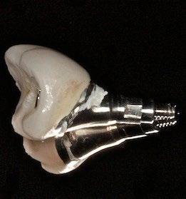
Take a look at the cemented restorations (15 has the fractured distal marginal ridge) and evaluate the cement specifically on the buccal aspect of the abutment for the restoration at 14 – presumably the easiest area to remove the cement. Compare the cement visible on the abutment to what you see on the pre-treatment radiograph (Figure 3).
The tissue attachment in the area surrounding a dental implant allows for cement to flow much farther apically than would be expected with natural teeth. Peri-implantitis, depending on how the literature is interpreted, could be more common today than periodontitis. Problems related to cemented dental implant supported restorations occur in a time range of about four months to nine years with an average time to adverse event of just under three years. There are two times more cytokines present around dental implants than natural teeth and appear to be the etiologic agent of the breakdown of the supporting structure. Interleukin-1 Beta is a potent bone resorbing cytokine that has been found in peri-implantitis. Figure 3

Dr. Bruce Houser has defined Cement Initiated Disease as iatrogenic with the disease process specifically initiated by excess dental cement. Considering the location (14 and 15) and required contours of the planned maxillary molar restorations as a result of the distal position of the dental implants, how effective is it to remove radiolucent or radio-opaque cement? How about screw-retained restorations in the posterior?
13


