
6 minute read
NEW CONTENT: RADIOLOGY: IMAGING IN ACUTE DISTAL BICEPS TEN
Imaging In Acute Distal Biceps Tendon Injuries.
Auckland Radiology Group
Advertisement
Below we have a new article piece which will be ongoing in 2020 - Thanks to Auckland Radiology Group for the content!
Distal biceps tears usually occur in middle aged men, heavy manual workers or in weight-lifters as a result of a lifting injury or eccentric loading. Treatment can be surgical or non-surgical, however untreated rupture leads to a loss of flexion and particularly supination power.
Clinical diagnosis can be simple in complete ruptures with retraction of the muscle belly (Popeye Sign). Imaging is often not required
History is important: typically, eccentrically loading the flexed elbow and suddenly feeling something snap with subsequent anterior elbow pain
Fig. 1 Ultrasound showing a normal distal biceps tendon inserting at the radial tuberosity.
Variations of injury exist and imaging can help with the diagnosis of incomplete tears or where there is rupture with an intact bicipital aponeurosis, and in
these cases the biceps muscle belly may not retract
Even with complete tears the elbow can still flex / extend / supinate
An ultrasound or MRI may help if the history and examination are atypical, if the patient is not clinically progressing, or as an aid for surgical planning
CLINICAL
Complete biceps tears are more common than partial. Injury most often occurs distally at the radial tuberosity attachment (tendon detaching from bone)
Partial tears may involve only the long or short head components
Mid tendon or musculotendinous junction tears (tendon detaching from muscle) are less common. Musculotendinous junction tears are often not surgically managed, as surgery is less successful at this site. Diagnosis of musculotendinous junction tears can prevent unnecessary surgical exploration
Rupture can cause an immediate 30% loss in flexion strength and 50% loss in supination
strength, which only partially recovers unless managed surgically
Non-operative Management:
In elderly and those not fit for surgery
In those who accept the cosmetic appearance and loss of strength
Surgical Management:
Indicated in those who cannot tolerate loss of supination strength or an altered cosmetic appearance
Outcomes are optimal with surgery performed within 1-3 weeks of injury
Delayed diagnosis beyond 6-8 weeks leads to muscle and tendon atrophy with an increased need for tendon graft or augmentation
IMAGING
X-Rays
Usually normal, but will exclude fractures and other radial abnormalities
Chronic tendinopathy can cause irregularity and sclerosis at the radial tuberosity
Ultrasound
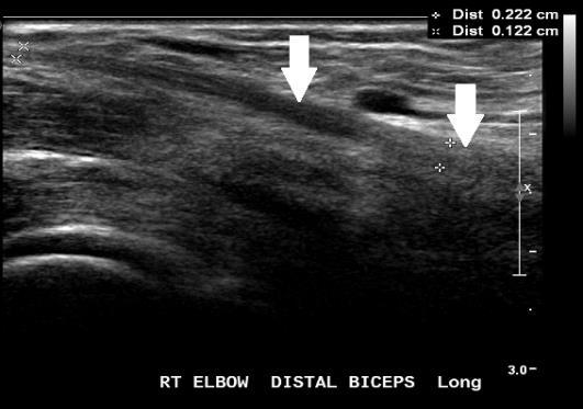
Can confirm the tendon rupture and locate the tendon remnant
Fluid or haematoma may be seen in a measurable tendon gap
On ultrasound the very distal attachment to the tuberosity may be difficult to visualise due to the oblique tendon path. Determining the grade of
distal partial tears can be more difficult in muscular patients
Ultrasound is operator dependent and most useful when performed by experienced sonographers and radiologists
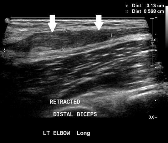
Fig. 4 Ultrasound showing biceps tendon retraction causing bunching and thickening of the torn tendon stump.
MRI •
Confirms biceps tendon rupture and locates the tendon stump, similar to ultrasound imaging
MRI provides greater sensitivity than ultrasound for partial and complex tears which may assist surgical planning
Multiplanar imaging of bone, cartilage and soft tissues in exquisite detail provides concurrent assessment of other alternative pathology, such as tendinosis, tenosynovitis, brachialis injury as well as ligamentous and osteochondral damage elsewhere in the elbow
Fig. 2 Ultrasound showing normal biceps tendon (white arrows). Fig. 3 Ultrasound: The distal biceps tendon is ruptured. There is fluid and haematoma at the tendon defect (red arrow), and the tendon is retracted proximally (white arrow).
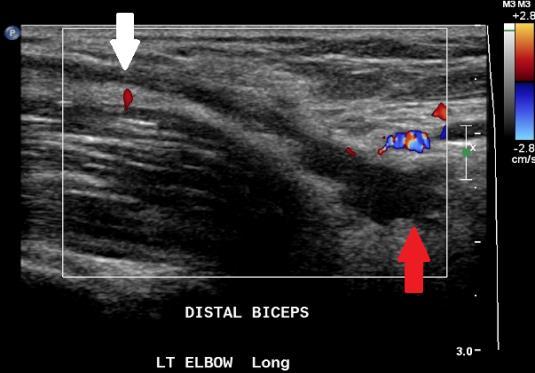
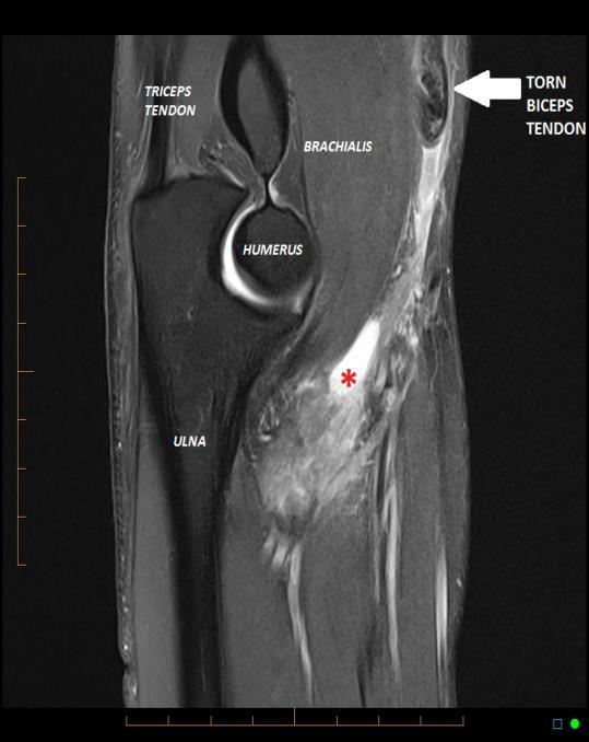
Fig. 5 Sagittal MR showing rupture of the biceps tendon with the tendon retracted proximally and bunched above the elbow joint line (white arrow). There is fluid in the tendon defect with surrounding oedema (red asterix).
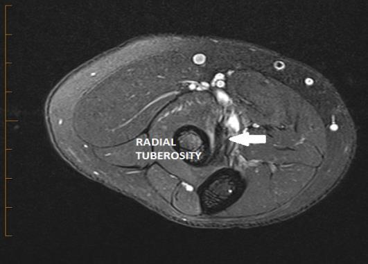

Fig. 7 Axial MRI showing rupture of the distal biceps. The tendon is absent from the radial tuberosity (red arrow). The bicipital aponeurosis has also torn causing subcutaneous and deep fascia oedema over the medial forearm (blue arrows). OTHER DISTAL BICEPS PATHOLOGIES
Imaging may identify other distal biceps pathology including: INSERTIONAL TENDINOSIS (from an acute event or chronic trauma):
the tendon is thickened and or hypoechoic with loss of the normal fibrillary pattern on ultrasound, or shows abnormal signal without tearing on MRI
BICIPITORADIAL BURSITIS:
the normal bursa surrounds the tendon in supination, and passes between the tendon and radial tuberosity in pronation, allowing tendon glide
bursitis can accompany biceps tendinosis and tears or be a stand-alone diagnosis. It typically causes swelling and anterior elbow pain
Fig. 6 Axial MRI showing a normal low signal biceps tendon inserting at the radial tuberosity.
BICEPS INSERTION CALCIFIC TENDINOSIS:
uncommonly seen at the biceps insertion
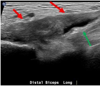
Fig. 8 Long section ultrasound image showing distal biceps tendinosis and bicipitoradial bursitis. The tendon is abnormally thickened and hypoechoic (red arrows) and there is fluid around the insertion in the bicipitoradial bursa (green arrow).

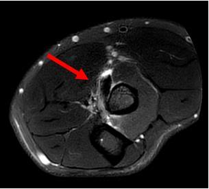
Fig. 10 X-ray showing deposit of amorphous calcium near radial tuberosity in patient with anterior elbow pain.
Fig. 9 Axial MRI showing thickened biceps tendon with fluid in the surrounding bicipitoradial bursa (red arrow).
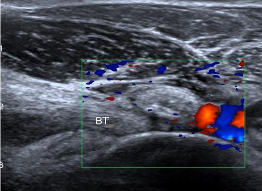
Fig. 11 Longitudinal ultrasound image showing calcific tendinosis with deposit of calcium next to biceps insertion.
NERVE IMPINGEMENT SYNDROMES:
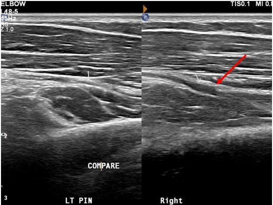
Radial nerve impingement / cubital tunnel syndrome can be associated with anterior elbow pain

Fig. 12 Longitudinal and axial ultrasound images showing a ganglion at the anterior elbow. The radial nerve is displaced (red arrows).
SUMMARY AND KEY POINTS
Many distal biceps tears will not be surgically managed, however early diagnosis is recommended for surgical candidates. Optimal time for surgery is within 1-3 weeks of injury.
Imaging is not required in many cases but may assist where:
history and examination are atypical
in diagnosing partial tears or biceps rupture without muscle belly retraction
in identifying alternative causes for symptoms such as tendinosis, bicipitoradial bursitis, or nerve impingement
X-rays are helpful for excluding elbow arthropathy or radial head pathology as an alternative cause for pain.
Ultrasound is a good modality for the primary investigation but is operator dependent.
MR provides excellent tendon detail for more complex injuries and surgical planning as well as when concurrent bone and cartilage assessment is also required.
Author:
DR BARNABY CLARK (MUSCULOSKELETAL RADIOLOGIST) AUCKLAND RADIOLOGY GROUP
Fig. 13 Longitudinal ultrasound images of anterior left and right elbow showing normal thickness posterior interosseous nerve (PIN) on left (white arrow), but a thickened right PIN (red arrow) at the margin of the Arcade or Frohse (not shown).


