6 COMPRESSED AIR IN THE PHARMACEUTICAL INDUSTRY: PART 1
This two-part series illuminates design and implementation principles for ensuring an efficient, reliable and cost-effective compressed air supply — which is often possible without major effort.
14 SARS-CoV-2 CAN INFECT EVEN MORE LUNG CELLS THAN WE THOUGHT
Scientists have reported that more lung cell types can be infected by SARS-CoV-2 than previously thought, including those without known viral receptors.
27 BLOOD TEST CAN TRACK THE BRAIN’S RECOVERY AFTER CONCUSSION
A blood test can accurately detect the ongoing effects of sport-related concussion and help determine when it’s safe to return to the field, according to an international study.
28 NANODROPLET DRUG CARRIERS TRIGGERED BY ULTRASOUND
By releasing the drug at exactly the desired spot in the body, this allows the total dose to be dramatically lower, which should minimise side effects.
14 24
18 THE POLITICS OF HEALTH: HOW ELECTIONS WILL IMPACT ON LIFE SCIENCES IN 2024
With elections this year in the US, the UK and India — all major players in the life sciences industry — what changes and benefits can we expect to see?
24 3D GENOME RECONSTRUCTED FROM FREEZE-DRIED WOOLLY MAMMOTH
Researchers have assembled a 3D reconstruction of the genome and chromosomal structures of a 52,000-year-old woolly mammoth, in what is understood to be a world first.
30 ‘ARTIFICIAL LYMPH NODE’ HAS THE POTENTIAL TO TREAT CANCER
The lymph node is implanted under the skin and is designed to teach immune system T cells to recognise and kill cancer cells.
Independence day
Back in July, the Australian Government announced the appointment of the new, independent Australian Research Council (ARC) board, which would be responsible for determining the priorities, strategies and policies for the ARC; advising Minister for Education Jason Clare; and approving research grants for many ARC funding schemes. The appointment came after the passage of legislation (introduced by Clare) to improve the governance and independent decision-making of the ARC, which followed a review to ensure the ARC could meet the current and future needs of Australia’s research sector.
Most significantly, the ARC board will be responsible for the approval of most research grants within the National Competitive Grants Program, meaning there will no longer be a ministerial veto on such grants. The Minister for Education will, however, be responsible for approving funding guidelines, which will be subject to parliamentary scrutiny, and retain the power to approve nationally significant investments that foster research capability for Australia. The Minister also has the power to direct the board not to approve a grant, or to terminate funding to research grants, based on national security concerns. They will be required to notify parliament of these decisions.
“Over the last decade, the ARC has been bedevilled by political interference and ministerial
delays,” Clare said. “That has made it harder for universities to recruit and retain staff, and it has damaged our international reputation.
“I promised to end the days of ministers using the ARC as a political plaything and … with the appointment of the new independent board, that’s what we’re doing.”
It’s certainly refreshing to see the federal government place such trust in a research funding body by putting itself at arm’s length, but one must wonder how other governments are treating their equivalent bodies, and whether this is likely to change with a change of leadership. Indeed, we must remember that 2024 is the biggest election year in history — with around 4 billion people, or more than half the world’s population, voting in over 70 national polls — and this is sure to have some flow-on effect for the global life science industry. Guest writer Ivor Campbell, Chief Executive of Snedden Campbell, shares his thoughts on page 18 of this issue.
Other highlights this issue include the 3D reconstruction of the genome and chromosomal structures of a 52,000-year-old woolly mammoth, understood to be a world first for any ancient DNA sample (page 24); and the development of an artificial lymph node that teaches immune system T cells to recognise and kill cancer cells (page 30). And as a new coronavirus variant, known as LB.1 or D-FLiRT, hits Australia, we reveal how a study in miniature lung organoids has found that more types of lung cells can be infected by SARS-CoV-2 than previously thought — including those without known viral receptors (page 14).
I’d like to close by acknowledging the work of Professor Lisa Harvey-Smith, Australia’s inaugural Women in STEM Ambassador, who left the role shortly before our last issue went to print following the news that the functions of the Ambassador would be amalgamated into other programs. Since 2018, Harvey-Smith and her office have contributed research, tools and resources to help break down structural barriers that prevent women and girls from participating in STEM education and careers. But February’s Pathway to Diversity in STEM Review: Final Recommendations report found that more effort was required to remove the barriers preventing people from all backgrounds entering STEM education and careers, recommending that the Ambassador program should not be extended. We can only hope that Harvey-Smith’s hard work will not go to waste, and will instead serve as one of many stepping stones towards the dream of finally achieving equality in STEM.
Lauren Davis
Compressed air in the pharmaceutical industry
Part 1
The extensive automation characteristic of modern production methods in the pharmaceutical industry makes compressed air a vitally important energy carrier. Supply failure can result in expensive downtime, whilst inefficiencies in production and distribution can mean unnecessarily high costs.
This two-part series from Kaeser Compressors illuminates design and implementation principles for ensuring an efficient, reliable and cost-effective compressed air supply — which is often possible without major effort.
Introduction
Compressed air is used widely throughout the pharmaceutical industry, from processes and transport of pharmaceutical materials through to packaging and use in medical devices. The huge variety of different requirements and individual design options characteristic of each production operation makes it impossible to make a one-sizefits-all final recommendation.
Major differences are evident simply in the floor plans and usage of space: different production sites are distributed over large areas, some housed in one or more buildings, whilst others may be meticulously planned in advance or the result of organic growth over time. Yet all share one commonality: an individual approach must be taken in each case.
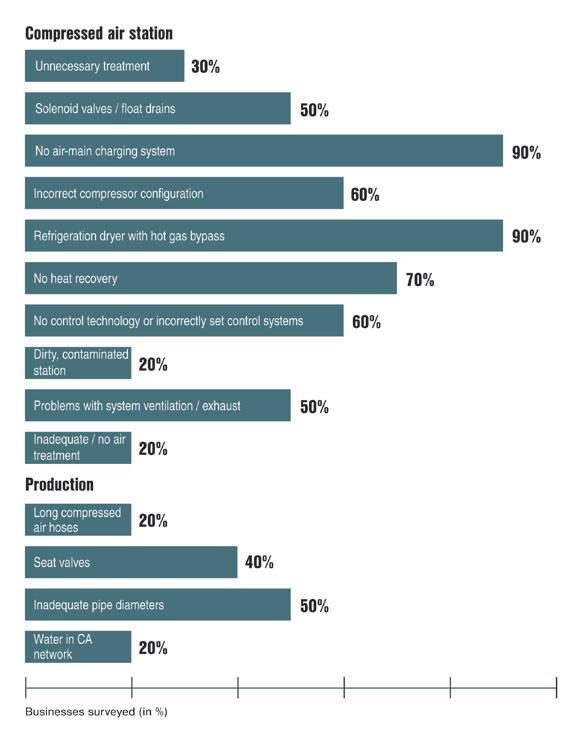
This plethora of possible differences results in each case having widely differing weak points and cost drivers (Figure 1). Thanks to some basic criteria, however, reviewing and even improving one’s own system is easy to accomplish. When produced under unfavourable conditions, compressed air can be very expensive; yet when the right framework is provided, compressed air can be extremely cost-effective.
Energy consumption and cost analysis, cost planning and ongoing management, as well as permanent optimisation based on actual circumstances, are all indispensable to any costeffective and reliable system. Innovative products and services recently launched on the market significantly facilitate the realisation of these key features whilst also ensuring that the compressed air supply is capable of accommodating future developments — provided several key points are taken into account.
Analysis: the first step
Although the ideal situation is to plan a completely new compressed air supply from the ground up, it’s more common for existing systems to be
assessed and optimised where possible. In either case, nothing meaningful can be accomplished until an air demand analysis has been performed — and this often presents the first stumbling block. Despite the significant progress achieved in compressed air technology in recent years, only a fraction of compressed air system operators actually know this crucial value in relation to their own system.
In a ‘mature’, running pharmaceutical operation, compressed air often remains a process element that’s simply ‘just there’ and doesn’t necessarily garner any further attention. Yet it’s precisely companies that have grown over time whose compressed air systems often harbour dramatic potential cost savings. These savings can be achieved in several different areas:
• Optimal adaptation and layout of the compressed air system based on the company’s actual needs.
• Energy cost reductions through the use of lowconsumption, highly efficient components.
• Optimisation of monitoring and service provision over the entire life cycle of the compressed air system.
Today, any business planning a new compressed air supply or renovating its existing one absolutely has to have the overall concept in mind, as well as the system’s continuing viability into the future.
Compressed air systems have a long average service life of some 20 years and are often used every single day. Some investments that may appear attractive over the short term can turn out to be very expensive in the long run. On the other hand, higher initial investment often pays for itself rapidly, then goes on to yield cost advantages year after year.
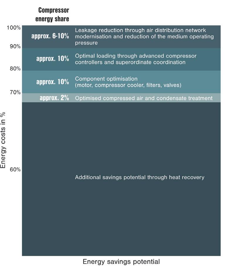
As a general rule, relatively high potential savings can be achieved through optimisation of compressed air systems. According to studies, savings ranging between 10 and 70% of energy costs can be achieved — regardless of the condition of the existing system (Figure 2).
A compressed air system comprises more than just the station itself; it’s rather like a chain, as in the case of electricity generation. In relation to compressed air, the chain begins with compressed air production, followed by treatment and distribution, and ends with the compressed air user. The whole chain must work synergistically to achieve the most cost-effective compressed air supply (Figure 3).
Figure 1: Inefficiencies in compressed air stations and production areas.
Since in most cases the compressed air required in the pharmaceutical industry must be highly pure, achieving synergies becomes especially important in this sector in particular, since compressed air loss or potential contamination can occur at all points along the chain. There’s no point in having the compressed air station produce top-quality compressed air unless this high quality level can be maintained throughout the downstream distribution network and by the end consumer. Ideally, optimised production, appropriate treatment, suitable distribution and constant monitoring go hand in hand.
The compressed air system ‘chain’
When planning or renovating a compressed air system, the first step is to answer some fundamental questions. How high is the air demand? What pressure is required? What processes will the compressed air be used for and what are the compressed air quality requirements of these processes? Are there some processes in which the waste heat from the compressors can be reused?
To determine the answers, the system is closely examined in great detail, from all angles. First the pressures, flow rates and compressed air quality of the consumers are determined; next it’s determined whether there is a decentralised treatment system. Finally, the pipelines are considered in terms of their length, diameters and materials.
When it comes to centralised treatment, the required compressed air quality levels play a role, along with ambient conditions, including
temperature, inlet pressure, inlet temperature and inlet moisture.
Following treatment, it’s time to consider the compressors. What type of compressor is involved and, if applicable, how is splitting implemented amongst the compressors? These are just a few of the important questions that have to be asked.
In order to ensure optimal efficiency of the compressed air supply, the system controller is absolutely critical. For larger stations, the ideal solution is a master control system that controls the entire station with maximum efficiency whilst also enabling modern energy management, data
analysis and remote servicing, including predictive maintenance. In any case, the compressors themselves should be equipped with a controller that supports integration options. When the entire system is taken into consideration, potential leaks also play a major role in the analysis.
The second part of this series examines these topics in detail and provides specific action recommendations to effectively achieve energy efficiency improvements.
To read Kaeser’s entire whitepaper series on compressed air in the pharmaceutical industry, visit https://bit.ly/3WWYyCs.
synergistically to achieve the most cost-effective
Figure 3: The whole chain must work
compressed air supply.
Figure 2: Compressed air production savings potential.
Loss of oxygen in ocean caused mass extinction of marine life
US and Italian researchers have discovered a clue in limestone that helps explain a mass extinction of marine life around 183 million years ago and may provide warnings about how oxygen depletion and climate change could impact today’s oceans. Their work has been published in the journal PNAS
During the Jurassic Period, when marine reptiles like ichthyosaurs and plesiosaurs thrived, volcanic activity in modern South Africa released an estimated 20,500 gigatons of carbon dioxide (CO2) over 500,000 years. This heated the oceans, causing them to lose oxygen. The result was the suffocation and mass extinction of marine species.
“It’s an analogue, but not a perfect one, to predict what will happen to future oxygen loss in oceans from human-made carbon emissions and the impact that loss will have on marine ecosystems and biodiversity,” said study co-author Mariano Remírez, an assistant research professor at George Mason University.
Studying limestone sediment that carries chemicals dating back to the time of the volcanic outburst, the researchers were able to estimate the change in oxygen levels in ancient oceans. At one point, oxygen was completely depleted in up to 8% of the ancient global seafloor — an area roughly three times the size of the United States.
“This event, and events like it, are the best analogues we have in Earth’s past for what is to come in the next decades and centuries,” said study co-author Michael A Kipp, an assistant professor at Duke University.
Since the Industrial Revolution began in the 18th and 19th centuries, human activity has released CO2 emissions equivalent to 12% of what was released during the Jurassic volcanism. But Kipp said that today’s rapid rate of atmospheric CO2 release is unprecedented in history, making it hard to predict when another mass extinction might occur or how severe it might be.
“We just don’t have anything this severe,” Kipp said. “We go to the most rapid CO2-emitting events we can in history, and they’re still not rapid enough to be a perfect comparison to what we’re going through today. We’re perturbing the system faster than ever before.
“We have at least quantified the marine oxygen loss during this event, which will help constrain our predictions of what will happen in the future.”
New MDMA variants could enable safe psychotherapeutic use
The use of the active ingredient 3,4-methylenedioxy-N-methylamphetamine (MDMA) to support psychotherapy for mental illnesses such as post-traumatic stress disorder is being discussed worldwide — with Australia and New Zealand having approved (and restricted) its controlled use by experts due to possible risks and side effects. Now, an international research team led by the Medical University of Vienna has identified three new variants of MDMA as promising alternatives for safer use in a controlled psychotherapeutic setting, with their research published in the Journal of Neurochemistry
MDMA has been known as the party drug ‘ecstasy’ since the 1980s, although the first patent for the substance was granted back in 1912. Due to its effect of promoting positive emotions and increasing interpersonal empathy, research in recent years has focused on the potential of MDMA to support psychotherapy for various mental illnesses. However, possible risks and side effects (tachycardia, high blood pressure, liver and nerve damage) have so far been an obstacle to its widespread therapeutic use. The newly
developed MDMA variants (ODMA, TDMA and SeDMA) have been modified in such a way that the positive effects are retained and the negative effects are reduced.
As studies carried out on human cell cultures show, the new chemical compounds have a similar effect to MDMA on the relevant clinical target structures in the brain (such as serotonin, dopamine and noradrenaline transporters), which are crucial for regulating
mood and emotion. In contrast to MDMA, however, the new substances have lower activity at certain serotonin receptors and are also broken down more favourably, resulting in fewer toxic breakdown products.
“This allows the conclusion that both the acute and long-term side effects of ODMA, TDMA and SeDMA may be lower than those of the conventional substance,” said study leader Harald Sitte.
“Since the MDMA analogs also have a weaker interaction with certain transport proteins in the body that are responsible for the absorption and excretion of drugs, the risk of interactions with other drugs could also be reduced,” added first author Ana Sofia Alberto-Silva.
Sitte said the new variants’ retention of the therapeutic potential of the conventional substance — while causing fewer side effects — “could advance the controlled use of psychoactive substances in neuropsychiatric illness”. At the same time, he acknowledged the need for further studies to comprehensively test the efficacy and safety of MDMA variants for use in a psychotherapeutic setting, for example in the treatment of post-traumatic stress disorder.
Observing dynamic changes in actin filaments during cell division
Laboratory equipment supplier Curiosis has used the Celloger Pro live cell imaging system to demonstrate dynamic changes in cell structure upon cytochalasin B treatment. By enabling real-time cell monitoring, the system allows for seamless observation and tracking of cellular dynamics without disrupting the environment.
Cytokinesis is the final stage in cell division, during which the cytoplasm of one cell is physically divided into two separate cells. Actin filaments play a crucial role in supporting cell structure and facilitating division; just before cell separation, these filaments constrict the cell membrane, leading to the formation of two daughter cells.1 Cytochalasin B, a compound widely used in cell division and movement research, significantly affects the structure and dynamics of actin filaments, primarily hindering cytokinesis by blocking the formation of contractile microfilaments.
Interestingly, cytochalasin B exhibits distinct effects on cell behaviour when employed at different concentrations, despite being the same drug. At high concentrations, it induces a transformation in cell morphology, including contraction of actin cables and rounding up of fibroblastic cells.2 At low concentrations, it inhibits cell migration and membrane ruffling without major morphological changes.3 Additionally, previous studies have shown that cytochalasin B can lead to incomplete cell division, resulting in the formation of multinucleated cells.4
Real-time live imaging is essential for monitoring various cellular motility and responses to drugs such as cytochalasin B — but this can be particularly labour-intensive when it comes to obtaining different images for different drug concentrations. Nevertheless, in order to understand the dynamic changes and structural characteristics of actin filaments induced by cytochalasin B during cell division, Curiosis used a HeLa cell line stably expressing tdTomato-tagged actin and observed it in real time over 48 h. Images were captured using the Celloger Pro with a 10x lens at 1 h intervals and were cropped for analysis.
In the control group, cells were divided from two cells to four daughter cells, undergoing a normal cell division. Conversely, cells treated with a low concentration of cytochalasin B (1.25 µM) failed to complete cell membrane separation, leading to the formation of multiple nuclei within a single cell. However, cells treated with high concentrations of cytochalasin B (10 µM) exhibited severe disruption in actin filament structures, leading to irreversible cell rounding. Quantitatively, a higher ratio of multinucleated cells was observed in the low-concentration group compared to the control group.
In conclusion, actin filaments within cells play a crucial role not only in cellular structure, but also in the processes of cell replication and division. Using the Celloger Pro’s multi-positioning features and user-friendly lens exchange capability, the Curiosis team were able to observe actin dynamics at high magnification, confirming cell structure changes due to cytochalasin B.
1. Pier Paolo D’Avino, et al. “Cytokinesis in Animal Cells”. Cold Spring Harbor Perspectives in Biology (2015): 7: a015834
2. J. W. Sanger. “The Use of Cytochalasin B to Distinguish Myoblasts from Fibroblasts in Cultures of Developing Chick Striated Muscle”. Proceedings of the National Academy of Sciences of the United States of America, Volume 71 no. 9, September (1974): 3621-3625.
3. Yahara, Ichiro, et al. “Correlation between Effects of 24 different cytochalasins on cellular structures and cellular events and those on actin in vitro”. The Journal of Cell Biology, Volume 92, January (1982): 69-78.
4. Awtar Krishan, “Fine structure of cytochalasin-induced multinucleated cells”. Journal of Ultrastructure Research, Volume 36, July (1971): 191-204. Capella Science www.capellascience.com.au
Urine test detects cervical cancer virus proteins
Cervical cancer is the fourth most common cancer in women, with almost all cases linked to high-risk human papillomavirus (HPV) infections. Current screening methods typically involve an invasive Pap smear or HPV DNA test, but emerging research suggests that measuring the cancer-causing activity of HPV may provide a more accurate assessment of cancer risk. Scientists have now found a way to detect HPV oncoproteins in urine, in a non-invasive approach that could encourage more women to participate in regular screening.
A team led by Waseda University’s Professor Etsuro Ito set out to develop an ultrasensitive enzyme-linked immunosorbent assay (ELISA) to detect high-risk HPV16 E7 oncoproteins in urine samples. The test was found to identify these proteins at extremely low levels in the urine of women with different stages of cervical intraepithelial neoplasia (CIN), a precursor to cervical cancer.
As documented in the journal Microorganisms, the ELISA test detected E7 proteins in 80% of women with CIN1, 71% with CIN2 and 38% with CIN3, suggesting that the presence of E7 oncoproteins correlates with lower-grade CIN lesions. The researchers theorise that this discrepancy may be due to variations in the HPV life cycle or oncogenic activity.
“We believe that the E7 oncoprotein is critical in the early stages of HPV-related cervical carcinogenesis and E7 may play a more significant role in the progression of CIN1 and CIN2 than in CIN3,” Ito said.
This innovative approach aligns with global health goals to reduce cervical cancer rates, especially in low- and middle-income countries where access to traditional screening methods is limited. With further development, the test could become a standard tool in the fight against cervical cancer, helping to save lives through earlier detection and treatment.
“We are optimistic that further development and validation of this assay will lead to its widespread use in clinical settings,” Ito said.
“This means that women may be able to screen for cervical cancer without the discomfort and inconvenience of a traditional Pap test.”
Dietary fibre supplement could suppress food allergies
Scientists from the University of Michigan have identified a potential new treatment for food allergies in inulin, a naturally occurring plant fibre commonly used as a supplement, a prebiotic in soft drink, a replacement for sweeteners and more. Their research, published in the journal Nature Materials, proposes that inulin gel addresses the root cause of food allergies, rather than just managing symptoms.
As many as one in three adults and one in four children have food allergies, a life-altering condition that is getting harder to manage as allergens can be hidden in a variety of foods and drinks. Food allergies have become a significant concern globally, especially in developed nations, as accidental exposure to allergens can trigger severe reactions, including death. And while there are various treatment options out there, these have seen low uptake due to adverse reactions and spotty effectiveness.
Inulins, meanwhile, are a group of polysaccharides and natural storage carbohydrates in more than 36,000 plant species — including wheat, onion, asparagus and chicory — which are often used to manufacture supplements. The fibre is also the subject of research and clinical trials investigating their role in treating or leading to better understanding of cancerous tumours, gastrointestinal illnesses, diabetes and other diseases.
The new research found that inulin gel, specifically formulated with an allergen, normalised the imbalanced intestinal microbiota and metabolites in allergic mice. This normalisation led to the establishment of allergen-specific oral tolerance, effectively suppressing allergic reactions to food allergens including peanuts, egg whites and milk.
“The therapy showed long-lasting protection even after the cessation of treatment, indicating its potential for sustained relief from food allergies,” said graduate student Fang Xie, who co-led the research.
The work was helmed by Michigan’s James Moon, who has studied inulin’s potential to treat disease for years. He said inulin gel-based therapy holds great promise due to its safety profile and potential for large-scale production, which together make it “a feasible and translatable option for clinical use”. And while further research and clinical trials are needed to test the findings, Moon said the study opens potentially life-changing new avenues for therapeutic interventions, thanks to its emphasis on the role of the small intestine’s microbiota and metabolites in food allergy regulation.
SARS-CoV-2 can infect even more lung cells than we thought
US and Australian scientists have reported that more lung cell types can be infected by SARSCoV-2 than previously thought, including those without known viral receptors. The research team also reported that the lung is capable of independently mustering an inflammatory antiviral response without help from the immune system when exposed to SARS-CoV-2.
The news comes as more than half of the states in the US report ‘very high’ or ‘high’ levels of COVID-19 infection, according to the US Centers for Disease Control and Prevention.
As noted by Professor Evan Snyder from Sanford Burnham Prebys, “Headlines have come and gone, but SARS-CoV-2 and COVID-19 have never left — and neither have the scientists studying it.”
In making the new discoveries, Snyder and his collaborators at Sanford Burnham Prebys, the University of California San Diego and elsewhere used a technique to transform cells taken from patients into cells resembling stem cells. These embryonic-like cells — known as induced pluripotent stem cells (iPSCs) — can then be turned into other types of human cells.
The team caused the cells to develop into a grouping of various lung cell types in a pattern that mimics the human lung at a smaller scale.
The advantage of this, as noted by Snyder, is, “With most models for studying respiratory infections, you can’t isolate a specific cellular response because you have all the immune system cells rushing in to help deal with the invaders.”
Another benefit, according to Associate Professor Sandra Leibel from UC San Diego, is that “we can choose the sex of the cells so we’re not just studying male-dominant or female-dominant lung tissue” — which is important as the lung responds differently during disease depending on a person’s sex. In addition, the team could make iPSCs from patients of different racial and ethnic groups to try to understand the disparity in this and other diseases in terms of susceptibility to infection, severity and responsiveness to various medications.
As reported in the journal PNAS, SARSCoV-2 was able to acutely infect many previously undescribed cell types in the mini lungs. This held true when testing different strains of SARS-CoV-2, although it was clear that certain strains were more effective at infecting specific cell types.
“People used to say that SARS-CoV-2 only infects cells with certain receptors, especially those with the ACE2 receptor known to interact with the infamous SARS-CoV-2 spike protein,” Snyder said. “We demonstrated that when a direct entry point was unavailable, the virus just punches through the cell membrane instead.”
“With the Delta variant having produced more severe symptoms, and the Omicron variant being less deadly but more contagious, we hypothesised that Delta may prefer the alveolar cells deeper in the lungs, while Omicron sticks more to the upper airways,” Leibel added. “While all strains were capable of infecting many lung cell types, we did see a distinct preference for these strains, as predicted.”
In fact, as the strains changed over time, the scientists could see what was reported in patients as the character of the pandemic changed. Earlier strains such as Delta caused more deadly pneumonias because they affected lower lung cells. Later strains like Omicron affected more upper lung cells and led to clinicians seeing less
pneumonia and more airway problems and sore throats. So, the mini-lung system may help the team predict patient outcomes.
In addition to demonstrating how the virus infects cells previously thought to be safe, the scientists found a way to block the virus’s unexpected flanking manoeuvre. The team demonstrated that apilimod — a drug currently being studied for potentially treating cancer, ALS, dementia and various viral infections — effectively blocked the backdoor entry of SARS-CoV-2 into cells lacking traditional entry points.
“Our data suggest that apilimod could be an adjunct therapy given early on to slow down the infection and enhance the effectiveness of other medicines and the innate immune response,” Snyder said.
In another surprising result, the team discovered that the mini lungs have their own intrinsic ‘first response’ system in reaction to sensing SARS-CoV-2. Even though the mini lungs lack any connection to an immune system, this study shows that lung cells can initiate many of
the same biologic and cell signalling changes in response to a viral threat that are observed when the immune system is present.
“We found that lung cells are capable of autonomously reacting to infection immediately while also subsequently calling for reinforcements from the immune system,” Snyder said.
“We showed that it’s not just the immune cells that are becoming over-activated and secreting too much of the pro-inflammatory cytokines that contribute to severe cases of COVID-19,” Leibel added. “The lung cells do this as well.”
The scientists learned that this inherent antiviral response system in the mini lungs was orchestrated by an unlikely source: one of the four proteins that mix with fats to form a soapy substance in the lungs’ air sacs that helps keep them open as we breathe. This detergent-like substance is called surfactant, and its constituent protein surfactant protein B (SP-B) turned out to be the most important player in the mini lungs’ attempts to ward off SARS-CoV-2. No prior research had suggested that SP-B played any cellular signalling roles.
“When we tested mini lungs genetically engineered to not express SP-B, we saw triple the number of cells infected with SARS-CoV-2,” Leibel said. “When we followed that up by treating these engineered mini lungs with SP-B in a similar way to how premature newborns with surfactant deficiency are treated, we noted a reduction in viral infectivity.”
“These findings suggest not just one but two potential novel drug applications with the possible clinical use of surfactant early in COVID-19 cases,” Snyder said. “This is important, as we only have two current proven antiviral drugs in Paxlovid and remdesivir.”
The team plans to follow up these findings with studies to determine exactly how surfactant is so effective at protecting cells against viral invasion. They are also investigating whether a rapid test for SP-B as well as certain characteristic pro-inflammatory cytokines may help quickly determine which people are at greater risk of more severe forms of COVID-19.
“This would help people make more informed decisions about travelling and attending public events during spikes of COVID-19, and it also would help physicians tailor treatments for those at an increased risk of serious disease,” Leibel said.
iStock.com/wildpixel
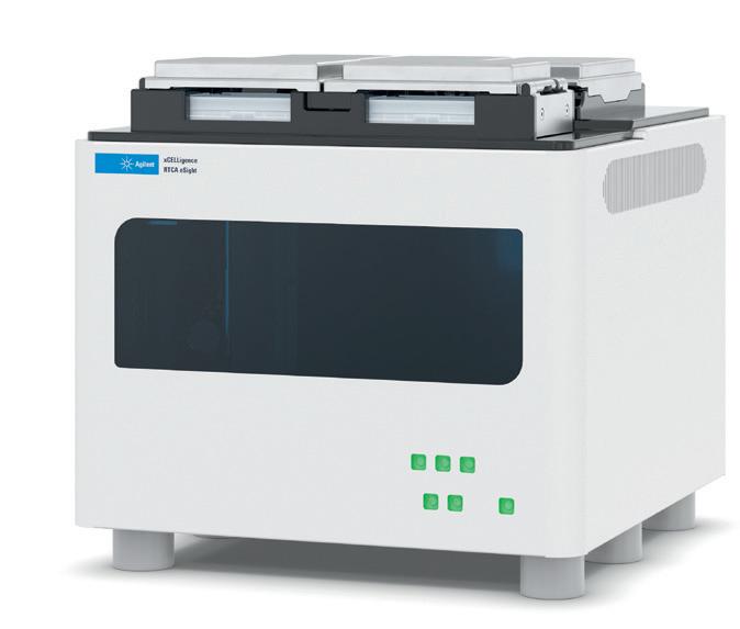
Real-time cell analyser
Monitoring cellular behaviour in real time is essential for understanding fundamental biological processes. The Agilent xCELLigence RTCA eSight Multimode Real-Time Cell
Analyser offers a sophisticated method that combines label-free impedance-based analysis with live cell imaging capabilities. This combination is designed to enable precise and comprehensive cell analysis, encompassing cell health, function and morphology, thereby facilitating deep insights into cellular dynamics.
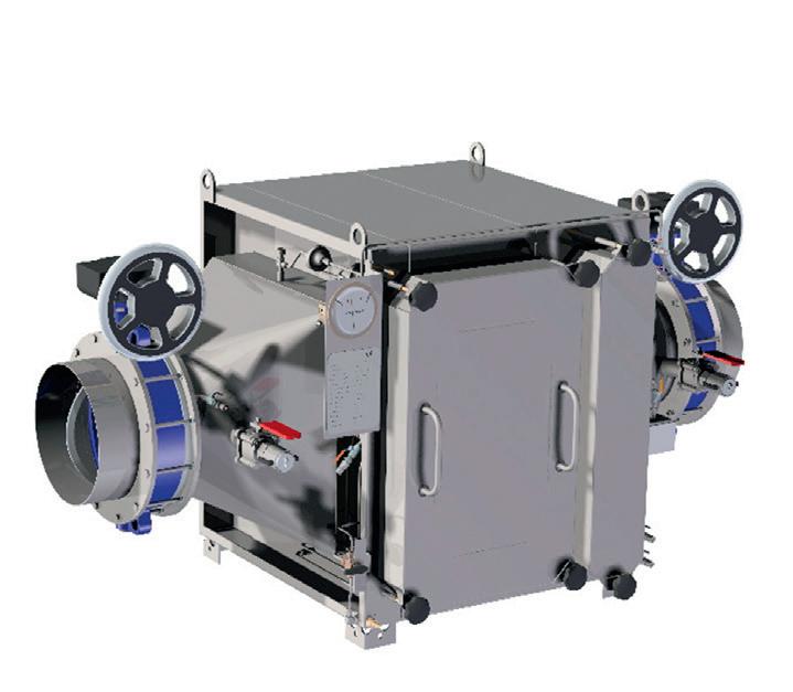
Filter housings
Stringent safety measures are essential in situations where isolating toxic, radioactive or bacterial substances is necessary, such as in the pharmaceutical industry, biotechnical equipment usage, BSL-3/BSL-4 laboratories and nuclear power engineering.
Central to the xCELLigence RTCA eSight Multimode Real-Time Cell
Analyser is its capacity to continuously monitor cell behaviour while simultaneously capturing live cell images in multiple channels, including red, green and blue. This dual-mode functionality allows for the realtime observation of changes in cellular morphology, proliferation and adhesion strength, complementing traditional impedance-based measurements with visual data. Furthermore, the system’s versatile imaging capabilities, encompassing brightfield imaging and three fluorescence channels, accommodate diverse experimental requirements, thereby enabling seamless adaptation of experimental protocols.
The workflow is both streamlined and robust. Following cell seeding and schedule set-up through the user-friendly software interface, researchers can introduce experimental treatments directly onto the cells. Subsequently, the system autonomously acquires data using its proprietary biosensor technology while concurrently overlaying live cell images in real time. This synchronised approach facilitates comprehensive analysis of cellular responses, enhancing the interpretability of experimental outcomes.
By bridging impedance-based analysis with live cell imaging, the xCELLigence RTCA eSight Multimode Real-Time Cell Analyser offers researchers a multifaceted perspective on cellular dynamics, helping to advance their understanding of fundamental biological processes. With its sophisticated capabilities, the innovative platform gives researchers the chance to explore new frontiers in cell biology and drive scientific discovery forward.
Millennium Science Pty Ltd www.mscience.com.au
Multiplexed gene fragments
The Camfil CamContain CS filter housings are designed to meet the highest safety standards. To ensure comprehensive documentation of air filtration, particularly in sensitive areas, the CamContain CS housing can come equipped with an integrated scanner. This allows for onsite testing of the HEPA filter’s efficiency and leakages, with results professionally documented. For applications requiring the filtration of dangerous microorganisms (BSL-3/BSL-4), the housing can be fitted with connections and devices for secure decontamination. Additionally, the maintenance bag replacement technology ensures added safety for operators.
Constructed from stainless steel, the housings are gas-tight welded, torsion-resistant and comply with the strictest sealing requirements, commonly seen in nuclear power plant engineering. The CamScan Mobile serves as a mobile analysis unit for automatically testing installed filters. Following the guidelines of standard DIN 1822, the integrated filter can be assessed for overall separation efficiency and potential leaks. The system’s integrated computer stores measurement values, facilitating seamless documentation.
Camfil Australia Pty Ltd www.camfil.com.au
Twist Multiplexed Gene Fragments offer a novel pooled format for up to hundreds of thousands of gene fragments, between 301 and 500 base pairs in length. The high-quality gene fragments enable a type of high-throughput screening that was previously considered unfeasible or cost-prohibitive for applications like ultra-complex CRISPR screening, antibody engineering, mRNA/vaccine development and more. Twist Bioscience says some of the product’s key benefits include its customisability (large-scale pooled fragment synthesis with several design options); scalability (lengths up to 500 bp for high-throughput screening); high quality (maximised pool quality with low error rates); and precision (even representation across fragment lengths and GC contents).
For research use only. Not for diagnostic procedures.
Twist Bioscience www.twistbioscience.com
Portable carbon dioxide probe with pump sampling
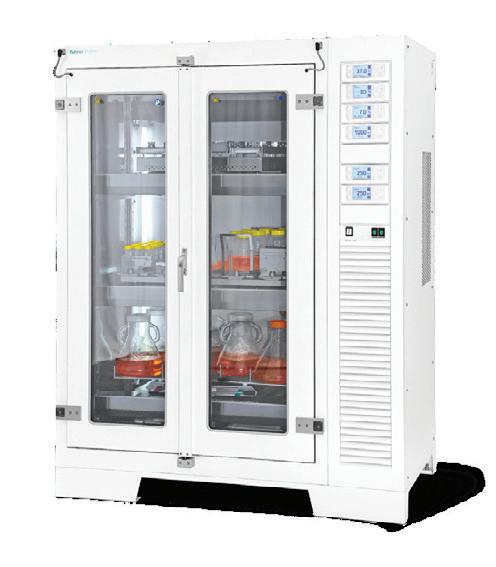
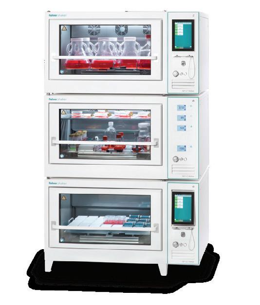
Vaisala’s CARBOCAP portable carbon dioxide probe with pump sampling, called the GMP80P, combines intelligent CO2 measurement with pump-aspirated sampling functionality. The robust and portable measurement device is designed for use in demanding applications such as life science incubators, where stable, reliable and accurate performance is required. It is suitable for CO 2 sampling from incubators, spot-checking fixed CO2 transmitters and sampling from areas otherwise difficult to access.
CO2 measurement data can be monitored with the compact Vaisala Indigo80 handheld indicator connected to the GMP80P. The indicator can be used for short-time logging of measurement data, as well as for calibrating and adjusting the probe.
The product has a measurement range of 0–20% CO 2 and an operating temperature range of 15–40°C. It offers easy CO2 sampling through standard incubator sampling ports and has stainless steel pipe and plastic tube options for sampling. Compatible with the Vaisala Indigo80 handheld indicator and Insight PC software, it offers good long-term stability and has a calibration certificate included.
Vaisala Pty Ltd www.vaisala.com
Nanobody-based reagents
Researchers involved in super-resolution imaging can achieve sharp images and high resolution with Proteintech’s recombinant Nano-Secondary reagents, which offer high species/sub-class specificity and reproducibility. The product portfolio includes NanoSecondary reagents that can specifically recognise either human or rabbit IgG.
The reagents are highly specific recombinant alpaca single domain VHHs (also known as Nanobodies) which recognise Fab or Fc fragments of primary antibodies. Their small size leads to minimal epitope-label displacement, resulting in sharp images and high resolution, making them suitable for super-resolution imaging applications (SRM).
Key benefits include good reproducibility due to high lot-to-lot consistency; minimal cross-reactivity; and validation for SRM, IF, WB and FC.
United Bioresearch Products Pty Ltd www.unitedbioresearch.com.au
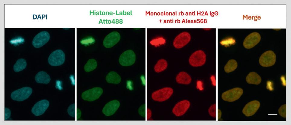
Image courtesy Dr Leila Nahidiazar, Dr Jop Kind and Prof Kees Jalink.
The politics of health
How elections will impact on life sciences in 2024
The global landscape for medical technology, biotechnology and life sciences is on the cusp of seismic change, as the confluence of geopolitical tensions, economic uncertainty and evolving social values has created a complex web of challenges and opportunities for these sectors around the world.
The broad life science industry too is in the midst of a period of significant transition. While the shadow of the pandemic recedes, its effects linger in the form of a market correction and an evolving healthcare landscape.
This, coupled with looming patent expirations, the impact of new drug pricing regulations and tighter universal regulation of medical devices presents a complex environment for both large and small life science companies. Amidst these challenges lies an undercurrent of excitement fuelled by groundbreaking innovations in advanced therapeutics, artificial intelligence and digital engagement strategies.
Navigating this new reality will depend significantly on the policy decisions of governments in major advanced economies. With elections this year in the US, the UK and India — all major players in the life sciences industry — what changes and benefits can we expect to see for companies and their investors, healthcare providers and for patients?
The looming shadow of economic uncertainty
A key concern for businesses is economic stability. The UK, under a new Labour government, will likely see increased public investment in healthcare infrastructure, potentially boosting domestic medtech and life sciences industries.
Last February, the Labour Party published its ‘Prescription for Growth’ — an official plan for the reinvestment and revitalisation of the NHS alongside the country’s life science industry. The strategy is aimed at keeping the sector competitive in a country that has recently been struggling to maintain relevance in the global medical device and clinical trials scenes.
At the same time, Labour has promised to end the rolling NHS junior doctor, nurse and senior consultant strikes that have crippled the NHS
amid sluggishly rising rates of pay for staff and international competition to identify and hire the most highly skilled medical staff. Presented by the new Secretary of State for Health, Wes Streeting, the plan set the tone for the new government’s determination to make good on promises to an industry on edge.
As part of that plan, Labour has pledged to strengthen the Office for Life Sciences while creating a more certain funding environment and a more streamlined funding process. The implementation of new, 10-year budgets for key R&D institutions to attract long-term investment is aimed at ending what Labour saw as the short-termism of its Conservative predecessor in government.
The 2024 US election: pharma in the crosshairs
In the US, both main parties are pledging to reform the pharma industry and, while their approaches may differ, both Donald Trump and now Kamala Harris are expected to focus on drug pricing and market competition, signalling potential upheaval for the industry, whoever wins.
The Democrats’ Inflation Reduction Act (IRA) — particularly its Medicare drug price negotiation provision — is being touted as a win against ‘Big Pharma’s price gouging’. While the industry has criticised this as ‘price control’, the election of the incumbent vice-president would signal consolidation of the IRA, putting further pressure on drug prices.
Despite criticising the IRA in the past, Trump is also prioritising a reduction in drug costs. His previous ‘most favoured nation’ executive order — later withdrawn — aimed to leverage international prices to lower US drug costs. While drug pricing isn’t as central to his current campaign,
his stance suggests a potential willingness to implement similar measures to the Democrats, if elected.
One area where the candidates disagree sharply is market competition. Harris supports ‘march-in rights’ to allow public access to patented drugs at lower prices, a stance vehemently opposed by the industry, which fears it will stifle innovation.
Conversely, Trump champions free market competition and biosimilars. His 2018 law bolstering the US Federal Trade Commission’s (FTC) oversight of biosimilar deals aimed to increase competition for expensive biologics. While this has benefited some companies with robust biosimilar pipelines, others, like AbbVie, have faced market share erosion for blockbuster drugs like Humira.
In India, the recent re-election of Narendra Modi as Prime Minister for a third consecutive term will fast-track his stated determination to make the country the world’s third-largest economy, up from fifth. Medtech, biotech and pharma will all be affected by Modi’s ‘made in India’ policy which aims to supercharge the country’s manufacturing base to help boost growth and create more jobs.
Projected revenues in India’s medtech market this year are expected to top US$8.71billion. With an anticipated annual growth rate of 7.61%, they are predicted to reach US$12.57bn by 2029. Yet despite this rapid growth — a result of increased government investments in healthcare infrastructure and rising demand for advanced healthcare solutions — India’s performance is dwarfed by the US, whose medtech sector is expected to generate US$210bn this year.
Biopharma companies on both sides of the Atlantic, meanwhile, are prepared for a protracted recessionary environment in the coming 12 months — while venture capital funding remains above pre-pandemic levels,
securing financing now requires stronger clinical data and longer negotiation periods. Private equity firms, increasingly partnering with venture capitalists, offer an alternative source of funding, as seen in KKR’s recent investment in Catalio Capital Management.
Artificial intelligence (AI) — through both machine learning and generative AI — is revolutionising the industry, with companies like InSilico Medicine and Relay Therapeutics leading the charge, accelerating the drug development process. AI promises to drive incremental but significant efficiency gains across operations, including clinical trial design, patient recruitment, manufacturing, supply chain management, competitive intelligence, and sales and marketing.
Meanwhile, new cell therapies are showing promise in oncology. Allogeneic therapies are gaining traction, and the application of CAR-T in autoimmune diseases is expanding. However, manufacturing bottlenecks and safety concerns, such as secondary T-cell malignancies, need to be addressed.
Across all areas, success will depend on companies being able to navigate market uncertainties, adapt to evolving regulations, harness the power of AI and embrace innovative engagement strategies. Those which can effectively leverage these trends will be better positioned to unlock the next stage of value creation and shape the future of health care.
The ethical minefield and its impact on innovation
Beyond economic considerations, governments will grapple with increasingly complex ethical dilemmas that directly impact the trajectory of these industries. Advancements in areas like gene editing, reproductive technologies and artificial intelligence raise profound questions about their application and potential consequences. In the contentious issue of abortion, the tension between reactionary policies of populist governments and rapidly advancing technologies has blurred the lines, challenging established ethical and legal frameworks.
Shifting demographics and the prioritisation of healthcare spending
Globally, governments face the dual challenge of aging populations and declining birth rates. This demographic shift will force difficult choices regarding healthcare spending priorities. Will governments prioritise geriatric care and technologies aimed at managing age-related diseases or will they focus on preventative care and technologies promoting the health and wellbeing of younger generations?
The potential for a government-led initiative to incentivise childbirth through improved maternal health care and childcare support underlines this critical dilemma. The outcome of this debate will have significant implications for the types of technologies and research that receive government support and funding.
The potential of emerging markets
While established markets grapple with these challenges, emerging economies, particularly in South-East Asia and South America, present a compelling alternative. These regions often boast rapidly growing populations, increasing healthcare expenditure and a burgeoning middle class with rising healthcare demands.
Governments in these regions are actively seeking to attract foreign investment and develop their domestic healthcare industries. Companies willing to navigate the complexities of these markets, including regulatory hurdles and infrastructure limitations, could find significant growth opportunities.
*Ivor Campbell is Chief Executive of Angus-based Snedden Campbell, a specialist recruitment consultant for the medical technology industry.
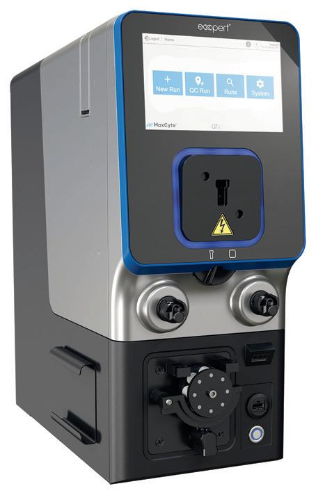
Electroporation-based cell engineering platform
Electroporation is a non-viral transfection method that applies an electric pulse to cells to cause transient permeability in the cell membrane. Diverse genetic engineering payloads such as DNA, mRNA, sgRNA and RNPs can travel through the membrane and enter the cell. Electroporation is a highly efficient delivery method that can be applied to a variety of cell types, from insect to mammalian.
MaxCyte’s STx cell engineering platform is based on Flow Electroporation technology, a universal transfection technology designed for the high-performance delivery of virtually any molecule to any cell, at any scale. It has the ability to transfect primary cells, stem cells and cell lines with minimal cell disturbance and transfection efficiencies routinely >90% for a variety of cell types. The company says its technology allows scientists to use the most physiologically relevant system facilitating the identification, development and manufacturing of cell therapies, biotherapeutics and small molecule candidates of the highest quality.
The platform provides a scalable electroporation technology for high-yield expression of complex proteins, vaccines and biologics, with the ability to transfect anywhere from 75,000 to 20 billion cells. These higher yields save time and cost in the development pathway.
MaxCyte has evolved the electroporation process from a single cuvette experiment to a Flow Electroporation protocol suitable for genetic engineering at any scale. The complete workflow, from development to manufacturing, can be performed on a single platform, meaning there is no need for repeat optimisation and validation when progressing from concept to clinic. This results in accelerated timelines with the added benefit of reduced manufacturing cost. A safer and more rapid therapeutic development pipeline can expedite therapies to patients, saving lives.
Bio-Strategy - Part of DKSH Group www.bio-strategy.com
Chromatin staining probe for IF and IHC
Chromotek’s Histone-Label is a high-performance, ready-to-use chromatin staining probe that provides low background levels and effectively differentiates between euchromatin and heterochromatin throughout the cell cycle.
The probe consists of an anti-histone Nanobody, conjugated to the fluorescent dye ATTO488, and is designed to provide several benefits, including good penetration (easily penetrating densely packed nuclei); high resolution (achieving clear, detailed images); and monovalent VHHs (preventing clustering of epitopes for correct results).
United Bioresearch Products Pty Ltd www.unitedbioresearch.com.au
Laboratory TOC analyser
The Hach QBD1200+ is a laboratory TOC analyser that supports a wide range of TOC measurements — from ultrapure and pharmaceutical water to cleaning validation.
The wet oxidation laboratory TOC analyser addresses common issues faced by testers in TOC measurement. It is designed to eliminate the need for extensive preparation before testing, reduce measurement time and minimise the costs associated with reagents.
The device offers stable TOC values, eliminating dispersion and providing correct measurement results. Startup is quick, daily inspections are smooth and calibration processes are made simple, enhancing the efficiency and effectiveness of TOC testing.
Other features include easy operability with the 10.4″ inch touch panel and compact design for easy lab installation. Overall, the product is a useful tool for laboratories and industries that require precise and efficient TOC measurement.
Hach Pacific Pty Ltd www.au.hach.com
Conductivity sensor
Knowing the conductivity of liquid samples is a key parameter to control product quality throughout the production process. The optek ACF60 conductivity sensor is designed to fulfil the technical requirements for high-quality and consistent measurements.
The sensor features a six-electrode, four-pole design. The arrangement of the four current electrodes around the two potential electrodes is designed to result in a more reliable and precise measurement. The design also provides reduced sensitivity to sensor fouling and polarisation. The combination of an optek C800 or C8000 universal converter and the ACF60 conductivity sensor allows a wide dynamic range from 0–10 µS/ cm up to 0–850 mS/cm with the same sensor (DIN EN 27888/ISO 7888 and ASTM D1125).
Applications include conductivity control for chromatography processes; pre/post column monitoring of pure, drinking and treatment water; TFF/ultrafiltration; heat exchanger control; and interface detection in food and beverage and CIP processes.
Flow cytometry is a powerful single-cell detection technique used in all aspects of scientific research to probe cell phenotype and function.
In traditional flow cytometry, dedicated bandpass filters are used to capture the maximum fluorescence emission. Although the design of multiparameter panels has had some success using this approach, limitations exist due to the size of the panel and use of certain dye/fluorophore combinations and resolution.
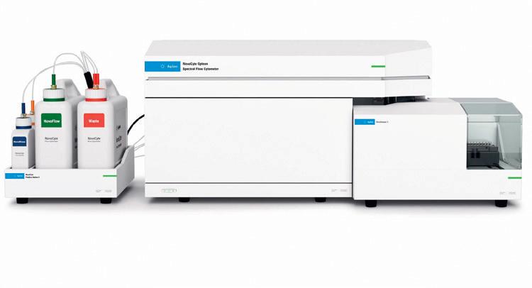
The advent of spectral flow cytometry has led to advancements in supporting more markers, greater flexibility and better data resolution. The Agilent NovoCyte Opteon is a cutting-edge spectral flow cytometer designed to revolutionise cell analysis research.
The NovoCyte Opteon Spectral Flow Cytometer is equipped with up to five lasers and 73 detectors. A dual 488 nm-SSC (B-SSC) and 405 nm-SSC (V-SSC) allows detection of particles as small as 80 nm without adjusting the settings to detect larger cells within the same sample. Furthermore, with this dual SSC, whole blood analysis can be separated into platelets, red blood cells and white blood cells without the need for any lysis protocols.
Real-time monitoring of instrument status enables uninterrupted data acquisition at varied high and low sampling rates. The intuitive Agilent NovoExpress software features flexible reference control set-ups and autofluorescence subtraction capability. This is all augmented by walkaway automation with the NovoSampler S autosampler, which is flexible (runs various plate and tube rack formats) and lab automation friendly.
The Agilent NovoCyte Opteon Spectral Flow Cytometer is designed to enable high-dimensional multiparametric cell analysis. Millennium Science Pty Ltd www.mscience.com.au
Multi-component, multi-range portable FTIR gas analyser
Measure 1000’s of gases with single unit
Software offers no-limit on number of gas measurements at once, using powerful PLS algorithms
Data can be downloaded and re-analysed offline for new gases
Built in O2 sensor, heated inlet filter and sampling control
Specific Applications for atmosFIR:
• Stack Emission Testing
• Incineration and Combustion Gas Monitoring
• RealTOC measurement
• Speciated VOC with use of high resolution
• Abatement plant efficiency – inlet and outlet
• Ambient air monitoring from 0.5ppm
• Siloxane Measurements
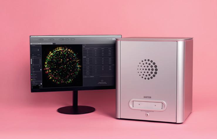
CytoDirect
The new era of cell counting
Cell counting is an important technique used in life science research, pharmaceutical R&D, diagnostic testing and more. Traditionally, this method is performed manually via microscope with cytotoxic reagents, or large, bulky automated cell counters — but what if there was a more convenient, safe and fast way to do this, all while maintaining a high level of accuracy?
METTLER TOLEDO’s newest product offering, CytoDirect, offers stain-free, automated cell counting in a portable device. CytoDirect leverages noninvasive technologies: digital holographic microscopy and machine learning algorithms. The advanced technology, together with an extra-large measurement area (31.2 mm2) and the elimination of cell staining, ensures measurement reliability in a short measurement time of only 15 seconds. A high degree of automation decreases human error, improves process standardization, and eliminates inter-operator variability.
Take it to your sample
CytoDirect is a portable device that enables semioperator-independent cell counting, viability tests, and cell size distribution assessments at the actual work location (i.e. laminar flow hood, lab bench, in production). CytoDirect also follows the researcher anywhere in the lab thanks to its small footprint, Wi-Fi connection, and integrated battery. The device’s portability helps to further reduce the risk of cell culture contamination by
minimizing user intervention and displacements across the workspace.
Say bye to trypan blue
Measuring cell viability typically involves additional biochemical procedures, such as staining to probe membrane intactness. This classical method involves extra incubation and handling steps with cytotoxic reagents, which increases error potential, experimental time, and operator exposure risk.
The unique combination of high-resolution digital holographic microscopy analyzed by a trained convolutional neural network allows reliable and reproducible cell analysis in a fully automated way1 without staining.
CytoDirect uses the morphological and compositional differences between dead and living cells to determine culture viability.
The digital holographic microscope can quantitatively assess changes in composition, e.g., reflecting protein turnover, making the percentage of viable cells directly accessible. This eliminates staining steps and reduces operation intervention to a minimum. Therefore, CytoDirect preserves the sample integrity and removes any cytotoxicity bias during the measurement.2,3
The machine learning algorithm is trained to reliably evaluate a variety of cell lines, making it a useful tool for many applications in biopharma, biotech, and academic research.
The application note “Holographic Microscopy and Machine Learning: The Revolution of Cell Analysis” contains more information about digital holographic microscopy.
Ensured reliability
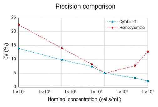
CytoDirect uses an extra-large measurement area — 30 times bigger than a standard hemocytometer — maximizing statistical significance in every measurement. Moreover, CytoDirect is robust and maintenance-free. It avoids wear-affected moving parts and does not require calibration.
The cell counting performance was assessed for many different cell types, including HeLa, PBMCs, and yeast, comparing linearity, accuracy, and reproducibility against manual counting using a hemocytometer. The experiments showed a high degree of linearity across the evaluated cell types. Therefore, it
Figure 1: Precision comparison of CytoDirect and the hemocytometer in different cell concentrations.
is shown that CytoDirect reliably detects and counts cells across a wide linear operating range, providing that it can be successfully applied in various cell-based experimental settings, ranging from established mammalian lines through primary cells to eucaryotic cells with walls. Moreover, CytoDirect shows higher precision than manual counting (Figure 1).
Keep it simple
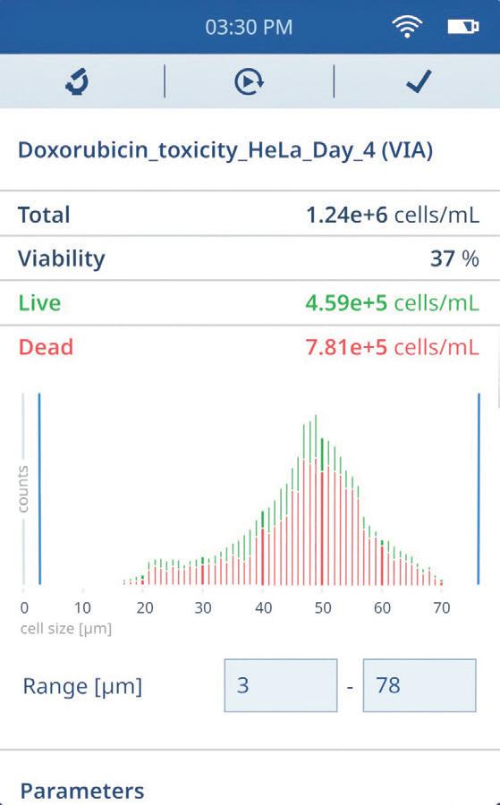
Operating CytoDirect is straightforward: the user pipettes 20 µL of a cell suspension without prior staining into the glass-made sample carrier. Then, the user can select an application (cell count or cell count and viability measurement).
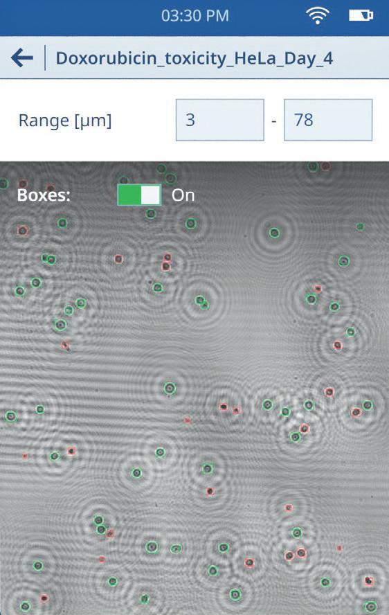
Guided by on-screen instruction, the user inserts the sample carrier in the adapter slot. The measurement starts automatically. Results are displayed after 15 seconds. The microscopic image can be displayed with a click, providing direct visual insight into the automatic detection of viable and dead cells, as shown in Figure 2.
Moreover, thanks to an integrated debris filter, CytoDirect easily recognizes cells vs. debris and other contamination, making the results even more reliable. The user still has the option to further filter the results by size by selecting the appropriate size range, as shown in Figure 2.
Connecting CytoDirect to LabX™ software will provide user management, store all measurements and metadata, and enable full GMP compliance in regulated environments such as biopharma.
The XDirect Portfolio
METTLER TOLEDO keeps innovating and adding additional life science tools, creating the XDirect portfolio, including (for now) CytoDirect and PlateDirect. PlateDirect A96 (Figure 3) features a robust, simplified, user-friendly plate reader and intuitive PC software that eliminates redundant features and moving parts. It provides everything needed to get ELISA, protein concentration, cell viability, or other absorbance measurements done while avoiding high capital, operational, and training costs.
The compact plate reader design allows you to run assays anywhere, share the device with colleagues, and easily stow it away when you are done. This efficiently uses lab space, minimizes contamination risks, and effectively promotes collaboration.
PlateDirect A96’s optical setup provides excellent optical accuracy and repeatability over a wide usable absorption range in the visible spectral region. As each well is measured with a dedicated photodiode, measuring a 96-well plate only takes 5 seconds.
The benefits of using METTLER TOLEDO’s CytoDirect and PlateDirect A96 are significant. CytoDirect provides a reliable, automated, and stain-free cell counting and viability testing solution. The instruments’ portability makes them ideal for various experimental settings while minimizing contamination risks and promoting collaboration. Overall, these devices offer a reliable and time-efficient cell analysis and plate reading solution.
1. Holographic Microscopy and Machine Learning –The Revolution of Cellular Assays, UserCom 28.
2. Riss, Terry L., et al. “Cell viability assays.” Assay Guidance Manual [Internet]. Eli Lilly & Company and the National Center for Advancing Translational Sciences, 2016.
3. Aslantürk, Özlem Sultan. In vitro cytotoxicity and cell viability assays: principles, advantages, and disadvantages. Vol. 2. InTech, 2018.
Mettler-Toledo Ltd www.mt.com
Figure 2: CytoDirect results display.
Figure 3: PlateDirect A96.
3D genome reconstructed from freeze-dried woolly mammoth
An international research team has assembled a 3D reconstruction of the genome and chromosomal structures of a 52,000-year-old woolly mammoth, in what is understood to be a world first for any ancient DNA sample.
As documented in the journal Cell, the fossilised chromosomes provide insight into how the mammoth’s genome was organised within its living cells and which genes were active within the skin tissue from which the DNA was extracted. This unprecedented level of structural detail was retained because the mammoth underwent freeze-drying shortly after it died, which meant that its DNA was preserved in a glass-like state.
“This is a new type of fossil, and its scale dwarfs that of individual ancient DNA fragments — a million times more sequence,” said corresponding author
Erez Lieberman Aiden, Director of The Center for Genome Architecture at Baylor College of Medicine. “It is also the first time a karyotype of any sort has been determined for an ancient sample.”
Knowing the three-dimensional architecture of a genome provides a lot of additional information beyond its sequence, but most ancient DNA specimens consist of very small, scrambled DNA fragments. Building off work mapping the 3D structure of the human genome, Aiden thought that if the right ancient DNA sample could be found — one with the 3D organisation of the fragments still intact — it would be possible to use the same strategies to assemble ancient genomes.
The researchers tested dozens of samples over the course of five years before landing on an unusually well-preserved woolly mammoth that was excavated in north-eastern Siberia in 2018. According to corresponding author Olga Dudchenko, also from The Center for Genome Architecture, “We think it spontaneously freeze-dried shortly after its death. The nuclear architecture in a dehydrated sample can survive for an incredibly long period of time.”
To reconstruct the mammoth’s genomic architecture, the researchers extracted DNA from a skin sample taken behind the mammoth’s ear. They used a method called Hi-C that allows them to detect which sections of DNA are likely to be in close spatial proximity and interact with each other in their natural state in the nucleus.
“Imagine you have a puzzle that has three billion pieces, but you don’t have the picture of the final puzzle to work from,” explained corresponding author Marc A Marti-Renom, an ICREA research professor and structural genomicist at the Centre Nacional d’Anàlisi Genòmica (CNAG) and the Centre for Genomic Regulation (CRG) in Barcelona. “Hi-C allows you to have an approximation of that picture before you start putting the puzzle pieces together.”
Then, they combined the physical information from the Hi-C analysis with DNA sequencing to identify the interacting DNA sections and create an ordered map of the mammoth’s genome, using the genomes of presentday elephants as a template. The analysis revealed that woolly mammoths had 28 chromosomes — the same number as present-day Asian and African elephants. Remarkably, the fossilised mammoth chromosomes also retained a huge amount of physical integrity and detail, including the nanoscale loops that bring transcription factors in contact with the genes they control.
By examining the compartmentalisation of genes within the nucleus, the researchers were able to identify genes that were active and inactive within the mammoth’s skin cells — a proxy for epigenetics or transcriptomics. The mammoth skin cells had distinct gene activation patterns compared to the skin cells of its closest relative, the Asian elephant, including for genes potentially related to its wooliness and cold tolerance.
“For the first time, we have a woolly mammoth tissue for which we know roughly which genes were switched on and which genes were off,” MartiRenom said. “This is an extraordinary new type of data, and it’s the first measure of cell-specific gene activity of the genes in any ancient DNA sample.”
Although the method used in this study hinges on unusually wellpreserved fossils, the researchers are optimistic that it could be used to study other ancient DNA specimens —from mammoths to Egyptian mummies — as well as more recently preserved museum specimens. For mammoths, the next steps would include examining the epigenetic patterns of other tissues.
“These results have obvious consequences for contemporary efforts aimed at woolly mammoth de-extinction,” said corresponding author M Thomas Gilbert, a paleo-genomicist at the University of Copenhagen and the Norwegian University of Science and Technology.
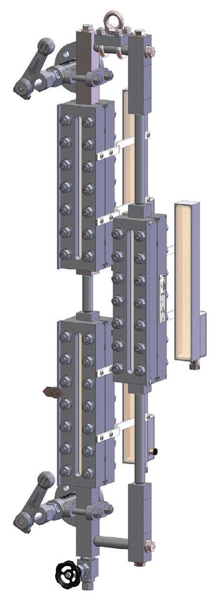
• LIQUID LEVEL indiCation
• INDIRECT leVel GauGeS
• BOILER WATER monitorinG
• GERmAN expertiSe & quality
Reader for analysis of analytes
The Mabtech IRIS 2 is an advanced FluoroSpot and ELISpot reader designed to analyse up to four analytes simultaneously with accuracy and speed. The device features intuitive software for easy data management and self-calibration and can integrate into automated workflows with robotic arms for larger laboratory set-ups and projects.
The plug-and-play system is designed to streamline workflow. Simply load the plate, select the assay and press “read”. The user-friendly Mabtech Apex software ensures a smooth onboarding process, making spot analysis easy for users of all experience levels. Its intuitive layout and automatic data export accelerates the transition from data acquisition to analysis.
After reading, users can modify parameters like brightness, contrast, spot intensity and size thresholds without affecting the original data, leveraging uncompressed spot signals captured directly from the camera sensor. The Apex software supports a variety of file formats to simplify data management.
The new FociSpot assay application enables automated foci counting in a 96-well plate format. It detects foci using immunostaining with virus-specific mAbs and a precipitating substrate reaction like ELISpot. Enhanced by the patented RAWspot technology integrated into the software, it optimises spot centre detection for precise counting and identification of cells secreting multiple analytes in FluoroSpot. Furthermore, it introduces the concept of relative spot volume (RSV), extracting a three-dimensional volume for each spot to accurately reflect the amount of secreted analyte.
Mabtech’s Performance Qualification (PQ) plate verifies instrument quality and ensures reproducibility, allowing users to conduct quantitative and qualitative tests to identify any inconsistencies.
Millennium Science Pty Ltd www.mscience.com.au
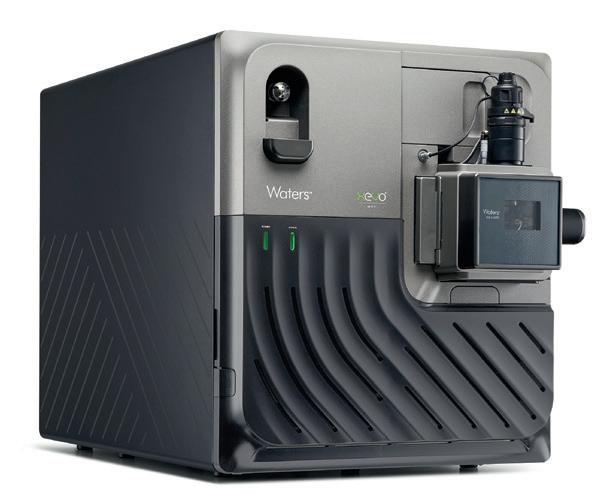
Mass spectrometer
Waters has unveiled the Xevo MRT, its highest-performing benchtop mass spectrometer (MS). Building on the innovative technology pioneered by the Waters SELECT SERIES MRT MS, the system combines multi-reflecting time-of-flight and hybrid quadrupole time-of-flight technologies within a flexible benchtop platform. It delivers 100,000 full width half maximum (FWHM) resolution at 100 Hz MS/MS scan speed and <500 ppb mass accuracy, enabling deep probing of biologically relevant concentrations with high levels of mass accuracy — independent of acquisition rate.
The innovative multi-reflecting time-of-flight design offers scientists the power to work at maximum resolution with high sensitivity and fast data acquisition rates. This helps users to identify analytes across a range of samples and complex matrices and generate comprehensive mass data for scientific interpretation. Users can thus run more samples in less time for large-cohort biomedical research and epidemiology-type studies, without compromising confidence in their data.
Waters offers complete workflows for metabolomic, lipidomic and metabolite identification for the MS, alongside the company’s high-throughput ACQUITY UPLC Systems and UPLC Column chemistries and waters_connect software for data acquisition, processing and reporting. The system supports universal data-sharing via mzML file formats with third-party informatics software, including popular applications from Mass Analytica such as its MARS, Lipostar2 and MassMetaSite software offerings.
Waters Australia Pty Ltd www.waters.com
Blood test can track the
brain’s recovery after concussion
A blood test can accurately detect the ongoing effects of sport-related concussion and help determine when it’s safe to return to the field, according to an international study led by Monash University.
Researchers measured two brainspecific proteins in the blood of 81 Victorian Amateur Football Association (VAFA) players who experienced concussion and compared them with 56 players who did not. By tracking levels of the blood biomarkers over time, they monitored how long it took the players’ brains to recover, otherwise known as ‘neurobiological recovery’, to help determine when it may be safe to return to play without elevated injury risk. Until now, there have been no well-established tools for tracking neurobiological recovery after sport-related concussion.
Published in JAMA Network Open, the cohort study delved into the dynamics of two brain cell proteins — glial fibrillary acidic protein (GFAP) and neurofilament light (NfL), which are released into the blood following brain trauma. While the team’s previous research demonstrated diagnostic potential of these blood biomarkers, this study aimed to reveal how their levels changed over time in concussed players.
The most striking finding was the variability in biomarker changes among individuals, with over 20% of concussion cases showing substantial and persistent increases in both GFAP
and NfL that remained elevated compared to non-concussed footballers for over four weeks. Individuals with these extreme biomarker changes were substantially more likely to have lost consciousness as a result of their head knock. Study lead Dr Stuart McDonald, from the Monash School of Translational Medicine, said while his team had investigated these biomarkers before, this was the first time a thorough profile of postinjury progress had been recorded.
“The unique thing about this study is not the measure, but how many times and how consistently we did it — eight times over six months for 137 athletes,” McDonald said. “With very few missing data points, due to our unique approach of going to the participants for home visits, we were able to get a thorough profile of the biomarker trajectories over time.
“We demonstrated that blood levels of GFAP are elevated in the vast majority of athletes with concussion at 24 hours, and we are now working to have this much-needed diagnostic test approved for use in the next few years.
“The next important step is demonstrating how and when we should measure these two proteins as return-to-play biomarkers. Our findings take us closer to this becoming a reality.”
While more work is needed to seek regulatory approval for these blood tests, Monash’s Dr William O’Brien said there was an important and immediate
takeaway message from this study: neurobiological recovery is likely to take longer in concussed athletes who experience loss of consciousness.
“Our finding of a strong association between loss of consciousness and substantial and prolonged biomarker changes supports the potential adoption of more conservative return-toplay timelines where this clinical sign is identified,” said O’Brien, who is first author on the study.
At the community level of Australian football, the latest policies mandate that the earliest a player can return to play is 21 days after the concussion, while this period is 12 days in the Australian Football League. These guidelines are based on self-reported symptom resolution.
“While return-to-play decisions after this period should consider symptom resolution, completion of a graded loading program and medical clearance, these mandated stand-down periods may not be adequate for all cases of concussion,” O’Brien said.
“Sport-related concussion symptoms are subjective, difficult to identify, and players may feel incentivised to not raise them. Furthermore, the brain continues to recover even after symptoms subside, and this ongoing recovery may make athletes more vulnerable to another concussion.”
More research is underway to create a much larger database on what is ‘normal’, which in turn will help identify what is abnormal. In the meantime, “We do have some good reasons to believe that elevated biomarker levels do indicate that the brain is still in a heightened state of vulnerability to repeated injury,” McDonald said.
Nanodroplet drug carriers triggered by ultrasound
Whether a drug is swallowed, injected, inhaled or absorbed through the skin, it ultimately diffuses to most parts of the body, including those where it isn’t needed — or where it even might cause harm. But what if the delivery could be targeted at exactly the right spot?
Scientists from The University of Utah have been perfecting a method that does just that, with their results published in the journal Frontiers in Molecular Biosciences
“Here we show a method to deliver drugs to specific areas of the body where they are needed,” said Matthew G Wilson, the study’s first author. “We do so using ultrasound waves, which trigger drug release from circulating nanocarriers when focused on the target.
“We developed a method to produce stable nanocarriers repeatably, and identified ultrasound parameters that can activate them.”
The nanocarriers are minuscule droplets, between 470 and 550 nm across, with a hollow outer shell composed of polymer molecules. These polymers have two distinct ends: a hydrophilic one, which mixes well with watery solutions like blood and which faces outward, and a hydrophobic one that doesn’t mix with water and which faces inwards.
Within the shell is an inner core of hydrophobic perfluorocarbons — molecules that consist mostly of fluorine and carbon, and which are mixed with an equally hydrophobic drug of interest. The shells keep the cores apart, preventing them from coalescing into a single
droplet, and form a barrier against the immune system. The effect is much like mayonnaise, where proteins from eggs form droplets of encapsulated oils, where otherwise the oil and water would separate completely.
To release the drug, the researchers played back an ultrasound — a sound wave with a frequency beyond the upper limit of human hearing — of 300 or 900 kHz. The beam of ultrasound can be steered across three dimensions, to focus on a desired area within the body that is only a few millimetres across. The ultrasound is thought to cause the perfluorocarbons to expand, stretching out the droplet’s shell and making it more permeable to the drug, which then diffuses out to the organs, tissues or cells where it is required.
The researchers compared the efficiency of delivery of a representative drug (the anaesthetic and sedative propofol) between three different perfluorocarbons: perfluoropentane (PFP), decafluoropentane (DFP) and perfluorooctylbromide (PFOB). The ultrasound was delivered to the nanodroplets in vitro, in 60 pulses of 100 ms over one minute. The results showed that the balance between stability of the nanodroplets and the efficiency of delivery was optimal for PFOB cores.
“Previous studies have focused on perfluorocarbons with low boiling points — usually lower than the human body temperature,”
Wilson said. “We found that droplets with a PFOB core, which has a boiling point of 142°C, are much more stable over time.
“Despite its high boiling point, PFOB can achieve similar levels of drug release when lowfrequency ultrasound of 300 kHz is applied. The ultrasound frequency turned out to be a critical factor in our study.”
To test for safety, the researchers injected a single long-tailed macaque with six doses of PFOB-based nanodroplets at one-week intervals, and monitored the evolution of a range of blood biomarkers for liver, kidney and immune response function. This experiment showed that the nanodroplets were well tolerated, with no detectable side effects. These experiments will need to be replicated in microdosing or phase 1 trials on human volunteers.
By releasing the drug at exactly the desired spot in the body, this allows the total dose to be dramatically lower, which should minimise side effects. Furthermore, said senior author Assistant Professor Jan Kubanek, the method can be applied to a variety of conditions depending on the drug used.
“For psychiatric applications, localised delivery of propofol could be used as a diagnostic tool to identify brain regions causally involved in disorders for individual patients. For more lasting treatment, ketamine delivery could be a potent method to rewire neural circuits,” he concluded.
Unlocking the Secrets of Intracellular Communication NanoSight Pro Masters
Extracellular Vesicle Analysis
Extracellular vesicles (EVs) have emerged as crucial biomarkers in various biological and medical research fields due to their ability to transport molecular signals between cells. This article highlights the significance of EVs and how the NanoSight Pro can be employed to explore various labeling approaches for unraveling different characteristics of EVs.
Understanding Extracellular Vesicles
Extracellular vesicles are small, membranebound particles released by cells into the extracellular environment and include exosomes and microvesicles each differing in size, origin, and function. Initially thought to be cellular waste, research has unveiled their pivotal role in influencing processes such as immune responses and tumor progression. Due to their presence in bodily fluids, EVs make promising biomarkers for diseases, ranging from cancer to neurodegenerative disorders and inflammatory conditions.
Importance of Characterising EVs
Characterising EVs involves determining their concentration, size distribution, and biochemical composition. The change in concentration of released EVs can indicate an increase in communication, which has diagnostic potential. The composition of EVs, including their cargo molecules such as proteins, lipids, and nucleic acids (including RNA and DNA), can reflect the physiological or pathological state of the cell they originated from. The ability to detect and quantify RNA within EVs serves as a valuable tool for QC to measure success of encapsulation for therapeutic applications.
NanoSight Pro: An Advanced Tool for EV Analysis
The NanoSight Pro is an essential tool that is increasingly utilised in EV characterisation, owing to its ability to track individual particles for size, size distribution and concentration measurements, as well as its fluorescent mode, which facilitates exosome detection through fluorescent labeling. It employs Nanoparticle tracking analysis (NTA) technology, which combines laser light scattering and Brownian motion to measure particle size and concentration simultaneously.
Quantify single EVs with confidence
NanoSight Pro provides high sensitivity detection and analysis of nanoparticles as small as 10 nm. This allows researchers to quickly gain comprehensive insights into the heterogeneity of EV populations. This is particularly important for studying smaller EVs, which are often challenging to characterise with traditional methods. Using real-time analysis, NanoSight Pro enables dynamic processes such as EV release and uptake to be monitored, which is valuable for time-sensitive studies and kinetic analyses.
In the NanoSight Pro, the laser is intermittently deactivated, and fresh sample is introduced between measurements to mitigate the effects of photobleaching. These novel features, combined with the trained neural network for particle identification and the continuous and controlled pumping of samples during the measurement, are essential for enhancing and ensuring the quality of fluorescence detection. The machine learning algorithms and smart
DON’T WAIT — BOOK YOUR PERSONAL DEMO & START USING NANOSIGHT PRO TODAY!
For more detailed information, contact ATA Scientific Pty Ltd | Ph: +61 2 9541 3500 | enquiries@ atascientific.com.au | www.atascientific.com.au
tools built into the software fuel productivity and increase confidence, while eliminating any concerns about subjectivity or human error.
A standout feature of the NanoSight Pro is its fluorescence detection capability. By using specific dyes such as CellMask™ Orange (CMO) and ExoGlow™, researchers can label and track EVs with high specificity. This enables the differentiation of various EV populations within a sample. In addition, simultaneous detection of multiple fluorescence signals is particularly beneficial for studying EVs derived from different cell types or carrying distinct biomarkers.
Using NanoSight Pro, biomarker detection methods developed using two different dyes, Alexa Fluor® 488 (488 nm excitation wavelength) and Pe/Cyanine (either 488 or 532 nm excitation wavelength), have confirmed the presence of CD9, CD63, and CD81 biomarkers. In addition, a novel cargo detection and quantification method has been established, using the Quant-iT™ RiboGreen dye, revealing RNA labeling inside EVs.
The NanoSight Pro, with its specialised fluorescence mode, holds limitless capacity in advancing R&D efforts concerning EVs for both diagnostic and treatment applications. As the field of EV research continues to grow, NanoSight Pro will be instrumental in unlocking new insights and advancing the development of new vaccines and drug delivery systems.
Malvern Panalytical. “Extracellular Vesicles and Nanoparticle Tracking Analysis.” https://www. malvernpanalytical.com/en/learn/knowledge-center/ whitepapers/wp240326-extracellular-vesicles-nta [Accessed 25 July 2024].
ATA Scientific Pty Ltd www.atascientific.com.au
‘Artificial lymph node’ has the potential to treat cancer
Scientists at Johns Hopkins Medicine have developed an artificial lymph node with the potential to treat cancer, according to a new study in mice and human cells.
The newly developed lymph node — a sac filled with immune system components — is implanted under the skin and is designed to act like a learning hub and stimulator to teach immune system T cells to recognise and kill cancer cells. Details of the team’s experiments have been published in the journal Advanced Materials
Lymph nodes — tiny glands throughout the body, mainly in the neck, armpits and groin — are part of the immune systems of mammals, including mice and people. They number in the hundreds so that immune cells in one area of the body don’t have to travel far to alert the immune system to impending danger.
“They are a landing spot where T cells — the immune system’s fighting cells — lay dormant, waiting to be activated to fight infections or other abnormal cells,” said Natalie Livingston, first author of the study and currently a postdoctoral
researcher at Massachusetts General Hospital. “Because cancers can trick T cells into staying dormant, the artificial lymph node was designed to inform and activate T cells that are injected alongside the lymph node.”
To create the artificial lymph node, the scientists used hyaluronic acid, a moisturising substance commonly used in cosmetics and lotions and found naturally in the body’s skin and joints. Hyaluronic acid has the ability to connect with T cells via a cell surface receptor, with a 2019 study from Johns Hopkins scientists showing that hyaluronic acid boosts T cell activation.
For the current study, the Johns Hopkins team used hyaluronic acid as the scaffolding, or base, for their new lymph node, and added MHC (major histocompatibility complex) or HLA (human histocompatibility antigen) molecules, which rev up T cells and other immune system components. Then, they also added molecules and antigens common to cancer cells to ‘teach’ T cells what to look for.
“By adding different antibodies to the artificial lymph node, we have the ability to control what the T cells are being activated to search for,” Livingston said.
The resulting artificial lymph node is about 150 µm in size, about twice the width of a human
hair. That’s small enough to remain under the skin and large enough to avoid being swept away in the bloodstream.
“An advantage to this approach over other cell-based therapies such as CAR-T is fewer manufacturing steps,” said Professor Jonathan Schneck, who led both the 2019 and current research teams.
Current cell-based therapies require extracting T cells from a patient, manipulating them outside of the body to recognise a particular type of cancer and injecting them back into the patient. “In our approach,” Schneck said, “we inject T cells along with an artificial lymph node, and the T cells get primed and educated by the artificial lymph node inside of the body. Then, the T cells can travel anywhere to destroy cancer cells.”
Livingston, Schneck and the research team tested the artificial lymph node in mice that were implanted with either melanoma or colon cancers. Six days after the tumours were implanted, the mice received injections of the artificial lymph node and T cells. The team compared these mice with ones receiving the artificial lymph node alone, those receiving T cells alone (which have not been activated by the artificial lymph node) and those receiving T cells in combination with a class of immunotherapy drug called anti-PD-1.
“An advantage to this approach over other cell-based therapies such as CAR-T is fewer manufacturing steps,” said Professor Jonathan Schneck.
Nine days later, mice with melanomas and colon cancers that received a combination of the artificial lymph node, T cells and the anti-PD-1 drug had the best survival rates (three of the seven mice were still alive at 33 days), compared with other groups that only lived to about 26 days. This group of mice also had the slowest cancer growth rate. It took 5–10 days longer for their cancers to double in size than the other groups.
The scientists also found that the artificial lymph node attracted an influx of other immune cells and acts as an ‘immunologically active niche’ to help to further stimulate the immune system. When T cells were injected into the mice alongside the artificial lymph node, T cell numbers grew as much as nine times more plentiful.
Livingston said the artificial lymph node approach is different from a cancer vaccine, which typically activates a dendritic cell — an immune system component that teaches T cells what to search for. People with cancer often develop malfunctioning dendritic cells and the artificial lymph node skips the dendritic cell to directly activate T cells.
The researchers plan to conduct additional laboratory studies to add more immune signalling molecules to the lymph node and recruit more of the host’s immune cells to the artificial lymph node environment. They have already filed for a patent involving the technology described in their research.
Electromagnetic flowmeter
Azbil MagneW PLUS+ electromagnetic flowmeters are designed to measure many types of liquid, including water, chemicals, slurries and corrosive liquids. The standard model has a mirror-smooth PFA liner for high adhesion resistance that enables durability for long-term use. It is available in an integrated type and a remote type and can be used in a wide range of settings, including explosionproof and outdoor environments.
Suppression of flow noise is 3.5 times that of the Azbil conventional model for improved stability in the presence of noise. The product is designed to achieve more reliable measurement in individual applications through features such as an excitation frequency change function, an optional auto spike cut-off setting, travel averaging and manual zero adjustment.
In addition to the serial number and production date on the product tag plate at shipment, the HMI enables checking in maintenance mode. Statuses that may be difficult to read on the product tag plate are backed up as electronic data.
A high-speed response function with a damping time constant of 0.1 s can be selected as an option. This enables compatibility with high-speed batch applications, allowing use with a pulse frequency of up to 3000 Hz.
Communication with CommPad is supported as in previous models. Communication superimposed on the analog signal can be used by selecting the HART communicator function.
Bio-Rad Laboratories has launched its ddSEQ SingleCell 3 ′ RNA-Seq Kit and complementary Omnition v1.1 Analysis Software for single-cell transcriptome and gene expression research.
Designed to be run on Bio-Rad’s droplet-based single-cell isolation system, the ddSEQ Cell Isolator, the kit delivers high-quality single-cell 3 ′ RNA-Seq libraries in a fast, efficient workflow, allowing researchers to easily conduct single-cell gene expression and regulation analyses. The subsequent QC, analysis and reporting of data generated from the kit is enabled by the accompanying Omnition v1.1
Analysis Software, a robust pipeline analysis tool. These valuable insights into single-cell transcription and gene expression support a broad range of research fields such as oncology, immunology, neurology and stem cell biology.
The kit and analysis software present a solution for single-cell transcription and gene expression studies, facilitating seamless integration into a broad spectrum of research applications and empowering research teams to undertake more experiments within the same budget.
Bio-Rad Laboratories Pty Ltd www.bio-rad.com
Cell culture plasticware
Pacific Laboratory Products’ (PLP) Servicebio plasticware is designed to ensure clear visibility of a user’s cell cultures with highly transparent walls to observe growth of their valuable cells. The cell culture plasticware is expertly crafted from premium-quality polystyrene (PS), which is designed to provide optimal conditions for cell growth. Versatile and suitable for a wide range of research applications, all the costeffective cell culture items are sterile and RNAse, DNAse and Endotoxin free. The range includes cell culture plates, flasks and dishes.
Cell culture plates feature 48 wells, a flat bottom and a tissue culture (TC)-treated surface with lid. They are sterile, individually packaged and designed to prevent cross-contamination with an effective gas exchange lid.
Cell culture flasks have a surface area 75 cm2, a vented cap, a TC-treated surface and an angled wide neck for easy access for serological pipettes and cell scrapers. They are stackable and available in bulk packs or individually packed.
Cell culture dishes are 60 mm, TC-treated and sterile. Available in 20 pieces per bag (with 25 bags to a box), they feature optically clear polystyrene for microscopy, a TC-treated surface and a unique lid for effective gas exchange.
Pacific Laboratory Products www.pacificlab.com.au
Sydney Analytical opens single-crystal X-ray diffraction facility
Sydney Analytical has opened its new high-performance single-crystal X-ray diffraction facility, featuring a newly installed SynergyCustom from Rigaku Oxford Diffraction — understood to be one of the most powerful and comprehensive systems of its kind. Custom-designed for Sydney Analytical’s research requirements, the system has a versatility that will see it serve researchers from multiple disciplines — such as molecular biology, structural biology, chemistry, chemical engineering, pharmacy and medicine — well into the future.
As The University of Sydney’s flagship facility for chemical, biological and materials analysis, Sydney Analytical maintains instruments across several locations to serve cutting-edge research. The SynergyCustom replaces an older system and will better serve research into areas such as macromolecular crystallography, MOFs, functional materials, catalysts, anion receptors, inorganic materials, organic chemistry and charge density studies.
The system is powered by a high-flux rotating anode X-ray generator, featuring a novel combination of copper and silver sources. It also has a dual port configuration allowing for experiments to be run on both ports simultaneously, which can cater for completely different experiments. On one side there is a 96 well-plate screener, ACTOR 2 sample-handling robot for maximum throughput and unattended operation, cryogenic sample facility and HyPix 6000HE hybrid photon counting (HPC) detector. The other side allows for high-pressure, ultralow-temperature experiments (down to 28 K) and features a HyPix Arc-150 HPC detector.
Any crystal structure collected by the system can be elucidated using Rigaku’s CrysAlispro software. With X-ray analytical power second only to the synchrotron, the new system also allows researchers to reduce the number of samples that need to be sent to the synchrotron through better optimisation and even avoiding the need in some cases where the data generated is of high enough quality.
“The system supports significant high-end science ranging from macromolecular samples, to framework materials and solid state compounds for a variety of research areas,” said X-ray Facility Manager Dr William Lewis. “The combination of a high brightness Cu source for routine and macromolecular samples and the short wavelength Ag source for high pressure, diffuse scattering and PDF measurements was key in our decision to go with this system.”
When asked about what new capabilities the system brought to the facility, Lewis replied, “The high-brilliance copper source allows significantly smaller crystals to be investigated and, in combination with the fast detectors and goniometers, also allow significantly faster data collection compared to previous infrastructure, greatly improving throughput.
“The plate screening attachment provides a capability that was previously unavailable, allowing crystallographers to screen crystals in situ, without having to go through the sometimes laborious process of mounting the crystals. The high-energy silver source improves the range of data collected for high-pressure, charge density and total scattering experiments, and also significantly speeds up these experiments.”
Professor Cameron Kepert, the chief investigator on the grant proposal that led to the acquisition of the system, added, “This is a tremendously exciting instrument to have and it will support numerous researchers across numerous faculties and bring these researchers together in ways that haven’t been possible previously.”
Rigaku Oxford Diffraction’s distributor in Australia, AXT, was responsible for the installation, commissioning and maintenance of the system. AXT Managing Director Richard Trett said, “This is a very significant installation for The University of Sydney, Rigaku Oxford Diffraction and AXT. We are extremely proud that we have been able to help bring the whole project together and look forward to seeing the fantastic research that it will enable.”
Dr Mark Benson, Head of Single Crystal Business at Rigaku Oxford Diffraction, added, “As one of the most comprehensive and powerful single-crystal diffractometers in the world, this installation shines a light on the importance of crystallography and the strength of Australian research. We are grateful to The University of Sydney for recognising the capabilities of our instruments and proud they have put their trust into our XRD instruments for over 10 years.”
The University of Sydney and Sydney Analytical are grateful to the Australian Research Council (ARC) and the Linkage Infrastructure Equipment and Facilities (LIEF) scheme that made the instrument acquisition possible.
AXT Pty Ltd www.axt.com.au
One side of the Rigaku Oxford Diffraction SynergyCustom, featuring the sample loading robot, cryo sample handling and HyPix 6000HE HPC detector.
17th APFCB Congress
October 31–November 3, Sydney
The Australasian Association for Clinical Biochemistry and Laboratory Medicine (AACB) will be hosting the 17th Asia-Pacific Congress of Clinical Biochemistry and Laboratory Medicine, APFCB 2024. The AACB’s 61st Annual Scientific Conference will be held as part of the congress. The exciting and informative scientific program will deliver the most recent scientific research related to laboratory medicine by worldrenowned experts. The congress enables those providing a clinical biochemistry testing service and users of biochemical analyses to update knowledge on recent developments and advances, share expertise and experience, and discuss problems in their field of practice. apfcbcongress2024.org
Acoustics 2024 — The Acoustical Society of New Zealand
September 2–4, Christchurch www.acousticsnz2024.co.nz
ASCIA 2024 Conference September 3–6, Adelaide and online ascia2024.com
Science at the Shine Dome 2024 September 9–12, Canberra www.science.org.au/news-and-events/events/ science-shine-dome/science-at-the-shine-dome-2024
AMSA-NZMSS 2024
September 15–20, Hobart www.amsa2024.amsa.asn.au
Biomolecular Horizons 2024 September 22–26, Melbourne www.bmh2024.com
2024 Boden Research Conference September 25–26, Sydney boden-conference-24.sydney.edu.au
2024 APHRS Scientific Sessions September 26–29, Sydney aphrs2024.com
11th International Conference on Legume Genetics and Genomics
September 30–October 3, Brisbane www.iclgg2024.org
BioProcessing Network 2024 Conference October 8–10, Brisbane www.bioprocessingnetwork.org.au/conference
Australian Society of Cytology Annual Scientific Meeting
October 11–13, Melbourne www.cytology.com.au/annual-scientific-businessmeeting
Australasian Cytometry Society Conference October 20–23, Hobart cytometryconference.org.au
Australasian Radiation Protection Society (ARPS) 2024 Conference
October 20–24, Coffs Harbour arpsconference.com.au
Blood 2024 October 27–30, Brisbane www.blood2024.com
AusBiotech 2024 October 30–November 1, Melbourne www.ausbiotechnc.org
ANBUG-AINSE Neutron Scattering Symposium 2024 November 4–6, Lucas Heights www.ansto.gov.au/whats-on/aanss2024-anbugainse-neutron-scattering-symposium-2024
Acoustics 2024 — Acoustics in the Sun November 6–8, Gold Coast www.acoustics2024.org.au
12th International Conference on Environment Pollution and Prevention November 8–10, Brisbane www.icepp.org
AIMS NSW North Coast Div Conference 2024 November 8–10, Port Macquarie www.aims.org.au/events/event/nsw-north-coastdiv-conference-2024
Energy Oceania 2024 November 13–15, Melbourne www.energyconferenceaustralia.com
AgCatalyst 2024 November 19–20, Brisbane events.csiro.au/Events/2024/March/25/AgCatalyst2024-save-the-date
Hendra@30 Henipavirus International Conference December 8–11, Geelong www.hendra30.com iStock.com/Niphon
Westwick-Farrow Media
A.B.N. 22 152 305 336 www.wfmedia.com.au
Head Office
Unit 5, 6-8 Byfield Street, (Locked Bag 2226) North Ryde BC NSW 1670, AUSTRALIA
Ph: +61 2 9168 2500
Editor Lauren Davis LLS@wfmedia.com.au
Publishing Director/MD Janice Williams
(Shy &) Retiring Proofreader Deborah Bailey
Art Director/Production Manager Linda Klobusiak
Art/Production Marija Tutkovska
Circulation Alex Dalland circulation@wfmedia.com.au
Ben Calvo Ph: +61 2 9168 2516 bcalvo@wfmedia.com.au
Tim Thompson Ph: 0421 623 958 tthompson@wfmedia.com.au
policy please email privacy@wfmedia.com.au
Start Separation at Ease
with Centrifuge Solutions from Eppendorf You deserve the best laboratory equipment to ensure that your separation tasks run smoothly every time. That’s why our goal is to provide optimised solutions for you and your requirements: From microcentrifuges and benchtop centrifuges to high-speed floorstanding- as well as ultracentrifuges, our extensive portfolio now o ers comprehensive solutions in the areas of molecular biology, biochemistry and cell biology. www.eppendorf.com/your-centrifuge-solution
Complemented by a wide range of rotors, adapters and consumables, as well as service o erings, our portfolio supports now all of your separation applications.
With our now complete centrifuge portfolio we give you the promise of separation at ease, enabling you to focus on what matters most: your research.