March - April 2023


March - April 2023
This Masterclass Guide is a concise overview of Moisture Associated
Skin Damage (MASD). MASD occurs when skin is exposed to moisture for prolonged periods of time, resulting in over-hydrated or eroded skin. This leads to trans epidermal water loss (TEWL) and an elevated skin pH that reduces the skin’s ability to maintain its barrier function.4,5 The end result is separation of the skin layers, which is also known as maceration.
Body/ Body Fluids
■ Direct skin contact with urine and/ or (liquid) faeces
■ Sweat on the skin surface
■ (Increased) wound secretions on the skin surface

■ Other body fluids such as mucus, (tracheal) secretions, or saliva on the skin surface
■ Increased dermal metabolism, elevated local temperature, abnormal skin pH, history of atopy, genetic susceptibility to contaminants, irritants, deep body folds, dermal atrophy, and inadequate sebum production
■ Pressure related injuries, such as immersion foot
Skin cleansing procedures and products
■ Repeated or excessive skin cleansing, strong friction or abrasive drying procedures, use of rough materials such as coarse towels
■ Repeated use of harsh skin cleansers
■ Ingredients in skin cleansers such as anionic tensides, fragrances, alcohol, preservatives, essential oils
Mechanical factors
■ Mechanical irritation (friction) from clothing or in skin folds
■ Occlusion, for example due to long periods of lying on non-breathable materials, wearing non-breathable clothing, incontinence pads
■ Pressure or shear forces
■ Skin damage from adhesive products, such as band-aids
Indirect risk factors
■ Old age
■ Care dependency
■ Immobility
■ Malnutrition
■ Obesity
■ Atopic diathesis
■ Microangiopathy and/ or macroangiopathy
■ Reduced sensory functions such as blindness, polyneuropathy, dementia
■ Immunosuppression
■ Wound
■ Wounds
■ Wound care
■ Moisture associated skin damage (MASD)
■ Trans epidermal water loss (TEWL)
■ Epidermal injury
■ Medical-adhesive-related skin injury (MARSI)
■ As the onset of MASD often goes undetected, it may first present as basic inflammation of the skin with or without skin breakdown7,8
■ It is often only when significant inflammation, maceration and/ or skin breakdown emerges that clinicians are able to notice and intervene
■ Protection from MASD can be achieved via the application of natural moisturizers containing pyrrolidone carboxylicacid, urocanicacid, propylene glycol, lactic acid, urea, dimethicone, and petrolatum9,10
■ Preventing MASD with barrier ointments and cyanoacrylates is key for at-risk skin and managing alterations to skin integrity such as IAD, ITD, and peri wound skin damage9,10
■ Preventing MASD requires replenishing the natural moisture of the skin with moisturizing products such as skin barrier ointments. The skin should be kept clean and free of excess moisture, and a moisturizer or skin barrier cream should be applied daily
Incontinence-associated dermatitis (IAD):
■ Incontinence-associated dermatitis is a type of irritant contact dermatitis found in patients with faecal and/ or urinary incontinence. The urea present in urine is transformed into ammonia by urease present on human skin
■ This reaction causes an elevation of pH that consequently compromises the skin’s acid mantle, thus reducing the chemical barrier effect of the skin. Faeces contains proteolytic and lipolytic enzymes highly corrosive to the epidermis, with liquid faeces having a higher concentration of these enzymes than formed faeces
■ These cofactors in combination with excessive exposure to moisture increase the risk of epidermal injury. Earlier literature supports a prevalence range of 5.6 - 50%, with the higher being related to faecal or dual incontinence (both faecal and urinary5,12
■ The reporting of MASD is often inconsistent, as many clinicians mistakenly document IAD as Stage 1 or 2 pressure injuries (PIs)7
Intertriginous dermatitis (intertrigo or ITD):
■ Intertrigo is the result of friction in the presence of moisture. Areas of the body most susceptible to intertrigo are those where the skin is warm, where moisture can accumulate, and where the skin is prone to friction. These areas include, but may not be limited to, the axilla, inframammary, abdominal and inguinal folds
■ For patients dealing with incontinence-related issues, the presence of lower body folds in the lower pelvic region may also contribute significantly to morbidity. Obesity and diabetes are two conditions considered to be related to an increased risk for ITD as they are both prone to physiological skin changes, including higher rates of TEWL and increased sweat gland activity
■ ITD tends to be more prevalent in geographic regions with hot and humid climates. In one acute care setting, ITD was prevalent in 2.66% of all reported cases of MASD13
■ The prevalence of ITD falls across a variety of sectors, with 20% of patients living in community dwellings, 17% in long-term care homes and only 6% in acute care settings10
Periwound MASD:
■ Periwound skin damage is multifactorial and often associated with irritant or allergenic contact dermatitis of the surrounding wound skin secondary to moisture. Literature pertaining to the prevalence of periwound MASD is low, and the exact burden remains elusive. It has been hypothesized that the impact of periwound skin MASD is substantial10
■ Wound exudate is created during the natural process of the inflammatory phase of wound healing due to infection, inflammation, or systemic edema. When a wound is stalled, the concentration of metalloproteinases (a proteolytic enzyme) present in wound exudate increases, resulting in periwound skin damage and increasing the opportunity for maceration to occur. Times in which factors such as inadequate compression or inappropriate selection of wound dressings are present, wound exudate may not be well contained and can accumulate on the surface of the skin
■ When moisture is trapped under a dressing there are two factors to consider: the length of time between cleansing the skin and applying the dressing may not be sufficient, or the dressing selected may not have adequate capacity to handle the amount of exudate present
■ Inadequate cover dressings may cause the wound exudate to seep back out of the dressing, especially as the level of compression over the dressing increases
Peristomal MASD:
■ The major determinant of skin damage around a stoma is the enzymatic-containing effluent, although other contributory factors can also play a major role. These include mechanical trauma or medical-adhesive-related skin injury (MARSI) from appliances, bacteria, underlying skin disorders such as psoriasis or eczema, and the possibility of allergies to chemicals or fabrics
■ A multifactorial aetiology is common, with mechanical trauma, moisture and stomal effluent all working in tandem to break down the epidermal barrier.
■ Peristomal MASD affects 17.4% of people with colostomies and 34% with ileostomies, as appliance leakage occurs in more than 50% of the ostomates10
■ A more recent study out of Japan concurs that those living with an ileostomy were more likely to experience peristomal MASD than those with a colostomy14
■ Immersion/ trench foot is a syndrome secondary to prolonged foot exposure to moisture and had initially been described by soldiers practising trench warfare in the early part of the 20th century. Recently, a rise in incidence has been noted among individuals who are homeless and those living with untreated serious mental health issues15
■ In IF, moisture damage to the stratum corneum, the outermost layer of skin, compromises barrier function. Prolonged exposure to wet conditions at temperatures above freezing results in peripheral neuropathy and microvascular damage
■ Progression to severe pain is most often associated with tissue ischemia. In more severe injury, cyanosis with significant swelling of the extremities has been well documented in published literature16
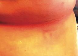
■ Moisture from urine and/ or stool leads to what is commonly called IAD (Figure 1)
■ This is predominately a chemical irritation caused by urine and/ or stool coming in direct contact with the skin. The alkaline nature of urine increases the skin’s pH, changing it from acidic (pH <7) to alkaline (pH >7)
■ In addition, the alkaline urine may promote the enzymatic activity of proteinases and lipases when faecal incontinence is present and further erode the skin’s surface. Maceration of the skin takes place, making the area prone to friction or shear damage. This is especially problematic in older adults with fragile skin that are subjected to sliding for transfer from bed to chair and similar activities
■ Intertriginous dermatitis occurs from moisture trapped between skin folds. Air is not circulated well in these areas, and therefore the moisture, usually as perspiration, remains trapped. Due to this, the skin is macerated, and friction damage from skin surfaces rubbing together may occur
■ This damage is mirrored on both sides of the skin fold. When the outer layer of the skin (stratum corneum) becomes macerated, the results of friction are increased. Consequently, this further erodes the epithelium and can progress to inflammation and breakdown. Thus, the area becomes a potential entry point for microorganisms and may lead to a secondary infection (Figure 2)


■ Skin surrounding a wound can develop toxic or allergic contact eczema, called periwound dermatitis (Figure 3). Periwound dermatitis may occur under wound dressings, due to insufficient management of exudation and longterm contact with the wound secretions.19 This eczema is limited to the areas that come into contact with moisture
■ Chronic wounds, usually stalled in the inflammatory stage, hold higher levels of proinflammatory cytokines and proteases and lower levels of growth factors. This causes an elevated pH (pH >7), and this alkaline environment makes the skin more susceptible to pathogens, resulting in extensive areas of redness surrounding the wound and more tissue destruction. Aggressive or frequent dressing removal, including any adhesive products, can also damage this fragile skin (Figures 4 and 5)

■ Peristomal dermatitis is a (usually toxic) eczema around the site of a colostomy (stoma). This occurs in 30 - 67% of all stoma patients22
■ It may result from a poor seal around the stoma, allowing stool or urine to collect under the seal. Inflammation and erosion (an incomplete loss of the epidermis caused by moisture that is circumscribed, and usually depressed) of the moisture-damaged skin can extend outward in a 10-cm radius. This can occur due to the fit of the pouch not being correct, or the person has a stoma in a difficult area to allow for adherence
■ The ostomy drainage is urine or stool, so the mechanisms of skin irritation are the same as that of IAD, but treatment is difficult because of pouching issues. Frequent removal of the skin barrier needed for pouch placement can further complicate skin issues
■ Medical adhesive–related skin injury is tissue trauma related to the use of medical adhesive products or devices. Adhesive is found within tapes, dressings, stoma barriers, electrocardiogram electrodes, and also medication patches. This includes any product that is used to approximate wound edges or affix a device to the skin

■ If correct placement and removal of such adhesive-containing items do not occur, then superficial layers of the skin are removed with the adhesive product
■ Despite cases where there is no visible irritation, some skin cell detachment still occurs, and a repeated process of application and removal compromises skin barrier function, initiating inflammation and the wound healing response. Medical adhesive–related skin injury is suspected if erythema or other forms of skin injury persist for 30 minutes or more after adhesive removal (Figure 6)

■ Shear, friction, or trauma can result in skin tears, caused by a separation of the skin layers. It usually presents as the epidermis is pulled away, resulting in a partial thickness wound, but in some cases it may be full thickness. Skin tears are classified by the International Skin Tear Advisory Panel (ISTAP) classification system as having no skin loss (type 1), partial flap loss (type 2), or total flap loss (type 3)24
■ Skin tears may occur during the removal of adhesive-based products, and also any maceration makes the skin more susceptible to friction, related to tearing of the epidermis (Figures 7 and 8)
■ Skin tears should be closely monitored and accurately described. Older persons or anyone with fragile skin should be taught prevention measures
■ If a skin tear is present, it should be carefully cleansed following assessment to remove debris. Skin tears are acute wounds and should be closed with primary intention. The skin flap (pedicle) should be approximated when possible, and a nonadherent dressing applied. Any dressing must be removed with caution to avoid additional skin injury

■ The skin injury takes place when skin-to-adhesive attachment is stronger than skin-to-skin attachment. This causes seperation of the epidermal layers or seperation of the entire epidermis from the dermis. Repeated application and removal of adhesive products may lead to skin injury. Trauma may be mechanical and can range from skin stripping, to tension or blisters, or to a skin tear. Irritant or allergic dermatitis may develop under the product, and maceration from trapped moisture or folliculitis can also occur
■ Risk factors for medical adhesive-related skin injury (or medical device-related pressure injury (MDRPI)) also include dry skin, and use of harsh cleaning agents. The use of moisturizers and skin barrier products helps mitigate the risk of skin damage caused by intrinsic and extrinsic factors. It has been demonstrated that a cyanoacrylate barrier film can protect at-risk skin from MDRPI caused by friction.23 When applied to hydrated skin, a cyanoacrylate barrier film significantly reduced the coefficient of friction between the skin and a simulated bed linen
1. North American Nursing Diagnosis Association. NANDA diagnoses. Risk for impaired skin integrity. 2022. Accessed March17, 2023. https://nandadiagnoses.com/risk-for-impaired-skin-integrity
2. DowsettDAL.Moisture associated skin damage made easy. 2013. www.wounds-uk.com/ made-easy/moisture-associated-skin-damage-made-easy. Last accessed June 9, 2017.
3. Gray M, Black JM, Baharestani MM, et al. Moisture-associated skin damage: overview and pathophysiology. J Wound Ostomy Continence Nurs 2011;38:233-41.
4. Bender JK, Faergemann J, Sköld M. Skin health connected to the use of absorbent hygiene products: A review. Dermatol Ther (Heidelb). 2017;7(3):319–330.
5. Voegeli D. Moisture-associated skin damage: Aetiology, prevention and treatment. Br J Nurs. 2012;21(9):517–518, 520–521.
6. Baranoski S, Ayello EA. Wound Care Essentials: Practice Principles. Philadelphia: Lippincott Williams & Wilkins; 2008.
7. Black JM, Gray M, Bliss DZ, Kennedy-Evans KL, Logan S, Baharestani MM, et al. MASD Part 2: Incontinence-associated dermatitis and intertriginous dermatitis: A consensus. J Wound Ostomy Continence Nurs. 2011;38(4):359–370; quiz 371–372.
8. Gray M, Black JM, Baharestani MM, Bliss DZ, Colwell JC, Goldberg M, et al. Moisture- associated skin damage: Overview and pathophysiology. J Wound Ostomy Continence Nurs. 2011;38(3):233–241.
9. Beeckman D, Campbell J, LeBlanc K, et al. Best practice recommendations for holistic strategies to promote and maintain skin integrity. February 28, 2020. Wounds International. Accessed March 17, 2023. https://www.woundsinternational.com/resources/details/best-practice-recommendations-holistic-strategies-promote-and-maintain-skin-integrityWoo KY, Beeckman D, Chakravarthy D. Management of moisture-associated skin damage: a scoping review. Adv Skin Wound Care. 2017;30(11):494-501. doi:10.1097/01. ASW.0000525627.54569.da
10. European Pressure Ulcer Advisory Panel, National Pressure Injury Advisory Panel, Pan Pacific Pressure Injury Alliance. Prevention and Treatment of Pressure Ulcers/Injuries: Clinical Practice Guideline. Haesler E, ed. EPUAP/ NPIAP/PPPIA; 2019.
11. Nobles T, Miller RA. Intertrigo. Treasure Island, FL: StatPearls Publishing; 2018 [cited 2019 Feb 5]. Retrieved from: www.ncbi.nlm.nih.gov/books/NBK531489.
12. Werth SL, Justice R. Prevalence of moisture-associated skin damage in an acute care setting: Outcomes from a quality improvement project. J Wound Ostomy Continence Nurs. 2019;46(1):51.
13. Nagano M, Ogata Y, Ikeda M, Tsukada K, Tokunaga K, Iida S. Peristomal moisture-associated skin damage and independence in pouching system changes in persons with new faecal ostomies. Wound Ostomy Continence Nurs. 2019;46(2):137.
14. Kuhnke JL, Wright G, Kapteyn R. Wound care in a drop-in and rehabilitation centre: A Calgary perspective. Wound Care Canada. 2015;13(2):6.
15. Bush JS, Watson S. Trench Foot. Treasure Island, FL: StatPearls Publishing; 2018. Retrieved from: www.ncbi.nlm.nih.gov/books/NBK482364.
16. Dissemond J, Bültemann A, Gerber V et al. WeitereDefini- tionen und Schreibweisenfür die Wundbehandlung. Hautarzt 2017; 68: 415–7.
17. Voegeli D. Incontinence-associated dermatitis: new insights into an old problem. Br J Nurs 2016;25:256, 258, 260-2.
18. Dini V, Janowska A, Oranges T et al. Surrounding skin man- agement in venous leg ulcers: A systematic review. J Tissue Viability 2020; 29: 169–75.
19. Rippon MG, Ousey K, Cutting K. Wound healing and hyper-hydrationVa counter intuitive model. J Wound Care 2016;25(2):68-75.
20. Gray M, Colwell JC, Doughty D, et al. Peristomal moisture–associated skin damage in adults with fecal ostomies: a comprehensive review and consensus. J Wound Ostomy Continence Nurs 2013;40:389-99.
21. Almutairi D, LeBlanc K, Alavi A. Peristomal skin complications: what dermatologists need to know. Int J Dermatol 2018; 57: 257–64.
22. Bernatchez SF, Mengistu GE, Ekholm BP, Sanghi S, Theiss SD. Reducing friction on skin at risk: the use of 3MTM CavilonTM No Sting Barrier Film. Adv Wound Care (New Rochelle). 2015;4(12):705710. doi:10.1089/ wound.2015.0628
23. LeBlanc K, Baranoski S, Christensen D, et al. International Skin Tear Advisory Panel: a tool kit to aid in the prevention, assessment, and treatment of skin tears using a simplified classification system. Adv Skin Wound Care 2013;26:459-76.
24. LeBlanc K, Baranoski S. Skin tears: state of the science: consensus statements for the prevention, prediction, assessment, and treatment of skin tears. Adv Skin Wound Care 2011;24(9 Suppl):2-15.
25. Arndt JV, Kelechi TJ. An overview of instruments for wound and skin assessment and healing. J Wound Ostomy Continence Nurs 2014;41:17-23.
26. Baranoski S, LeBlanc K, Gloeckner M. CE: preventing, assessing, and managing skin tears: a clinical review. Am J Nurs 2016;116(11):24-30.
27. International Skin Tear Advisory Panel (ISTAP). 2023. Available from: www.skintears.org.

How
Masterclass Guide: Moisture Associated Skin Damage. Wound Masterclass. Volume 2. No 4. March 2023.
