When a young person dies suddenly
Information for the family and relatives of a young person who has died of Sudden Arrhythmis Death Syndrome (SADS)
by Dr Elijah R Behr MDProduced by Cardiac Risk in the Young
 Text by Dr Elijah R Behr
Senior Lecturer and Honorary Consultant Electrophysiologist
Cardiac and Vascular Division, St George’s University of London.
Wordworks, London W4 4DB for plain English editing and the cover design.
Particular thanks to Professor William J McKenna for his involvement in the initial development of this booklet.
Illustrations by Louise Roberts
Medical Photography and Audiovisual Services
St George’s Hospital Medical School
Remastered by Nat Jenkins
Communications and Publications Manager
Cardiac Risk in the Young
Produced by Cardiac Risk in the Young - CRY
1140B The Axis Centre Cleeve Road, Leatherhead Surrey.
KT22 7RD
Web: www.c-r-y.org.uk www.sads.org.uk
Phone: 01737 363222
Fax: 01737 363444
E-mail: cry@c-r-y.org.uk
Registered charity number: 1050845
For further free copies of this booklet please contact CRY, or you can download thebooklet from www.c-r-y.org.uk, www.sads.org.uk
CRY will be updating this booklet regularly. Any comments on its text or design would be welcomed.
Special thanks for the cardiological support of CRY’s programme to:
Dr Elijah R Behr
Dr Navin Chandra
Dr Michael Papadakis
Dr Hariharan Raju
Professor Sanjay Sharma
Copyright Cardiac Risk in the Young 2003
5th Edition - 2017
You may be reading this booklet because a young relative of yours - perhaps a member of your own family - has died suddenly and unexpectedly. This is not only a tragedy for the person and all your family, but a great loss for society too. You may still be asking why it happened, and how it could have happened to someone so young and who perhaps seemed so healthy. Or maybe your doctor has suggested that you should have some tests to find out if you have inherited the same medical condition as the person who has died.
This booklet outlines the possible causes of sudden death in young people and children. It concentrates on the medical conditions responsible for a sudden unexpected death where a definite cause cannot be found, even after a postmortem. This is called Sudden Arrhythmic Death Syndrome, or SADS.
The booklet:
• describes what happens after someone has died suddenly and unexpectedly
• explains how the heart works and how it can cause sudden death
• explains what causes SADS and why it is important that the close blood relatives of the person who has died should have a medical examination and tests to find out if they have inherited the same condition
• describes the tests your doctor may ask you to have,
• offers advice on how to live a healthy lifestyle if you are found to have one of the conditions that can sometimes lead to sudden death.
We have tried to explain medical and technical terms as we go along but, if you find a word you do not understand, you can look it up in the list of Technical terms on page 28.
We hope that this booklet will help you and your family understand what has happened, and hopefully help you come to terms with the event. If you need further help or information see page 27.
What is the difference between Sudden Arrhythmic Death Syndrome (SADS) and Sudden Cardiac Death (SCD)?
Sudden Cardiac Death (SCD)
Sudden cardiac death is a dramatic and/or spontaneous death that is thought to be (and usually is) caused by a heart condition.
Sudden Arrhythmic Death Syndrome (SADS)
In about 1 in every 20 cases of sudden cardiac death, no definite cause of death can be found, even after the heart has been examined by an expert cardiac pathologist. This is then called Sudden Arrhythmic Death Syndrome. (In the past it has also been called Sudden Adult Death Syndrome or Sudden Death Syndrome but, because it affects children too, the term Sudden Arrhythmic Death Syndrome is now used.) It is thought that cot death (Sudden Infant Death Syndrome, or SIDS) may be partly due to the same causes responsible for SADS.
What happens after an unexpected sudden death in a young person?
After an unexpected sudden death it is usual that the coroner of the area where the death has happened will ask for a post-mortem to be performed. This involves the body being examined by a pathologist. Small samples of tissue from organs including the heart are often taken and examined under a microscope. Usually the pathologist can easily detect any abnormality like significant coronary artery disease (furring of the arteries) or pulmonary embolus (a clot on the lung). The coroner will take into account the circumstances of the death and, if necessary, will do tests for signs of any medications or drugs in the body. If it is difficult to assess the heart or to detect any abnormality in it, the pathologist may ask for the help of an expert cardiac pathologist (one who specialises in the heart) to determine the cause of death. The pathologist or coroner may suggest that small samples of heart tissue, spleen and/or blood are kept, sometimes in frozen form, for further testing in case a genetic or inherited cause of sudden death is suspected. This is known as ‘molecular autopsy’ and will only take place with agreement from the family.
How the heart works, and how it can cause sudden death
In order to understand why sudden death can happen, it helps to understand how the heart works.
The heart is a specialised muscle that contracts regularly and continuously, pumping blood to the body and the lungs. The pumping action is caused by a flow of electricity through the heart that repeats itself in a cycle. If this electrical activity is disrupted - for example by a disturbance in the heart’s rhythm known as an ‘arrhythmia’ - it can affect the heart’s ability to pump properly.
The heart has four chambers - two at the top (the atria) and two at the bottom (the ventricles). The normal trigger for the heart to contract arises from the heart’s natural pacemaker, the SA node, which is in the top chamber. (See the diagram opposite.) The SA node sends out regular electrical impulses causing the atrium to contract and to pump blood into the bottom chamber (the ventricle). The electrical impulse then passes to the ventricles through a form of ‘junction box’ called the AV node (atrio-ventricular node). This electrical impulse spreads into the ventricles, causing the muscle to contract and to pump blood to the lungs and the body. Chemicals which circulate in the blood, and which are released by the nerves that regulate the heart, alter the speed of the pacemaker and the force of the pumping action of the ventricles. For example, adrenaline increases the heart rate and the volume of blood pumped by the heart.
The electrical activity of the heart can be detected by doing an ‘electrocardiogram’ (also called an ECG). An ECG recording looks something like the ones shown on page 11 (figure 3).
A death is described as sudden when it occurs unexpectedly, spontaneously and/ or even dramatically. Some will be unwitnessed; some may occur during sleep or during or just after exercise. Most sudden deaths are due to a heart condition and are then called sudden cardiac death (SCD). Up to 95 in every 100 sudden cardiac deaths are due to disease that causes abnormality of the structure of the heart. The actual mechanism of death is most commonly a serious disturbance of the heart’s rhythm known as a ‘ventricular arrhythmia’ (a disturbance in the heart rhythm in the ventricles) or ‘ventricular tachycardia’ (a rapid heart rate in the ventricles).
This can disrupt the ability of the ventricles to pump blood effectively to the body and can cause a loss of all blood pressure. This is known as a cardiac arrest. If this problem is not resolved in about two minutes, and if no-one is available to begin resuscitation, the brain and heart become significantly damaged and death follows quickly.
Adrenaline in the blood Right Atrium
Adrenaline from the nerves
Left
Figure 1: How the heart functions electrically The heart’s natural pacemaker - the SA nodesends out regular electrical impulses from the top chamber (the atrium) causing it to contract and pump blood into the bottom chamber (the ventricle). The electrical impulse is then conducted to the ventricles through a form of ‘junction box’ called the AV node. The impulse spreads into the ventricles, causing the muscle to contract and to pump out the blood. The blood from the right ventricle goes to thelungs, and the blood from the left ventricle goes to the body.

What causes sudden death in young adults and children?
A sudden death in a young person can be caused by:
• heart disease, including cardiomyopathy, congenital heart disease, myocarditis, genetic connective tissue disorders, mitral valve prolapse or conduction disease
• medication-related causes,
• other causes. We explain these below.
Heart disease
Heart disease is the most common cause of an unexpected sudden death in all age groups. In people aged 30 or over, the heart disease is usually due to ‘furring’ or ‘blockages’ of the blood vessels that supply the heart, i.e. coronary artery disease. But in younger people and in children the cause is much more often something other than coronary artery disease. The main causes are listed below. Some of these are inherited conditions. Some are detected easily while the person is alive, while others may go unnoticed until a tragic sudden death occurs.
Cardiomyopathies
These are abnormalities of the heart muscle and are usually inheritable.
Hypertrophic cardiomyopathy (HCM) The walls of the heart become abnormally thick without any other cause being identifiable. Even if there is not any thickening, the arrangement of the heart’s muscle cells (myocytes) are disorganised and disrupted.
Arrhythmogenic right ventricular cardiomyopathy (ARVC) This condition causes the heart’s muscle to become thin because of an abnormal amount of fat and scar tissue in its wall. It affects mainly the right side of the heart but can also affect the left side and frequently causes heart rhythm disturbances. Dilated cardiomyopathy (DCM) The left and right sides of the heart become enlarged and pump less efficiently, sometimes progressing to heart failure when the heart cannot meet the body’s requirements.
Congenital heart disease
This group includes abnormalities of the structure of the heart which have been present since birth. Some of them may be inherited conditions. They include:
Valvular and more complex disease
Abnormality of the heart’s valves that can be associated with other abnormalities of the heart’s structures such as a ‘hole in the heart’ (for example, Fallot’s Tetralogy).
Anomalous coronary arteries
When there is an abnormal arrangement of the arteries that supply blood to the heart muscle.
Myocarditis
Myocarditis is inflammation of the heart’s muscle. It is usually due to a viral infection although it can be a complication of other medical conditions or exposure to drugs. It is not inheritable.
Genetic connective tissue disorders
These are inheritable conditions affecting the structures that give support, strength and elasticity to the walls of the major blood vessels and, to a lesser extent, the heart muscle - for example Marfan’s Syndrome and Ehler-Danlos. These can occasionally cause sudden death by arrhythmias or more commonly due to the sudden rupture of a major blood vessel such as the aorta (the major blood vessel that leaves the left side of the heart and supplies blood to the body).
Mitral valve prolapse
The mitral valve can sometimes be ‘floppy’ in appearance. This will show up on an echocardiogram (see Tests on page 18). This is very common and affects around 1 or 2 in every 20 people. It is usually an asymptomatic and benign condition. In some rare cases mitral valve prolapse can be inherited in a family and can then be associated with arrhythmias and sudden death.
Conduction disease
This includes abnormalities in the way that the electrical impulses are conducted through the AV node due to disease (for example as in myotonic dystrophy), or because there are additional or ‘accessory’ pathways as in Wolff-Parkinson-White (WPW) Syndrome.
Medication-related causes
Prescription, over-the-counter and illegal drugs can have potentially dangerous but usually rare side effects, particularly if too much is taken (an overdose). These effects include arrhythmias (disturbance in the heart’s rhythm) and sometimes a sudden death.
Other causes
Research suggests that sudden death may be caused infrequently by conditions such as fits (epilepsy) and severe asthma attacks. Pulmonary embolus (a clot to the lungs) has become better known recently due to its association with staying immobile for long periods during air travel. It can cause a sudden collapse and a rapid death. (For more on this see page 32.)
Sudden Arrhythmic Death Syndrome (SADS)
In around 1 in every 5 cases of sudden cardiac death under 35 - up to 600 every year in the UK - no cause can be found, despite examination of the heart by an expert cardiac pathologist. The cause of death is therefore described as ‘unascertainable’. This is called Sudden Arrhythmic Death Syndrome, or SADS.
In the next section we describe some of the conditions responsible for SADS.
What causes SADS?
The conditions responsible for SADS cause a cardiac arrest by bringing on a ‘ventricular arrhythmia’ (a disturbance in the heart’s rhythm), even though the person has no structural heart disease.
There is a group of relatively rare diseases called ion channelopathies that affect the electrical functioning of the heart without affecting the heart’s structure. This means that they can only be detected in life and not at post-mortem. Ion channelopathies are probably responsible for 4 in every 10 cases of SADS. There are several different types of ion channelopathies including:
• Long QT syndrome (LQTS)
• Brugada syndrome
• CPVT (catecholaminergic polymorphic ventricular tachycardia)
• PCCD (progressive cardiac conduction defect)
• Early repolarisation syndrome
• Mixed sodium channel disease.
• Short QT syndrome
We describe each of these on pages 8-15.
Structural heart disease is sometimes found to be a cause of SADS (between 1 and 2 in every 10 cases). For more on this see page 15.
Ion channelopathies
Ion channelopathies are rare genetic conditions that are caused by abnormalities of the ‘DNA’ known as ‘mutations’. They are usually inherited from parents although they can occur for the first time in a family. (If they occur for the first time they are described as ‘sporadic’.)
The mutations affect certain genes - specific segments of the DNA that are responsible for the production of cardiac ‘ion channels’. An ‘ion’ is a chemical substance - such as sodium or potassium - that carries an electrical charge and forms the basis of the movement of electricity through the heart muscle. An ‘ion channel’ is the route that the ions take in and out of the heart muscle cells to allow the movement of electricity. The ion channels regulate the flow of electrical charge. If these channels do not behave normally, the electrical function of the heart becomes abnormal. The person can then be prone to arrhythmias (disturbances in the heart’s rhythm) that can cause blackouts, cardiac arrest and in some cases sudden death.
Below we describe the different types of channelopathies, the tests needed to diagnose them and the treatment that may be needed for each one.
Long QT syndrome (LQTS)
LQTS is the most common and best understood type of channelopathy. It occurs in about 1 in 5,000 people. In 7 in every 10 people with LQTS, the ion channels involved have been identified. In most cases two of the potassium channels that regulate the movement of potassium ions from the inside to the outside of the cell are affected. In a small proportion of people with LQTS, a sodium channel that regulates the flow of sodium ions from the outside to the inside of cells is affected.
In people with potassium channel associated LQTS, the channels do not behave as efficiently as normal. They let potassium ions into the cell too slowly. If the sodium channel is affected, too many sodium ions are allowed into the cell. (See the LQTS diagram on page 9.) This results in an electrical disturbance in the cells of the heart called ‘prolonged repolarisation’. This can be seen on an ECG recording as a lengthening of the time period known as the ‘QT interval’. (We show this in the diagram on page 11 - figure 3.) This is where the name long QT syndrome comes from.
Rare forms of LQTS known as Andersen’s and Timothy syndromes have been associated with potassium and calcium channel abnormalities respectively.
What are the symptoms?
LQTS varies greatly in severity. Symptoms vary according to the type of channel involved, whether the person is male or female, their age, and the length of the QT interval on the ECG. Males are more likely to have symptoms before puberty, while females are more likely to have them in adolescence and early adulthood. Relatives from the same family who have inherited the same mutation may have very different experiences. For example, some may have a normal QT interval and not have any symptoms; some may have a very abnormal QT interval but no symptoms; and some may have a very abnormal QT interval and have many events that put them at risk.
The most common symptom of LQTS is blackouts. Sometimes palpitations due to extra or ‘ectopic’ heartbeats can be a problem.
Potassium channel LQTS is associated with sudden death which is related to exercise or when the person has been startled or awoken suddenly (‘sudden arousal’). The sodium channel form is associated with death while asleep.


Are there any physical signs?
There are no physical signs of LQTS. However, people with Andersen’s Syndrome may also have muscle weakness or minor abnormalities of the skull, chin, fingers and toes.
How is it diagnosed?
Diagnosis involves having an ECG. Sometimes it is possible to tell which ion channel has been affected just by looking at the ECG recording (see page 11). Unfortunately, in many people who might be carriers, the ECG does not show
These diagrams show the flow of potassium and sodium ions in and out of the heart’s cells. The black arrows represent a normal flow. The thick red arrows represent too much flow. The thin red arrows represent a reduced flow.
A In a normal heart - Potassium flows out of the cell to ‘repolarise’ the heart, and sodium flows into the cells to activate the heart.
B In people with LQTS - The flow of potassium is usually reduced. In some people with LQTS, the flow of sodium may be increased.
C In people with Brugada Syndrome or PCCD - The flow of sodium into the heart cells is reduced.
any sign of the condition. Repeated ECGs, exercise tests and 24-48 hour tape monitoring may be needed before any hint of the condition is seen, and even then there may be no sign of it. (We describe all these tests on pages 17-23.)
Genetic testing can sometimes identify carriers of LQTS (see page 23). Unfortunately, this form of testing is limited at the moment, as 3 in every 10 people who are known to have LQTS do not have mutations of the genes known to be associated with LQTS. An additional problem is that many families who do have the mutations appear to have a specific change to the DNA code which is not found in other families (known as a ‘private’ mutation). This sometimes makes it difficult to decide whether a mutation is causing the disease or not. Things are further complicated by the fact that people with the same mutation can have effects that vary greatly in severity. All of this makes it very difficult for doctors to decide on the best way to treat people with this condition.
Treatment and advice
If you have LQTS, your doctor will advise you to avoid excessive exercise or strenuous athletic activities. He or she will also advise you to avoid certain drugs that can make the condition worse and which could increase the risk of blackouts and sudden cardiac death. It is also important to avoid low blood potassium levels, known as hypokalaemia (see General Lifestyle Advice on page 22).
The level of risk of sudden death helps decide on the need for treatment. Those who are statistically at greatest risk of sudden death are people with one or more of the following features:
• a previous cardiac arrest
• blackouts
• a very long QT interval on the ECG
• young adult women
• specific genetic forms of the condition. Children who are most at risk tend to be young boys before puberty, and girls who are passing into puberty.
Drugs
The first line of treatment is with drugs. The most commonly used drugs are betablockers. These block the effects of adrenaline and associated natural chemicals in the body that make the heart pump harder and faster. They therefore also block the effects of exercise on the heart. They are effective in the most common forms of LQTS as they reduce symptoms and the risk of sudden death. However, they are less effective in people with the sodium channel form of LQTS.
There are other more recent trends in drug treatment that look promising, but their longterm benefits are unknown. These involve using antiarrhythmic drugs. These drugs block disturbances in the heart rhythm that can cause sudden death. Potassium supplement pills and ‘potassium sparing’ water tablets (meaning that potassium is not lost in the urine as with most water tablets) have also been tried with occasional success.
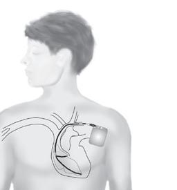
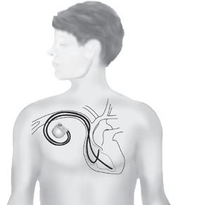
Pacemaker or ICD
If you are at high risk (for example if you have already had a cardiac arrest), or if drugs have failed to control your symptoms, your doctor may advise you to have a pacemaker or an implantable cardiac defibrillator (ICD) fitted, as well as taking your medication. A pacemaker and an ICD both consist of an electronic box that is inserted under the skin and attached to the heart by special electrical ‘leads’. A pacemaker controls the heart rate and stops any excessive slowing of the heart that could trigger an arrhythmia. An ICD acts in the same way as a pacemaker but it can also identify any dangerous arrhythmias and deliver an electrical shock to reset the heart. (For more information on pacemakers see page 31, and for more on ICDs see page 30.)
Surgery
Another option is to perform surgery to disrupt the nerves that release adrenaline and related chemicals at the heart. This is known as ‘cervical sympathectomy’ and involves operating on the left side of the neck. For more on this see page 29.
As it would appear in one lead in the following circumstances:
A: Normal
B: LQTS
C: Brugada Syndrome
The ‘P’ wave reflects the electrical activity of the pacemaker and atrium while the ‘QRS’ reflects the electrical activity of the ventricle pumping. The ‘T’ wave represents the ventricle resetting itself ready for electrical activation again. This is also known as ‘repolarisation’ and is measured using the ‘QT’ interval (the beginning of the QRS to the end of the T wave). In B the repolarisation is prolonged and hence so is the QT interval. In C the area of the ECG tracing connecting the end of the QRS and the T wave (the ST segment) is elevated abnormally with some broadening of the QRS itself (right bundle branch block).
Brugada syndrome
This condi on was first iden fied in the early 1990s. It is an uncommon condi on in the western world but seems to be much more common among young men in South East Asia. In the western world it affects mainly young and middle-aged adult men. It has been associated with muta ons in the same sodium channel that is affected in LQTS, but this appears to account for only 1 in every 5 people with the condi on. The sodium channel behaves abnormally in that movement of sodium ions into the cells is restricted. (See the Brugada syndrome diagram on page 9.) This results in par cular changes on the ECG (as shown in the diagram on the le - figure 3C) but no abnormali es in the structure of the heart.
What are the symptoms?
Some people with Brugada syndrome may have no symptoms at all. In others, the most common symptoms are blackouts. Some people may no ce palpita ons due to ectopic beats. Sudden death may occur. If it does, it usually happens while the person is sleeping or doing very li le.
Are there any physical signs?
There are no associated physical signs.
How is it diagnosed?
Diagnosis involves having an ECG. The changes characteris c of Brugada syndrome may appear on the ECG con nuously or intermi ently, or they may not show at all. Some mes the presence of a fever can bring out the ECG changes. If they do not show up on the ECG, there are tests that can make the ECG changes visible. These are called ‘provoca on tests’ and involve having a short injec on of an an arrhythmic drug while you are having an ECG (see page 19). The drugs most commonly used for this are ajmaline and flecainide. There is some controversy, however, about how much reassurance a nega ve result should give. Researchers have found that, in some carriers who have already been iden fied by gene c tes ng, changes on the ECG are not seen even with a provoca on test. However, in these people the level of risk does appear to be low.
Gene c tes ng is not very useful for diagnosing Brugada syndrome because muta ons have been found in only a small propor on of people known to have the syndrome.
Treatment and advice
The outlook for people with Brugada syndrome can be poor in people who get symptoms or have already had a cardiac arrest, the highest rates of sudden cardiac death being found among young male adults and par cularly those who originally come from South East Asia. It is therefore standard prac ce for high risk carriers to
have an ICD fi ed as this is very successful form of protec on. For more informa on on ICDs see page 27. To date medica on has not been shown to be protec ve in people with Brugada Syndrome although there is research into this possibility.
Unfortunately it can be very difficult for doctors to decide how to treat those people who do not get symptoms but who have an abnormal ECG. An EPS (an electrophysiological study) may help to iden fy those people who do or do not need an ICD but its usefulness is increasingly disputed . Research has suggested, however, that adults with normal ECGs and no symptoms have such low risk that an ICD should not be needed. It is highly unusual for children to be at high risk. Carriers are advised to take fever lowering drugs such as paracetamol or ibuprofen due to the poten al effect of fever on the ECG. They are also advised to avoid certain medica ons and low blood potassium levels, known as hypokalaemia (see General Lifestyle Advice on page 24).
CPVT (Catecholaminergic polymorphic ventricular tachycardia)
CPVT is a rare condi on found in young people and children. It causes a par cular type of arrhythmia. It has been associated with two genes that make proteins found inside the cell - the human ryanodine receptor (a calcium ion channel) and calsequestrin (a protein that interacts with the channel). These regulate the release of calcium ions into the rest of the cell. If these do not func on normally, the level of calcium inside the cell becomes too high, resul ng in the arrhythmias characteris c of CPVT.
What are the symptoms?
Some people with CPVT have no symptoms at all. Others may have blackouts. Sudden death may occur while the person is exer ng themselves or suffering emo onal stress. The condi on can affect children and seems to cause more blackouts in males than in females.
Are there any physical signs?
There are no physical signs.
How is it diagnosed?
The diagnosis is usually made a er the chance recording of arrhythmias that are characteris c of CPVT, while the person is doing exercise. Gene c tes ng is useful to help make a diagnosis and in cases where a member of the same family has already been found to carry a muta on and is showing the signs of the condi on.
Treatment and advice
Your doctor will advise you to take beta-blockers (a type of drug) and to restrict the amount of exercise you do. This combina on greatly improves the outlook for people with CPVT. About 1 in every 3 people with the condi on will also need to have an ICD
fi ed. (For more on ICDs see page 30).
PCCD (Progressive cardiac conduction defect)
PCCD is a rare condi on. In people with PCCD, the heart’s electrical impulses are conducted very slowly and this results in the gradual development over me of ‘heart block’. (Heart block is a failure of the heart’s electrical impulse to conduct properly from the top chambers [the atria] to the bo om chambers [the ventricles]. The severity of the condi on and its associated risk can vary.) PCCD can cause arrhythmias - either because the heart’s rhythm is too sluggish (bradycardia and asystole), or because of rapid rhythm disturbances (tachycardia) arising from parts of the heart that have escaped normal regula on. In some people PCCD has been associated with sodium channel muta ons that cause changes in channel behaviour similar to those found in people with Brugada Syndrome (see the Brugada Syndrome diagram on page 9figure 2C).
What are the symptoms?
Dizziness and blackouts are the usual symptoms. Sudden death may also occur.
Are there any physical signs?
There are no physical signs.
How is the diagnosis made?
The ECG abnormali es may be detected either on a standard ECG or with Holter monitoring. An electrophysiological study may also help the doctor make a diagnosis. (We describe all these tests on pages 17-23). If a sodium channel muta on is iden fied in affected members of a family then it may also be found in other rela ves.
Treatment and advice
If you have PCCD you will need to have a pacemaker fi ed in order to stop dangerous bradycardia from occurring. This may not prevent ‘escape tachycardias’ so you may also need to take an arrhythmic drugs. Some people may need to have an ICD fi ed instead of a pacemaker. (For more on pacemakers see page 31, and for more on ICDs see page 30.) Medica on alone does not help.
Early repolarisation syndrome
Early repolarisa on is seen on the ECGs of around 5% of all normal people but i t has recently been associated with an increased risk of sudden death. In par cular some survivors of a cardiac arrest who have no signs of structural heart disease, long QT and Brugada syndromes or CPVT , have clear early repolarisa on changes on their ECG that have worsened drama cally immediately before their cardiac arrest. This has been described as early repolarisa on syndrome and in a few cases muta ons of potassium and calcium ion channel genes have been found. The diagnosis and treatment of this condi on is s ll unclear but an ICD is needed in pa ents who have suffered a cardiac arrest.
Sodium channel disease
There are specific sodium channel muta ons that can cause Long QT Syndrome, Brugada Syndrome and/or PCCD in the same family. They can be diagnosed and treated as described above and can be iden fied by gene c tes ng.
Short QT syndrome
Short QT intervals are seen on the ECGs of some families and can be associated with blackouts and sudden death due to ventricular arrhythmias. It is a very rare condi on and as in long QT syndrome, the potassium channels are affected, except that too much potassium is allowed to flow out of the cells. If you have short QT syndrome, your doctor may prescribe quinidine or even an ICD to treat your condi on.
Structural heart disease
In some cases, the pathologist cannot confirm a diagnosis of structural heart disease - either because there is no evidence of it, or because there is not enough evidence and the heart is felt to be relatively normal. So the death will be recorded as SADS. This may happen even in cases where evidence of inherited structural heart disease is subsequently detected in other members of the victim’s family. The presence of very subtle structural heart disease in the victim may, however, have been enough to cause sudden cardiac death.
In these circumstances the most common causes of death are:
• arrhythmogenic right ventricular cardiomyopathy (ARVC)
• dilated cardiomyopathy (DCM)
• hypertrophic cardiomyopathy (HCM)
• mitral valve prolapse (MVP)
• Wolff-Parkinson-White Syndrome (WPW).
We explain each of these on pages 5-6.
If you are a close blood relative of someone who has died of SADS Why you need to have tests?
If you are a close blood relative of the person who has died of SADS, it is important that you have tests to find out if you have inherited the same medical conditions as the SADS victim. We describe all the tests on pages 17-23. If there have been any sudden or suspicious deaths in your family, including cot death, this further suggests that there may be an underlying inherited condition.
What if nothing is found in your family?
In families where someone has died of SADS and the remaining family members are tested, about 4 in every 10 families show no sign of inherited heart disease. This can be due to two reasons:
1 The SADS victim may not have inherited any abnormality from his or her parents. Either the victim was the first to suffer a mutation in the family (and therefore the victim’s children are the only relatives who are at risk), or there was another cause which has not been identified and which is not inherited.
2 Some family members may be carriers but show no signs of any disease. It is impossible to give 100% reassurance that a relative is not a carrier except in cases where a mutation has been identified in the person who has died and the victim’s relative is genetically tested to see if he or she has the same mutation. However, people who do not have any symptoms or signs are at low risk of sudden death, so in these cases the doctor can give some reassurance. The people at highest risk are those who have symptoms, or have already had a cardiac arrest, or have significant abnormalities on their ECG.
In the meantime, there is little evidence that repeated testing of relatives of someone who has died of SADS is helpful - unless the relative develops new symptoms, or the technology for detecting these conditions improves.
What if something is found in your family?
If you are a relative of someone who has died of SADS and you have been diagnosed with one of the conditions described in ‘What causes SADS’, you will need regular follow-up - whether you receive treatment or not - unless your doctor believes that you are at very low risk.
If you are young and have been tested, and your doctor thinks that you are not affected, you should have another review in the future. This is because ECG changes can become more obvious with age in children and young adults or they may show up on some ECG recordings but not on others. So, for example, children will need some follow-up until they pass through puberty.
If a recognised mutation was found in the person who died of SADS (or if another relative with signs of inherited heart disease is found to carry one), and if you are found not to have the mutation, then you can be fully assured that you are not affected.
Tests
Because the conditions that cause SADS can be inherited it is important that, if you are a blood relative in the immediate family of someone who has died of SADS, you are evaluated for signs of these diseases, particularly the ion channelopathies. There may also have been other sudden or suspicious deaths in your family, including cot deaths, suggesting that there may be an underlying inheritable condition. Below, we explain what is involved in the evaluation and describe the tests you may need to have.
Molecular autopsy
When and if DNA from the SADS victim has been kept from the post-mortem it can be tested for mutations that cause genetic heart disease. This may help in 2 ways. Firstly, it can confirm that a mutation found in blood relatives caused the sudden death of the SADS victim. Secondly, if a comprehensive range of genes is tested then the genetic test may be able to diagnose the actual cause of death. This currently only happens in a research setting but may be available routinely in the future.
Medical history
It is vital that a clear history of the victim and his or her death is established, using the family’s and friends’ recollections as well as the reports of the coroner, pathologist, GP and police. For example, fits brought on by exercise can be due to an underlying channelopathy such as LQTS or CPVT, or a sudden cardiac death during sleep may have been caused by sodium channel LQTS or Brugada Syndrome. It is also important to find out about any medications and any potentially dangerous drugs that the person may have taken before they died. Your doctor may ask you if you have ever had symptoms such as blackouts or palpitations as these may suggest underlying heart disease.
Medical examination
A medical examination may help to discover if there is an inheritable structural heart disease in the family. For example, if there is mitral valve prolapse with leakage from the valve this will cause a ‘murmur’ that a doctor can hear through a stethoscope.
Your doctor may suggest that you have some of the tests we describe below.
ECG (electrocardiogram) *
This is the most basic test. It involves taping electrical leads onto your legs, arms and chest to take readings of the electrical activity of your heart. These are printed out onto a piece of paper for the doctor to examine. If the first ECG does not show
any sign of a channelopathy, the test can be repeated later.


Electrical leads from the ECG machine arte taped to the chest, legs and arms and a recording is made of the electrical activity of the heart.

Signal averaged ECG *
This is an ECG that adds together the electrical readings from at least 250 heartbeats so that any very subtle variations can be seen - for example if the electrical impulses in the heart are being conducted more slowly. It is useful for diagnosing Brugada Syndrome, PCCD or ARVC.
Echocardiogram * (Also called an ‘echo’.)
This test uses ultrasound waves to look at the structure of the heart. It is useful for people whose ECG shows changes that could be caused either by a channelopathy or by uninherited heart disease that has damaged the heart - for example a previous heart attack that you may not have even been aware of. An echocardiogram can also detect inheritable conditions such as cardiomyopathy and mitral valve prolapse.
The operator puts some clear gel on your chest and then places an ultrasound probe on it. The probe sends ultrasound beams into your body and their reflections are detected and used to generate images of the heart. You can see different parts of your heart on a screen as the probe is moved around on your chest. The test is similar to the ultrasound scan that is used to examine a pregnant woman’s unborn baby. It is completely painless.
Exercise test * (Also called an Exercise ECG.)
This test is the same as the ECG described on page 17 but is recorded before, during and after a period of time spent exercising on a treadmill or an exercise bike. This allows the doctor to examine any changes in the electrical patterns that occur with exercise, and analyse any abnormalities. This test is particularly useful in detecting some of the features that are characteristic of LQTS or CPVT.
Figure 6: Exercise Test
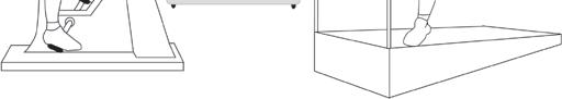
Electrical leads from the ECG machine are taped to your body and you are monitored while you exercise either on an exercise bike or treadmill. If you are having a ‘cardiopulmonary exercise test’, your doctor will ask you to breathe in and out of a special piece of equipment while you are doing the exercise, in order to monitor how efficiently your body uses oxygen.

Cardiopulmonary exercise test
Some hospitals may also ask you to do a cardiopulmonary exercise test. This test analyses the efficiency of the heart muscle by measuring the amounts of oxygen your body uses during exercise. You will be asked to breathe into special equipment while you are exercising. If the efficiency of your heart is low, this may suggest that you have cardiomyopathy (inefficient pumping action of the heart).
Holter *
The Holter is a recording device that comes in two different forms:
• a small portable tape recorder (like a walkman), or
• a small digital device the shape of a pager.
You wear the device on a belt round your waist. Four or six ECG leads from the
device are taped to your chest. The device records the electrical activity of your heart for 24 to 48 hours, or for up to 7 days if a digital one is used. The doctor can then analyse the electrical activity and rhythm of your heart to find out if you have any arrhythmias (for example, the arrhythmias typical of LQTS and CPVT), or some of the other features characteristic of LQTS.
Cardiomemo and event recorder *
Figure 7: Holter
The Holter monitor is attached by 4 or 6 electrical leads to your body. It monitors your heart’s electrical activity over a period of time.
These are more sophisticated versions of the basic Holter. Whenever you have an attack of symptoms, you can activate the device to record your heart’s rhythm. (You can also do this with the digital Holter.) The advantage of the cardiomemo is that it doesn’t have any leads, so you can just place it on your chest when you get symptoms, without having to put any leads in position.

Reveal© device
When it is difficult to assess or record a symptom because it only happens infrequently - as with blackouts - a Reveal© device can be used. The device, which is the size of a packet of chewing gum, is placed under the skin at the left shoulder. You will need to go into hospital as a day case to have this done. A small cut about 2 cm long (just under one inch) is made and the device is inserted. The device monitors the heart’s rhythm and can record any abnormal events that it is programmed to detect. If anything happens, a small box with a button can also be placed on the surface of the skin over the Reveal© device. The device may then be activated by pressing the button, causing it to record the preceding 15 minutes of the heart’s activity. The device can then be ‘interrogated’ by a computer at the hospital and the doctor can examine the recording. The device has a battery that can last up to two years if necessary.

Provocation tests (Ajmaline, flecainide, adrenaline and adenosine tests)
You may be asked to have this test if your doctor suspects Brugada Syndrome. While you are having an ECG test you will be given an injection of ajmaline or flecainide (antiarrhythmic drugs). The test may show changes on the ECG that are typical of one of the channelopathies.
A fine plastic tube is inserted into a vein at the front of your elbow. The drug is
injected over a short period of time (5-10 minutes) and you will be monitored for 20 minutes or a few hours afterwards, depending on the drug used. There is, however, a risk in 1 in 200 Brugada Syndrome carriers or their immediate blood relatives of causing a potentially life-threatening arrhythmia during the injection. The test is therefore always performed with appropriate facilities to protect patients from this risk. Ajmaline is preferable as it lasts a shorter period of time in the circulation.
Under the same circumstances adrenaline may be given to try and diagnose the long QT syndrome or CPVT while adenosine (another short-acting chemical) is given if Wolff-Parkinson-White Syndrome (WPW) is considered a possible diagnosis.
Cardiac Magnetic Resonance (CMR) scan *
This is a special kind of scan used to examine the structure of the heart and the nature of its muscle. It uses a Magnetic Resonance scanner that creates intense fluctuating magnetic fields around your body while you are inside the scanner. This generates the signals that make up the pictures produced. It may be useful for detecting the presence of fat and scarring in the heart muscle that is associated with ARVC.
Other tests
Coronary angiography and electrophysiological study (EPS)
Depending on the results of the above tests, your doctor may suggest that you have other tests such as coronary angiography or an electrophysiological study (EPS). Both these tests are performed in an X-ray laboratory that allows the body and any medical tools (such as cardiac catheter tubes or pacing wires) to be seen using an X-ray camera. You will be asked to lie down on a special moving table and will be given a local anaesthetic in your groin. The doctor will then place fine tubes, called cardiac catheters or electrodes, into blood vessels in your groin. These are gently passed through to the heart.
During coronary angiography the coronary arteries (the arteries that supply blood to the heart muscle) are injected with a dye to reveal any furring or blockages - coronary artery disease. (The ECG changes that are characteristic of Brugada Syndrome or LQTS can sometimes be caused by coronary artery disease.)
An EPS (electrophysiological study) involves placing electrical leads inside the heart to analyse its electrical properties and induce arrhythmias. It may be useful in diagnosing Wolff-Parkinson-White Syndrome (WPW) and PCCD and deciding on what treatment to give people with Brugada Syndrome. If the extra pathway seen in WPW is detected at EPS it can be treated there and then by ‘burning’ it away using high frequency radio waves. This procedure is called ‘RF ablation’.
There are other tests that may be used to provoke ECG features in LQTS such as ‘cold pressor tests’. A stimulus such as placing your hands in ice-cold water can bring out the ECG features of the condition. This does not appear to increase significantly the likelihood of making a diagnosis but is still used at some centres.
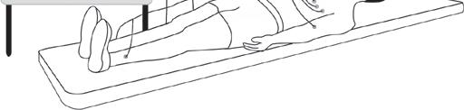
Tilt-table testing
Tilt-table testing is used to identify other common conditions that can cause blackouts - such as Vasovagal Syndrome (see page 33) or simple fainting - that tend to particularly affect young women and girls but have a very low risk of causing sudden death. These symptoms are very similar to the symptoms of more rare and potentially life-threatening conditions like the channelopathies, so it is important to discover the cause of the blackouts so that the doctor can give appropriate treatment. While you lie flat on a table, your blood pressure, pulse and ECG are monitored. The table is then tilted to an angle of 60 to 75 degrees and monitoring is continued. If nothing happens, a spray of a substance called GTN is given under your tongue as a stimulus and you will be monitored for another 10-15 minutes. The table will then be returned to the flat position and the leads disconnected. The whole test takes around 45 minutes. If your blood pressure falls at the same time as you suffer your usual symptoms, this means that you have Vasovagal Syndrome or a related condition.
Figure 8: The tilt table test

The tilt table test involves monitoring the ECG, pulse and blood pressure while you are lying flat on a table, then when the table is tilted to 60-75 degrees, and then lying flat again.

Genetic testing
In most of the inherited conditions known to cause SADS, mutations of specific genes have been detected and are thought to cause a specific disease. So in principle, if we could identify these mutations, we would be able to make a diagnosis in any DNA sample including any obtained from SADS victims at their autopsy or from their relatives who have given blood. Unfortunately this cannot be done at the moment because we don’t have complete knowledge of all the genes involved in any condition. For example, 7 in every 10 people known to have LQTS have mutations of known identified genes. Also, many variations in the DNA code are found in a large number of people and do not necessarily cause any disease. Many families with LQTS have mutations specific to them (‘private’ mutations) which can also make it difficult to decide whether it is the mutation that is causing the disease or not. As research progresses, more genes will be identified and there will be better tools to decide whether the impact of a mutation causes a disease.
* Tests marked with a * are non-invasive.
‘Non-invasive’ means that it does not invove penetrating the skin or body.
 Alison Cox MBE CRY Founder