SPRING 2019
WHAT’S INSIDE: DINNER WITH A DOCTOR 18
IMAGING EVOLVES 22
FINAL RIGHTS 30

SPRING 2019
WHAT’S INSIDE: DINNER WITH A DOCTOR 18
IMAGING EVOLVES 22
FINAL RIGHTS 30
The Vega String Quartet, in residence at Emory, performs Mozart’s Divertimento in D major, K. 136 (1772) and other classical selections in the lobby of the medical school, an annual spring tradition.


We can use 3-D printing to craft medical devices and body parts, we can predict the onset of some diseases with big data, we equip patients with bionic limbs that can detect neuromuscular signals, we can perform robotic heart surgery, we can implant stimulation devices in the brain to diminish depression and epileptic seizures.


But as John Naisbitt foretold with the concept of “high tech, high touch” in his 1982 bestseller Megatrends, in a world of digitalized, computer-augmented reality, people long for personal, human contact.
In this as in every issue of Emory Medicine magazine, we showcase ways our medical teams are making others’ lives better, in research and in practice. You’ll learn about promising immunotherapies for treating cancer (p. 12), why your allergies might get worse the longer you live somewhere (p. 18), and what six Emory doctors suggest for end-of-life medical directives (p. 30).
While our physicians do indeed use the latest therapies and techniques, these are not the only tools in their doctor’s kit.
As Emory cardiologist Laurence Sperling says, in an article about advances in medical imaging (p. 22), effective clinical care requires “listening to, talking to, and touching the patient. We are taking care of people, not pictures of people.”
To view the digital version of Emory Medicine, with bonus content, go to emorymedicinemagazine.emory.edu.

Editor Mary Loftus
Art Director Peta Westmaas
Director of Photography Jack Kearse
Contributing Writers Janet Christenbury, Quinn Eastman, Jennifer Johnson, Sidney Perkowitz, Melva Robertson, Kellie Vinal, Sylvia Wrobel.
Production Manager Carol Pinto
Advertising Manager Jarrett Epps
Web Specialist Wendy Darling
Intern Laura Briggs 19C
Exec Dir, Editorial Karon Schindler
Associate VP, HS Communications Vince Dollard
Emory Medicine is published twice a year for School of Medicine alumni, faculty, and staff, as well as patients, donors, and other friends. © Emory University

Emory University isanequalopportunity/equalaccess/affirmativeaction employerfullycommittedtoachievingadiverseworkforceandcomplies withallapplicablefederalandGeorgiastatelaws,regulations,andexecutive ordersregardingnondiscriminationandaffirmativeactioninitsprograms andactivities.EmoryUniversitydoesnotdiscriminateonthebasisofrace, color,religion,ethnicornationalorigin,gender,geneticinformation,age, disability,sexualorientation,genderidentity,genderexpression,orveteran’s status.InquiriesshouldbedirectedtotheOfficeofEquityandInclusion, 201DowmanDrive,AdministrationBldg,Atlanta,GA30322.Telephone:404727-9867(V)|404-712-2049(TDD).
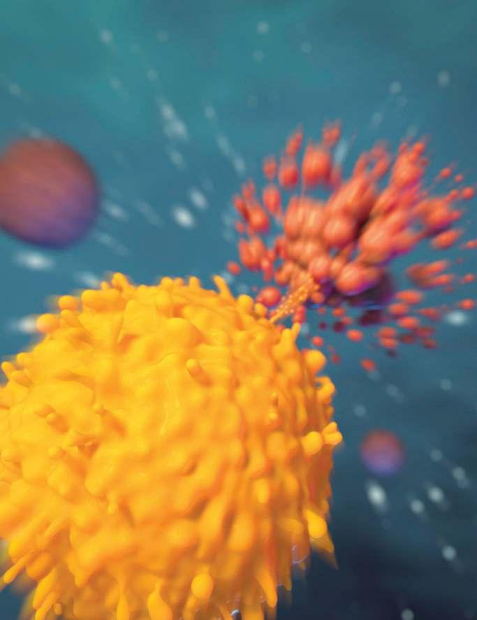
Education team leader, on the immunotherapy illustration on p. 12: “I always strive to be accurate, even when simplifying a complex cellular scene. Here, the T cell is releasing granules of perforins and granzymes in a ‘lethal hit’ to a cancer cell. The ‘explosion’ is a slightly dramatized depiction of apoptosis, or cell death. The contents of the cell don’t just spill out, but are left in smaller packages that can be cleaned up by other cells. The cells were hand drawn with 3D computer software, using micrographic images as reference.”
Cell Wars 12
PD-1 checkpoint inhibitors. CAR T-cell therapy. Cellular immunotherapy is gaining ground as an effective cancer treatment—and it works by revving up the patient’s own immune system.
DinnerwithaDoctor:SeasonalAllergy 18
Ah (or rather, Ah-choo), the joy and horror of springtime in Atlanta. Dr. Marissa Shams, allergist/immunologist, answers our group’s questions about seasonal inhalant allergies.

The Better to See You With 22

Medical imaging has evolved over the years, from shoe store X-rays to cancertracking nanoparticles. Take a trip through radiology’s dangerous past to its luminous present with physicist Sidney Perkowitz.
FinalRights:AdvanceDirectives 30
Most people don’t die where and how they want. But there are ways to make your voice heard near the end. Six Emory physicians talk about the importance of advance medical directives and share details of their own.

THE BARE BONES
Letters 4
Experts Weigh In 4
Briefs 5
AND MORE
Your Smartwatch 10
High-tech health tracker or talisman?
Medical Ethics 36
When a baby is being kept alive artificially and a parent must make the hardest decision.
18
“ I cried. Everybody [in my family] cried. But by day two, I was like, okay, I got sick. What are we going to do about it?”
—Ricardo Parks, Winship patient
Pet allergens can be isolated from pet dander, skin cells, saliva, and urine, all of which can trigger allergic symptoms.”
Thanks for giving me an opportunity to tell your creative team specifically how much I appreciated (still do!) the graceful writing and engaging photos in the piece about Dr. Vin Tangpricha [Emory Medicine, Autumn 2018, “Seen & Heard.”] Since he recently took office as WPATH’s president, being able to share this article with our global membership is a fantastic way of introducing him to health care providers around the world who are in various stages of their careers working with transgender patients. You really brought out his humanity and his dedication. Thank you so much!

 Jamison Green Communications director World Professional Association for Transgender Health (WPATH)
by Emily Sullivan in the Autumn 2018 issue of Emory Medicine magazine was a frightening story, yet the ending was a truly happy one thanks to Emory and Children’s Healthcare of Atlanta. Childhood is indeed both adventurous and dangerous. A serious accident such as this affects everyone, and the article presented a full picture of how the whole family and Dr. Andrew Reisner and his staff all came together to give Brayden a second chance at childhood.

 Edye Bradford Clayton, Georgia
and Nancy Writebol. I am very grateful that they are still in my life today, which is a direct result of the care they received at Emory.
Dr. Nathalie MacDermott Wellcome Clinical Research Training Fellow Imperial College, LondonWe like to hear from you. Send us your comments, questions, suggestions, and castigations. Address correspondence to Emory Medicine magazine, 1762 Clifton Road, Suite 1000, Atlanta, GA 30322; call 404-727-0161; or email mary.loftus@emory.edu
“Boyhood Disrupted”
“
“
For a very emotionally fraught conversation, telemedicine might not be the way to do it.”
Emorymedical ethicist John Banja, to FierceHealthcare.com, on the outcry about the use of livestreaming in a hospital to tell an elderly patient he was close to death.
This is not a Republican or a Democrat issue. This is an American issue. . . . We were in a state of inertia about HIV. Hopefully this will light a fire.”Emory professor of medicine and global health Carlos del Rio to Georgia Health News, about the new White House initiative to stop HIV/AIDS.
Unrelenting mental stress, whether caused by work or personal pressures, spurs the body to release hormones that can increase blood pressure and heart rate and constrict blood vessels, increasing the risk of heart disease.”
Emory cardiologist Laurence Sperling, director of Emory’s Preventive Cardiology program
Nearly five years after housing its first Ebola patient, the Serious Communicable Diseases Unit (SCDU) in Emory University Hospital continues to be of keen interest.
Four patients with Ebola virus disease were successfully treated in the SCDU by a highly trained Emory Healthcare team in 2014. Since, more than 60 groups—including infectious disease and public health experts, scientists, military and government officials, ministers of health, and students—have toured the high-level isolation unit.
In January, NBA Hall of Famer and philanthropist Dikembe Mutombo, a Congolese American who built a hospital in the Democratic Republic of Congo (DRC), toured the unit. The current Ebola outbreak in the DRC has infected more than 1,100 people and killed about 750. “I am delighted to see this facility I’ve heard so much about,” said the former Atlanta Hawks player. “We are about to launch a new lab at our hospital so I’m very interested in the attached clinical laboratory. “
Mutombo (above, and right) tried on a powered air purifying respirator under the direction of nurse Jill Morgan, who cared for the first Ebola patient in the unit. He also tried out virtual reality programs created with a 360-degree camera. The immersive experience helps prepare health care workers to care for patients with Ebola and other
infectious diseases—while wearing cumbersome protective suits. “The VR simulation does a good job of portraying your limited view and dexterity,” said infectious disease physician Aneesh Mehta.

In June, physician Ian Crozier accompanied a group from the American Society for Microbiology’s annual conference as they toured the unit where he spent 40 days recovering from Ebola virus disease. Crozier was the sickest of the four patients treated at Emory.
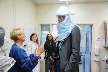
“As a physician, if you had told me that I would develop encephalopathy, respiratory failure, kidney failure, and cardiac arrhythmia, I would have expected my chances of survival to be almost zero,” Crozier says. “What happened inside those four walls changed the nature of how we treat Ebola patients.”
Emory’s efforts to prepare for future outbreaks have continued through research, improved patient care protocols, and training health care workers and first responders around the country.
“The government has called on Emory to care for patients with special pathogens in the U.S.,” says infectious disease physician Colleen Kraft. “We would like to take what we learned about preventing transmission of diseases in a biocontainment unit out into every health care setting and interaction.” Active drills continue for the SCDU staff, who must be prepared to reactivate the unit at a moment’s notice. n
The National Academy of Medicine (NAM) has elected Emory President Claire Sterk to its 2018 class of 75 leading health scientists and 10 international members.

A pioneering public health scholar, Sterk became Emory’s 20th president in 2016 and is Charles Howard Candler Professor of Public Health. Before becoming president, she served as the university’s provost and executive vice president for academic affairs.
Sterk’s work has focused on social and health disparities, addiction and infectious diseases (specifically HIV/AIDS), community engagement, and the importance of developing women leaders.
As a leader in higher education, she emphasizes the choices and responsibilities of research universities and their real-world impact. She is leading efforts to broaden research across the university, including enriched opportunities for student research. Emory’s new strategic framework includes a heightened focus on serving both Atlanta and global communities through enhanced research partnerships.
Membership in the NAM is considered one of the highest honors in the fields of health and medicine. Emory currently has 31 members.

The new Winship Center for Cancer Immunology, directed by immunobiologist Madhav Dhodapkar, aims to integrate basic science research, clinical trials, and immune-based therapies to guide the field forward.
Previously, says Dhodapkar, it wasn’t clear whether the immune system had anything to do with cancer. Now, with the majority of newly approved cancer drugs and active clinical trials centering on immune-based therapies, the proof of principle has been achieved.
“Cancer is a very complex disease,” says Dhodapkar, the Anise McDaniel Brock Chair and Georgia Research Alliance Eminent Scholar in Cancer Innovation and professor in hematology and medical oncology at Emory. “In retrospect, it should not be a surprise that to tackle a complex, adapting disease you need a complex, adaptive system.”
An expert in the treatment of multiple myeloma, Dhodapkar came to Emory last year from Yale Cancer Center, where he was co-director of the Cancer Immunology Program. Between Winship’s team of experts, the new cancer immunology center and Phase I Clinical Trials Unit, and a cell therapy facility currently in the works, Emory is aiming to make important advances in cellular immunotherapy.
“Each tumor has an Achilles’ heel from the perspective of the immune system,” Dhodapkar says. “Our job is to figure out what that is and to target it, as opposed to taking a one-size-fits-all approach. It requires a different way of looking at patients, as well as resources and infrastructure devoted to better understanding the immune system.”—Kellie Vinal (See related story on p. 12)
“Each tumor has an Achilles’ heel ... our job is to figure out what that is.”
Neuroscientists have discovered a focal pathway in the brain that, when electrically stimulated, causes laughter followed by a sense of calm and happiness.
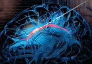
These effects were observed in a 23-year-old woman with epilepsy who was being monitored for a seizure diagnosis, and the technique was then used two days later to help calm her during brain surgery while she was awake.
The behavioral effects of direct electrical stimulation of the cingulum bundle—a tract of neural fibers in the brain—were confirmed in two other epilepsy patients undergoing diagnostic monitoring. Emory neurosurgeons see the technique as a potentially transformative way to calm patients during awake brain surgery.
For protection of certain brain functions, patients may need to be awake and not sedated during surgery so that doctors can talk with them, assess their language skills, and gauge when they are getting close to critical areas.
“Even well-prepared patients may panic during awake surgery, which can be dangerous,” says Kelly Bijanki, assistant professor of neurosurgery. “One of our patients was especially prone to it because of moderate baseline anxiety. Upon waking from global anesthesia, she did indeed begin to panic.
ActorLukePerry’srecentdeathat 52fromastrokereceivedalotof attention,largelybecauseastrokeis thoughtofasaconditionthataffects olderpeople.Butdoctorsarereportingajumpinyoungerstrokepatients, between35and55,whowereunaware theyhadunderlyingproblemslikehigh bloodpressure.Aboutone-thirdofpeoplehospitalizedforstrokeareyounger than65.AtGradyHospital’sMarcus StrokeandNeuroscienceCenter,about 1,200patientsayeararetreated.

“Bloodpressure,Ialwayssay,isrisk factorone,two,andthreeforstroke,” saysAaronAnderson,assistantprofessorofneurologyatEmory.“Ifwewere
When we turned on her cingulum stimulation, she immediately reported feeling happy and relaxed, told jokes about her family, and was able to tolerate the awake procedure successfully.”
Understanding how cingulum bundle stimulation works could lead to better ways to treat depression, anxiety disorders, or chronic pain via deep brain stimulation. The stimulation, says doctors, immediately provoked “mirthful behavior, including smiling and laughing, and reports of positive emotional experience.”

“The patient described the experience as pleasant and relaxing and completely unlike any component of her typical seizure or aura. She reported an involuntary urge to laugh that began at the onset of stimulation and evolved into a pleasant, relaxed feeling.”
The cingulum bundle (depicted above during stimulation by Emory medical illustrator Bona Kim) lies under the cortex and curves around the midbrain like a girdle or belt —hence its Latin name. It links many brain areas together, like a “superhighway with lots of on and off ramps,” says Jon Willie, assistant professor of neurosurgery and neurology at Emory, who performed the surgeries.
The area that appears key to laughter and relaxation lies at the top front of the bundle.—Quinn Eastman
abletonotonlydiagnosebuttreathigh bloodpressure[inall],wecouldprevent probably60percent ofstrokes.”
Otherriskfactors includediabetesand highcholesterol.To furtherlowertheirrisk, peopleshouldstayat ahealthyweight,get regularexercise,and stopsmoking. Symptomscanbe rememberedbythe remindertoActFAST: Facialdrooping,Arm weakness,Speech
difficulties,Timetocall911.Evenif symptomsgoaway,itdoesn’tmeanthe dangerhaspassed.
Atransientischemic attack(TIA)isastroke thatlastsonlyafew minutes,whentheblood supplyisbrieflyblocked, butmedicalcareshould stillbesoughtimmediately.“ATIAcanbea warningofalargestroke andstrokephysicians canperformariskfactor evaluationtohelpprevent adevastatingstroke,” Andersonsays. n
Prenatal exposure to alcohol can result in abnormalities in the fetus’s developing heart.
Indeed, half of all children with fetal alcohol syndrome have congenital heart defects, such as arrhythmias or structural abnormalities.
Chunhui Xu and colleagues recently published a paper in Toxicological Sciences on how human cardiac muscle cells, derived from induced pluripotent stem cells, can be used as a model for studying the effects of alcohol.
Alcohol-induced cardiac toxicity is usually studied in animal models, but human cells are different, and a cell-culture based approach could make it easier to study the effects of alcohol and possible interventions more easily.
Xu and her colleagues observed that high levels of alcohol did damage cardiac muscle cells and put them under oxidative stress. Even at relatively low concentrations of alcohol, the researchers saw disruptions in cells’ electrical activity and the ability to contract, which reasonably matches the effects of alcohol on human heart development.
The lowest level tested was 17 millimolar—the legal limit for driving in most states (0.08% blood alcohol content).
 Quinn Eastman
In a survey of parents in pediatric ERs, most believed their children would be able to tell the difference between a toy gun and a real gun—as did the children themselves.

But when Emory physician Kiesha Fraser Doh, assistant professor of emergency medicine in pediatrics, and colleagues asked children 7 to 17 to distinguish a real weapon from a toy one, only 41 percent made the correct choice.
Also, they found, only a third of parents who own guns follow the American Academy of Pediatrics’ guidelines for safe gun storage: Keep weapons locked, unloaded, and separate from ammunition. “The majority of parents are storing their firearms insecurely, and children cannot tell the difference between a real gun and a toy one,” Fraser Doh says. “It behooves pediatricians to continue to educate families on how to store firearms safely, and is a reminder to parents to check on how firearms are being stored in their own home and in homes their children are visiting.” Holly Korschun

If Alzheimer’s disease is, as yet, incurable, what is the use of getting yourself or a family member diagnosed?
“Early diagnosis is in fact, even without a treatment, very important,” says neurologist Allan Levey, director of the Goizueta Alzheimer’s Disease Research Center at Emory. About 1,000 people a year come to the center to be evaluated for the disease.
Most Americans living with Alzheimer’s—or related dementia disorders—never receive a diagnosis, and even if they do it is usually several years into the illness since memory loss is often seen as a normal part of aging.
About 5.7 million Americans age 65 or older are living with Alzheimer’s or related diseases. By 2060, it is estimated that will increase to 13.9 million Americans.
Risk factors: age, genes, head injury, vascular disease, diabetes, hypertension, or high cholesterol, sedentary lifestyle.
Factors that might reduce the risk: exercise, healthy diet, mental stimulation, and control of high blood pressure, heart disease, and diabetes.
Alzheimer’s is the most expensive disease in America, costing $18 million an hour. The costs to families and society total more than $277 billion a year, with Medicare and Medicaid picking up the vast majority of expenses.
Many patients list a penicillin allergy on medical forms, but often these allergies are unconfirmed or low-risk.
Early diagnosis allows someone to:
n make their choices for the future known, before the disease progresses
n put financial and legal plans into place and draw up and sign legal documents
n decide on living arrangements for now and the future, with increased levels of assistance
n reduce the chance of elder abuse
n avoid unnecessary hospitalizations and procedures.
Patients are encouraged to first see a primary care physician for a medical exam, blood work-up, and a simple memory screening. This physician can determine if the patient’s memory loss is due to Alzheimer’s or another condition, such as a vitamin deficiency or improper medications.
At the Goizueta Alzheimer’s research center, or one of five satellite centers, doctors review patient symptoms, changes in symptoms, past medical conditions, and family history. The patient will have another physical examination, possible blood work, a memory assessment, and a brain scan. If a diagnosis of Alzheimer’s is made, the doctor, patient, and patient’s family sit down to come up with a treatment plan.
Patients can volunteer to participate in clinical trials and patient registries. A network of NIH-funded Alzheimer’s disease research centers, including Emory’s, is working to find a treatment for the disease.
“Ideally, we would be able to prevent it, and the first prevention trials are under way,” says Levey, chair and professor of neurology at Emory. “We’re trying to develop the next generation of biomarkers that can improve diagnosis to see if we can slow down or prevent the disease.” n
Emory researchers say that 10 percent of hospitalized patients report a penicillin allergy, but recent studies indicate that approximately 98 percent of these patients are not acutely hypersensitive or are actually tolerant of penicillin. In fact, according to the American Academy of Allergy, Asthma & Immunology, most people lose their penicillin allergy over time—even patients with a history of severe reaction such as anaphylaxis (a potentially life-threatening allergic reaction).
Emory researchers are trying to combat penicillin allergy labels in patients in these categories. They have determined that a direct oral amoxicillin (a penicillin antibiotic) challenge in the doctor’s office, without prior skin testing, is acceptable in patients with unconfirmed or low-risk penicillin allergy. Their findings were published in the journal Allergy & Asthma Proceedings.
“Unconfirmed penicillin allergy has emerged as a public health issue, and an evaluation of penicillin allergy labels is recommended to improve antibiotic stewardship,” says Merin Kuruvilla, assistant professor of pulmonary, allergy, critical care, and sleep medicine. Other Emory researchers in the study include assistant professors of medicine Jennifer Shih and Kiran Patel, and internal medicine resident Nicholas Scanlon.
“Penicillin is a very effective drug used to treat many different illnesses, yet many people avoid it because of their reported allergy,” Kuruvilla says. “Our research should help patients determine if their penicillin allergy exists, and if not, they can remove it from their listed allergies in their health record.”—Janet Christenbury
 BY Michelle Au AND Andrew Bomback
Think of the stereotypical representations of medicine, as they might appear on a television show: the crisp white coat, of course, and the stethoscope dangling at the ready. Syringes and intravenous lines, maybe. An X-ray or a CT scan slammed theatrically into a light box.
But any medical scene is incomplete without an electrocardiogram (EKG) machine running in the background, its jagged line tracing across the screen.
The EKG is the backbeat of many hospital scenes on television. Important medical things are happening here, it says.
To tap into that potent association, many private medical practices, urgent-care clinics, community hospitals, technology companies, and health care-product designers use EKG imagery in their advertising.
Most of those images bear little resemblance to actual EKG tracings. The spikes and bumps generated for signs or emblems (like the logo of the daytime talk show The Doctors, for example) mostly amount to arbitrary peaks and valleys. They do not reflect the output of a human heart, healthy or diseased.
But accuracy might be less important than allegory. Like the white coat or the caduceus, the EKG has become talismanic, more valuable for
the symbolism it provides than any diagnostic information it can convey.
Now that EKGs are making their way into smartwatches, their symbolic purpose could risk overtaking their medical one.
Wearable medical technology promises a new, better way to manage personal health.
Whether it’s Fitbits counting steps and calories burned, blood glucose monitors aiding insulin dosing for diabetic patients, or Bluetooth earpieces offering round-the-clock heart rate and body temperature tracking, wearable devices sell the promise of the coldly clinical made portably intimate.
Intermittent EKG monitoring, like that available in smartwatches, might seem like a small technological leap, putting what was once the sole purview of hospitals and doctor’s offices neatly around a consumer’s wrist.
But EKG monitoring is a little different from other, more discrete medical information.
Unlike devices that measure more cleanly numerical metrics—step counts or target heart rates or blood glucose levels—a wearable EKG display doesn’t give the user an easy sense of hitting targets or falling short.
Reading an EKG tracing is nuanced and interpretive, more art than math. A Fitbit gives you a number. An EKG paints a picture.
The 12-lead EKG, the gold standard of the diagnostic, measures the flow of current from 12 points on the patient’s body, offering a 360-degree view of the heart’s electrical activity. Its tracing reports the patient’s heart rate, rhythm, and regularity.
Because the various parts of the heart produce different shapes of electrical activity owing to their size and muscularity, the EKG can also
The wearable EKG offers the comforting weight of medicine itself, worn on the wrist like an amulet warding off evil.”
detect which chambers are beating at what time, and whether these chambers are correctly synced up and beating effectively.
The larger a muscle is, the stronger its electrical impulses, so the size of an EKG wave can also indicate whether parts of the heart muscle are enlarged or dangerously thickened.
The most urgent diagnostic use of the device determines the presence and location of cardiac damage due to decreased blood flow.
Areas of the heart getting less oxygen will show changes in their electrical conduction, and the 12-lead EKG provides real-time information: not just indicating whether a patient is having a heart attack, but also which coronary vessels are most likely blocked.
The 12-lead EKG can also detect the location of scarring left behind by prior, sometimes silent heart attacks. An EKG tracing will clearly show an area of dead heart muscle no longer conducting electrical signals. Dead meat don’t beat, as cardiologists put it.
Still, there’s plenty that a snapshot EKG can’t do, including diagnosing intermittent problems with rhythm or changes that only occur with certain activities.
An EKG doesn’t capture the shape or function of the heart’s valves, nor can it diagnose precarious plaques in the coronary arteries that could signal heart attacks waiting to happen.
The fewer number of leads an EKG has, the less information it can give you.
A one-lead EKG, such as the kind that appears in some smartwatches, gives just a single vantage point. For the diagnosis of some cardiac abnormalities, that’s akin to solving one side of a Rubik’s Cube. A watch endowed with this kind of EKG feature likely won’t
have a large public health impact.
By incorporating an EKG into smartwatches and their marketing, electronics makers use EKG to brand their watches in the same way that health care providers use EKG to establish trust in their clinics, or television shows use EKG to communicate medical professionalism.
An EKG feels weighty and complex, standing in for doctors and their expected acumen. Don’t worry, the purveyors of an EKG are communicating, we’ve got this.
needed a ready supply of water to cool its massive electromagnets.
The machine was virtually impossible to use and its readouts equally difficult to interpret, yet it became a near-immediate sensation in the medical world, and Einthoven won the Nobel Prize in Medicine for the contribution.
The modern 12-lead EKG is more streamlined, and the future (if the current crop of wearables is any indication) promises further downsizing of the device, with even more potential for unobtrusive integration into daily life.
Whether it is room- or wristsized, the EKG has always attempted to record the size, shape, and timing of the heart’s intrinsic electrical impulses, giving people the ability to review, track, and potentially intervene on its functions.
Put differently, the EKG tries to reframe the heart as a machine that can be monitored and, to some extent, mastered.
Shrinking and wearing an EKG is a symptom of technology’s drive to subsume health and wellness, and it renews a belief in humanity’s mastery of the heart, that most important muscle.

EKGs might start to seem capable of producing meaning on their own, instead of producing pictures to be interpreted by medical professionals.
The history of the EKG has always pitted cutting-edge medical advances against the placebo of quasi-medical reassurance.
The first electrocardiograph was developed in 1901 by Dr. Willem Einthoven, a Dutch cardiologist.
Einthoven’s original “string galvanometer” weighed about 600 pounds, required five skilled operators, and
That aligns the single-lead, wearable EKG with the set designer’s intentions for the symbol.
The EKG—especially the 12-lead device—offers real diagnostics, but not nearly as often as its traces convey the symbology of health.
The wearable EKG offers the comforting weight of medicine itself, worn on the wrist like an amulet warding off evil, whether it ever gets used or not. n
 BY Kellie Vinal n ILLUSTRATION BY Michael Konomos
In early 2016, Ricardo Parks had been experiencing stomach aches and unexplained vomiting spells for a few months. A local doctor prescribed tablets for an upset stomach, but by March, things had gotten even worse. “Anything I ate, I threw up,” he says.
He lost weight, developed a sore throat, and lost his voice. Parks decided to take action. He enrolled in a health insurance marketplace plan just before the deadline, called around to see what providers took his new insurance, and—mostly by chance—found himself in an Emory GI clinic.

Physician assistant Susan Rubio noticed a swollen lymph node on Parks’ neck from across the exam room. As troubling as his digestive and upper respiratory symptoms were, she told him, “That lump on your neck is what you should be worried about.” Enlarged lymph nodes are often a telltale sign that something is wrong.
Parks lives in Snellville, Georgia, but grew up in the Caribbean, where he says there’s a home remedy for almost every ailment. “In the Caribbean, we get bumps all the time!” he says. “You get a bump, you just brush it off and keep going.” Rubio ordered a biopsy of his lymph node and within a week, the results came back: it was stage IV cancer. Parks was stunned. “I cried,” he says, “everybody [in my family] cried. But by day two, I was like, okay, I got sick. What are we going to do about it?”

colon, which needed to be removed as quickly as possible. A genetic test revealed that he had microsatellite instability, which is associated with Lynch syndrome. It meant one of the DNA repair genes he inherited—that typically acts like a “spell check” as DNA replicates and divides—was faulty. This explained his unexpected diagnosis at such a young age.
Under the guidance of Joshua Winer, surgical oncologist at Emory’s Winship Cancer Institute, and Bruce Goldsweig, medical oncologist at Winship, Parks underwent a colostomy procedure to remove the tumor, started chemotherapy, and navigated complications with his chemo port. “I never knew it was going to be that rough,” says Parks. “Chemo, you know, it’s
killing everything. It’s like a scattershot.”
Despite the challenges, by November 2016, he was in remission.
Then, in January 2017, Parks went in for a follow-up CT scan and the doctors found another lump. The cancer was back. This time, five months of chemotherapy didn’t work. Despite the grim prognosis, Parks kept up his spirits. “I was lucky, no, I was blessed, to meet the right people,” he says.
Bassel El-Rayes, associate director for clinical research at Winship, who specializes in translational research for gastrointestinal cancers, works at the cutting edge of treatments for patients like Parks.
El-Rayes has seen patients with microsatellite instability respond very well to PD-1 checkpoint inhibitor drugs, which take the brakes off the immune system by targeting a mechanism that cancer cells use to cloak themselves. Blocking the mechanism enables the immune system to recognize and attack the cancer cells.
He invited Parks to join a new clinical trial to determine the effectiveness of using a combination of two checkpoint inhibitor drugs, a PD-1 inhibitor with a LAG-3 inhibitor. “He’s had a very remarkable response,” says El-Rayes, a professor in hematology and medical oncology at Emory School of Medicine.
Since he joined the clinical trial, Parks’ cancer has continually improved, with almost no side effects along the way.

Doctors caution that individual response to immunotherapy treatments vary, with checkpoint inhibitors being so effective in only a small subgroup of GI patients. However, the response in Parks’ subgroup—those with high microsatellite instability colorectal tumors—is about 50 percent.
Parks is delighted with his results. “It’s the best thing ever,” he says. “When I asked Dr. ElRayes how long the clinical trial is normally, he said, ‘Two years.’ At that time, I wasn’t thinking I had two years [to live], so even that was good news. I’m so happy to be on this trial, I’ll do it for 10 years if they want me to.”
With chemotherapy or targeted drugs, the doctor relies on the medication to produce the effect and the patient is “sort of like a bystander— the drug is working on the tumor,” says El-Rayes.
With immunotherapy, the patient’s own immune system is fighting the cancer. “So that’s why after a certain point, even if the drug goes away, the immune system is onto the cancer,” El-Rayes says. “It’s like, ‘I got this. I don’t need you anymore.’ ”
These benefits aren’t uniform across all types of cancer, and substantial long-term studies have yet to be conducted. But for now, the advances in cancer treatment are significant, especially for patients like Parks. “What is unique about the outcomes we’re seeing with immunotherapy [is] the sustainability of the benefit,” says El-Rayes. “It’s very exciting to see that we have patients who are two, three, sometimes four years out—their cancers are still under control and they’ve only used immunotherapy.”
As of now, Parks has no evidence of disease. “There’s a good chance it won’t be active again,” El-Rayes says.
Parks is back to enjoying family time with his wife, Taniesha Blake, and 9-year-old daughter, Recayna. “I want people to know not to give up,” he says.
The first PD-1 checkpoint inhibitor drugs were game-changers for advanced lung cancer and melanoma patients, but a significant portion of cancer patients do not respond to them.
One of the next steps was to combine them with other therapies that could enhance their effectiveness.
Steven Hatfield’s doctors decided to try this tactic on his cancer, after other options had failed. Hatfield’s health had taken several twists and turns over the years. At a routine medical exam at his aluminum smelting plant job in the 1990s, he discovered he had diabetes. A decade later, he underwent gastric bypass surgery, lost 200 pounds, and kept the weight off. He’s had a pulmonary embolism, partial thyroid removal, a hernia, and other ailments.
So, when he visited his endocrinologist for a routine check-up, he wasn’t too alarmed when the doctor told him he ought to get the knot on his neck checked out. He waited a few months before getting it examined.
In late 2015, a biopsy determined that the
lump was cancerous: stage IV oropharyngeal cancer. Just as he was about to start chemotherapy and radiation, a bout of painful stomach cramps landed him in the hospital. Hatfield had a leak in his small intestine that required a colostomy procedure.
He began chemotherapy and radiation on a tumor at the base of his tongue in February 2016, as soon as he healed from the previous surgery. “The chemo didn’t seem to bother me that much, not that I noticed,” he says, “but the radiation, oh my gosh.”
By the end of six weeks of treatment, Hatfield found it so painful to swallow that he stopped eating. He became weak, dehydrated, and feverish, developing pneumonia. By March 2016, however, there was no cancer seen on his scans. “They couldn’t find it anywhere,” he says.
A year went by. Then, in August 2017, the cancer came back. This time, it had spread throughout his chest and was dangerously close to his heart and lungs, which meant that surgery and radiation were out of the question.
Hatfield met with Nabil Saba, the Winship medical oncologist who oversaw his initial chemotherapy. Saba is an expert in the treatment of head and neck cancer.

Not long before Hatfield’s diagnosis, several checkpoint inhibitor drugs had been approved for use in head and neck cancer by the FDA, after a series of clinical trials in which Emory was a participant.
Patients who receive nivolumab [PD-1 inhibitors] have double the chance of being alive one year after the start of treatment, compared with patients on chemotherapy, says Saba. “Not only that, but they basically also have preservation of their quality of life, whereas the patients who receive standard chemotherapy have deterioration of their quality of life.”
Saba says head and neck cancer is among the toughest to handle for patients, due to symptoms of the disease itself, the toxicity of treatment, scarring, and the proximity to vital structures. “I think immunotherapy stands to make a major impact in head and neck cancer, possibly more so than other tumors,” he says.
At this point, Hatfield’s cancer was considered to be metastatic, incurable stage IV. But his cancer also happened to be the perfect candi-
“So that’s why after a certain point, even if the drug goes away, the immune system is onto the cancer. It’s like ‘I got this. I don’t need you anymore.’ ”
—Dr. Bassel El-Rayes
date for one of the clinical trials that Saba was working on, testing the effectiveness of a PD-1 inhibitor in combination with a CTLA-4 inhibitor.
“I would say that we’ve entered the era of combination therapy,” says Saba. “We’re past the single agent approvals, and now we’re in a phase where we are exploring ways of banking on this success that we’ve had with single agent checkpoint inhibitors.”
Even though Hatfield was an ideal candidate for the study, Saba says, there was no guarantee that he’d receive the immunotherapy treatment. These trials are randomized, with some patients receiving immunotherapy and some receiving chemotherapy, to compare the differences.

Hatfield was intimidated by the pages of possible side effects on the consent form. “I felt like I didn’t have a choice, you know,” he says. “I really felt like it could be a positive note for me. And if not, well then, I wasn’t afraid of death.”
In August 2017, Hatfield began the clinical trial and was selected for the immunotherapy arm. He receives one drug every two weeks, and a second drug every six weeks, undergoing routine scans to check the progression of his tumor along the way.
His tumor has gone down and stayed down. And he hasn’t experienced any side effects from the immunotherapy. “I apologize to the nurses and doctors all the time, saying, I’m sorry I don’t have any side effects to report!” he laughs. “It’s unbelievable how well it’s worked.”
“I feel very fortunate, very lucky,” Hatfield says. He spends time between treatments going camping with his family. “I’ve got three grown children, and they’ve been the biggest part of my life,” he says. Looking ahead, Hatfield will stay on the clinical trial for up to two years, at which point Saba will evaluate his progress and determine if immunotherapy is still a good fit. Clinical trials, both at Emory and internationally, are showing promising results in favor of immunotherapy as a first-line treatment for head and neck cancers.
Saba, like other oncologists, has high hopes. “What we used to think of as impossible to cure may be within reach of curability with immunotherapy,” he says.
Marylee VanKeuren, a 64-year-old nurse at Emory University Hospital Midtown and a clinical nursing instructor at Emory’s School of Nursing, knew there was something wrong when she started bruising easily without bumping into anything. She was also short of breath, even without exertion. “Just doing normal things,” she says.
When she had a screening mammogram, her lymph nodes were so large you could see them on the scan. The doctor did a biopsy. Despite her fears, she was pretty sure it would turn out to be nothing. “When you take care of other people, you don’t think anything can happen to you,” she says.
But her diagnosis came back as diffuse large B-cell lymphoma, the most common subtype of non-Hodgkin lymphoma. After a course of recommended chemotherapy, she had a good scan. But a scan two months later was not as good, and more chemotherapy was required.
She had an autologous stem cell transplant, using her own stem cells. During follow-up testing at 100 days, the lymph nodes in her neck showed up as visibly enlarged—so much so that people at her work and church noticed them. “That’s when my doctors started talking about immunotherapy,” she says. “You had to have two previous failed therapies. You don’t just jump right in to CAR-T.”
CAR-T stands for chimeric antigen receptor T-cell therapy, which uses the patient’s own, modified T-cells to attack their specific cancer.
“Mrs. VanKeuren had an aggressive cancer from the beginning,” says Jonathon Cohen, medical oncologist at Winship and assistant professor of medicine at Emory. “Until now, autologous stem cell transplants represented the best chance at a cure for patients with relapsed non-Hodgkin lymphoma. Unfortunately, she had progressive disease again despite the transplant, and this made her a candidate for the CAR-T treatment. This was particularly exciting as only a few years ago we would have not had such encouraging treatment options for her.”
For CAR-T therapy, T-cells are obtained from a patient by collecting their blood through apheresis. The cells are then engineered to recognize the patient’s cancer. “They are infused
“It is as if we are training the immune system to identify the cancer and attack it.”
—Dr. Jonathon Cohen
“What we used to think of as impossible to cure may be within reach of curability with immunotherapy.”
—Dr. Nabil Saba
back into the patient where they go to work, hopefully targeting the patient’s cancer cells,” Cohen says. “It’s as if we’re training the immune system to identify the cancer and attack it.”
Winship has a dedicated cell therapy program, with several physicians who have extensive experience in administering CAR-T therapy. “We’re using it more frequently with each passing month as patients become aware of its potential benefits,” Cohen says. “At this time, it’s only available for patients whose disease has progressed after more traditional therapy, but Emory is one of several centers investigating the use of CAR-T earlier in the disease course, as well as in other lymphoma subtypes and cancers.”
It was a stressful time for VanKeuren—her 88-year-old father had just died, and she was facing another cycle of chemo to make her body less likely to fight the CAR-T cells. “You think you have empathy with your patients, but now I really, really do,” she says.
After the transfusion, she had two of three serious possible side effects of CAR-T: low blood pressure with an elevated heart rate and neurotoxicity, which temporarily affected her cognitive abilities and memory. “I lost a few days,” she says. “My kids came to visit and said, ‘Who am I? You didn’t know yesterday.’ ”
Patients getting immunotherapy sometimes experience side effects due to the “cytokine release”—an inflammatory response caused by the surge of immune cells and their activating compounds (cytokines). “Patients must be monitored closely while they are in the hospital and also in the first few weeks after they are out,” says Cohen. “Fortunately, we are getting better at foreseeing these toxicities and managing them.”
After the initial shock, VanKeuren’s body got to work, fighting the cancer with her turbocharged immune system.
A scan just a few weeks later showed no enlarged lymph nodes, and nothing lighting up, for the first time in years. “It was always just incremental improvements before,” she says. “They gave me a copy of the results, and I had to keep reading it. It was hard to believe after all that time.”
“Mrs. VanKeuren is doing great and has had a complete remission,” says Cohen. “She is

several months out from the treatment, and we will be checking on her regularly to ensure she remains in remission.”
Patients without any evidence of disease at the six-month mark have tended to stay in remission for a long time, Cohen says, and potentially could be cancer free.
VanKeuren went from feeling fragile and shaky to exercising and doing water aerobics. She and her husband, Gary, are finally taking an Alaskan cruise that they had to cancel twice during her illness.
“I had made plans when things were not going well…” she says. “Now, my plans are different.” n

With the pollen count set to skyrocket into the thousands in Atlanta by early april, it was the perfect spring evening for a Dinner with a Doctor on seasonal allergy.

You could practically sense the pine and oak pollen grains floating on the breeze. Our group gathered on an open-air patio overlooking a park with an expanse of grass, trees, and decorative foliage.
Suddenly, a man at the next table sneezes. “Seasonal allergies,” he says apologetically.

The panelists at our table, all inhalant allergy sufferers, nod sympathetically.
“Allergies, also known as allergic rhinoconjunctivitis, are specific to where you live and can worsen over time,” says Marissa Shams, an allergist and immunologist at Emory Healthcare.

Shams, also an assistant professor at Emory’s School of Medicine, is leading tonight’s Q&A. “Climate varies with region, and so does the local flora,” says Shams. “For example, someone in Phoenix is exposed to different plants than someone in Atlanta and would have different allergic sensitizations. When you are seen in our office, we will test you to locally relevant allergens.”
In the South, different plants pollinate during different seasons—pollen blooms for trees occur in early spring, grasses in late spring, and weeds in the fall, she says.
“I am lucky enough to experience all three,” says one panelist, who pulls out five small nasal spray bottles, some prescription and some over the counter: CVS Extra Strength Sinus Relief,
Afrin, Flonase, Dymista, and ipratropium bromide. “I have been prescribed or tried each of these medications, and I still feel congested. Nothing works.”
“And what about the rebound effect?” asks another panelist.

Rebound nasal congestion can occur with the overuse of decongestants, including oral and nasal products using oxymetazoline hydrochloride or phenylephrine hydrochloride, says Shams. “This drug-induced nasal inflammation will fail to improve until the decongestant is discontinued,” she says.
Nasal steroid sprays are first-line management for seasonal allergies, as they help control inflammation from allergen exposure. This inflammation contributes to many of the bothersome symptoms of seasonal allergies, including congestion, post-nasal drip, sneezing, and runny nose.
In the past few years, many of these medications have become available over the counter, including fluticasone proprionate, budesonide,
and mometasone furoate, Shams says. Others—nasal anticholinergic agents and nasal antihistamines—are also helpful in treating symptoms but are still available only by prescription.

Some participants swear by one or another of these sprays. “As long as I take my daily dose of Nasacort, one spray per nostril, I am bulletproof,” says a panelist.
A few others add that they love their neti pots, especially with the onset of allergy or sinus symptoms. Shams cautioned them to use distilled water in the pots as opposed to tap water, to guard against potential parasite infection.
One of the panelists says he had been allergic to nearly everything when he was younger but seems to have outgrown many of his allergies. “Some people do outgrow allergies as their immune systems mature,” says Shams. “The biggest risk factors for allergies are personal history of other allergic conditions (asthma, atopic dermatitis, food allergy), family history of allergy, and frequent exposure to known air allergens.”
Several of the panelists mentioned that they received shots for their allergies. Oftentimes, patients who don’t respond to medications require allergen immunotherapy, or customized “allergy shots,” for long-term symptom control and reduction in sensitivity, Shams says. “Allergy shots are tailored for a patient’s specific allergic sensitization,” she says. “First, patients are tested for specific allergens to pinpoint the offending triggers.”
Production of allergy shot extracts—the components of allergy shots—is a highly refined process to isolate specific allergens such as
pollen grains or house dust mites. (That’s right, it’s not the dust people are allergic to, it’s the mites that live in the dust.)
In fact, the group has experienced allergic reactions to a whole host of creatures, great and small.
“When I was younger, my parents were real animal people, and they decided to raise Himalayan cats. They had about 20 cats, and my job was to clean out the litterbox,” says a panelist. “When I was 11, I was frequently hospitalized for recurrent asthma attacks, and no one could identify the reason.”
Finally he was seen by an allergist who deduced that exposure to cats was the potential trigger and recommended allergy testing. “No one previously had made the connection,” he says. “After I tested positive to cat dander, my parents were advised to remove the cats and had the home cleaned, and I improved.” He says he still knows if a cat has been in someone’s home though. “First my chin itches, then my nose runs.”
“Pet allergens can be isolated from pet dander, skin cells, saliva, and urine,” says Shams, “all of which can trigger allergic symptoms.”
While there is anecdotal testimony from patients regarding decreased sensitivity to non-shedding or short hair furry animals (such as poodles), there are no true “hypoallergenic breeds” of dogs or cats, she adds.
One panelist says he read about success in reducing pet allergy symptoms by bathing cats, which got a laugh from the table. “Bathing would help reduce dander and likely improve symptoms,” says Shams, “but would require frequent bathing several times per week. Once a week is not enough.”


You can also be allergic to birds, rodents, and pests such as insects, she adds. “Some people develop allergy symptoms after working with research animals such as mice, rats, and rabbits.”
“What are some preventive steps you can take to avoid allergies?” a panelist asks.
“That depends on the specific allergen,” Shams says. “Those allergic to pollen, for example, should keep their windows and sunroofs closed during their offending seasons and rinse off after coming in from the outdoors. Those allergic to animals may consider having the air ducts cleaned before moving into a home with a prior pet occupant.”
“How do I prevent my seasonal allergies from turning into chronic sinus infections?” asks a panelist.

“The secret is to find the right regimen to minimize nasal and sinus inflammation and to keep the nasal/sinus passageways open and dry,” Shams says.
One of the panelists asks Shams why she became an allergist. “Do you have allergies?”
“I don’t,” she says. “I was fascinated by the field and the ongoing scientific discoveries made about immune system function, and I was attracted to the ability to develop long-term relationships with patients.”
Everyone nods appreciatively.
“What about allergies to strong odors, like perfume?” one asks.
Some people report allergy to perfume, detergents, or other strong odors, Shams says, but these are actually more of an irritant reaction than a true allergic reaction and are best managed through avoidance. “Unfortunately, there is no specific testing or desensitization that can be used in the management of this type of sensitivity,” she says.
“Is Atlanta getting worse for people with allergies?” asks a panelist. “My seasonal allergies weren’t bad when I first moved here, but now they are.”
Remember that allergy can build with exposure over time, Shams says. “Also, newly imported, non-native plants can influence our local pollen counts,” she says. For example, the recent influx of Chinese elms over the past decade has increased fall pollen counts in Atlanta.
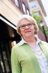
“Please, no more trees!” says a panelist, only half-joking.
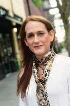
“There are some good things about trees too,” replies Shams, smiling.
After dinner wraps up and desserts are shared, the allergy-sufferers rise to go out into the evening air, armed with an arsenal of nasal sprays and new information, to continue their love/hate relationship with these, the cruelest months—springtime in Atlanta n


 BY Sidney Perkowitz
THE FIRST TIME I HAD MY HEART CHECKED BY ULTRASOUND AT THE EMORY CLINIC, I DIDN’T KNOW WHAT TO EXPECT. The technologist pressed a small device against my chest and moved it around, using a gel-type substance as a conductor, until an image appeared on a computer screen off to the side. Craning my neck, I could just make out a moving shape. It took me a second to realize what I was seeing. It was my own heart, beating even as I watched it— the closest I’ve ever come to an out-of-body moment.


As a physicist, I understand the mechanics of medical imaging. Put simply, images from inside your own body are gathered using sound waves, X-rays, gamma rays, or radio waves and a magnetic field.


These physical probes enable ultrasound, X-ray computed tomography (CT), positron emission tomography (PET), and magnetic resonance imaging (MRI)—medical tools developed by physicists, engineers, and physicians that are now essential to patient care.

Jon Lewin, executive vice president for health affairs at Emory and CEO of Emory Healthcare, is a professor of radiology and imaging sciences and biomedical engineering, and an innovator in the field.
What drew Lewin to medical imaging was its trifecta of tech, biomedical science, and patient impact. “When I got involved in the mid-1980s,” he says, “imaging was going from being very static—looking at X-rays, shadows on a screen—to interrogating and visualizing
the human body as never before, even during surgery.”
And the level of detail just keeps accelerating, he says. “The kind of information it is possible to learn through current imaging is transforming how medical care is provided across almost every specialty.”
Take, for example, functional brain imaging—“how thoughts are translated into electrical impulses and blood flow,” says Lewin.
Or the advances made possible by interventional radiology. “Minimally invasive, image-guided therapies have changed what used to be major surgery and a week in the hospital into outpatient surgery through a needle hole,” Lewin says. “I had to tell one patient to at least take the weekend to recuperate.”
Long before these techniques existed, physicians had to try to heal patients without knowing much about what was inside their bodies.

Anatomists and physicians eventually learned more through invasive surgery, by dissecting dead animals and people, and by using tools like the microscope. But it took an accidental observation in 1895 to allow doctors to finally peer inside the living human body without cutting into it.

German physicist Wilhelm Röntgen was working with a novel lab device, a glass tube pumped free of most of its air, with a metal electrode at each end. When high voltage was applied to the electrodes, a glow appeared between them. That was not new, but something else was. Though the tube had an opaque covering, Röntgen noticed that whenever it was turned on, a fluorescent screen three meters away would light up. Röntgen had discovered an unknown kind of penetrating radiation he dubbed “X-rays.” They generated universal wonderment.
Only later did physicists learn that

X-rays are energetic electromagnetic waves emitted by electrons streaming within the tube. The medical value of these new rays became immediately clear when Röntgen displayed an X-ray photo of the bones in his wife’s hand [FIG 1]. In less than a year, American physician William Morton published The X-Ray: or, Photography of the Invisible and Its Value in Surgery as a guide to medical use of the technique [FIG 2].
The early medical use of X-rays did much good but also harm.
With their high energies, X-rays penetrate the soft flesh of the body until they are absorbed by the denser bones, which appear as shadow images in an X-ray photo. Those high energies also can damage DNA and harm living cells. Before this was understood, X-rays were indiscriminately applied. As late as the 1970s, they were used in shoe stores to show a customer’s foot within a shoe [FIG 3].
X-rays were also causing cancer in some researchers who worked with them.
Now we carefully limit X-ray dosages for practitioners and patients. Still, the potential harm from X-rays highlights the need for alternative imaging methods
with much lower potential for tissue damage.
Computed tomography ( X-ray images of cross sections of the body that a computer then assembles into a detailed image. When this is done from different angles, changes in density can be converted into pictures of soft tissue, along with bones and blood vessels. CT can diagnose conditions from cancer and cardiovascular disease to spinal problems and trauma.

At Emory, biomedical engineer Amir Pourmorteza, assistant professor
while reducing X-ray dosages by more than 30 percent.

CT scanners have become lower in radiation, faster, and more precise in two ways: less noise (grainy appearance) and higher image resolution,” Pourmorteza says. “We’re hoping to double resolution.” Think of it like TVs or digital cameras, he says—the same improvement in resolution has taken place in CT scans as well. “You can detect tumors sooner. And you can actually see differ-
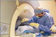
A 43-year-old sports reporter had a venous malformation involving the sole of his left foot since childhood. He underwent two surgeries as a child to correct this malformation, but had a recurrence after both. He spent 30 years of his life suffering from chronic foot pain that kept him from being active. Approaching his mid-40s, he started to suffer the consequences of his sedentary life with weight gain and high cholesterol level. He sought help at Emory Orthopaedics, but additional surgery was not an option. He was referred to the Emory interventional MRI program, which uses state-of-the-art MRI technology to provide minimally invasive diagnosis and treatment of challenging medical conditions. The patient was found to have a scarred sole with a very tender venous malformation. He was given two sessions of percutaneous MRI-guided sclerotherapy, where MRI determines the exact anatomy and extensions of the malformation and then is used for real-time guidance of the procedure. This ensures precise delivery of treatment, reduces the number of sessions and complications, and avoids radiation exposure. The patient responded well, ending up pain free for the first time in 30 years. He is now able to run five to 10 miles a day. “I lost so much weight that people do not recognize me,” he says. Also, his cholesterol level dropped to normal without the need for medications. —case provided by Dr. Sherif Nour


A 61-year-old man developed progressive lower back pain with accompanying pain and numbness in his legs. An MRI showed dilated veins in his lower spinal canal that indicated an abnormal connection between arteries and veins called a spinal dural arteriovenous fistula (dAVF). A catheter angiogram evaluating 31 vessels leading to the spinal canal could not find the site of the fistula. A time-resolved MR angiogram (MRA)—an MRI study of the blood vessels—was performed that showed the exact site of the fistula to be within the left L2 segment neural foramen. A repeat catheter angiogram targeting just that vessel confirmed the site of the fistula, and the patient underwent successful surgical treatment of the dAVF. The patient’s symptoms improved following treatment. Since this case, the MRA technique is now used prior to catheter angiography for all suspected spinal dAVFs, which has resulted in more accurate diagnoses and localization of the fistula level, reducing procedure time, radiation dose, and iodinated contrast dose during catheter angiography.

—case provided by Dr. Amit Saindane
ent colors now, which helps us differentiate diseases,” he says. “It’s like trying to separate an apple from the background leaves—if they both look grey, it can be difficult. But if you can separate the red and green, you can see the apples and the leaves, or the differences between two dense tumors.”
Xiangyang Tang, associate professor of radiology and imaging sciences and director of the Laboratory of Translational Research in CT, works with other X-ray detectors and computer algorithms to turn CT data into detailed images of organs like the heart [FIG 5 ], potentially at lower X-ray exposures.
“Each modality has its own advantages and disadvantages. Our responsibility, as radiologists, is to make sure we do the right thing for the patient,” Tang says. “One of the most exciting progressions in CT is in cardiac imaging. Rapid heartbeats used to cause blurring, but increased scanner speeds mean physicians don’t have to administer beta-blockers to reduce a patient’s heart rate. The new technology can cover the whole heart in one rotation.”
Like CT, positron emission tomography (PET) also assembles 2D images into 3D, in this case to locate normal and abnormal metabolic processes in the body.
Positrons are elementary particles first suggested in 1928 when French physicist Paul Dirac predicted that an electron has a kind of fraternal twin, exactly the same but with the opposite (positive) electrical charge. This was confirmed when positrons were found in cosmic rays. Now, of course, we know that every kind of elementary particle has an antiparticle and that antimatter exists along with ordinary matter.
Antimatter is a staple of science fiction because of a dramatic fact: when antimatter meets matter, they annihilate each other in a burst of energy. That burst makes PET the only practical
application of antimatter so far.
In PET, the patient is injected with a biologically active and weakly radioactive compound, frequently a special formulation of the sugar glucose. As this is taken up by bodily tissues, it emits positrons, each traveling a millimeter or less before meeting an electron resident in the body. The particles mutually annihilate and generate two gamma rays, powerful electromagnetic waves that, like X-rays, must be carefully monitored. The gamma rays travel in opposite directions until they are detected outside the body, at which point a computer tracks their paths back to their internal point of origin. Adding up many such calculations produces images of regions with strong metabolic activity, as shown by their glucose uptake.
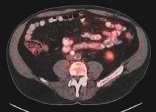
A high metabolic rate typically shows the presence of a tumor, so a main use of PET is to identify and monitor cancers. Brain activity also involves glucose metabolism so PET can diagnose brain tumors, guide brain surgery, and confirm diagnosis of brain deterioration, as in Alzheimer’s disease.
Carolyn Meltzer, chair of radiology at Emory, specializes in PET, and has shown that the best results come when a patient is evaluated by a combined PET/ CT scanner—the resulting near-perfect alignment of the two sets of images outperforms separate scans.
For instance, when squamous cell carcinoma, the second-most prevalent type of skin cancer, is suspected in the head and neck, a PET/CT scan facilitates the ability to differentiate between cancer and other abnormalities.
Radiology has medical applications far beyond what people normally think of, says Meltzer. “We are interested in expanding work with imaging agents that have both diagnostic and therapeutic functions, as well as further innovation in image-guided interventional treatments for such wide-ranging conditions as obesity, pain, and tumor ablation.”
Interventional radiology (IR) is, indeed, a field to watch. Image-guided ablation is an effective treatment for early kidney cancer. Interventional radiologist Sherif Nour also has performed many successful ablations of liver and prostate cancers at Emory. Last year, he traveled to Egypt to teach the technique.
And David Prologo, associate professor of radiology and imaging sciences and an interventional radiologist at Emory Johns Creek Hospital, is helping to develop an IR program in Tanzania. Prologo is also investigating the use of IR in reducing obesity by freezing a portion of the vagus nerve that sends hunger signals to the brain.
But much of his and other interventional radiologists’ work is pain related. “We are using our skill set to solve new, long-standing problems, like chronic pain, phantom limb pain, cancer pain. We are able to make dramatic changes in patients’ quality of life,” he says.
One of Prologo’s patients had metastatic lung

cancer in her spine and intractable nerve-related pain for more than a year. She underwent CT guided cryoablation of the cancer and the nerve, to block the pain signals. “I ran into her at Publix and she was crying because she was so relieved,”

“We do our procedure while viewing the CT on a screen, in real time,” Prologo says. “It allows us to guide a needle in safely. We inject the nerve with the anesthetic bupivacaine, a temporary block, and if it works on the patient, we feel justified in doing something more permanent,
Interventional radiologists work with doctors from all subspecialties— surgeons, ob/gyns, generalists, orthopedists, and bariatric, pain, and palliative care physicians. “We can do more for the patient,” says Prologo, “when we all work scans require can be an issue for pregnant women or for patients who require frequent scans for diagnosis or follow-up. That’s where ultrasound and magnetic resonance imaging (MRI) come in.
MRI grew out of nuclear magnetic resonance, invented in 1939 by the American physicist Isidor Rabi to study atomic nuclei.
Like tiny magnets, nuclei placed in a strong magnetic field align themselves in specific configurations with different energies. When excited by radio waves, the nuclei emit their own electromagnetic waves at frequencies corresponding to their energies, which act as markers for the particular type of nucleus.
This evolved into the biomedical MRI, which exposes the patient to electromagnetic radiation in the form of radio waves that are too weak to harm cells. The scan also requires a magnetic field thousands of times stronger than the Earth’s. This normally does no harm, although MRIs cannot be used on patients with metallic implants, such as pacemakers.
MRI is a versatile tool for diagnosis and treatment of the neural, cardiovascular, musculoskeletal, and GI systems.

For example, two different MRI studies of

Image-guidedinterventionalradiology canbeusedfortumor ablationsandtotreat conditionssuchas obesityandpain, saysCarolynMeltzer, WilliamP.Timmie Professorandchairofradiology. Lung cancer, 3D CT scan Metastatic ovarian carcinoma, PET CT scan
X-rays are a form of electromagnetic radiation with high energy that are used to generate images of tissues and structures in the body.
CT or CAT (computerized axial tomography) scans are computerized X-ray imaging that use multiple images to form detailed pictures of body organs.
PET scans (positron emission tomography) use electrons and their antimatter versions, positrons, to image inside the body, using a special dye containing radioactive tracers.
MRI (magnetic resonance imaging) is an imaging method for soft tissue that doesn’t use ionizing radiation.
Ultrasound developed from sonar and is now used for all kinds of imaging, from fetuses in pregnant women to real-time motion pictures of a beating heart.
RadiologistJohn Oshinskihasused MRItotrackstem cellstaggedwith nanometer-sized particles.

methods like this for the central nervous system.
Magnetic nanoparticles also played roles in recent MRI investigations by Hui Mao, professor of radiology and imaging sciences, showing how nanoparticles carry drugs into tumors and other targeted locations.
MY HEART WILL GO ON Medical ultrasound differs from the other techniques in using sound waves, not electro-
sound waves into water and register the echoes as the waves are reflected from objects, similar to echolocation as used by dolphins and bats. The result was sonar.
Sonar, in turn, inspired the Scottish gynecologist and obstetrician Ian Donald, who learned about it during his military service. In the 1950s, improvising with industrial ultrasound units that detect flaws in metal, he imaged ovarian cysts in women and published a paper showing the first pictures of a fetus’s head in a pregnant
woman. Commercial medical ultrasound units soon followed.
The sound waves used in medical ultrasound have frequencies far beyond the upper limit of human hearing, 20 kilohertz. They are typically in the range 1 to 18 megahertz, with the higher frequencies sensing finer details and the lower values providing deeper penetration into the body. A vibrating transducer—the device the technologist at Emory used to examine my heart—is placed against the body or sometimes in a body cavity to send out ultrasound waves. Like light waves, these are reflected, scattered, refracted, or absorbed as they encounter organs and tissues. The returning echoes are detected, and a computer analyzes the results to form a visual image of what the sound is passing through.
With its lack of damaging radiation, obstetric ultrasound is widely and safely used to examine the fetus in pregnant women, allowing prospective parents to see inside the womb. The portability of ultrasound equipment and its real-time results mean it can quickly image tendons, muscles, joints, blood vessels and organs. For instance, it can be used to examine the size, shape, and pumping capacity of the heart right in the doctor’s office.
Successful ultrasound imaging depends on putting sound energy into the body with efficient transducers and obtaining high-contrast images. These can be enhanced by using contrast agents—gas-filled microbubbles that strongly reflect sound waves and improve the contrast when blood vessels are being examined.
Researchers at the Coulter Department of Biomedical Engineering (BME) at Emory and Georgia Tech
address both of these issues. Brooks Lindsey, assistant professor of BME, is using Georgia Tech’s clean room facilities and other resources to design better transducers and contrast agents in cooperation with Emory physicians.
And Stanislav Emelianov, professor of electrical and computer engineering and BME and director of the ultrasound imaging and therapeutics research lab, has developed a new laser-based contrast agent. In 2017, his team showed that nanodroplets filled with a specific liquid can be vaporized by laser pulses to form highly reflective, gas-filled microbubbles. These quickly re-condense into liquid bubbles that can be reactivated over multiple cycles. This method produces superior high-contrast images of lymph nodes, which can show the spread of cancer.
“The radiology department provides access to imaging for researchers throughout Emory, not only clinical, but also brain health, public health, and animal research,” says Elizabeth Krupinski, professor and vice chair for research in radiology and imaging sciences. “A lot of key studies in developing biomarkers, contrast agents, and tracers have to be done in animals first.”
The future of imaging science will undoubtedly involve artificial intelligence and big data. But imaging can never completely take the place of good clinical skills, says Laurence Sperling, who directs Emory’s preventive cardiology program. Sperling reminds students and young physicians that effective clinical care requires “listening to, talking to, and touching the patient. We are taking care of people, not pictures of people.”
A man treated for prostate cancer in the past presented with elevated PSA levels. Axumin, a diagnostic imaging agent developed at Emory that can help detect prostate cancer across local and distant sites, showed recurrent cancer in the prostate, but also in lymph nodes outside the pelvis and bone. The ability to determine if a recurrence is in the prostate only, versus outside the prostate, improves the therapy that can be offered to the patient.
—case provided by Dr. David Schuster in nuclear medicine
A female patient came to us after her son expressed concern for her health because she was obese. She proceeded to lose more than 200 pounds and had plastic surgery to remove excess skin. That surgery was performed elsewhere. She awoke to what felt like a knife in her abdomen. An ultrasound showed a lesion on her right kidney. She came to Emory for an MRI, which confirmed stage I grade III carcinoma, an aggressive form of cancer. She then went to Emory Radiology, where doctors agreed she was an ideal candidate for laser ablation. The cancer was eradicated, with excellent margins. She went home the same day, and her three-week scan and blood work were clean. Her kidney shows no reduced function. She needed no pain medication after the procedure except ibuprofen.
—case provided by Dr. Sherif Nour
To support this important work in medical imaging contact ashley.michaud@emory. edu or call 404-778-1250.
Most people don’t die how or where they want. But there are ways to make your voice heard near the end. Six Emory physicians share details of their own advance medical directives.
Uncomfortable as it may be to consider our own death, most of us plan ahead for that inevitable event in some way.
We do what we can to make sure our wishes will get carried out if we die or become incapacitated: Wills that specify what we want done with money and other resources. Documents that declare who would be responsible for our young children. Powers of attorney that give a person or institution authority to manage our financial affairs if we no longer have the mental capacity to do so.
All of these preparations can be helpful, says Robin Everhart, associate general counsel for Emory Healthcare and Emory University, but fewer than one in three Americans prepares a guide for their relatives as to what medical measures they want in the event of an irreversible injury or illness.
Before Everhart became a health care lawyer, she was an intensive care nurse. She was at the bedside of unconscious patients, watching stunned family members struggle to decide the right course of action when considering measures that would prolong life but had no chance of producing a recovery.
How could they be the ones to make the decision—no ventilator, for instance—that would effectively end Mom’s life?
How could they choose an option, like feeding tubes, that would keep Papaw with them a little longer but might increase his suffering?
What would their relative have wanted them to do?
That’s exactly what advance medical directives are designed to convey, says Everhart.
Previously known in Georgia as a living will and durable power of attorney for health care, the advance directive for health care allows people to designate someone to make health care decisions on their behalf, if and when they have lost decision-making capacity.
Fewer than one in three Americans prepares a guide for their relatives as to what medical measures they want in the face of an irreversible injury or illness.
An advance directive can, if the person chooses, reflect which end-of-life treatments, if any, the patient wants, in the event that they have an incurable or irreversible condition that may result in death in a relatively short time.
Robin Everhart, associate general counsel for Emory Healthcare and Emory University
Advance directives for health care are written when people have decision-making ability and only go into effect when and if they lose that capacity.
If someone is still able to make decisions, they remain the ultimate decision-maker, overriding the advance directive document.
If they lose the capacity to make health care decisions, then such
decisions will be made based on their stated wishes in the advance directive, by the person they have designated as their surrogate health care decision maker.
The patient’s attending physician must determine and document whether the patient lacks the capacity to make their own health care decisions. The attending physician, working with a second physician, also determines and documents if the patient’s condition is actually
a terminal condition or a state of permanent unconsciousness.
Doing nothing—allowing a patient to die a natural death when technology exists to keep them alive — does not always come easily to physicians, who are trained to protect and enhance life.
Without direct guidance from the patient, relatives and caretakers may err on the side of continued life, no matter its quality or quantity.
A push for advance directives

Emory
University HospitalSusanne Hardy and her husband began end-of-life planning as part of their “safety net” after having children.
An only child, Hardy had been through the loss of both her parents before she turned 36. She was grateful that she understood and had documentation of her parents’ last wishes.
It was a step she knew she and her husband needed to take early. “These are difficult decisions and I encourage everyone in my life to complete an advance directive,” she says. “And not just complete it but discuss it openly with care providers and family.”
Her and her husband’s directives, for example, express that neither wants to be kept alive if only by life-support methods like ventilators and tube feedings, but both would want medications to assure maximum comfort until a natural death occurs.
Hardy, assistant professor of emergency medicine at Emory, knows well that these plans need to be written down and discussed with family. After particularly difficult cases in the emergency room, she sometimes passes colleagues in the hall shaking their heads, murmuring, “If I’m ever in that situation, please don’t keep me alive.” She often nods in agreement. But real life in the ER is more complicated than that. “We realize that we are privileged to be involved in only a snippet of patients’ lives and that every situation is different,” she says. “In the ER we do everything we can to save a patient’s life. That’s our job.”
It is still unusual for ER doctors to have an advance directive in hand stating the patient’s wishes regarding life-extending measures, Hardy says. “But we are grateful when we do.”
If there is any question about what the patient’s wishes are, the team does its best to give the family time and space to decide. “We want them to be comfortable with these end-of-life decisions. If people need more information about what medicine can or cannot do, we give it to them,” she says. “If they ask for time for other family members to drive or fly to the hospital to be there for support, we try to accommodate that.”
“These are difficult decisions and I encourage everyone in my life to complete an advance directive.”
began in the late 1960s in response to increasingly effective life-sustaining technologies, many of which were used on unconscious or unresponsive patients.
The concern was that dying patients could not control decisions about their own medical care. Horror stories about patients trapped in medical limbo, undergoing unnecessary, invasive, and often expensive procedures at the end of life, abounded.
In 1971, California became the
Atlanta Veterans Affairs Medical Center
first state to legally sanction living wills. Twenty years later, all 50 states had legalized some form of advance directive. The U.S. House of Representatives enacted the Patient Self-Determination Act, which stipulates that all hospitals receiving Medicaid or Medicare reimbursement (which includes all Emory hospitals) must ask whether patients have or wish to have advance directives.
Creating an advance directive when you’re young and healthy
doesn’t mean you’re planning for something bad to happen, says Kimberly Curseen, director of supportive and palliative care outpatient services for Emory Healthcare.

“And making one when you are seriously ill or injured doesn’t mean you lack hope or faith that you can get better,” she adds. “It means taking and maintaining control by making responsible plans. I don’t expect my house to burn down, but I buy insurance in case it does.” n
Like most younger doctors, Antonio Graham made his first advance directive as a resident because the institution where he was training—Johns Hopkins, in his case—demanded that all residents and fellows complete one. If they had not yet spelled out their own end-of-life wishes, how could they advise their patients to do so?
Having a baby only reinforced Graham’s initial decisions. If there were a chance to extend his life with some level of quality, he would “take whatever medicine could offer.”
But, he added, if after one week there was no evidence he would recover, then he would want “all lifesaving care de-escalated and comfort care intensified.”
Graham’s wife, his designated agent, agrees totally. His mother, however, fears her only child won’t get good treatment if the doctors see him “giving up.”
That’s not how it works, Graham reassures her. He gently reminds her that he is a doctor who fights hard for his patients but also tries to take their wishes into account—as he did when his father refused ventilation. It was a peaceful death, helped by morphine and oxygen.
Graham, an assistant professor of medicine at Emory, believed his father was making the right decision, and he was grateful that he knew what his dad wanted. But it was still incredibly hard.
“It’s one of the most difficult things you’ll ever face: having the courage to honor a person’s wishes, whether you agree with their decisions or not,” he says.

“It’s one of the most difficult things you’ll ever face: having the courage to honor a person’s wishes, whether you agree with their decisions or not.”


It’s become something of a Thanksgiving tradition. After the dishes are cleared, Ashima Lal checks in with family members about what everyone’s wishes are in the event of an irreversible condition. They may tease her —“What, again?”—but they understand. As a palliative care physician frequently consulted in Grady’s ICU, Lal sees patients with non-survivable injuries or illnesses, as well as their next of kin having to make crucial decisions at a completely unexpected, shocking time. There’s always anxiety and often conflict. To avoid that for their own families, Lal, assistant professor of medicine at Emory, and her husband prepared advance directives, discussed them with everyone, and keep copies in their cars’ glove compartments and the drawer next to the refrigerator. “Making decisions at this point in our lives is fairly easy. We would be okay with physical limitations,” she says, “but not huge cognitive ones.” To her family, patients, and friends, she explains that the documents can be revised if things change with their health or the person they want to be their designated health care decision maker. Advance directives are especially important, she says, if you want someone other than your next of kin making health care decisions for you if you are not able. Legally, without a document explicitly stating otherwise, the brother you may not have spoken to for years will have more authority over your end-of-life care than your fiancée or long-time partner, for example.
Benjamin Stoff, dermatology Senior Faculty Fellow, Emory Center for Ethics
As a dermatologist, even one specializing in skin cancer, Stoff doesn’t encounter as much death as many of his colleagues.
But having a graduate degree in ethics and being part of the Emory Ethics Center has helped him better understand the moral basis for both informed consent (medical decision making by the patient, when they have the capacity to do so) and advance directives regarding end-oflife treatments (for when patients can no longer make or express those decisions). Both protect patient autonomy, he says.
Because physicians faced with hard decisions sometimes ask for consultations with ethics center professionals, he also has seen the full range of complexity and conflict that can occur when no one knows or family members disagree about what the patient would have wanted.
Stoff, associate professor of dermatology and pathology at Emory, and his wife are each other’s designated agents, but they deliberately involved family members as witnesses, to emphasize how seriously they took their decisions and to encourage others in the family to follow suit.
Like most of Stoff’s colleagues, they choose to allow their natural deaths to occur without medications or machines, when a return to meaningful life is not possible. In the section of the document for additional statements, both wrote: “My top priority at end of life is comfort. If to keep me comfortable requires high levels of pain medication that may hasten my death, that is acceptable to me.”
“If keeping me comfortable requires high levels of pain medication that may hasten my death, that is acceptable to me.”
“We would be okay with physical limitations, but not huge cognitive ones.”
As head of special diagnostic services in the Paul W. Seavey Compre hensive Internal Medicine Clinic at Emory, Clyde Partin and his team see patients with mysterious symptoms and undiagnosed illnesses. Often called “Dr. House” by jocular patients and friends, Partin has seen “both sides of the end-of-life coin”—cases where patients have made their desires expressly known, and those where families are left to struggle with questions about what their relatives would have wanted. To spare their own families that dilemma, Partin and his wife have talked to everyone involved about their wishes.
An associate professor of medicine at Emory, Partin teaches his residents to be aware of cultural and individual differences. In some families, for example, senior members automatically take on larger roles in end-of-life decisions. Having advance directives is especially helpful if adult children need to argue for what their parent or sibling wanted, even when it is different from the opinion of a patriarch or respected aunt. More than once, patients faced with a critical situation have sent Partin their quickly written advance directive, so he would have it if they later arrived in the ER. Advance directives don’t have to be the original— copies are perfectly legal, whether in your purse, in the hands of your family, scanned into your medical records, or as a photo/PDF on your cell phone. So make a lot of copies and distribute broadly.

 Kimberly Curseen, palliative care Supportive Oncology Clinic, Winship Cancer Institute


Watching patients struggling with illness or traumatic injury made Kimberly Curseen think about what her family would go through if something catastrophic happened to her and she were incapacitated. “I wanted to absolve my husband and parents of the difficulties of trying to decide how I would have made decisions if I were able,” says Curseen, associate professor of medicine at Emory. “I wanted them to hear my voice even when I could no longer speak for myself.” And she wanted them to be as comfortable as possible with her decisions to forgo life-extending measures. In her advance directive, she wrote that it is important to her that her family have consensus. If that doesn’t happen quickly, as she fears it might not, then “it would be okay to prolong my life for a reasonable amount of time in hopes of achieving that consensus.”
Advance directives are living documents, and Curseen recommends that people revisit them every five years, or when their health or life events change.
“Humans have the ability to live with significant disability and still find beauty and wonder in life,” she says. “I redid my plan when my son was born because his birth made me think about what I could accept as a minimal quality of life—the amount and types of disability I could or could not accept—in order to watch him grow up.”
“I wanted [my husband or parents] to hear my voice even when I could no longer speak for myself.”
“Advance directives do no good if no one knows they exist or where to find them.”
An infant is born to Ms. L, who is 19. After delivery, the infant, Baby J, does not breathe and has a low heart rate. The neonatology team resuscitates her. A brain study shows severe brain damage. Her prognosis is poor: A lifelong need for a ventilator and feeding tube. She will not be able to communicate and probably will not recognize her mother.
The neonatal team and Ms. L discuss withdrawing life support. They ask Ms. L what she wants to do, but she can’t decide. The neonatology team wonders how to ethically proceed.
According to the American Academy of Pediatrics, parents and neonatologists may opt to forgo life-sustaining treatment when the burdens of treatment outweigh the benefits if the infant has a high risk of permanent and severe neurodevelopmental disability and a poor quality of life.
As a vulnerable new mother, Ms. L has difficulty deciding what she wants for Baby J.
The neonatal team members have a clear preference toward withdrawing life-sustaining measures. They think Baby J will have an unacceptable quality of life.
The team asks Ms. L to decide whether to continue or withdraw life support. Her inability to decide creates a de facto choice to continue life support.

In the U.S, the patient’s best interests guide ethical decision-making.

in the quality of life. Have you thought about your values?” asks the neonatologist.

Ms. L takes time to contemplate the question. A week later, she asserts that she believes Baby J will have a poor quality of life.
Ms. L asks the neonatologist what she should do.
Case presented by Michael Arenson, who has a master’s in bioethics from Emory and is a medical student, and April Dworetz, associate professor of pediatrics in the division of neonatology and a senior faculty fellow at the Emory Center for Ethics. This medical ethics series is based on real Emory cases, with some details changed to protect patient identities
The parents (or in Baby J’s case, the mother) represent their infant’s best interests. The neonatologist cannot ethically base her decision on her own values.
In this case, the physician’s moral obligation lies with the mother’s choice of best interests.
Ms. L asks for help from the team. She does not know how
The neonatologist says, “From what you have told me, you do not want Baby J to live the life she will have. We can make her comfortable, disconnect the ventilator, and allow her to die peacefully.”
Collaborative decision-making enables an ethical process.
In this case, the medical staff and family communicated throughout the patient’s course of treatment.

The parent(s) reveal values, beliefs, and cultural preferences, and shared decision-making takes place.
In the end, after the infant was given a dose of morphine for comfort, Ms. L held Baby J while the physician disconnected the ventilator. Baby J died in her mother’s arms n
Angel Flight has brought hope to thousands of families by arranging free flights to lifesaving medical treatment. We’ve flown over 35,000 missions serving over 300 medical facilities. To schedule a flight or to learn more go to AngelFlightSoars.org or call 1.877.4.AN.ANGEL

