ALSO IN THIS ISSUE
Common Concerns with Refractive IOL Procedures
Panel discusses how expanding IOL choices give surgeons more to consider.
Stromallenticuleextraction, implantation expand refractiveoptions

The Environmental Impact of the OR Ophthalmology has a unique opportunity to make procedures more sustainable.
Come Earlier, Stay Later
The 2023 Congress packs plenty of punch on the first and last days.
JUNE 2023 | VOLUME 28 | ISSUE 5
https://congress.escrs.org/

BINKHORST MEDAL LECTURE
Jorge L Alió MD, PhD, FEBOphthal will speak on “Corneal Regeneration: The Future of Corneal Surgery.” Dr Alió is a professor and chairman of ophthalmology at the University Miguel Hernández de Elche in Alicante, Spain, and a leader in the emerging topic of corneal regeneration. He conducted the first worldwide clinical trial for the treatment of corneal dystrophies and particularly keratoconus with autologous mesenchymal stem cells, and he has published a series of important scientific papers on this innovative type of corneal surgery.
SMART AND @CTIVE MONDAY
A new addition to the ESCRS Congress, Smart and @ctive Monday will feature “brushups” on retinal surgery, glaucoma surgery, and oculoplastics, a medical writing workshop for young ophthalmologists, an ophthalmic anaesthesia symposium, a surgical video session, and practice management sessions. A “digital track” will offer a “Continents Going Digital” symposium, symposia on automating eye surgery and the digital operating room, and talks and panel discussions about artificial intelligence in ophthalmology.

HERITAGE LECTURE
Marie-José Tassignon, MD, PhD, FEBOS-CR will speak about “The Enigma of the Anterior Interface.” Dr Tassignon is the emeritus head and chief of the department of ophthalmology of the Antwerp University Hospital and University of Antwerp. The first female president of ESCRS (2004–2005), Dr Tassignon developed bagin-the-lens cataract surgery for the paediatric population and holds 10 patents (three earned since 2019). She has been published more than 370 times in peer-reviewed journals and is the author of 27 book chapters and two full textbooks in ophthalmology.
iNOVATION DAY
Building on the success of the inaugural iNovation® Day in 2022, ESCRS is hosting the second iNovation Day on Friday, 8 September. Sessions will focus on the most urgent clinical needs and barriers to success in anterior segment care and how new technologies may address those barriers and clinical needs within the next 5–10 years. Of special interest is a new feature, “The Innovators Den: EyeCare Pioneers,” which will highlight entrepreneurs who have personally developed unique ideas to address some of the biggest unmet needs of Congress attendees.
Thomas Kohnen MD, PhD, FEBO

2 20
IOLs
Advantageous Phakic
EUROTIMES | JUNE 2023
G Holz FEBO, FARVO; Daniele Veritti MD; Valentina Sarao MD
12 Cover
lenticule
expand refractive
June 2023 | Vol 28 Issue 5
Best of Both Worlds Stromal
extraction, implantation
options
Publishers
Jemilah Senter
Mariska van der Veen
Mark Wheeler
Executive Editor
Stuart Hales
Editor-In-Chief
Sean Henahan
Senior Content Editor

Kelsey Ingram
Creative Director
Kelsy McCarthy
Graphic Designer
Jennifer Lacey
Circulation Manager

Mariska van der Veen
Contributing Editors
Cheryl Guttman Krader
Howard Larkin
Dermot McGrath
Roibeárd O’hÉineacháin
Contributors
Soosan Jacob
Leigh Spielberg
Timothy Norris
George Liu
Colour and Print
W&G Baird Printers
Advertising Sales

Roo Khan
MCI UK
Tel: +44 203 530 0100 | roo.khan@wearemci.com


EuroTimes® is registered with the European Union Intellectual Property Office and the US Patent and Trademark Office.


Published by the European Society of Cataract and Refractive Surgeons, Suite 7–9 The Hop Exchange, 24 Southwark Street, London, SE1 1TY, UK. No part of this publication may be reproduced without the permission of the executive editor. Letters to the editor and other unsolicited contributions are assumed intended for this publication and are subject to editorial review and acceptance.
ESCRS EuroTimes is not responsible for statements made by any contributor. These contributions are presented for review and comment and not as a statement on the standard of care. Although all advertising material is expected to conform to ethical medical standards, acceptance does not imply endorsement by ESCRS EuroTimes. ISSN 1393-8983


3 ALSO IN THIS ISSUE 40 Leadership and Business Innovation: Learn to Juggle Management Roles at ESCRS Congress 42 Newsmaker: Who is Looking Out for Ophthalmology? An interview with Professor Mor Dickman 44 Industry News 45 JCRS Highlights 46 Index of Citations 49 Upcoming Events ALSO IN THIS ISSUE 2023 JUNE | EUROTIMES 42 38 28 22 16 Learn more about EuroTimes or connect with ESCRS at ESCRS.org
Remembering Dan Epstein
The sad news about Dan Epstein was heard by the officers of the ESCRS during one of our Council meetings, of which he had until recently been a longstanding member. All of us were upset, and it is hard to grasp he is no longer with us. We all knew he had health problems but had hoped they were not that serious. I would like to express here my personal sorrow and the sorrow of our—and his— Society. Dan was a leading member of our Society and meant a lot to all of us.
Dan was a serious and brilliant scientist, expressing his intellectual honesty in ophthalmology, and ophthalmic surgery in particular. His lectures at meetings were full of objective data and taught us to read the actual outcome of surgeries and procedures without prejudice. His capacity for analysis was evident in evaluating published papers— especially in preparing the FEBOS-CR exam. The papers he authored always presented clear and precise data, and many remain landmark papers in ophthalmic literature.
As an ESCRS officer, Dan lived for decades within the Society, always seeking the best in administration and meeting organisation. I learnt from him that to receive an invitation to our symposia, a doctor must be a serious scientist, must know the topic better than others, and must be a good speaker. We owe Dan’s rules a substantial part of our success. His ability to criticise without being rude was invaluable and always led to improvements in our programmes and decisions.
Dan was a good friend to all of us. He used to help those who needed help, encourage those who were uncertain, and acknowledge and congratulate personal successes. His
love for ophthalmology turned into a love for ophthalmologists, with special sympathy for the young. He put his deep knowledge of our world at the service of those asking his opinion, his suggestions, his help.

We will miss Dan like we would miss an older brother who held a great place in our life. We join his wife, Beatrice, and his daughter in a virtual hug, participating in their sorrow. His memory will not fade within the Society and the world of ophthalmology in general.

 Roberto Bellucci
EDITORIAL BOARD
Oliver Findl ESCRS President
Noel Alpins (Australia)
Bekir Aslan (Turkey)
Roberto Bellucci (Italy)
Hiroko Bissen-Miyajima (Japan)
John Chang (China)
Béatrice Cochener-Lamard (France)
Oliver Findl (Austria)
Nino Hirnschall (Austria)

Soosan Jacob (India)
Vikentia Katsanevaki (Greece)
Daniel Kook (Germany)
Boris Malyugin (Russia)
Marguerite McDonald (US)
Cyres Mehta (India)
Sorcha Ní Dhubhghaill (Ireland)
Rudy Nuijts (The Netherlands)

Leigh Spielberg (The Netherlands)
Sathish Srinivasan (UK)
Ulf Stenevi (Sweden)
Marie-José Tassignon (Belgium)
EDITORIAL 4
Paul Rosen Medical Editor
José Güell Medical Editor
Thomas Kohnen Chief Medical Editor
EUROTIMES | JUNE 2023
2023 ESCRS Pioneer Award
True innovation comes from good research.
The ESCRS Pioneer Award is an initiative sponsored by the ESCRS to support and encourage independent clinical research in the field of cataract and refractive surgery.
The competition is open to young ophthalmologists up to the age of 40 with at least three years of ESCRS membership holding a full-time clinical/research post at an EU-based clinical or academic centre.
https://www.escrs.org/education/grants-awards/pioneer-research-awards/
Going to Vienna? Come Earlier, Stay Later
Boxing matches have caused many an eye injury, so a meeting of eye surgeons is not a place you would expect to find a boxing ring. But the ESCRS Programme Committee wanted to create a venue for speakers at the 2023 Annual Congress to debate issues while also allowing for audience interaction— and their solution was creating a stage in the middle of a room and placing a screen on the wall where audience questions can be displayed.

“They will be very vivid, interactive discussions,” says Dr Oliver Findl, president of the ESCRS and chair of the Programme Committee. “The speakers will be pro and con, just a couple of minutes on each topic, followed by a debate. People in the audience can write comments, and the comments will come up on a big screen and be fielded by a moderator.”
The boxing ring debates are just one of several interesting features awaiting attendees at the ESCRS 2023 Annual Congress in Vienna. Two other highlights are the “bookend days” of the Congress—Friday, 8 September, and Tuesday, 12 September—which will offer more educational content than in previous years.
“Focused Friday,” as it’s being called, will include Cornea Day, Glaucoma Day, and iNovation Day, as well as two sections of wet labs, the opening of the exhibit hall, and a symposium titled “Who owns ophthalmology?” Tuesday will feature the popular “Best of the Best” session, symposia on cataract surgery and postoperative complications, an IOL exchange workshop, and two free paper sessions.
“If attendees want to have the full impact of the Congress, they need to arrive on Thursday night and leave on Tuesday evening or Wednesday morning,” Findl says. “Saturday and
Sunday, like always, are fully packed. But on Friday and Monday, we’ve really tried to enhance the programme, and on Tuesday, we have the ‘Best of the Best’ session, which wraps everything up and puts it into perspective.”
Positioning for the future
Monday is being billed as “Smart & @ctive Monday” thanks to a “digital track” highlighted by symposia on the continents going digital, the digital operating room, automated robotic eye surgery, and the newest from artificial intelligence. Monday will also feature a mini-symposium on ophthalmic anaesthesiology, a full day of practice management workshops, a medical writing workshop, numerous instructional courses, and the ESCRS Heritage Lecture. Three 45-minute “brush-up” sessions focusing on glaucoma, retina, and oculoplastics are a novelty.
“Sometimes you have a patient who says, ‘I’m taking these new drops,’ and you think, ‘Oh, I should know about them,’ or she asks you, ‘I heard there’s a new drug coming out that is supposed to do this or that,’ and you think, ‘I should have heard about it, but it’s not my field,’” Findl says. “The idea of the brush-up sessions is to get on top of things again.”
A highlight of Saturday will be a “near-live” surgery session featuring challenging cases filmed by an ESCRS audiovisual team. The film crew visited surgeons in their home operating theatres using their own materials and equipment, and the filmed surgeons will be present at the session to discuss the cases.
“We’ll see more interesting and challenging cases than you usually have for live surgery sessions,” Findl says. “With live surgery sessions at conferences, you’re not in your own operating
theatre, it’s not your patient, it’s not your equipment, maybe not even your nurse. Everything is new, everything is different. And then you have 2,000 people watching. So I think this new concept is ethically more sound and will be more educational.”
6
INSIDE ESCRS
If attendees want to have the full impact of the Congress, they need to arrive on Thursday night and leave on Tuesday evening or Wednesday morning.”
The
2023 ESCRS Annual Congress will ‘pack more punch’ on the opening and closing days.
STUART HALES REPORTS
EUROTIMES | JUNE 2023
ESCRS CONTENT
Another new concept is a series of “100-second pearls,” which will take place in the boxing arena and feature short videos with special tips for surgical techniques. Like the pro-con debates, the pearls will be interactive, with audience members sharing real-time comments.
“We’ve never done it before at ESCRS, and I’ve never seen it in ophthalmology,” Findl says. “It’s something new that we can probably position for the future.”
The pearls, the boxing ring debates, the near-live surgeries, the brush-up
sessions, and other new features are critical to keeping the ESCRS Annual Congress fresh.
“After people have attended a few conferences, they think they’re pretty much all alike,” Findl says. “So we asked ourselves, what new things can we do for this one? What would draw somebody in for the first time if they’ve never been, or get them to come back if they’ve attended before? And I think they’re going to find a lot of things to like.”
What’s Vienna without a Ball? Vienna is famous for its balls—more than 400 take place there during winter alone. The 2023 Vienna ball calendar will be a little more crowded than usual, as ESCRS will host one at its Annual Congress on Saturday, 9 September.
“Balls in Vienna are very different from balls elsewhere,” says ESCRS President Dr Oliver Findl, who lives and works in Vienna. “The men wear tuxedos and the women wear long gowns, and of course we’re known for the Viennese waltz.”
The ESCRS ball will be held at the Hofburg Palace, formerly the residence of the Habsburg monarchy. A live orchestra and operetta singers will provide the music, and a Quadrille, a traditional French dance, will take place at midnight. The entertainment will include a casino and a disco with a deejay, and several cash bars will be available.

“People will be able to walk around, meet and greet others, and dance, of course,” Findl says. “It’s a unique and memorable way to connect with peers and build relationships.”
In keeping with tradition, the ball will adhere to a strict dress code: floor-length evening gown, dinner jacket/tuxedo with bow tie or dark suit with bow tie, or formal national costume (below the knee). No short skirts or dresses, cocktail dresses, or casual attire will be permitted.
“It’s really designed to keep the event elegant,” Findl says. “You can’t come in lederhosen or jeans—it’s black tie or white tie. But we will allow a dark suit or a formal national costume, such as from India or Africa.”
Attendees are encouraged to bring spouses and partners. Advance registration is required through the Congress registration portal.
“Vienna balls are quite amazing, and I think people will love it,” Findl says. “It’s an occasion that people will be talking about for a long time to come.”
7 2023 JUNE | EUROTIMES
ESCRS CONTENT
Resources on ESCRS.org by Topic
CATARACT CORNEA GLAUCOMA OSD PAEDIATRIC REFRACTIVE PRESBYOPIA RETINA TORIC IOLS
400 350 300 250 200 150 100 50 0 ESCRS AT A GLANCE ESCRS CONTENT 8 EUROTIMES | JUNE 2023
1,26645941,020 TOTALS:
ARTICLES POSTERS PODCASTS VIDEOS
ESCRS CONTENT 9 2023 JUNE | EUROTIMES
The Devastating Toll of Ocular Injuries in Ukraine
 DERMOT MCGRATH REPORTS
More than one year after the Russian invasion of their territory, Ukrainian ophthalmic surgeons continue to deal with a devastating range of traumatic eye injuries inflicted on the military and civilian population under the most challenging of circumstances.
The extent of these challenges, and the horrific toll on the Ukrainian people in devastating eye injuries incurred during the conflict, were outlined in a recent webinar devoted to the theme of “Defense Vision: experience in providing ophthalmological care in wars and armed conflict,” organised by the ophthalmology department of the National Military Medical Clinical Centre in Kyiv.
Welcoming online delegates to the conference, Bogdan Zhupan MD, Head of the National Military Medical Clinical Centre, said Ukrainian ophthalmologists were doing their best to provide care to their patients despite the many challenges of operating in a conflict zone.
“Ukraine continues to defend its independence and freedom against Russian aggression. We continue to work despite the many difficulties we face, and this conference is one more example that we are winning this fight,” he said. “We may come from different countries, but we are united in the goal of learning from each other and providing better ophthalmological care to our citizens.”
Robert A Mazzoli MD, FACS opened the conference with a talk on the blast eye, detailing how the incidence of ocular trauma due to blast forces has increased dramatically with the introduction of new explosives technology into modern warfare.
“Blast creates complex ocular polytrauma—and concussive injuries are as worrisome as penetrating injuries but may be subclinical, leading to delayed diagnosis of significant injury,” he said. “Blast injuries evolve, so it is important to temper surgical enthusiasm in the acute setting and try to minimise anaesthetic episodes by working closely with other services. And remember, blast never forgets, so it is a long-term risk factor for many diseases and conditions.”

Mykhailo Gavura MD discussed repeated injuries in patients with traumatic retinal detachments, pointing out the advice in medical journals to deal with ocular trauma as soon as possible after the injury is not always the best strategy for patients injured on the battlefield.
“If a patient comes from the battlefield with multiple injuries to the body and face, we must take account of this in our treatment plan, which needs to be phased over time,” he said. “If a patient has purulent discharge of the orbit, I usually limit myself to adequate primary surgical treatment of the anterior segment to deal with the eyeball. Only after the general condition stabilises can we move on to further stages of treatment.”
In other presentations, Ivan Gulko MD shared a clinical case of penetrating eye injury with intraocular metallic foreign bodies and preserved high vision, while Oksana Sidak-Petretska MD discussed simultaneous damage to the anterior and posterior parts of the eye during vitreoretinal surgery. Andri Ruban MD outlined the many challenges in combat eye trauma surgery, focusing on blast proliferative vitreoretinopathy (BPVR), and Robert MacLaren FRCOphth, FACS looked at the various options for restoring vision with electronic devices. Lesya Shuba MD rounded off the webinar with a talk on traumatic glaucoma.
10 UKRAINE EUROTIMES | JUNE 2023
2023 ESCRS Clinical Research Awards
Real-world studies with improved patient outcomes.
The ESCRS Clinical Research Awards (the “Awards”) is an initiative sponsored by the ESCRS to support and encourage independent clinical research in the field of cataract and refractive surgery.
The competition is open to all clinicians and researchers with at least three years of ESCRS membership holding a full-time clinical/ research post at an EU-based clinical or academic centre.
https://www.escrs.org/education/grants-awards/clinical-research-awards/
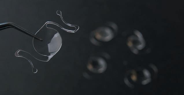
Stromallenticuleextraction, implantation expand refractiveoptions

COVER ARTICLE EUROTIMES | JUNE 2023 12
ince the first stromal lenticule extraction more than a decade ago, the procedure has grown in popularity, and is now one of the most widely used corneal refractive procedures to correct myopia. Conversely, stromal lenticule implantation also shows promise for indications including hyperopia, astigmatism, presbyopia, and keratoconus.
Speaking at the ESCRS Winter Meeting in Vilamoura, Dr Mario Nubile provided a broad overview of the technology. Stromal lenticule extraction has evolved through several iterations from femtosecond lenticule extraction (FLEx), in which (Carl Zeiss Meditec) femtosecond laser created a flap and cut out an underlying stromal lenticule, to the more recent small incision lenticule extraction (SMILE, Carl Zeiss Meditec), with the stromal lenticule extracted through a small 2.0 to 4.0 mm corneal incision without the need for a flap.
13 2023 JUNE | EUROTIMES
Until recently, he added, it has been difficult to draw any meaningful conclusions in comparing lenticule extraction to the gold standard of femtosecond LASIK and corneal lenticule extraction procedures.
“That picture has now changed greatly. We now have more than 750 publications on lenticular extraction from all over the world,” he said. “Many comparative studies and meta-analyses have demonstrated the efficacy, predictability, and safety outcomes of SMILE for the treatment of myopia are comparable to those of both LASIK and femtosecond LASIK.”
Both LASIK and refractive lenticule extraction are safe and effective procedures with their own advantages and drawbacks. SMILE may also have a slight edge over LASIK in treating high myopia.
“Unlike a LASIK procedure where tissue is being ablated, there is no great difference with a lenticule extraction to treat -10.0 D of myopia compared to a patient with -1.0 D of myopia,” he said. The energy delivery to the stroma is the same.”
SMILE also offers more rapid postoperative visual rehabilitation and a possible reduction of dry eye sensation, as fewer corneal nerves are cut during surgery than excimer laser ablation.
“We see some variability in the results in the literature for SMILE due to issues of centration and cyclotorsion control, as well as astigmatic correction and coma aberration,” Dr Nubile noted. “Many of these issues have since been resolved with the second-generation platforms that are now available.”
Another recent development has been the addition of other laser platforms offering lenticule extraction procedures for refractive correction—both Schwind and Ziemer received European Union CE mark approval in 2020 for treating myopia and astigmatism.
“It is nice to have these different platforms because each has its characteristics and features that improve some aspects of the treatment,” he said. “And the enhanced competition will probably also facilitate further growth of the technology worldwide.”
cule banking as a valuable resource of tissue, now wasted in our refractive procedures,” said Dr Nubile at a presentation during the 40th ESCRS Congress in Milan.
Intrastromal implantation of a human lenticule for treating hyperopia was first done in 2014 with expectations the procedure would have better stability, less regression, fewer aberrations, and less postoperative dry eye than LASIK for correcting moderate hyperopia. Although several studies demonstrate its feasibility and safety, implanting cryopreserved tissue in heterologous patients, the results also show the remaining need for improving refractive predictability, Dr Nubile said.
Full circle
While SMILE and related procedures involve the removal of the lenticule, several procedures now under investigation go the other direction—implanting a lenticule. The femtosecond laser has made it possible to precisely dissect stromal lenticules in which keratocyte viability and collagen structural integrity are maintained, even after cryopreservation, and therefore opened the door for the development of a variety of therapeutic applications using human tissue for intrastromal implantation.
“There is a big push towards investigating procedures using tissue from donor corneas and living patients. And in the near future, there is the chance that we will have lenti-
Stromal keratophakia for keratoconus
Intrastromal lenticule implantation shows promise for treating corneal ectatic disease. Within this field, researchers are investigating several approaches, including implantation of planar lenticules, ring segments (Cornea Allogenic Intrastromal Ring Segments, CAIRS), negative meniscus lenticules (Stromal Lenticule Addition Keratoplasty, SLAK), doughnut-shaped lenticules, and Bowman’s layer.
“Each technique has its potential applications, advantages, and disadvantages,” Dr Nubile said. “So far, none has shown superiority compared to others, and we are still working to understand what the best shape implant is to use.”
Under development at the University Gabriele d’ Annunzio of Chieti Pescara under the direction of Dr Nubile and Dr Leonardo Mastropasqua is the implantation of a lenticule thinner in the centre and thicker in the periphery, implanted at approximately 130 μm depth to treat keratoconus with central cones. It has been shown to have excellent biocompatibility and improve spherical equivalent and both uncorrected and best-corrected visual acuity in moderate to advanced keratoconus.
“There is also an increase in epithelial thickness favoured by the keratoconus flattening, which allows for better contact lens tolerance, and stromal thickness increases even more than expected because of keratocyte activity inducing fibrosis,” Dr Nubile said. “The combination of epithelial and stromal thickening leads to significantly increased corneal

14 COVER ARTICLE EUROTIMES | JUNE 2023
Each [platform] has its characteristics and features that improve some aspects of the treatment.
thickness, which can allow a cross-linking procedure in a patient who previously was not a candidate because of a thin cornea.”


Confocal microscopy studies show a mild wound healing reaction, stable interface reflectivity, and near absence of immune stromal rejection, which could be improved using decellularised lenticules.


Lenticule implantation to address eccentric keratoconus remains a big challenge since the femtosecond laser technology needed to create the customised lenticule profiles is not yet available but under investigation. Lenticule customisation is possible with an excimer laser, but using two different instruments for the procedure makes it more complicated and less appealing.
He noted while the CAIRS technique has shown very good biocompatibility, the inability to sculpt the ring segments precisely has limited its predictability.
Correction of presbyopia is another application for intrastromal implantation of human lenticules. Using tissue from SMILE, the PrEsbyopic Allogenic Refractive Lenticule (PEARL) technique—developed by Dr Soosan Jacob—is one such approach that improves near vision by inducing changes in central corneal power and asphericity.


High-mixed astigmatism continues to challenge corneal refractive surgeons. Intrastromal lenticule rotation is a potential technique for correcting high-mixed astigmatism. In this procedure, an autologous lenticule is created and rotated 90 degrees to add tissue in the preoperative steep meridian and reduce tissue in the preoperative flat meridian.


The future


Decellularisation of lenticules that removes the source of donor immunity, erases any trace of donor DNA, and leaves only collagen has the benefit of overcoming ethical issues with using human tissue, particularly as it applies to lenticule implantation for refractive surgery procedures. It is also the foundation for future applications, including stromal repair and drug delivery.
“Additionally, keratoplasty using a decellularised lenticule enriched with stem cells is an interesting concept that may be effective for improving quality of the stroma in eyes where keratocyte activity is impaired, such as in a patient with a history of keratitis,” Dr Nubile said.
A newly established international research group will coordinate lenticule implantation research. The group includes Dr Nubile, Dr Leonardo Mastropasqua, Dr Harminder Singh Dua, Professor Béatrice Cochener-Lamard, Professor Jorge L Alió, Dr Jorge L Alió del Barrio, Dr José L Güell, Dr Jesper Hjordtal, and Dr Jodhbir S Mehta.


15 2023 JUNE | EUROTIMES
Mario Nubile MD, PhD is an ophthalmologist at the Excellence Eye Research Center University Gabriele d’ Annunzio of Chieti Pescara, Italy. mario.nubile@unich.it
Western practices can learn waste-reduction strategies from lower-resource settings.
DERMOT MCGRATH REPORTS
ccording to the World Health Organization, the climate crisis poses the most significant threat to global public health. Ironically, the healthcare system is a major contributor to climate change, reported Dr David F Chang.
“The healthcare system accounts for nearly 10% of greenhouse gas emissions in the United States,” he said. “If the global healthcare sector were a country, it would rank number five in total emissions.”
Ophthalmology is not exempt from these considerations. The International Agency for the Prevention of Blindness predicts a significant rise in visually disabling diseases directly attributable to the climate crisis in the coming years.

“This will disproportionately affect the poorest and most vulnerable societies,” he said.
As operating rooms account for a major share of the healthcare sector’s emissions, Dr Chang highlighted the pressing need to address surgical waste in ophthalmology. “With cataract surgery and intravitreal injections, we have the highest procedural volumes in medicine,” he said. “As the global population continues to age, we will be challenged to make eye care delivery both economically and environmentally sustainable.”
Much of this surgical waste in the United States and other Western nations, Dr Chang said, stems from procuring the supplies and drugs discarded after a single use.
The Environmental Impact of the OR A
He suggested learning more efficient and sustainable practices from lower resource settings that don’t have the luxury of wasting money or resources. For example, the Aravind Eye Hospitals in India have been able to provide 60% of their care and cataract surgery for free to patients who cannot afford it.
“To do this, they maximise safe but efficient and cost-effective practices. They routinely reuse many surgical products, devices, and pharmaceuticals rather than discarding them after a single use, as we do in the West.”
For example, surgeons don’t change surgical gowns after each case. Despite these routine practices that classify as infection control violations in the West, Dr Chang said their registry study from Aravind reported a 0.04% rate of endophthalmitis in more than 2 million consecutive cataract surgeries—identical to the US rate in an overlapping period from the American Academy of Ophthalmology’s (AAO) Intelligent Research in Sight (IRIS) Registry.
“Through this comparison, you reach the infuriating conclusion that discarding all of our surgical supplies and devices after a single use did not make cataract surgery safer but instead generated an enormous amount of unnecessary waste.”
Yet, there is growing acknowledgement in Western practices to act now to reduce surgical waste. Dr Chang introduced EyeSustain (eyesustain.org), a web-based coalition of ophthalmology societies and their members collaborating to advance more sustainable practices in the industry, as such an example. Co-sponsored by ASCRS, ESCRS, and AAO, the website centralizes resources and information about sustainability in ophthalmology. It outlines immediate waste-reducing steps surgical facilities can take, such as using multidose topicals and reusable instruments and eliminating unnecessary items from surgical packs.
Dr Chang emphasised the importance of ophthalmologists educating themselves and their staff to promote a team approach to sustainability in the OR.
“Because we have the highest surgical volumes, ophthalmology has a unique opportunity and an obligation to make our procedures more sustainable,” he concluded.
16 CATARACT & REFRACTIVE EUROTIMES | JUNE 2023
Dr Chang presented at the 40th Congress of the ESCRS in Milan.
David F Chang MD is clinical professor at the University of California, San Francisco, US. He chairs the advisory board of EyeSustain. dceye@earthlink.net
Changing Patterns for IOL Exchange
Delving deeper into IOL explantations.
Advances in cataract surgery techniques and technologies combined with improved IOL designs and materials have lowered the rate of IOL exchange surgery and altered the percentages of its main indications, according to Dr Matthias Gerl.
“IOL dislocation remains one of the most common indications for IOL explantation, followed by incorrect IOL power, patient dissatisfaction due to halos and glare, IOL opacification, and capsular contraction,” he said. “In the past, the principal indications for IOL exchange surgery were IOL dislocation, anterior uveitis, and unpredicted high refractive errors. However, IOL exchange surgery today appears to have a good safety profile and good visual outcomes for the majority of these patients.”
Dr Gerl’s retrospective study included more than 26,000 eyes that underwent phacoemulsification with IOL implantation between 2016 and 2022 at the Augenklinik in Ahaus, Germany. The goal was to assess the incidence and causes of IOL exchange after cataract surgery, in addition to reporting the visual outcomes, complications, and the different techniques used for secondary IOL implantation.
Over six years, 26,377 eyes underwent cataract surgery—of which 59 eyes had an IOL exchange, with a cumulative incidence of IOL exchange of 2.2 per 1,000 cataract operations.
“This rate of IOL exchange is quite low and aligns with the findings from other studies, which have reported a range between 0.19% to 0.77%,” Dr Gerl said.
The main indication for IOL explantation was IOL dislocation in 24 cases (41%), followed by incorrect IOL power in 21 (35%), patient dissatisfaction due to halos and glare in 9 (15%), IOL opacification in 4 (8%), and capsular contraction in 1 patient (2%).
Glaucoma was the most common pre-existing ocular condition, accounting for 11 (19%) of all cases, followed by high myopia in 10 (16%), pseudoexfoliation syndrome in 6 (10%), diabetic retinopathy in 4 (7%), floppy iris in 5 (8.5%), and age-related macular degeneration in 3 (5%).
Glaucoma was the most common pre-existing ocular condition, accounting for 11 (19%) of all cases.
26,377
Over six years, 26,377 eyes underwent cataract surgery.
59
59 eyes had an IOL exchange, with a cumulative incidence of IOL exchange of 2.2 per 1,000 cataract operations.
19% 10.5
The types of secondary implanted IOLs were monofocal in 32%, iris-claw in 30%, three-piece in 20%, EDOF in 13%, and multifocal in 3%. The IOL was implanted in the capsular bag in 56% of cases, followed by iris fixation in 30% and sulcus in 14%. The mean visual acuity improved from 0.29 (logMAR) preoperatively to 0.18 after implantation, while the mean IOP increased from 15.13 mmHg before surgery to 15.83 mmHg after surgery.
The mean time interval between the cataract surgery and the IOL exchange was 10.5 months. Vitreous prolapse was the most common intraoperative complication in 35%, followed by capsular tear (10%) and zonular dehiscence (3%). Secondary cataract was the most common postoperative complication, occurring in 15% of cases, followed by IOP spike (8%), cystoid macular oedema (3%), and corneal decompensation (2%).
10.5 months.
17 2023 JUNE | EUROTIMES
DERMOT MCGRATH REPORTS
Dr Gerl gave this presentation at the 27th ESCRS Winter Meeting in Vilamoura, Portugal.
Matthias Gerl MD is an ophthalmologist at Augenklinik in Ahaus, Germany. m.gerl@augenklinik.de
The mean time interval between the cataract surgery and the IOL exchange was
Allograft Inlays for Presbyopes
Modern epikeratophakia offers new option in presbyopia.

ROIBEÁRD O’HÉINEACHÁIN REPORTS
Intrastromal lenticules derived from donor tissue appear to provide a safe, effective, and predictable treatment for presbyopia without the complications associated with previous synthetic corneal inlays, said Dr Arthur B Cummings.
“Corneal inlays have had a chequered past. Nobody can deny that,” he said. “However, we understand the problems, and we know what the issues were. We have learned, and I currently don’t see any corneal inlays that are not allografts or biosynthetic collagen, and they now do provide an exciting option.”
When corneal inlays such as the Kamra (AcuFocus) and the Raindrop (ReVision Optics) first came to the European market, refractive surgery clinics advertised the devices as an amazing, quick solution for presbyopia—thousands of inlays were implanted over a couple of years. However, the commercially available corneal inlays have since fallen out of favour, mainly because they induce haze that persists after explantation, he explained.
Dr Cummings noted he was the first in Europe to implant the Kamra inlay, having implanted 13 of the devices, 7 of which he has since removed. He has also implanted 18 Raindrop inlays, 15 of which he has removed. In a recent survey he conducted of Refractive Surgery Alliance Society members, 65% said they had implanted Kamra and/or Raindrop inlay before, but 90% said they do not do so currently.
On the other hand, 80% said they would use an allograft for presbyopia if it was available.
Previous problems with corneal inlays stem from the poor biocompatibility of the synthetic material used to compose them, he said. In contrast, years of experience with epikeratophakia have demonstrated the safety and biocompatibility of allografts composed of donor stromal tissue. Moreover, advances in laser and imaging technology and tissue preservation have made it possible to produce sterile stromal lenticules with high precision.
Phase II study yields encouraging results
Dr Cummings and his associates have been part of a phase II prospective multicentre study involving 101 patients who underwent implantation of an Allotex TransForm™ lenticule corneal inlay in the nondominant eye. TransForm obtains the corneal stromal tissue under strict ethical standards from an Eye Bank Association of America (EBAA)-approved eye bank. The tissue is sterilised with electron beam radiation and shaped into the desired dimensions with a femtosecond laser.
The patients in the study were all emmetropic presbyopes who required at least a +1.75 D add to read. The lenticules were 2.65 mm in diameter and 22 microns thick with a +2.5 D add. They were placed under a LASIK flap 110 microns thick and centred visually over a light-constricted pupil.
18 CATARACT & REFRACTIVE EUROTIMES | JUNE 2023
The 6-month data is available for all 45 patients who received the inlays at Dr Cummings’ centre, and the 12-month data is available for 43 patients. So far, he has removed two inlays because of patient intolerance of the refraction difference between the two eyes, despite a visual acuity of 6/7.5 for distance and N5 for near in the treated eye. A further two eyes have undergone LASIK enhancements, one to increase myopia and the other to decrease it.
The lenticules are easy to manipulate when wet, and— once put in place to dry—they remain securely where positioned, Dr Cummings said. No lenticule moved postoperatively, and when incorrectly placed, they can be easily recentred by lifting the LASIK flap and placing it in the correct position. In addition, the corneas have remained optically clear, and the implants are invisible under the slit lamp.
Design Innovations that Incorporate Operator and Patient Comfort with Gentle Measurements
• Fully-automatic measurement
• Gentle voice guidance (available in 9 languages)
• Reliable tono/pachymeter
• Flexible and space-saving design
• A variety of options to meet your needs
Overall study results showed that in the treated eye of 34 patients with 12 months of follow-up, decimal uncorrected visual acuity (UCVA) was 0.63 for distance and 0.8 for intermediate and near. In 29 treated eyes with four years of follow-up, UCVA was 0.83 for distance, 0.94 for intermediate, and 0.82 for near.
In terms of safety, none of the treated eyes lost more than two lines of best-corrected distance visual acuity. There was only a 6.6% probability of losing more than one line and less than a 1% probability of losing more than two. In addition, the four-year results in 28 hyperopes and 29 presbyopes with the TransForm inlay showed that none developed haze, compared to 112 (75%) of 150 eyes with the Raindrop inlay.
“The conclusion from this study is these inlays are very quiet in the cornea,” he said. “There is reduced uncorrected distance acuity in the treated eye on day one, but that improves as the weeks go by. Reading vision improves quicker than distance vision, and binocular distance visual acuity remains unchanged. Moreover, biocompatibility is excellent, and at three years, you still cannot see these inlays under the slit lamp. I see this being used an awful lot more in combination with LASIK.”
Non Contact Tono/Pachymeter New
www.nidek.com ET 93 x 266mm
Dr Cummings presented his findings at an ESCRS eConnect webinar.
Arthur B Cummings MD, FRCSed, FWRCS is based at the Wellington Eye Clinic, Dublin, Ireland. ABC@WellingtonEyeClinic.com
Corneal inlays have had a chequered past. Nobody can deny that.
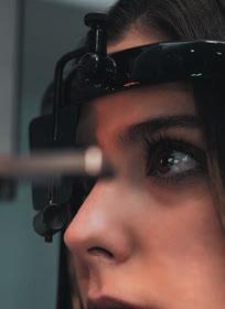
Advantageous Phakic IOLS
Underutilised PIOLs safe and effective for moderate to high myopia.
DERMOT MCGRATH REPORTS
Posterior chamber phakic IOLs offer surgeons a safe and effective option for treating moderate to high myopia and present a viable alternative in patients where surface ablation procedures are contraindicated, according to Professor Thomas Kohnen.

“When I look at my myopic cases, I probably do twothirds on the cornea and one-third on the lens,” he explained. “I think phakic IOLs are an underestimated procedure that offers a lot of advantages. For a start, it is the only reversible procedure, and also one where we don’t experience any problems with postoperative power calculations for intraocular lenses because we are not changing the cornea.”
In a broad overview of the available phakic IOL options, Prof Kohnen said he has personally implanted more than 2,500 phakic lenses, including all three principal types: angle-supported anterior chamber lenses, iris-fixated, and sulcus-fixated IOLs.
Despite good early results, angle-supported PIOLs have not lived up to their initial promise, with the Cachet lens (Alcon) withdrawn from the market due to concerns about abnormally high endothelial cell loss, Prof Kohnen explained.
“The 10-year follow up study of more than 1,123 implanted eyes in 7 countries reported a 10% explantation rate, most because of endothelial cell loss over time in patients with a shallow anterior chamber,” he said. “I have witnessed some patients who have experienced no cell loss at all after 15 years, but all patients need to be monitored closely.”
Likewise, iris-fixated IOLs (Artisan IOL, Ophtec) have encountered problems of long-term endothelial cell loss, despite good visual and refractive outcomes.
“A study by Rudy Nuijts’ group looked at 5- and 10-year data for the Artisan phakic lens and found a high level of endothelial cell loss over the follow-up period. So again, these anterior chamber lenses need to be monitored, as we can see
problems after 10 years or even later,” he explained. “We conducted a smaller study of the same lens in high myopes and found that the endothelial cell loss was higher in patients with an anterior chamber depth (ACD) below 3.0 mm than in lenses with ACD greater than 3.4 mm.”
Prof Kohnen said his preference is to use a posterior chamber lens, either the Visian implantable collamer lens (ICL V4c, Staar Surgical) or the implantable phakic contact lens (IPCL, Care Group), for those patients outside the dioptric range of the ICL.
“The implantation is a little bit more difficult than an anterior chamber PIOL but still only takes about four to five minutes,” he noted. “I would estimate between 50% to 75% of the ICLs I implant now are toric models so I can correct up to 0.5 D of astigmatism at the same time. There is also a cosmetic benefit to patients in that the lens is completely invisible in the eye.”
Looking at the evidence in the scientific literature, Prof Kohnen said that the lens has solid safety and efficacy data out to 10 years.
“We are currently in the process of publishing our own five-year results in 45 eyes which showed good predictability and stability over time, with a high index for safety and efficacy,” he concluded. “We found no significant change in endothelial cell count. I think it’s currently our best option for a phakic IOL.”
20 EUROTIMES | JUNE 2023
Prof Kohnen gave this presentation at the 27th ESCRS Winter Meeting in Vilamoura, Portugal.
CATARACT & REFRACTIVE
Thomas Kohnen MD, PhD, FEBO is professor and chairman, Department of Ophthalmology, Goethe University, Frankfurt, Germany. kohnen@em.uni-frankfurt.de
Presbyopic LASIK
LASIK treatment combining anisometropia with spherical aberration treats presbyopia with little compromise.
Combining modest anisometropia with a therapeutic amount of spherical aberration Presbyond Laser Blended treatment with the Zeiss Meditec MEL 80 excimer laser can provide presbyopes with a high degree of spectacle independence and minimal compromise in terms of visual quality or stereoacuity, said Professor Dan Z Reinstein.
“This modified binocular vision procedure has the advantages of the safety and accuracy of LASIK and the lowest side effects of any presbyopic procedure,” he said. “It maintains contrast and stereoacuity, is adjustable, and is entirely reversible with a spherocylindrical enhancement.”

In studies he and his associates have conducted involving patients who have undergone the procedure, 20/20 binocular visual acuity for distance and J2 for near was achieved in 95% of those with myopia up to -8.50 D, 77% of those with hyperopia up to +5.75 D, and in 92% of emmetropes. Additionally, no patients have lost two or more lines of best-corrected visual acuity (BCVA), and only 8% of myopes, 17% of hyperopes, and 13% of emmetropes lost one line. Furthermore, 36% of myopes, 19% of hyperopes, and 27% of emmetropes gained one or more lines of BCVA.
Prof Reinstein noted more than 250 centres conducted more than 320,000 Presbyond procedures. Multiple publications from multiple sites worldwide have reproduced the results achieved at his centre. As an example, he cited a study by Sri Ganesh FRCS that showed the satisfaction rate was nearly 100% for myopes and hyperopes, spectacle independence was nearly 100%, and the dysphotopsia rate was nearly zero. The study also showed the reading speed was faster with presbyopes treated with Presbyond compared to matched presbyopes treated with trifocal surgery with the Zeiss trifocal.1
Not monovision
Prof Reinstein stressed Presbyond should not be thought of as monovision or even mini-monovision, as the vision it achieves is a result of both eyes working together. It involves targeting the dominant eye for plano and the nondominant eye for -1.50 D of myopia while also inducing around 0.6 microns of spherical aberration in both eyes. The treatment is based on research showing stereoacuity maintained at that level of anisometropia—spherical aberration increases depth of focus but does not reduce contrast sensitivity so long as it is 0.6 microns or below.
“With monovision, there is poor tolerance—not everyone can do it, and you have to choose whether you will have near vision or intermediate,” he noted. “Whereas if you use spherical aberration with a small anisometropia, you can have good night vision, good contrast, very high tolerance, and good near, intermediate, and distance acuity.”
Dr Reinstein presented his findings at an ESCRS eConnect webinar.
Moreover, mean contrast sensitivity increased slightly but significantly in all three groups, and all eyes retained 400 arcseconds of stereoacuity. Some 89% of myopes, 88% of hyperopes, and 71% of emmetropes retained 100 arcseconds.
For citation notes, see page 46.
21 2023 JUNE | EUROTIMES
ROIBEÁRD O’HÉINEACHÁIN REPORTS
Dan Z Reinstein MD, MA (Cantab), FRCSC DABO, FRCOphth, FEBO is based at the London Vision Clinic, EuroEyes Group, London, UK. dzr@londonvisionclinic.com
This modified binocular vision procedure has the advantages of the safety and accuracy of LASIK.
The Digital Ophthalmologist
Online medical education: pros, cons, and future looks.
DR GEORGE LIU REPORTS
In December 2019, whispers of coronavirus disease 2019 (COVID-19) began to spread. Initially, like most services around the world, medical education suffered, and subsequently, so did students. When the inevitable happened, we adapted. Our modern world of social media and the rise of online services allowed medical educators to communicate and rethink how they could educate.
Webinars
The rise of webinars, Zoom calls, and virtual conferences ensued. Alongside university-prescribed webinars, an abundance could be found online. Every day one would be spoilt for choice for supplementary materials. However, this real-time evolution did not come without its cons. Initially, there was no way of distinguishing self-proclaimed medical educators from accredited ones. There was often repetition between webinars. And after some time, prescribed educators became preoccupied with their clinical workload, and live webinars morphed into pre-recorded sessions. The combination of these factors contributed to “webinar fatigue.”
In an interview, Professor Pearse Keane, Consultant Ophthalmologist at Moorfields Eye Hospital, London, told EuroTimes he finds webinars awkward, removing much of the spontaneity live teaching can provide. Prof Keane encourages camera use and prefers “icebreaker” introductions when running artificial intelligence journal clubs with his colleagues.
From an academic viewpoint, the proliferation of free webinars also posed difficulties for traditional seminars, which normally incurred a fee. Professor Christopher Liu OBE, Honorary Treasurer of The Royal College of Ophthalmologists (RCOphth), stressed the importance of these fees as a source of income for bodies such as RCOphth for carrying out statutory duties for the profession.
Mr Ahmed Shalaby Bardan, Consultant Ophthalmologist at Leeds Teaching Hospitals, describes his preferred method of online interactive live courses as more engaging, enabling active discussion. He also supports recording lectures so students can keep the material and review it when they want to refresh their training. Mr Bardan also recommends sharing content with trainees at the right stage of their career, as pitching information at the right level is key to engaging an audience.
Social media
Social media adjuncts to medical education also contributed to the education evolution. YouTube surgical videos have become a mainstay for all trainees. Whilst a variety of Instagram and Facebook medical pages, such as “Zero to Finals”, offered excellent resources such as spot questions followed by disease summaries, students shouldered a never-ending supply of medical education resources infiltrating their personal social
media feed. This disruption to work-life balance lasted 24/7 and perhaps contributed to the increased stress levels throughout the UK during COVID-19.
Prof Keane, an avid user, describes Twitter as by far the best platform for identifying new ideas and hot topics. Similarly, Mr Bardan uses social media for live broadcasting and sharing knowledge and experience. He says social media has the advantage of showing different techniques and hosting open discussions with audiences whilst concurrently responding to questions and getting feedback. Mr Bardan believes social media—if used properly for teaching—can be a very strong aid to the teacher and an asset for the student.
Accredited online medical education platforms
In-person ophthalmology conferences offer a plethora of educational gems for students and consultants alike. However, prior to COVID-19, expenses, travel time, annual leave, and sustainability were some factors limiting accessibility. Now, with virtual access to many conferences, this barrier has mostly been overcome. Yet still, there is scope for development as delegates miss out on the physical aspects, such as wet labs and networking.
The Royal College offers its members INSPIRE—an online, comprehensive learning platform that covers the full spectrum of ophthalmology integrated into a unified learning provision. INSPIRE gives its users access to learning anytime and anywhere, harnessing various modalities best suited to both the type of learning and the learner.
22 CATARACT & REFRACTIVE EUROTIMES | JUNE 2023
Pros of online medical education Cons of online medical education
1. Ease of access
2. Further reach as an educator
3. Greater variety of surgical techniques available as a trainee
4. Content can be reviewed if recorded
5. Access to live research
In addition to its two annual in-person conferences, the ESCRS offers its members a host of online learning opportunities: ESCRS iLearn, ESCRS research portals, landmark articles, EuroTimes podcasts, ESCRS on demand, EuroTimes conference coverage in ESCRS Today, JCRS online case reports, the Video Journal of Cataract, Refractive, and Glaucoma Surgery, and access to the FEBOS-CR exam.
1. Unaccredited courses
2. Risk of losing education/life balance, culminating in burnout or webinar fatigue
3. Lack of real-life surgical knowledge
4. Cost of access to accredited education
learning that could mimic current teaching norms through immersive technology. In theory, virtual, augmented, or mixed reality are excellent tools to simulate theory, surgery, and critical thinking in a patient-safe environment. But one cannot ignore the looming cost of virtual reality hardware and software and the wavering reliability of one’s internet connection. Despite these challenges, there is certainly room for development, either as a discrete unit or potentially paired with artificial intelligence to generate a self-adapting education tool for all.
Yet online education platforms have limitations. Whilst one can learn theory for any operation, real-life experience is paramount, preferably initially supervised by an experienced trainer, says Prof Liu. Therefore, proficient surgical theory does not denote surgical competence.
The future of medical education

Cue the metaverse. Whilst this may still be a sandbox project, the metaverse shows great potential for interactive

23 2023 JUNE | EUROTIMES
Table 1. Pros and cons of social media education
The Power of Osmolarity Testing in the Palm of Your Hand PORTABLE ✦ PRACTICAL ✦ PRECISE Visit us at www.GetScoutPro.com SP Euro Times Journal Ad_R0_042723_NB.indd 1 4/28/23 9:49 AM
George Liu BSc Hons is a 4th year Medical Student, Chelmsford, Essex, Tongdean Eye Clinic, UK. GeorgeLiu.Surg@gmail.com
ESCRS Learning Resources RCOphth
Postop Follow-up Zooms Ahead
Telemedicine passes test for routine cataract follow-up.
DERMOT MCGRATH REPORTS
Telemedicine software can provide a safe and effective means of following-up patients after routine cataract surgery, according to a recent UK study.
“Our experience showed the video consultation was effective, safe, accurate, and satisfactory in this cohort of patients following routine cataract surgery,” said Dr Lucia Pelosini. “There were no adverse effects or complications missed in the group subject to video consultation at two weeks compared to the patients reviewed in the clinic during the same period. The novel process of integrating remote video consultation into routine medical care has the advantage of reducing hospital attendance and the cost of travelling for patients.”
The current practice in the United Kingdom is to review the patient within two to four weeks after routine cataract surgery, usually in nurse-led clinics or with trainees in national healthcare institutions, Dr Pelosini noted.
“The COVID-19 pandemic has pushed us to look for alternative ways to follow our patients safely,” she said. “During the pandemic, we invested a lot of time in telephone consultations and follow up. This type of telephone follow-up after routine procedures has also been described in many other specialties.”
The telemedicine software (AOS, Advanced Ophthalmic Systems) allows the clinician to perform live video consultations, assess images of the anterior eye, and complete an interactive live visual acuity measurement following surgery.
“We wanted to look at this software, which combines the use of information obtained from the patient—equivalent to that obtained in a telephone conversation—but also adds some objective measurements,” she said.
Dr Pelosini’s study assessed the software in 65 patients at King’s College Hospital, London, two weeks after routine cataract surgery and compared this cohort with an equivalent group of patients who attended a face-to-face consultation two weeks after cataract surgery.
Patients selected for a follow-up by video consultation tended to be younger (73 years versus 85 years), have better general health, and be more familiar with using devices such as computers and smartphones. The video consultation allowed a reliable assessment of symptoms, compliance with medication, and examination of the external eye in all cases. Only 4% of patients seen by video consultation required a clinic review due to postoperative medicine intolerance. Some 85% of patients had a distance visual acuity of 6/12 (Snellen) or better, which is in line with normal cataract follow-up, Dr Pelosini said.
The vast majority (97%) of patients said they were very satisfied with the video assessment, while 3% said they were satisfied with the consultation but experienced some technical difficulties in the connection. There was no adverse effect or missed postoperative complications in the group assessed by video consultation when assessed at the final clinic review five weeks after surgery.
“The only real limitation in taking this approach is we lose some refractive outcomes for audit purposes of our surgery, and we also lose some opportunities for teaching junior doctors and nurses,” she said.
97% of patients said they were very satisfied with the video assessment.

24 CATARACT & REFRACTIVE EUROTIMES | JUNE 2023
Dr Pelosini gave this presentation at the 27th ESCRS Winter Meeting in Vilamoura, Portugal.
Lucia Pelosini MD, FRCOphth is a Consultant Ophthalmic Surgeon at King’s College Hospital NHS Foundation Trust, Queen Mary’s Hospital, London. luciapelosini@googlemail.com or misspelosini@theclinic.co.uk
Research. Education. Innovation.
ESCRS’s vision is to educate and help our peers excel in our field. Together, we are driving the field of ophthalmology forward.
Common Concerns with Refractive IOL Procedures
Expanding IOL choices increase treatment options.
SEAN HENAHAN REPORTS
Who should be considered for presbyopia-correcting IOLs and who should not? Panellists addressed a series of key issues surrounding this question during a symposium at the ESCRS Winter Meeting in Vilamoura, Portugal.
Current estimates suggest 10% of cataract patients have glaucoma. The first question to the panel addressed the suitability of implanting presbyopic correcting IOLs in patients with glaucoma. A preliminary audience survey showed strong reluctance, with 68% opting not to use these lenses in glaucoma patients.
“We need to consider what we mean by glaucoma—severe glaucoma with visual field defects is different than, say, a little hypertension, and are controlled with drops,” said Professor Thomas Kohnen. “Nowadays, we have many more options to keep the pressure down over the long term.”
Professor Oliver Findl noted the importance of contrast sensitivity in these patients, saying that even if there is a patient with ocular hypertension, full visual fields, and normal optic nerve, or a patient with very early disease with no reduction in the visual field, contrast sensitivity issues can still appear later.
“These patients have a high life expectancy. When we operate on a 65-year-old, they may live another 20 or 25 years,” he commented. “We don’t know how the glaucoma will progress over that time. You generally don’t want a lens that will com-
promise contrast sensitivity, so it also depends on which type of presbyopia-correcting IOL you are talking about.”
Prof Kohnen concurred, adding, “We now have a variety of presbyopia-correcting IOLs. From monofocal plus and EDOF lenses to diffractive multifocal IOLs. I would aim most of the time for non-diffractive EDOF lenses because you have a lower chance of contrast sensitivity loss.”
Presbyopia IOLs for retinal disease?
The panel next addressed presbyopia IOL use in patients with retinal pathology. A majority of the audience, 86%, opposed the idea. Contraindications for presbyopia lenses in this group include severe retinal pathology or disease, patient age, and visual status.
What percentage of your cataract patients present as asymptomatic of any ocular surface disease prior to surgery but develop symptoms postoperatively?
On average, 22% of cataract surgery patients present free of any ocular surface disease prior to surgery but develop symptoms postoperatively.
26 CATARACT & REFRACTIVE EUROTIMES | JUNE 2023
1% 1% 28% 36% 21% 9% 4% 0% 76–100% 1–10%11–25%26–50%51–75% Don’t perform cataract surgery
There is a broad spectrum of retinal pathology. There are restrictions, but I would not say it is an absolute no-go.
“There is a broad spectrum of retinal pathology. There are restrictions, but I would not say it is an absolute no-go. Trifocal IOLs are a no, but monofocal plus IOLs might be a good option in some cases,” Prof Kohnen said. “An EDOF lens might be considered, depending on which type, though never a diffractive lens.”
Ocular surface disease is not uncommon in cataract patients. The panellists agreed OSD should be managed first before considering presbyopic lens options.
“We need to do the diagnostics. This is more important than ever. You have to look not only at the tear film but at the thickness of the epithelium,” Prof Kohnen explained. “For me, severe OSD is always a contraindication. In milder cases, I support pretreatment and multiple diagnostic assessments. Then I would go with a more forgiving lens.”
Night vision problems
Night vision dysphotopsia continues to be one of the most prominent concerns among potential presbyopia IOL patients.
“The average percentage of patients with no residual refractive errors and healthy ocular surface who go on to have postop aberrations ranges from 5–7%. It is higher for bifocal and trifocal lenses,” Prof Findl told the symposium. “From our perspective, it is really important to inform patients beforehand that they may encounter these symptoms, depending on the type of IOL. With a trifocal, for example, you will definitely expect halos, maybe less so with an EDOF lens.”
Prof Kohnen added the challenge now is matching the IOL to the patient.
“You have to manage expectations. If I have a patient who has to drive five times per week from Frankfurt to Munich at night, I would probably not give him a trifocal lens. I would go for a non-diffractive EDOF,” he said. “But you have to remember many patients do not have this problem with trifocal IOLs.”
Neuroadaptation is another piece of the puzzle in these cases. Prof Kohnen and his group have completed a study, now in press, that concludes halos—the most bothersome aspect of dysphotopsia—tend to diminish in some cases between three and six months after surgery.
Should monofocal plus IOLs should be the new standard monofocal IOL?
“It really depends on our healthcare systems. If monofocal plus has better outcomes, then they really should replace standard monofocals. But you need healthcare systems to adopt— and possibly pay more—for them,” Prof Kohnen said.
Prof Findl responded enhanced monofocals are essentially the same price in his country, Austria, and they have become the standard choice for all his patients.
27 2023 JUNE | EUROTIMES
Oliver Findl MD, MBA, FEBO is the Chair of the Department of Ophthalmology, Hanusch Hospital, Vienna, Austria. He is the current president of the ESCRS. oliver@findl.at
2.2% 7.1% 4.9% 4.0% 3.2% monovision patient, with two monofocal IOLs bifocal presbyopiacorrecting IOL trifocal presbyopiacorrecting IOL extended depth of focus (EDOF) presbyopiacorrecting IOL enhanced monofocal presbyopiacorrecting IOL Average % of monofocal/bifocal/trifocal/EDOF/ enhanced monofocal patients with no residual refractive error and a healthy ocular surface having functionally significant visual aberrations at night.
Thomas Kohnen MD, PhD, FEBO is professor and chairman, Department of Ophthalmology, Goethe University, Frankfurt, Germany. kohnen@em.uni-frankfurt.de
Innovative Corneal Imaging Device
Prototype shows rich potential of high-resolution aberrometry.
DERMOT MCGRATH REPORTS
Anovel high-resolution device capable of performing reliable, precise measurements of ocular aberrations could potentially lead to new screening or follow-up methods for patients with keratoconus or other corneal diseases, according to Mr Gonzalo Velarde-Rodríguez.
“The ocular aberrations measured by this device are reliable, precise, and well correlated with the corneal aberrations. Furthermore, the extraordinary high-resolution measurements revealed micro-alterations in the wavefront phase of keratoconus patients that varied with the disease stage,” he said.
Typically, keratoconus cases are detected in clinical practice using the slit lamp or classified using topography or aberrometry devices, usually based on Hartmann-Shack technology.
However, these approaches have limitations, with Hartmann-Shack devices, for instance, restricted to sampling the phase map at up to 2,600 measurement points within the pupil, or approximately 175 µm of lateral resolution for a 9-mm pupil diameter. This constrains its usefulness in abnormal eyes, which are precisely the most interesting to characterise, he noted.
The aberrometer prototype (t-eyede, Wooptix) was developed originally for the astrophysics field with sensors capable of detecting propagating light waves. The wavefront phase imaging (WFPI) sensor allows the user to acquire millions of data points within the pupil of the human eye with a lateral resolution of 8.55 µm, which is several orders of magnitude higher than current industry standard ophthalmic devices.
Mr Velarde-Rodríguez’s cross-sectional study included 43 eyes from 25 healthy patients and 43 from 27 keratoconus patients analysed by a corneal tomography system and the aberrometer prototype. Corneal aberration values were provided by the tomography device and compared with ocular aberration scores obtained from intensity images captured by the t-eyede device.
“We wanted to use the prototype to measure real patients, including healthy and highly aberrated eyes, and to compare the corneal and ocular optical aberrations,” he said. “We also set out to study the small details of the ocular wavefront because of the huge lateral resolution of this device.”
The results showed all ocular and corneal optical aberrations were statistically significantly higher in the keratoconus group than in the control group.
“As we expected, keratoconus and healthy eyes were different. Keratoconus eyes have higher amounts of coma but also higher amounts of astigmatism and higher-order aberrations. The ocular and corneal aberrations were also different for keratoconus eyes, especially for astigmatism and coma,” he said.
Analysing the ocular wavefront using the high pass filter map helped obtain the two main patterns.
“The first pattern we identified as ‘smooth,’ with around 95% of the healthy eyes belonging to that group,” Mr Velarde-Rodríguez explained. “However, we found another pattern that we call ‘rough’, and 77% of the keratoconus eyes belonged to that group. It also seems like the disease severity played a role in the resulting pattern, as more advanced stage 3 and 4 keratoconus eyes were more likely to show this rough pattern.”
The study did have some limitations, he added.
“We made the analysis using a 3-mm pupil because the larger pupils are prone to fail,” he said. “Also, the wavefront micro-alterations are detected in the ocular wavefront, and we cannot distinguish between corneal and lens contributions in those alterations.”
CORNEA 28 EUROTIMES | JUNE 2023
Mr Velarde-Rodríguez gave this presentation at the 27th ESCRS Winter Meeting in Vilamoura, Portugal.
Gonzalo Velarde-Rodríguez OD, MSc is an R & D researcher at the Jiménez Díaz University Hospital in Madrid, Spain. gonzalo.velarde@quironsalud.es
Increased Mask Use May Aggravate Dry Eye Symptoms
Healthcare workers show high symptom scores.
DERMOT MCGRATH REPORTS
The increased use of face masks in the wake of the COVID-19 pandemic led to a marked increase in dry eye symptoms and signs, Portuguese researchers report.
“Ophthalmologists should advise their patients of the potential ocular surface health risks related to face masks,” said Dr Ana Pereira. “And fitting strategies should be adopted to try to minimise the discomfort and lessen any symptoms that may result.”
Explaining the rationale for her study, Dr Pereira noted emerging research suggests the widespread use of face masks led to an increase in the prevalence of ocular symptoms during the COVID-19 pandemic. With face mask use, the upward flow of air during expiration or the limited movement of the lower eyelid has been shown in some studies to promote faster tear evaporation, which may aggravate dry eye symptoms.
Dr Pereira’s study included 40 eyes of 20 health professionals with a mean age of 47 years. An Ocular Surface Disease Index (OSDI) questionnaire assessed dry eye disease (DED) symptoms. In each answer, the participants were asked to self-report the symptoms pre- and post-COVID-19 when face mask use became generalised.
In addition, all volunteers underwent a non-invasive ocular surface workup by means of TearCheck ® (ESW Vision) on the same day at two different times: firstly, at the beginning of the work shift before wearing a face mask and then after six hours of continuous face mask use. The system calculated eye redness score (range 1–4), non-invasive breakup time (NIBUT), tear meniscus thickness, and tear film stability evaluation (TFSE) variation between the two measurements.

The results showed a mean increase in the OSDI score of 15.33 when comparing pre- and post-COVID-19 periods. The eye redness score also showed a statistically significant increase of 0.75 after six hours of continuous face mask use. The tear meniscus thickness score decreased 0.04 mm between the two evaluations, and the non-invasive breakup time test also showed a reduction of 2.20 between the assessments. Although the tear film stability evaluation did not show a statistically significant difference, the values were higher for the second evaluation, indicating greater instability, Dr Pereira noted.
Putting the results into context, Dr Pereira said the mean increase of more than 15 points in the OSDI scores seems to confirm the initial reports of mask-associated dry eye (MADE).
“When evaluating non-invasive ocular surface parameters, a six-hour period of face mask use worsened all the measured variables, including ocular redness score, tear minus thickness, non-invasive breakup time, and tear film stability,” she said.
She added the study has some limitations, including a small sample size.
“In addition, we know that perhaps some other assessments could have been carried out, such as tear osmolarity measurements,” she noted. “We also could not examine the impact of taping the upper mask edge, nor did we account for diurnal variation of tear meniscus volume.”
29 2023 JUNE | EUROTIMES
Dr Pereira gave this presentation at the 27th ESCRS Winter Meeting in Vilamoura, Portugal.
Ana Pereira MD is an ophthalmologist at the São João University Hospital, Porto, Portugal. u016823@chsj.min-saude.pt
Improving Dry Eye Diagnosis and Management
Dry eye severity and type accurately predicted by machine learning.
DERMOT MCGRATH REPORTS
Machine learning shows high potential in automatically detecting and classifying dry eye disease (DED), reported Dr Karl Stonecipher.
“The models proposed in our study accurately predicted dry eye severity and type,” he said. “Such models may improve health outcomes and provide early alerting to potentially prevent the progression of DED. We are looking at predicted diagnosis and treatment directed towards the diagnosis to improve non-surgical and surgical outcomes.”
The study used real-world clinical data captured over 18 months to develop two machine-learning diagnostic models for the severity and type of dry eye. Researchers drew the data from more than 25,000 assessments performed in more than 253 clinics currently using the CSI Dry Eye software (https://csidryeye.com), which incorporates the machine learning algorithm. CSI Dry Eye combines machine learning and complex evidence-based algorithms to determine the root cause and associations behind DED.
Patients filled out a detailed questionnaire covering their medical history, symptoms, and signs before coming to the clinic. The team fed this data into the software for analysis alongside the diagnostic test results.
“There are about 50 questions on the questionnaire, which dovetails into the software automatically, saving time for you and your staff,” Dr Stonecipher explained. “It also allows us to look at different things, like verified medications. I am often surprised at how many medications pop up that are implicated in dry eye issues. That is also important in the bigger picture of the patient’s systemic health.”
One of the key advantages of the software is the practitioner is not obliged to perform every type of dry eye test to arrive at an accurate diagnosis.
“If you don’t perform all the available tests—whether it is fluorescein staining, tear breakup time, or matrix metallopro-
teinase 9 testing—it is not a problem, as machine learning will fill in the gaps,” he said. “But at the same time, the more data you put into the model, the better it will perform.”
With all entered data, the software works to understand patterns of abnormality to suggest an accurate and appropriate treatment plan.
“We have developed two models based on Support Vector Machines (SVM), which was the best machine learning technique to fit our data: a dry eye severity model and a dry eye type model,” he explained. “Both models were successful in predicting different dry eye severity and type cases based on ROC curve analysis.”
In the future, Dr Stonecipher said incorporating more data will further enhance the performance of the prediction models.
“The more data we put into machine learning, the better the outcomes. At that level of patient data, we get into deep learning. The better we get into deep learning, the more we’re going to get into artificial intelligence, and this will really help not just with diagnosis but treatment,” he said. “Since we started using the software in our clinic, the patients have been extremely happy.”
Karl Stonecipher MD is Clinical Professor of Ophthalmology, University of North Carolina, Chapel Hill, US; Clinical Adjunct Professor of Ophthalmology, Tulane University, New Orleans, US; and Medical Director, Laser Defined Vision, Greensboro, North Carolina, US. ldv2020@gmail.com
30 EUROTIMES | JUNE 2023
Dr Stonecipher gave this presentation at the 27th ESCRS Winter Meeting in Vilamoura, Portugal.
CORNEA
Disclosure: Dr Stonecipher consults and conducts research for CSI Dry Eye. No funds were provided for this presentation.
2023 ESCRS iNovation Day
Friday 8 th September 2023
Messe Wien Exhibition & Congress Centre, Vienna, Austria
Reasons to Attend
Learn of the latest innovative technologies, including presentations from several emerging companies, and perspectives on how they will change the future of European ophthalmology over the next several years.

56 Faculty Members
Including leaders from the medical, industry and financial communities
30 Minute Panel Discussions on
• Presbyopia • Cataract extraction • MDR
• Digital health • Glaucoma • Sustainability
• Industry leadership • Financial future of eyecare
Multiple Networking Opportunities Review Results of ESCRS Clinical Trends Survey
Data from over 1,000 delegates will be reviewed on key clinical opinions and practice patterns impacting the integration of the latest technologies
Participate in onsite attendee survey of prevailing trends and barriers
www.inovation.escrs.org
#ESCRS2023 #ESCRSiNovation
Participate in this second iNovation meeting in European Ophthalmology
Glaucoma Treatment Paradigm Shift
A focus on the place of canaloplasty.
BY DR KARL MERCIECA
Surgical treatment for glaucoma has dramatically changed within my career as a glaucoma surgeon. Early in my training, the treatment paradigm split between filtration surgery, with trabeculectomy the [most common] procedure, and topical therapy involving various drugs and target mechanisms. Whilst trabeculectomy is no doubt highly effective, it can also carry a substantial complication risk. On the other hand, medications may not be effective enough, and their chronic use can lead to ocular surface disease, exacerbating the very problem they were designed to address. The gap between the two options was extremely wide.
Micro-invasive glaucoma surgery (MIGS) was introduced around 2012 to bridge the gap. And in the last decade, we have witnessed a flourish of new options, roughly split into three areas based on target anatomy: Schlemm’s canal and/or the trabecular meshwork, supraciliary space, and subconjunctival space. Most of these approaches respect the essence of MIGS: minimal trauma with an ab-interno approach, favourable efficacy, a high safety profile when compared to more invasive glaucoma surgery, and rapid recovery.1
With such variety, there is ongoing debate regarding the criteria for selecting the most appropriate MIGS procedure for each patient. The pathway in Figure 1 (“Single-Track” algorithm) broadly reflects the selection criteria. As with any treatment algorithm, this is subjective to the clinician; no system—including the one depicted in Figure 1—is rigid or linear. However, my clinical and research experiences have shown me a systematic graded approach to glaucoma management allows a safe and reasoned way to care for patients.
Overall, MIGS can be classified into those that cut, stent, and dilate/restore patient trabecular outflow anatomy and
performing the procedure properly and safely and examine the postoperative follow-up requirements. The variables are many, and it is, at present, challenging to provide a definitive criterion. However, I do believe there are generic guidelines we can recommend and follow.
Canaloplasty first
First and foremost, always think about the disease pathophysiology and elevated pressure, typically found at the trabecular meshwork (TM). The pathology involves abnormal accumulation of extracellular matrix in the TM over time, resulting in chronic inflammation, fibrosis, and sclerosis. If left untreated, the situation becomes irreversible over time; at that point, bypassing the stiff and fibrotic TM with a stent using a procedure such as iStent or Hydrus makes sense. These devices should be in the glaucoma surgeon’s armamentarium, but of course it would be desirable not to have a foreign body in the eye—it is best to work with the natural ocular physiology and restore natural anatomy wherever possible.
Based on my stepwise approach, if one could intervene before this fibrosis, one can restore natural flow via a restorative/dilating MIGS procedure. One such procedure, canaloplasty, combines the effectiveness of pressurised viscodilation and catheterisation over the entire conventional outflow pathway. It works by catheterising Schlemm’s canal, pushing out herniations, and removing outflow resistance in these areas. This step combines with the pressurised injection of a high molecular-weight ophthalmic viscosurgical device (OVD), further stretching the TM trabecular plates, simultaneously dilating Schlemm’s canal and the collector channels.2–4 We still lack a preoperative test allowing TM stiffness and alteration measurement, as well as one showing the degrees of patency along Schlemm’s canal.
Via ab-interno or ab-externo?
physiology—with surgeons sometimes targeting more than one simultaneously. Something exciting about MIGS is the possibility of taking a truly nuanced approach to glaucoma surgery. The first thing to consider is the preoperative features: anatomical eye structure, type of glaucoma, patient-related concerns, medication burden, target pressure, disease comorbidity, rate of progression, and whether treatment will combine with cataract surgery. Moreover, we must consider
Canaloplasty can be performed via an ab-interno approach with two devices: the iTrack Advance microcatheter (Nova Eye Medical, US), which delivers +100 μl of OVD over 360 degrees of the canal; and the OMNI (Sight Sciences, US), which delivers 5.5 μl across 180 degrees and can couple with ab-interno trabeculectomy. Alternatively, in more severe cases, an ab-externo technique where a tensioning suture placed in Schlemm’s canal ensures prolonged aqueous outflow and a higher IOP reduction—offers efficacy approaching that of trabeculectomy and reduces IOP and medication dependence in moderate to severe glaucoma patients while maintaining a good safety profile.5 While both techniques result in outflow system restoration that delivers a significant and sustained IOP reduction and medication burden,6–8 the ab-interno technique preserves conjunctiva and can thus occur earlier, reducing the risk of scarring and failure if a future subconjunctival filtering procedure is needed.
32 GLAUCOMA EUROTIMES | JUNE 2023
If one could intervene before this fibrosis, one can restore natural flow via a restorative/dilating MIGS procedure such as canaloplasty.
Glaucoma Therapy Algorithms


SINGLE TRACK (“STANDARD THINKING”) ALGORITHM


MULTI TRACK (“STANDARD THINKING”) ALGORITHM

The glaucoma treatment pathway
In using the stepwise but flexible approach depicted in Figure 1 (“Multi-Track” algorithm), I always try to control IOP with the least number of medications, which may mean also offering SLT in some cases. The target IOP is important, but it depends on the many variables affecting the magnitude and rate of glaucoma progression. When drops (with or without SLT) are not enough, particularly if a patient requires more than two medications, ab-interno canaloplasty is the procedure that most closely adheres to the “MIGS principles.” Stenting and/or cutting would subsequently find their place within the algorithm, followed by more invasive bleb-forming procedures.
This diagrammatical treatment algorithm incorporates the many secondary features MIGS has brought to glaucoma surgery. Canaloplasty is not always preferable, namely, when the TM is already fibrotic, when the surgeon expects complicated cataract surgery, or when the glaucoma type is not right. Other factors in selecting a particular MIGS relate to risk analysis and disease magnitude and progression; in some cases, possibly pushing for stronger and consequently more invasive procedures earlier. On the other hand, there are exceptions in favour of canaloplasty—in pseudophakic patients with early glaucoma, where the evidence for microstents is not very strong, ab-interno canaloplasty would be my current first choice.
Conclusion


Every MIGS has its place in the glaucoma paradigm, and its true value is enabling the surgeon to customise the procedure to each patient. To realise the true benefits of each MIGS device, surgeons must carefully consider the preoperative and postoperative characteristics, review the published evidence, and engage in open and ongoing dialogue with their peers to determine patient selection criteria, procedure pearls, protocol suggestions, and best practice.
I also believe greater MIGS awareness among cataract surgeons would greatly help in the interventional management of ocular hypertension and early glaucoma. Coupling some MIGS procedures, such as ab-interno canaloplasty, more often when these patients undergo cataract removal could be an important step in preventing glaucoma development or progression—and may delay or avoid more invasive procedures.
For citation notes, see page 46.


33 2023 JUNE | EUROTIMES 20 23
Invasiveness Single medication and/or SLT Non penetrating glaucoma surgery Trabeculectomy or glaucoma drainage implant Multiple Medications and/or SLT Ab-interno MIGS MIGS subconjuntival
Single medication and/or SLT Glaucoma Drainage Device (GDI or tube) Multiple Medications and/or SLT MIGS sub conjunctival Non penetrating glaucoma surgery Trabeculectomy Phaco Ab
Lens Status Age, Mobility, angle configuration Lens Status Lens Status o High IOP despite prior Sx o Neovascular glaucoma o Previous Pars Plana o Vitrectomy o ICE syndrome o Cicatrizing conjunctivitis o Aphakia o ASD/congenital glaucoma o Previous keratoplasty o Contract lens wear o Intravitreal treatments Contract lens wear Intravitreal treatments o Normal Tension Glaucoma o Angle Closure Glaucoma
Karl Mercieca MD, PGCME, FRCOphth, FEBOS-GL currently works as a senior consultant ophthalmic surgeon and is Head of the Glaucoma Secto r at the University of Bonn Eye Clinic in Bonn, Germany. Karl.Mercieca@ukbonn.de.
Corvis ST Helpful in Glaucoma Assessment
AI model shows high accuracy in predicting glaucoma progression.

 DERMOT MCGRATH REPORTS
Visual field progression in open-angle glaucoma (OAG) can be predicted with relatively high accuracy using an artificial intelligence (AI) prediction model applied to measurements obtained using an ultra-high-speed tonometry device (Corvis ST, Oculus), according to a study presented at the 27th ESCRS Winter Meeting.
“Visual field progression can be predicted with relatively high accuracy using Corvis ST measurements registered one month after prostaglandin analogues treatment,” said Dr Marta I Martínez-Sánchez. “The moment of maximum concavity of the cornea seems crucial for estimating the risk of progression.”
The early identification of potential fast progressors could have an evident clinical benefit, she noted.
“Identifying fast progressors would allow better optimization of resources by focusing more on these at-risk eyes,” she explained. “In addition, the prognosis of our patients could be improved because we can be more aggressive in the treatment and follow-up in those eyes with predicted high risk of progression.”
Explaining the background to the study, Dr Martínez-Sánchez said she and her co-workers set out to create an AI algorithm to predict the risk of visual field progression in 65 eyes of newly diagnosed and treatment-naive patients with early open-angle glaucoma or ocular hypertension using measurements derived from several different sources: Goldmann applanation tonometry, Ocular Response Analyzer
(ORA, Reichert), and Corvis ST Dynamic Corneal Response (DCR) parameters. The measurements were taken at baseline and one, three, and six months after initiating therapy with prostaglandin analogues.
The team tested different AI models to achieve the best predictions evaluated by their accuracy and area under the curve (AUC). The predictive variables were then analysed to assess which were more relevant for the final prediction of the visual field progression as determined by Humphrey VF analyser (Carl Zeiss Meditec).
“Defining visual progression is not easy, so we took into account different ways of evaluating progression,” she said. “We included the Advanced Glaucoma Intervention Study (AGIS) defect score as well as trend analysis, expert analysis, and event-based analysis. In the artificial intelligence algorithm, we included all forms of defining VF progression, although we gave more importance to event-based analysis as a marker of progression.”
The most accurate data predictors were those registered at the one-month follow-up using the Corvis ST parameters, predicting glaucomatous progression with an accuracy of 86.2%. The main variables involved in the prediction were the arc length at the moment of maximum concavity, the time from deformation onset to maximum concavity, and the corneal deflection length at the time of maximum concavity.
“This was consistent across different algorithms, and we also found the results did not improve by including baseline variables or additional variables such as corneal hysteresis or IOP values,” she said.
34 GLAUCOMA EUROTIMES | JUNE 2023
Dr Martínez-Sánchez gave this presentation at the 27th ESCRS Winter Meeting in Vilamoura, Portugal.
Marta I Martínez-Sánchez MD is an ophthalmologist at the Hospit al Universitario Infanta Leonor in Madrid, Spain. martamartinezmedicina@hotmail.com; martaisabel.martinez@saludmadrid.org
Identifying fast progressors would allow better optimization of resources by focusing more on these at-risk eyes.
35 2023 MONTH | EUROTIMES YOUNG OPHTHALMOLOGISTS ESCRS https://escrs.org/special-interest-groups/yos/
CID: A New Pathway for Glaucoma Surgery
Novel cilioscleral device shows promising results for IOP management in treating open- and narrow-angle glaucoma.
TIMOTHY NORRIS REPORTS
Encouraging clinical results in the two first-in-human SAFARI 1 and SAFARI 2 studies in open-angle glaucomas (Shaffer 3–4) showed Cilioscleral Interposition Device (CID) safety and efficacy, promising new standards for the future of IOP management and glaucoma surgery.
The CID is a 25% hydrophilic acrylic, 6.0 by 4.0 mm, 200 μm thick device surgically inserted 2.0 mm behind the limbus through two scleral incisions. For the first time, a glaucoma implant is intended to lower IOP without entering the anterior chamber or creating subconjunctival filtration.
“It is a very low-risk device. Placed ab externo between the sclera and the ciliary body, it opens an uveoscleral pathway between the two tissues, draining the aqueous humour directly from the uveal trabecular meshwork towards the supraciliary space,” said Ciliatech’s co-founder and medical director Dr Philippe Sourdille, who added the operation is performed under topical anaesthesia.
The 42 patients implanted with the CID in clinical studies had a mean IOP lowering of 32% at year two, with a measured 16.2 mmHg (range 12.0 to 20.0 mmHg) pressure average. The consistent year-one results presented at the ESCRS iNovation Day 2022 were confirmed in year two.
“This is a very important milestone because many promising devices usually fail to deliver on their promises at this point,” Dr Sourdille said. “This device could change the surgical approach to glaucoma.”
The two-year outcomes from the SAFARI 1 and SAFARI 2 studies will be officially presented at the 41st ESCRS Congress in Vienna, though more research and development are underway to refine this implant further.
pressure compared to the SAFARI 1 and 2 mean values at six months, but the same IOP lowering was present in the narrow-angle group. This is the most interesting finding about this study so far,” Dr Sourdille said.
“I can only speak for the first six months, but we are very encouraged because these are very good numbers, and I am confident we will be able to widen our field of indications to narrow-angle glaucoma as well.”
Possible option for narrow-angle glaucoma
The CID could become an especially important option for glaucoma surgeons dealing with narrow-angle glaucoma when the gonioscopy-assisted ab interno approach is not possible—and the lens does not need to be removed to lower the IOP.
No operations have been needed thus far in the series, but the CID can easily combine with other surgical techniques and devices for treating glaucoma. Should it be indicated, filtrating or non-filtrating operations, such as trabecular stents (with ab interno and ab externo approach), could be performed without a loss of chance for the patients. A cyclodialysis implant could also be used in a different surgical site.
“Following a slight modification to the implant design, we launched the SAFARI 3 trial in June 2022, which has 44 patients enrolled,” he explained. “The two previous studies considered only open-angle glaucoma; this time, half of SAFARI 3 patients have narrow-angle glaucoma (Shaffer 1–2).”
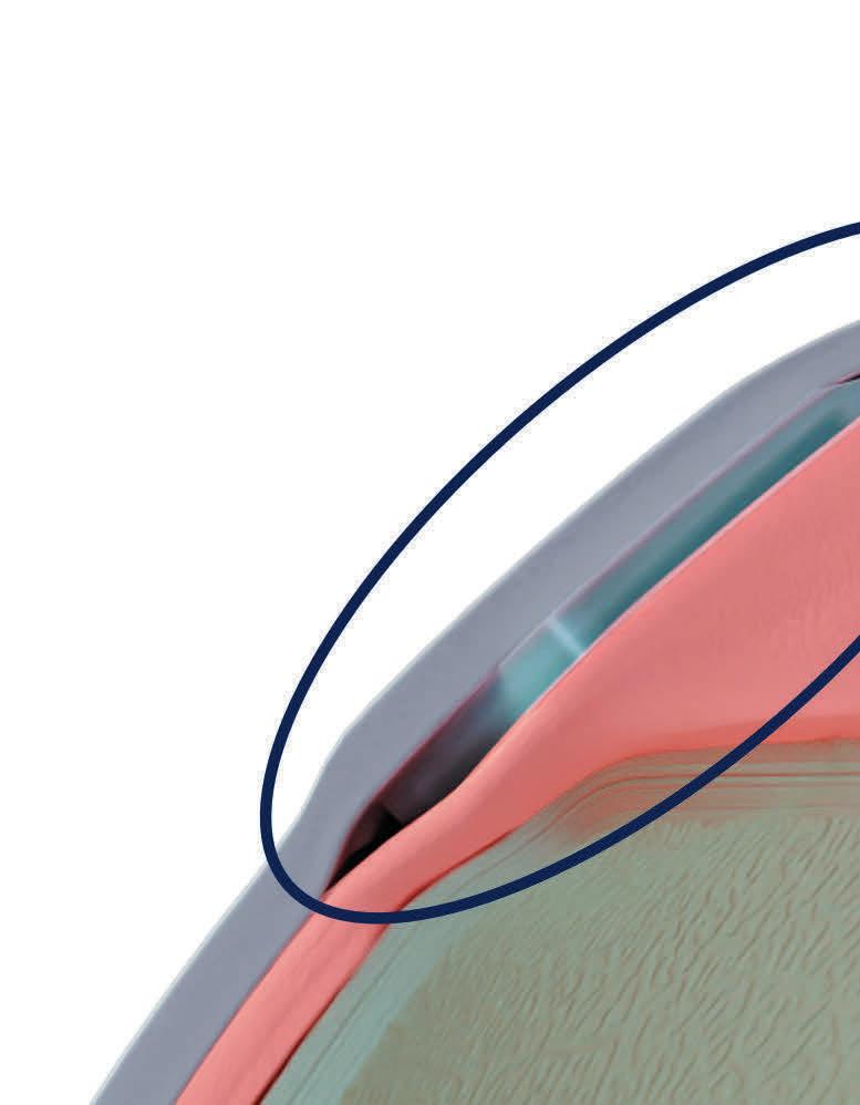
And so far, the preliminary results are promising. “Not only were we able to achieve a 1.6 mmHg lower intraocular
This is made possible, he said, by optically sparing the conjunctiva and the anterior segment of the eye using only two radial conjunctival incisions over the scleral incision sites. And the CID location neither prevents access to the corneoscleral trabeculum nor the subconjunctival filtration.
CID implantation takes no longer than 15 minutes. The implantation requires the surgeon to perform two manual penetrating scleral incisions. They must apply scleral sutures
36 GLAUCOMA EUROTIMES | JUNE 2023
This is a very important milestone because many promising devices usually fail to deliver on their promises at this point.
32%
The 42 patients implanted with the CID in clinical studies had a mean IOP lowering of 32% at year two, with a measured 16.2 mmHg (range 12.0 to 20.0 mmHg) pressure average.
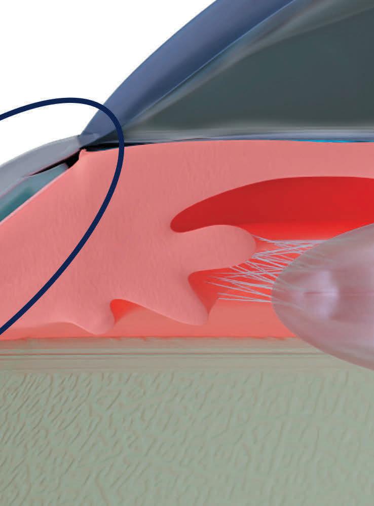
after the implantation to avoid any subconjunctival bleb, he explained.
“I can tell from the experience of Dr Lilit Albert Voskanyan, Chief of Glaucoma Surgery at the Malayan Ophthalmological Center in Yerevan—who conducted and performed the three series—that her maximum time for the implantation of a CID device is set around nine minutes. By doing just two radial conjunctival incisions, this surgical technique minimises the risk of conjunctival scarring and the failure risk of a second operation.”

Ciliatech hopes to apply for the CE mark before the end of the year or possibly next year.
“We are already consulting with the FDA to define the best approach to obtain US market approval,” Dr Sourdille concluded.
Learn more at ESCRS.org
37 2023 JUNE | EUROTIMES
Philippe Sourdille MD is an ophthalmologist, researcher, inventor, and medical director and co-founder of Ciliatech in Chavanod, France. p.sourdille@cilia.tec
To be the leading community and trusted source for science, education, and professional development in the fields of cataract and refractive surgery.
New Option for GA Patients
Treatment approval paves the way to exciting new therapies.
TIMOTHY NORRIS REPORTS
The US FDA approval of the Complement C3 Inhibitor Syfovre (pegcetacoplan injection, Apellis) for treating geographic atrophy (GA) sets a remarkable milestone for both retina specialists and patients suffering from macular degeneration, leading the way to new and effective therapeutic solutions for a steadily increasing health challenge.
“For many years, the treatment for GA has been a significant unmet need in ophthalmology,” said Dr Valentina Sarao. “Geographic atrophy is a significant cause of visual impairment and blindness with a growing prevalence worldwide, particularly in developed countries with an ageing population. A prevalence that is expected to double in the next two decades, with a proportionally burdening economic and social impact. The approval of Syfovre is a landmark transition in the retina field, comparable to the introduction of anti-VEGF therapies for neovascular AMD.”
According to Dr Daniele Veritti, this recent approval by the FDA is only the first step in a rapidly expanding therapeutic pipeline.
“Significant advancements are expected in the next few years, with novel approaches and compounds to effectively treat the disease,” he said. “The traditional classification of advanced age-related macular degeneration into treatable neovascular AMD and untreatable geographic atrophy may soon become obsolete as novel therapies targeting GA will become widely available.”
The FDA decision follows the positive results of the OAKS and DERBY trials. Eyes treated with pegcetacoplan demonstrated a reduction of lesion growth compared to the sham group—19% in DERBY and 22% in OAKS with monthly injections, and 16% in DERBY and 18% in OAKS with injections every other month. Significant acceleration of the treatment effect was reported between months 18 and 24.
“The Phase 3 OAKS and DERBY trials, including 24 months of data, demonstrated a slowing of geographic atrophy lesions growth based on fundus autofluorescence lesion area quantification,” said Dr Frank G Holz. “Overall, pegcetacoplan was well tolerated with a safety profile generally in line with other therapeutics administered intravitreally. However, new-onset exudative AMD cases were reported higher with pegcetacoplan compared with sham.”
“Choroidal neovascularisation and new onset AMD represent the most important safety concerns, with a combined rate of 11.9% at month 24, but that is the only significant side effect observed,” said Professor Anat Loewenstein. “While still a matter of concern, we all know how to manage these side effects.”
Moving forward
Although not overwhelming, the overall results of pegcetacoplan represent an important glimmer, a “crack in a wall called geographic atrophy,” according to Prof Loewenstein.
“Giving injections to patients with 6/60 (20/200) vision will not restore visual acuity, but the mere fact that a drug able to slow GA progression has been FDA-approved gives it a lot of credibility,” she said. “Until now, GA patients were not usually seen by retina specialists, but rather by optometrists and general ophthalmologists. We now must acquaint ourselves more with these healthcare providers for the benefit of our patients and set up the right expectations to spare them from bitter disappointment. Therefore, we will have to explain the treatment will not restore their vision but will certainly slow down the disease and the progressive loss of central vision.”
New solutions for GA treatment are expected to follow shortly, tackling the disease from different angles and methods. The FDA granted the Breakthrough Therapy status to Zimura (avacincaptad pegol, Iveric Bio) in November 2022, “a novel drug with a safety and efficacy profile similar to pegcetacoplan that works on the inhibition of the C5 complement, instead of the C3,” Prof Loewenstein said.


Anat Loewenstein MD, PhD, MHA is Chair of the Department of Ophthalmology, Tel Aviv Sourasky Medical Center, and Professor and Vice Dean at Tel Aviv University, Israel. anatlow@tasmc.health.gov.il
Frank G Holz FEBO, FARVO is Professor and Chair of the Department of Ophthalmology, University of Bonn, Germany. Frank.Holz@ukbonn.de
Daniele Veritti MD Department of Medicine-Ophthalmology, University of Udine, Italy. moc.liamg@eleinadittirev
Valentina Sarao MD, Instituto Europeo di Microchirurgia Oculare (IEMO), Udine, Italy. moc.liamg@anitnelav.oaras
RETINA 38 EUROTIMES | JUNE 2023
LEADERSHIP AND BUSINESS INNOVATION PROGRAMME VIENNA 2023
ESCRS Leadership & Business Innovation Masterclass
Sunday, 10 September
Strauss 3, 08.30–18.00
The Fine Art of Juggling
Ophthalmologists must be master jugglers. Most of these skills are learned on the job with no formal training. The expectation is they will manage them all—and do them well! In this highly interactive and didactic workshop, ESCRS Consultant Kristine Morrill and an expert panel will cover topics including Finding the Hidden CEO, Managing Money, and Encouraging Creativity.
Contributing panel: Omid Kermani, Co-founder, Augenklinik am Neumarkt, Germany; Daniel Kook, Co-founder, Prof Kook & partner, Germany; James Ball, Founder, Custom Vision Clinic, UK; Paul Rosen, Consultant Ophthalmic Surgeon at the Oxford Eye Hospital, Oxford University Hospital Foundation Trust, UK; Michael Mrochen, Founder & CEO, IROC Science, Switzerland; Damien Gatinel, Chief, anterior segment, France; Detlef Holland, Co-founder, Augenzentrum One, Germany.
ESCRS Leadership & Business Innovation Workshops
Monday, 11 September
Kristine Morrill, France Chairman Paul Rosen, UK
TOPICS INCLUDE:
Strauss 3, 09.00–18.00
• Business Skills For Young Ophthalmologists
• Mastering The Art Of Negotiation
• Building And Developing Your Private Practice


• Value-Based Healthcare
• The Cost And Savings Of Sustainability In Ophthalmology
• Getting An Ophthalmological Business Back On Its Feet During Wartime
• Managing Patient Flow In A Busy Ophthalmological Practice
FOR FULL PROGRAMME, VISIT: https://congress.escrs.org/programme/programme-overview/
LEARN TO JUGGLE MANAGEMENT ROLES AT ESCRS CONGRESS
Leadership and Business Innovation Masterclass aims to expand the skill sets of all ophthalmologists.
BY HOWARD LARKIN
Whether running a private or public clinic, conducting medical research, or developing new technologies, building a successful ophthalmology practice requires mastering skills beyond those taught in medical school. Juggling multiple management roles in a rapidly changing environment is the focus of this year’s Leadership and Business Innovation Programme Masterclass at the 41st Congress of the ESCRS in Vienna.
Designed for all ophthalmologists, the interactive event features clinicians from across Europe sharing what they’ve learned building and managing successful practices, departments, and companies. “The panel members are in ophthalmology practice today, living this in the trenches, not just pontificating from on high. It will be an open exchange of ideas, not just lectures,” said session co-organiser and ESCRS consultant Kristine Morrill.
Breakout sessions will include:
Finding your hidden CEO


Omid Kermani MD and Daniel Kook MD, PhD, who both launched successful clinics with partners in Germany, will share their insights into developing leadership skills needed to lead from opposite ends of the career journey. Nearing retirement, Dr Kermani is planning for what comes next after a long successful run, while Dr Kook shares his experience building a profitable private practice in the past three years. “It will be an interesting conversation about how their paths have followed each other and what they’ve learned along the way,” Morrill said.
Money management—Balancing profitability with patient care

James Ball MA (Cantab), FRCOphth, discusses his process of balancing the
costs with the added patient benefits of building a cataract surgery suite in his office in the UK. With better patient service and care as his guiding light, Dr Ball calculated the financial margin he needed to make the improvements and priced his private services to reflect their added value. “It’s refreshing to talk to someone who is not a finance manager about this part of the practice-building equation,” Morrill said.
Human resource mistakes and what we learned from them
Paul Rosen BSc, MB ChB, FRCS, FRCOphth, MBA, who ran a large public ophthalmology department in the UK, and Morrill, who manages an ophthalmology business consulting firm from France, discuss their experiences with human resources. Even if based in a large clinic with an HR department, ophthalmologists still deal with employee issues daily, Morrill pointed out. These include hiring, training, managing, developing, and firing employees for optimal productivity and morale—all while complying with EU and local
laws and regulations. The session will feature practical advice on managing HR issues without spending a fortune and
40 LEADERSHIP AND BUSINESS INNOVATION EUROTIMES | JUNE 2023
avoiding what can be very expensive HR mistakes, such as making a bad hire.
Ophthalmologists as inventors— Moving from idea to reality
Whether a new device, service, or medication, most ophthalmologists have ideas about improving patient care and practice efficiency. This panel includes three who moved their inventions from idea to product.
Florent Costantini MD of France discusses how he and his brother Mathieu, also an ophthalmologist, developed Glasspop, an automated subjective refractor; Nick de Pennington MA, BM Bch, MBA of the UK talks about Ufonia, an AI-based system for following up with patients by phone; and Robert McLaren FMedSci, FRCOphth, FRCS, FACS, VR covers his work with Nightstar Therapeutics/Biogen developing gene therapies. “We will cover all the steps from idea to creating a prototype, raising money, getting CE marking and addressing regulatory challenges, and pitching and partnering with financial backers and other firms to develop and market products,” Morrill explained.
Understanding the EU regulatory environment
Clinical studies are popular and essential for advancing ophthalmic practice, particularly given the requirements to maintain CE marks under the EU Medical Device Regulation (MDR). But a lot of doctors don’t realise they can’t just collect data and do anything with it, Morrill said. “We get documents with patient information, names, and addresses all the time. Doctors really need to know this information is protected by the EU General Data Protection Regulation,” or GDPR. Michael Mrochen MD, PhD of Switzerland will walk colleagues through current regulations for clinical studies, such as GDPR, MDR, and compliance with Good Clinical Practice and local ethics committee review. He will also discuss quality management, including ISO:9001 compliance and certification.
Embracing creativity
A hobby that makes the ophthalmologist happy and provides a different perspective on life is critical to be an effective surgeon, businessperson, and family member.
Damien Gatinel MD, PhD of France presents his music and photography; Detlef Holland MD of Germany sailing; and Florian Auerbach MD, FEBO of Germany visual arts. “Hobbies give your life balance. Make sure to take the time,” Morrill advised.
The Masterclass will take place from 8:30 through 17:30 Saturday, 10 September 2023, at the Messe Wien venue in Vienna, Austria. Save the date!
CTF/TCT optic designed to:


REDUCE GLARE & HALOS1

TOLERATE THE KAPPA ANGLE2
TOLERATE DECENTRATION 3

TOLERATE MISALIGNMENT 4


PRESBYOPIA & ASTIGMATISM CORRECTION REINVENTED


Kristine Morrill is co-founder and president of Medevise Consulting,
bourg, France, and a consultant to ESCRS. kris-escrs@medevise-consulting.com
41 2023 JUNE | EUROTIMES
1) Broader Toric meridian designed to be more tolerant of misalignment. White paper: Evaluation of a new toric IOL optic by means of intraoperative wavefront aberrometry (ORA system): the effect of IOL misalignment on cylinder reduction. By Erik L. Mertens, MD Medipolis Eye Center, Antwerp, Belgium 2) The misalignment tolerance and the use of segments instead of concentric rings reduces photic phenomena, helping patients to adapt more naturally to their new vision. 3) The central zone of 1.4 mm in diameter is larger than most available mIOLs and allows a wider tolerance so that the visual axis passes through the wider central segment avoiding visual disturbances. 4) In cases of tilt or misalignment, the patient can still benefit from good near and far vision, as the segmented zones allow a balanced far/near light distribution in a steady optical platform. OPHTEC | Cataract Surgery
Stras
WHO IS LOOKING OUT FOR OPHTHALMOLOGY?
It is hard enough to keep up with the daily demands of a busy ophthalmology practice, much less with the sometimes bewildering EU regulatory landscape. Nonetheless, policy decisions made in the EU parliament affect everyone doing research or clinical work. EuroTimes Editor-in-Chief Sean Henahan spoke with Mor Dickman MD, PhD, who provided insight on current initiatives affecting ophthalmology.
Busy clinicians barely have the time to read the newspaper, much less lobby the EU. What do they need to know?

There is a lot of frustration among our clinical colleagues because there is little understanding of how the European mechanism works and where we can influence matters that are important to us, even in the short and medium term. You achieve that only by engaging with the regulators and policymakers to explain what is important, what should be done, what should not be done, what would be cumbersome and unnecessary, and what is really needed.
In terms of a regulatory landscape in Europe, who is looking out for ophthalmology?


An organisation called the European Alliance for Vision Research and Ophthalmology (EU EYE) engages with regulators and policymakers to advance the interests of the ophthalmology community and ensure we are represented in the wide context of EU health policies. This alliance of major ophthalmology societies is a non-profit, pan-European advocacy organisation. It is important to mention the ESCRS played a key role in establishing this. Peter Barry was the first president of EU EYE, and Paul Rosen is continuing this work.
The gamechanger was the recognition of EU EYE as an eligible organisation for the EMA, the European Medicines
Agency. I want to congratulate Ioanna Psalti on her work in achieving this. I represent the EU EYE at Health Care Professional Workgroup (HCPW) at the EMA. This is an interesting forum where we gather medical specialists from all the specialties in Europe and provide the EMA and its scientific committees with recommendations related to medicines and medical devices.
You are also involved with the European Alliance for Transformative Therapies (TRANSFORM). Tell us about that.
TRANSFORM is a multi-stakeholder alliance that connects members of the EU parliament with policymakers, medical experts, patient groups, medical associations, scientists, and industry. All these stakeholders are involved in this very exciting field of advanced therapy medicinal products (ATMPs). These therapies have tremendous potential to influence how we may treat eye diseases, but also the implications for the sustainability of our healthcare systems. It is about all stakeholders coming together for a common goal.
For ophthalmology, this relates to cell therapies. The field of ophthalmology has been quite pioneering in this area. Holoclar, the first stem cell therapy approved by EMA in 2015, was for treating limbal stem cell deficiency. One of the first gene therapies approved by EMA was Luxturna to treat retinal degeneration related to a mutation in the RP65 gene.
Since 2007, all the regulatory work related to ATMPs is coordinated by one body, the EMA. Within the EMA, the
committee for advanced therapies is responsible for proving scientific opinions on product quality, safety, and efficacy. In one of the most interesting developments in recent years, the EMA is inviting clinical researchers—especially academics and non-profit organisations—to engage with the regulatory bodies at a very early stage to gain an understanding to help them overcome obstacles they may encounter. Initiatives such as the PRIME project focus on driving these products quickly from the lab to the bedside.
What ophthalmology ATMPs are in the pipeline now?
Several ATMPs are in various stages of clinical development, including cell therapies for age-related macular degeneration, inherited retinal disorders, and corneal diseases. A second group are gene therapies using adenovirus vectors,

42 EUROTIMES | JUNE 2023
NEWSMAKER
The gamechanger was the recognition of EU EYE as an eligible organisation for the EMA, the European Medicines Agency.
An interview with Professor Mor Dickman
developed mostly for inherited retinal diseases, including choroideremia and Leber congenital amaurosis. A third and very exciting category is RNA-based therapies for inherited retinal diseases such as Stargardt disease and cornea diseases such as Fuchs’ dystrophy.

What about the costs of these new therapies?
Cost is a very important question. Normally, you price a new treatment against an alternative, but these conditions have no alternative, which means it will be expensive. Research and development
costs are very high, and the regulatory pathway is also quite expensive. We are experimenting to see if we can improve this situation by using the hospital exemption rule more often—a European legislative initiative that guarantees patient access to novel therapies based on the use of ATMPs in the member states.


Mor M Dickman MD, PhD is professor of ophthalmology at the University Eye Clinic, Maastricht UMC, Netherlands. m.dickman@maastrichtuniversity.nl
43 2023 JUNE | EUROTIMES
Eye
EU
www.eueye.org
The European Alliance for Transformative Therapies transformalliance.eu
learn more at escrs.org
The leading community and trusted source for SCIENCE, EDUCATION & PROFESSIONAL DEVELOPMENT in the fields of cataract and refractive surgery.
Slit Lamp Competition
Haag-Streit is giving eye care professionals across the globe an opportunity to showcase their slit lamp imaging skills in their “Slit Lamp Imaging Competition 2023”. Judging criteria includes image quality, technical execution, and disease interest. In addition to receiving a trophy, the competition winner will feature in an ophthalmic journal and receive a Nikon Z 6 II with NIKKOR Z 24–120 mm F/4 camera and NIKKOR 26 mm f/2.8 lenses. Second prize is an Apple Watch Ultra, and third prize is an Ona Brixton leather camera bag. The competition is open until 11 August 2023. To enter, visit www.hsimagingcompetition.com
www.haag-streit.com
New Corneal Ectasia Display for Eyestar 900
Haag-Streit announced the release of a new corneal ectasia display for its Eyestar 900 sweptsource-based precision OCT analyser. The new display provides clinicians with OCT-based topography of the cornea’s anterior and posterior surface, pachymetry maps and profiles, and Belin ABCD grading. It provides corneal specialists with a highly precise and sensitive means of detecting ectasia and the earliest signs of keratoconus in both eyes in a 40-second examination. The new display adds to the Eyestar 900’s Anterior Chamber Suite high-quality 18 mm diameter OCT images of the anterior chamber—including the crystalline lens—for visual inspection of key structures such as lens position, ICL vault, or chamber angle.
www.haag-streit.com
Oertli Collaborates with Lucerne University
Oertli Instrumente AG has joined the Lucerne University of Applied Sciences and Arts to launch the “Oertli Ophtha Lab”. The lab gives Oertli access to the latest technical developments, such as artificial intelligence, blockchain technology, and robotics, enabling the company to integrate such research into its development projects. By linking research and education, the Ophtha Lab provides students from various engineering, medical, and biological disciplines with an innovative and collaborative research laboratory to complete their studies.
www.oertli-instruments.com
New Tonometer from Nidek
NIDEK announced the launch of a new addition to its NT-1 series of non-contact tonometers. In response to market demand, the new NT-1/1e model is similar to the NT-1p non-contact tonometer/pachymeter but without the pachymetry function. The NT-1e includes the other standard features of the NT-1 series, such as the 3D auto-tracking function and a screen with a continuous range of tilt and swivel, allowing the clinician to place the device anywhere in an examination room.
44 INDUSTRY NEWS NEWS IN BRIEF EUROTIMES | JUNE 2023
REDUCING WASTE IN OPHTHALMIC SURGERY
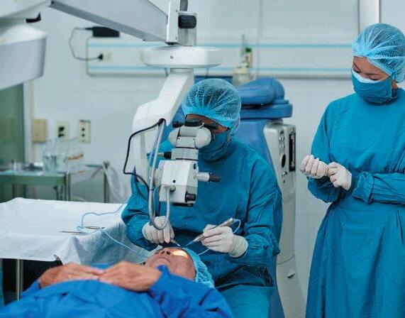
Dr David F Chang outlined the growing consensus among American and European ophthalmic surgeons that operating room (OR) waste is excessive and should be reduced, pointing to a 2019 report showing the healthcare sector is responsible for 4.4% of greenhouse gas emissions worldwide and nearly 10% in the US. ORs account for a major share of the healthcare sector’s emissions and waste. Yet, in an ESCRS survey and a similar survey conducted by the Ophthalmic Instrument Cleaning and Sterilization (OICS) Task Force, 43% of North American and 32% of European cataract surgeons said they were completely unaware of the sector’s environmental impact. Most respondents in both surveys want their medical societies to advocate for more sustainable eye care delivery. To reduce unnecessary ophthalmic surgical waste, ASCRS, ESCRS, and AAO launched EyeSustain.org. Experience at India’s Aravind Eye Care System (AECS) demonstrated it possible to safely reduce carbon emissions in cataract procedures by 95% by implementing waste-reducing practices such as reuse of surgical gowns, gloves, irrigation bottles, phaco cassettes, irrigation-aspiration tubing, and more. Such practices are forbidden by US infection control regulations, even though AECS hospitals and clinics show the same endophthalmitis rates (0.04%). “Due to their staggering economic and environmental impact, overzealous and unproven infection control practices and regulations in ophthalmic surgery will ultimately do more harm than good to the overall health of our society,” he concluded.
DF Chang, et al., “Tackling the challenge of needless surgical waste in ophthalmology”, 49(4): 333–338.
SURVEY SHOWS MOTIVATION TOWARDS ENVIRONMENTAL SUSTAINABILITY
The majority of 458 respondents to an ESCRS survey of cataract surgeons shows 92% felt operating room waste is excessive and should be reduced, with most expressing a strong desire for more reusable options for instruments, devices, and supplies. Their responses closely mirror those from an identical survey of North American cataract surgeons published in 2020. Regarding global strategies to reduce surgical waste, 94% of ESCRS survey respondents said they want manufacturers to use recycled content in packaging, 92% want manufacturers to offer more reusable instruments and supplies, and 89% would prefer manufacturers and regulatory bodies allow more surgeon discretion in reusing products. Like their North American counterparts, most ESCRS survey respondents are either reusing or willing to reuse topical or intraocular pharmaceuticals and many surgical supplies. However, more ESCRS surgeons than North American surgeons currently reuse intraocular antibiotics (48% vs 32%), miotics (28% vs 20%), lidocaine (39% vs 30%), capsular dye (21% vs 10%), phacoemulsification tips (48% vs 38%) and tubing (21% vs 7%), irrigating solution (26% vs 8%), cystotomes (32% vs 13%), and disposable surgical devices (16% vs 9%). Compared to their North American counterparts, more ESCRS respondents considered endophthalmitis risk a major factor (64% vs 48%)—though fewer felt strongly influenced by cost savings (47% vs 63%), efficiency (49% vs 63%), and malpractice liability (41% vs 51%). DF Chang, et al., “Survey of ESCRS members’ attitudes toward operating room waste”, 49(4): 341–347.
ESCRS iLearn is an online learning platform, free for ESCRS members.
Visit elearning.escrs.org to access over 30 hours of interactive, assessed, and accredited e-learning content, including surgical videos, diagrams, animations, quizzes, and forums.
45 JCRS HIGHLIGHTS 2023 JUNE | EUROTIMES
iLEARN
Cited in this Issue
Presbyopic LASIK
Page 21
1. S Ganesh, et al., “Visual and Refractive Outcomes following laser blended vision using nonlinear aspheric micro monovision”, J Refract Surg, 2020; 36: 300–307.
Glaucoma Treatment Paradigm Shift
Page 33
1. Saheb H, Ahmed IIK. “Micro-invasive glaucoma surgery: current perspectives and future directions.” thalmol.
2. Grieshaber MC, Pienaar A, Olivier J, Stegmann R. “Clinical evaluation of the aqueous outflow system in primary open-an gle glaucoma for canaloplasty.” 51(3): 1498–1504. doi:10.1167/iovs.09-4327
3. Fellman RL, Grover DS. “Episcleral Venous Fluid Wave in the Living Human Eye Adjacent to Microinvasive Glaucoma Sur gery (MIGS) Supports Laboratory Research.” 28(2): 139–145. doi:10.1097/IJG.0000000000001126

4. Smit BA, Johnstone MA. “Effects of viscoelastic injection
into Schlemm’s canal in primate and human eyes: potential relevance to viscocanalostomy.” Ophthalmology. 2002; 109(4): 786–792. doi:10.1016/s0161-6420(01)01006-5
5. Brüggemann A, Despouy JT, Wegent A, Müller M. “Intraindividual comparison of Canaloplasty versus trabeculectomy with mitomycin C in a single-surgeon series.” J Glaucoma. 2013; 22(7): 577–583. doi:10.1097/IJG.0b013e318255bb30
ESCRS Academies
Committee representatives of ESCRS organise and present sessions at meetings organised by our national and sister societies. These sessions are typically delivered by a group of speakers on a current topic selected by ESCRS in person or virtually.
These sessions provide useful education as well as collaboration between societies promoting and sharing benefits across both memberships.

46
CITATION INDEX
EUROTIMES
escrs.org/education/academies/
Our IOL Calculator is now live on the ESCRS website!

This first-of-its-kind web application for IOL power calculations uses multiple modern formulas simultaneously, and suggests lens constants for a wide range of IOL models.
Find out more at iolcalculator.escrs.org/
47 2023 JUNE | EUROTIMES
ESCRS Educational Forum is supported by multiple industry partners to provide independent didactic education on selected therapeutic areas. The platform combines presentations from ESCRS Winter and Annual Congresses, selected EuroTimes articles, videos, and webinars to provide an in depth overview on current clinical outlooks.
escrs.org/education/forum/

Forum
Education
Upcoming Events
June 8–10
35th APACRS Annual Meeting
Singapore

June 8
Throughout June
The ESCRS Moving Simulator
Romania
June 14
ESCRS eConnect Webinar
Trials and tribulations of the small pupil: achieving (and maintaining) the intraoperative mydriasis
June 15–17
European Society of Ophthalmology (SOE)
Prague, Czech Republic

June 28–July 1
World Glaucoma Congress
Rome, Italy

Sep 8–12
41st Congress of the ESCRS
June 15
Vienna, Austria Sep
June 28

49
2023 JUNE | EUROTIMES
8
MINIMALLY INVASIVE GLAUCOMA SURGERY WITH TRABECULAR MICRO BYPASS MAY SLOW FUNCTIONAL PROGRESSION
Kevin Gillmann MD, PhD. MBBS. MBA. FEBOphth. MArch. MCMI. PgCert. (Harvard) Genève Ophtalmologie, Geneva, Switzerland Byers Eye Institute, Stanford University, USA

The role of true endpoints in glaucoma trials and the effect of iStent devices on functional progression
To date, intraocular pressure (IOP) remains the only modifiable risk factor for glaucoma progression, and as such, is the cornerstone of all glaucoma treatments. Intraocular pressure measurements are also readily available, convenient, and inexpensive, making them attractive endpoints for glaucoma trials. Yet, most ophthalmologists would agree that the primary goal of any glaucoma treatment is not to lower IOP, but to preserve patient’s functional vision and quality of life. Besides, while IOP is an important factor in glaucoma management, it was never formerly validated as a surrogate for true endpoints or a predictor for functional loss in glaucoma. This is the main reason why a number of authors and guidelines, including the World Glaucoma Association consensus have been calling for a shift in the focus of glaucoma clinical trials towards biomarkers that would better represent disease stability, such as structural, functional or even composite endpoints.1,2
glaucoma.4 In addition, static IOP endpoints rarely take into account other factors responsible for glaucoma progression such as endogenous and exogenous IOP fluctuations, patient compliance, or non-pressure dependent mechanisms.
“In the case of the iStent devices, the observed functional outcomes suggest that early intervention may have a role to play in mild glaucoma where MIGS procedures could help slow progression”.
This specific example shows that, as clinical trial methods evolve and data accrue, true endpoints are gradually becoming a reality in glaucoma. Functional and structural endpoints take a different perspective on treatment outcomes. The greater emphasis on the broader picture they provide may lead to new, more clinically-relevant conclusions.
Finally, and generally, these observations are another reminder that, as clinicians and researchers, we should strive to shift our focus away from traditional static endpoints, towards more patient-centered measures such as functional stability and quality of life.
Minimally invasive glaucoma surgery (MIGS) are good examples of how focusing solely on IOP endpoints may lead to a biased approach to decision making in glaucoma. In an ongoing meta-analysis of the effect of the iStent devices on functional progression, we identified 24 individual trials reporting pre-operative and postoperative visual field data for a total of 1305 eyes.3 When looking at the mean deviation (MD), half of these studies reported either functional stability or improvement between baseline and the last timepoint. Considering all the identified studies, the overall weighted mean progression rate was well under 0.1 dB per year, over a mean duration of 40 months – below most reported progression rates for treated glaucoma. Interestingly, while mean IOP reduction ranged widely across the studies [15.2%42.7%], it correlated poorly with functional progression measures, further supporting the idea that, while an important measurement, IOP may not be a suitable surrogate endpoint for functional stability. A number of explanations have been put forward by various authors, including the fact that, within physiological values, IOP does not appear to be directly correlated with functional progression in mild
3. Gillmann K. Unpublished data. 4. Yohannan J, Boland MV, Ramulu P.The Association Between Intraocular Pressure and Visual Field Worsening in Treated Glaucoma Patients.J Glaucoma. 2021 Sep 1;30(9):759-768
iStent inject® IMPORTANT SAFETY INFORMATION
2019. 11th Consensus Meeting: Glaucoma Surgery. 2. Medeiros FA. Biomarkers and surrogate endpoints in glaucoma clinical trials. Br J Ophthalmol. 2015 May;99(5):599-603.
INDICATION FOR USE: The iStent inject, is intended to reduce intraocular pressure safely and effectively in patients diagnosed with primary open-angle glaucoma, pseudo-exfoliative glaucoma or pigmentary glaucoma.The iStent inject, can deliver two (2) stents on a single pass, through a single incision. The implant is designed to stent open a passage through the trabecular meshwork to allow for an increase in the facility of outflow and a subsequent reduction in intraocular pressure. The device is safe and effective when implanted in combination with cataract surgery in those subjects who require intraocular pressure reduction and/or would benefit from glaucoma medication reduction.The device may also be implanted in patients who continue to have elevated intraocular pressure despite prior treatment with glaucoma medications and conventional glaucoma surgery. CONTRAINDICATIONS: The iStent inject System is contraindicated under the following circumstances or conditions: • In eyes with primary angle closure glaucoma, or secondary angle-closure glaucoma, including neovascular glaucoma, because the device would not be expected to work in such situations. • In patients with retrobulbar tumor, thyroid eye disease, Sturge-Weber Syndrome or any other type of condition that may cause elevated episcleral venous pressure. WARNINGS/PRECAUTIONS: • For prescription use only. • This device has not been studied in patients with uveitic glaucoma. • Do not use the device if the Tyvek® lid has been opened or the packaging appears damaged. In such cases, the sterility of the device may be compromised. • Due to the sharpness of certain injector components (i.e. the insertion sleeve and trocar), care should be exercised to grasp the injector body. Dispose of device in a sharps container. • iStent inject is MR-Conditional; see MRI Information below. • Physician training is required prior to use of the iStent inject System.• Do not re-use the stent(s) or injector,as this may result in infection and/or intraocular inflammation,as well as occurrence ofpotential postoperative adverse events as shown below under “Potential Complications.” • There are no known compatibility issues with the iStent inject and other intraoperative devices. (e.g., viscoelastics) or glaucoma medications. • Unused product & packaging may be disposed of in accordance with facility procedures. Implanted medical devices and contaminated products must be disposed of as medical waste. • The surgeon should monitor the patient postoperatively for proper maintenance of intraocular pressure. If intraocular pressure is not adequately maintained after surgery, the surgeon should consider an appropriate treatment regimen to reduce intraocular pressure. • Patients should be informed that placement of the stents, without concomitant cataract surgery in phakic patients, can enhance the formation or progression of cataract. ADVERSE EVENTS: Please refer to Directions For Use for additional adverse event information. CAUTION: Please reference the Directions For Use labelling for a complete list of contraindications, warnings and adverse events. Glaukos®, iStent®, iStent inject® and iStent inject® W are registered trademarks of Glaukos Corporation.All rights reserved. ©2023. PM-EU-0245
Figure 1 – Mean change in visual field mean deviation (MD) in decibel per year across all 24 identified studies.
References: 1. Weinreb, R.N., Ramulu, P., Topouzis, F., Park, K.H., Mansouri, K. and Lerner, F.,
ADVERTISEMENT
“The primary goal of any glaucoma treatment is not to lower IOP, but to preserve patient’s functional vision and quality of life”.


