Introduction
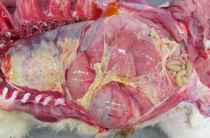
Polyserositis in calves is defined as inflammation of multiple serosa (peritoneum, pleura or pericardium), due to a bacterial infection. In veal calves, submitted to Royal GD by veal calf farmers for necropsy in 2022-2023, 83 cases of fatal polyserositis were identified of which 54 with a Mannheimia haemolytica (M. haemolytica) infection and second most frequent with Escherichia coli. The high involvement of M. haemolytica was reason to further explore this relationship. In polyserositis in these calves, serotype A2 completely dominated the situation and serotype A1/A6 were absent. To answer the question if the dominance of serotype A2 was the result of a high prevalence of A2 in veal, we performed culturing and whole genome sequencing (WGS) for M. haemolytica from 11 tissues of 54 calves with Mannheimia polyserositis and controls calves after necropsy.
Materials and methods
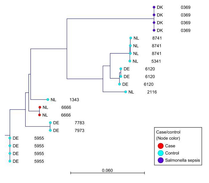
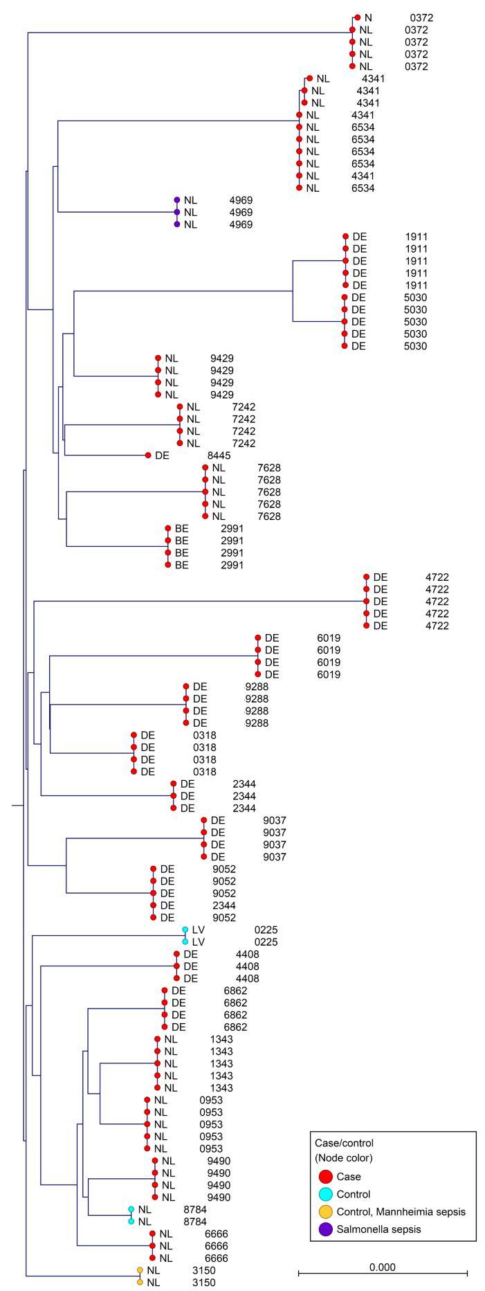
In these 54 cases of polyserositis, pleura, peritoneum, pericardium, nasal cavity, tonsils, bronchia, spleen, retropharyngeal lymph nodes, bronchial lymph nodes, mesenterial lymph nodes and cerebellum were sampled to isolate M. haemolytica. The same, excluding serosa, was done for 50 control calves without polyserositis. M. haemolytica isolates were used for WGS, to identify serotype and relatedness of the isolates derived from the different tissues. Due to the high number, WGS was applied to a selection of 98 isolates from the serosa, spleen, nasal cavity, retropharyngeal and bronchial lymph nodes of 23 cases. All 19 isolates derived from 14 controls were sequenced. The remaining controls were M. haemolytica negative.
Results
Serotyping based on WGS resulted in 96 times serotype A2, obtained from nasal cavities, spleens, serosa, bronchial lymph nodes and lung tissues. Two were serotype A1, which were retrieved from the nasal cavity and the retropharyngeal lymph nodes. In contrast, 15 control calf isolates were serotype A1 and four were serotype A2. These were mainly found in tonsils, nasal cavity and bronchial bifurcation. In addition, the results show that per individual calf, one specific M. haemolytica strain is causing the disease.
Conclusions
M. haemolytica serotype A2 is widely spread in calves with M. haemolytica polyserositis. In contrast, in control animals mainly serotype A1 was found. This is an indication that the linkage between polyserositis and serotype A2 is not simply caused by a wider distribution of this serotype A2 in the Dutch veal calf industry but must have another cause.
Acknowledgements
The project was financed by the producers organization for veal (SBK, Zeist, The Netherlands).

e.v.engelen@gdanimalhealth.com www.gdanimalhealth.com