Parkinson’s disease is the fastest-growing neurological disorder globally and is the second most common after Alzheimer’s disease. Parkinson’s is a neurodegenerative condition, meaning it worsens progressively, and there is currently no cure or drugs
Cont’d on page 4
Novel Blood Test Detects Hidden Inflammation Across Multiple Diseases
Inflammation is a key factor in almost every disease, yet existing blood tests are unable to specify which organs or tissues are affected. In a groundbreaking discovery that could revolutionize how doctors diagnose and monitor a variety of conditions, researchers
have now identified unique chemical markers that could enable blood tests to detect inflammation in specific organs.
The research, conducted by scientists at Case Western Reserve University (Cleveland, OH, USA; case.edu), focuses on compounds
Cont’d on page 8
POC Test Prevents Antibiotic Overuse
TDNA Sequencing-Enabled Metabolite Detection Could Transform Diagnostics
Metabolite Detection Could Transform Diagnostics
novel platform for small molecule sequencing utilizes short DNA sequences called aptamers to detect metabolites, thus ushering a new breakthrough in diagnostics by rendering metabolite detection as easy and rapid as DNA sequencing.
New Platelet Counting System Helps Prevent Diagnostic Errors
Accurate platelet count testing is a significant challenge for laboratories. Inaccurate results can lead to misdiagnosis, missed diagnoses, and delayed treatment for a variety of potentially fatal conditions, including acute massive hemorrhage, coagulation disorders, infections, autoimmune diseases, and
Traditional diagnostic methods for autoimmune diseases and other immunological conditions typically combine physical examinations, patient history, and laboratory tests to detect cellular or molecular abnormalities. However, this process is often
time-consuming and complicated by misdiagnoses and ambiguous symptoms. These methods generally do not take full advantage of data from the patient’s adaptive immune system, particularly from B cell receptors (BCRs) and T cell receptors (TCRs). In response
he misuse of antibiotics— such as taking them unnecessarily, for too long, or in incorrect doses—leads to antimicrobial resistance (AMR), making infections harder to treat and increasing the risks of severe illness and death. AMR causes 700,000 global deaths annually, and many people either expect antibiotics from doctors or misuse them by not following prescribed dosages.
Cont’d on page 10
Predicting Response to Cancer Treatment
Breast cancer is the most common cancer among women worldwide, with 2.3 million new cases diagnosed each year. In the era of personalized medicine, targeted therapies for different types of breast cancer are now available. However, despite these advancements, some tumors remain difficult to treat. As a result, even with significant progress in early diagnosis and treatment
Cont’d on page 2
Predicting Response to Cancer Treatment
options, 670,000 women worldwide still die from breast cancer annually. Historically, predicting how well a patient’s tumor will respond to available treatments has been challenging. This uncertainty means some patients undergo ineffective and unpleasant treatments. There is a need for more accurate lab testing methods to help doctors improve clinical outcomes. Researchers have now developed a technique that could predict how well certain breast cancer patients will respond to chemotherapy and antibody-directed treatments.
In their study, researchers at the University of Leicester (Leicester, UK) applied advanced digital pathology and multi-immunofluorescent techniques to examine changes in patient tumor samples in the lab. Building on their previous work with non-small cell lung cancer and endometrial cancer patient-derived explants (PDEs), the team discovered a significant link between explant responses to chemotherapy drugs and patient outcomes. This finding set the stage
for their current research. Published in Nature Scientific, the new study involved measuring the stability of tumor explants from 55 breast cancer patients over time. The tumors were ‘treated’ with either chemotherapy or the HER2 antibody therapy trastuzumab in the lab.
The results showed that using the PDE technique, the tumor architecture was preserved for up to 72 hours during testing, and the immune microenvironment remained intact. This was particularly encouraging, as other testing methods have struggled with maintaining these characteristics, providing the researchers with confidence that their testing was meaningful. The team then compared their observations of the tumor explants’ response to treatment in the lab with the clinical progression of the same patients. They found a similar pattern in the explant’s response to treatment and the patient’s progression. This is especially promising, as it suggests that the tumor explant in the lab responded in a manner similar to the patient’s tumor in the clinic.
“Our findings suggest that our patient derived explant technique could be a suitable preclinical testing platform for some breast cancer patients,” said Gareth Miles, Lecturer in Cancer Patient Derived Explant Technology at the University of Leicester. “It could provide a more accurate method to predict how they may respond to particular treatments. Doctors can avoid giving ineffective treatments, saving time and ultimately improving a patient’s clinical outcome.”
Image: The technique predicts how well some breast cancer patients will respond to chemotherapy (Photo courtesy of Shutterstock)
Discovery Improves Parkinson’s Diagnosis
that can slow or stop its progression. The diagnosis of Parkinson’s primarily relies on clinical evaluation of motor symptoms, which can result in diagnostic delays or even misdiagnosis. Early detection is critical, as current pharmacological treatments are more effective when administered in the early stages. A new discovery involving blood immune cells has brought researchers closer to identifying a blood biomarker that could help doctors personalize treatments for Parkinson’s disease. In a collaborative effort, researchers from the WEHI Parkinson’s Disease Research Centre (Victoria, Australia; www.wehi.edu. au) identified a new connection between blood immune cells and Parkinson’s disease. This research was part of a project that analyzed data from over 500,000 people, marking it as the largest study of its kind. White blood cells play a key role in immune responses and inflammation, and it is known that people with Parkinson’s disease exhibit higher levels of neuroinflammation. The researchers found that changes in the levels of certain blood immune cells could serve as a potential marker for Parkinson’s disease progression, bringing them closer to developing a blood test for diagnosing and monitoring the disease. Mitochondrial dysfunction has long been associated with Parkinson’s, and one potential biomarker for assessing mitochondrial health has been the measurement of the ‘mitochondrial DNA copy number’ (mtDNA-CN).
However, the new study, published in NPJ Parkinson’s Disease, challenges the previous understanding of the link between mtDNA-CN and Parkinson’s disease, suggesting it may not be due to mitochondrial dysfunction as previously thought. Mitochondria, the energy-producing structures in cells, have their own DNA, which is distinct from nuclear DNA. Each cell contains multiple mitochondria, each with multiple copies of mitochondrial DNA. Some cells, such as those in the
heart, require a large number of mitochondria, while others need far fewer. Given the significant role of mitochondrial dysfunction in Parkinson’s disease, it was previously believed that measuring mtDNA-CN in blood samples could serve as a useful biomarker. Since accurately counting the number of mitochondrial DNA copies is nearly impossible, the WEHI team developed a software algorithm to estimate the count from DNA sequencing data obtained from blood samples. They initially tested this method on a dataset of over 10,000 participants.
Their findings revealed that lower levels of mitochondrial DNA in the blood were not directly linked to an increased risk or severity of Parkinson’s disease, contrary to previous assumptions. This apparent connection disappeared when the researchers accounted for the different types of cells in the blood. Instead, they found a stronger association with certain immune cells, specifically neutrophils and lymphocytes, which are types of white blood cells. This suggests that the earlier reported link between mtDNA-CN and Parkinson’s is actually driven by immune responses in the blood, rather than mitochondrial dysfunction. Building on these findings, the researchers replicated their study using data from the UK Biobank, which included nearly 500,000 participants, making it the largest study of mtDNA-CN in Parkinson’s disease to date. This replication confirmed their results and underscored the robustness of their methodology. The researchers have made the specialized software, called mitoCN, available to other research teams worldwide, with the aim of advancing studies beyond Parkinson’s disease.
“The ultimate goal is to be able to screen for Parkinson’s disease in a similar way to the national screening program for bowel cancer, so people can get access to medication sooner,” said Professor Melanie Bahlo, one of the researchers who led the study.
Graham
Hernán
Argentina
Bernard Gouget France
Maurizio Ferrari Italy
Tahir S. Pillay South Africa
Andreas Rothstein Colombia
Praveen Sharma India
Rosa I. Sierra-Amor Mexico
Peter Wilding United States
Andrew Wootton United Kingdom
LabMedica lnternational is published eight times a year and is circuIated worldwide (outside the USA and Canada) without charge and by written request, to clinical laboratory specialists and administrators, and other qualified professionals allied to the field.
To all others: Paid Subscription is available for a two-year subscription charge of US$120. Single copy price is US$20. Mail your paid subscription order accompanied with payment to Globetech Media, P.O.B. 800222, Miami, FL 33280-0222. For change of address or questions on your subscription, write to: LabMedica lnternational, Circulation Services at above address; or visit: www.LinkXpress.com
LabMedica International
Image: Peripheral immune cell abundance differences link blood mitochondrial DNA copy number and Parkinson’s disease (Photo courtesy of Adobe Stock)
Cont’d from cover
New Platelet Counting System Helps Prevent Diagnosis Errors
cancers. Pseudothrombocytopenia, a false low platelet count, can occur during platelet aggregation or due to improper handling of samples. It may also occur when large or giant platelets are present, which conventional methods fail to identify. As a result, test results may show low platelet levels, even though the actual count is normal. Such misdiagnoses can result in unnecessary anxiety, additional tests, and potentially inappropriate medications or transfusions. Surgical delays can also occur if procedures are postponed due to inaccurate results. On the other hand, pseudothrombocytosis, or a false high platelet count, typically occurs when red blood cell fragments or microcytes are misidentified as platelets. This can cause low platelet counts to be reported as normal or elevated, and normal counts to be misinterpreted as high. If low platelet counts are reported as normal or high, the risk of bleeding may go undetected, delaying treatment and potentially threatening the patient’s life. Additionally, normal platelet values reported as high could lead to incorrect diagnoses and inappropriate treatments.
Mindray (Shenzhen, China; www.mindray.com) has introduced a high-precision platelet counting technology designed to reduce risks that could lead to errors in cancer diagnoses. Available across Europe for the first time, this technology is supported by artificial intelligence (AI) and has been tested on hundreds of thousands of samples. The technology is intended to help laboratories perform more accurate and efficient plate let counting, a critical process in the detection of many serious diseases. It is designed to help laboratories address these challenges, improve accuracy, and meet the increasing demands placed on laboratories. The CAL 8000 Cellular Analysis Line combines platelet technologies to support high-quality reporting. This system automates platelet analysis, allowing laboratory professionals to report on aggregated samples in as little as 30 minutes, compared to the typical two-hour wait.
The cellular analysis line features PLT-H, a new platelet detection technology that uses high-precision optics and innovative algorithms to reduce interference and enhance accuracy without increasing costs. Self-de-aggregation technology, which involves heating, stirring, and disaggregation, can break down most platelet clumping caused by eth ylenediaminetetraacetic acid (EDTA). Samples flagged as abnormal by Mindray’s cost-effective technology can be further analyzed with PLTO. This fluorescent staining method provides accurate results for low platelet counts. When optical methods detect low platelet samples, the PLT-O instrument automatically boosts particle counting by eight times without needing to resample. This significantly improves the detection of low platelet counts.
Image: The high precision platelet counting technology reduces risks that can lead to errors in cancer diagnoses
are getting more and more help from machines able to count cells with minimal supervision, alerting us and providing us with more time to spend looking after patients. Accurate evidence behind low platelet count could allow patients to have neurosurgery, or inform clinicians if patients don’t need a blood transfusion. Mindray has evidently listened to customers in the development of its latest technologies. Its disaggregation protocol and optical staining of platelets will help with productivity – reducing the time medical scientists need to spend examining low platelet counts to identify platelet clumping. Removing that process will be good for everyone – scientists, clinicians and patients.”
Mindray’s PLT-M technology, integrated into its digital morphology analyzer, automatically estimates platelet counts through advanced morphological imaging. The technology also includes high-definition, high-speed scanning to identify platelet aggregation in samples by detecting platelets at the body, edge, and tail of blood smears. Known as PLT-Pro, this technology scans slides in under a minute—30 times more efficiently than traditional methods. Research into the effectiveness of this technology has been highly favorable, with findings on platelet counting presented by specialists from France and Poland at the 2024 International Society for Laboratory Hematology’s International Symposium on Technical Innovations in Laboratory Hematology.
“Abnormal platelet counts are sometimes a precursor to life threatening illnesses, including cancers such as leukemia or lymphoma,” said Huan Qi, Director of Clinical Research, Medical Affairs, Mindray. “Inaccurate platelet counting can lead to significant and potentially deadly consequences. Through innovative technology, we are now equipping laboratories with modern tools to enable efficient, high-quality, and cost-effective blood cell analysis. Through a combination of innovative tools, automation, and sophisticated algorithms, laboratories have the potential to enable 99.9% of samples to be reported with accurate platelet count results, without the need for manual intervention.”
“The latest developments in platelet counting technology could help a lot of people in treating hematology disorders,” added Professor Marie Christine Béné from the Faculty of Medicine at Nantes University. “We
Designed + manufactured with P E O P L E in mind.
Puritan® specimen collection and transport devices. Our extensive selection of swabs and transport systems — including foam and patented flock swabs, as well as liquid Amies and universal transport media — are crafted for both convenience and patient comfort.
MORE ONLINE info.puritanmedproducts.com/specimen-2025
ANTIPSYCHOTIC TDM ASSAYS
SALADAX BIOMEDICAL
Saladax’s CE mark Antipsychotic TDM Assays provide rapid, precise, and reproducible results. Its test menu covers the six common antipsychotic drugs: clozapine, olanzapine, risperidone, paliperidone, aripiprazole, and quetiapine.
DNA Sequencing-Enabled Metabolite Detection Could Transform Diagnostics
Metabolites play a vital role as biomarkers that provide insights into our health, and when their levels go awry, it can lead to diseases such as diabetes and phenylketonuria. Quantifying metabolites remains challenging due to their biochemical diversity, making them difficult to amplify using methods like PCR. The major hurdle in metabolomics is to efficiently measure a broad range of molecules across various samples, such as tissues, plasma, or single cells, rapidly and effectively. Researchers have now created a method that leverages DNA sequencing to measure metabolite or drug levels, thus incorporating the capabilities of DNA sequencing into metabolomics.
The new DNA sequencing-based approach for metabolite measurement was developed by scientists at the University of Toronto (Toronto, Canada; temertymedicine.utoronto. ca), and their findings were published in Nature Biotechnology. This method facilitates the swift and precise analysis of biological compounds, including sugars, vitamins, hormones, and numerous other metabolites crucial to health. The novel platform for small molecule sequencing, named “smol-seq,” utilizes short DNA sequences called aptamers to detect metabolites. Each aptamer is specifically engineered to bind to a target metabolite and carry a unique DNA barcode. When an aptamer binds to its designated target, the aptamer’s structure changes and releases its DNA barcode. For instance, an aptamer designed to detect glucose releases one barcode, while an aptamer targeting the stress hormone cortisol releases a distinct barcode. By sequencing these released barcodes, researchers can deter-
mine which aptamers have successfully found their targets. The more of a metabolite present in the sample, the more barcodes are released, providing a way to measure the concentration of various molecules within a mixture.
Although aptamers have been previously used to measure metabolites, those methods generally only allowed the measurement of a limited number of metabolites at once. The researchers recognized that by using DNA barcodes as tags for metabolites, they could measure hundreds or even thousands of metabolites simultaneously. With the smol-seq platform now operational, the next phase is to develop aptamers for metabolites with potential biomedical significance. Over time, the expanding aptamer database will support machine learning approaches for predicting new aptamer designs capable of binding novel metabolite targets. In addition to enhancing the aptamer database, the research team will refine the platform to improve the precision of aptamer binding. This will be achieved by fine-tuning aptamer development at the nucleic acid level, ensuring the specificity required as the platform’s capacity to study an increasing number of metabolites grows.
“DNA sequencing is millions of times faster than it was 20 years ago, and we wanted to harness that power for metabolite detection,” said Andrew Fraser, principal investigator on the study and professor of molecular genetics at U of T’s Temerty Faculty of Medicine. “Smol-seq could transform diagnostics and biotechnology by making metabolite detection as easy and rapid as DNA sequencing.”
CLINICAL CHEMISTRY CONTROL RANDOX LABORATORIES
The Acusera Smart Liquid Chemistry Control is intended for IVD use in quality control of diagnostic assays. Three clinically significant levels are available. Covering 98 analytes, it enables effective consolidation and cost savings.
Cutting-Edge Device Purifies and Concentrates Urine for Rapid Cancer Diagnosis
Agroundbreaking medical device designed to purify and concentrate urine has the potential to revolutionize research and diagnosis of various health issues, including cancer.
Developed by Bee Robotics (based in Wales, UK; www.beerobotics.com) in collaboration with Bangor University (Gwynedd, UK; www.bangor.ac.uk), this prototype effectively processes urine to generate concentrated samples suitable for biological analysis. By collaborating with Bangor University, Bee Robotics was able to analyze and evaluate the automated procedure, resulting in more reliable test outcomes through the concentration and purification of urine samples. The automation aspect of this technology distinguishes it from traditional sample analysis methods, enabling faster processing times.
Complete automation minimizes the need for human involvement, marking a significant advancement in the diagnosis and research of various medical conditions. Although the research is still in its early phases, the scientific principles underlying the process have been a key focus for the team. Researchers intend to continue refining the methodology” and advancing the prototype, which allows for sample concentration while minimizing inhibitors to improve the sensitivity of diagnostic tests.
Cont’d from cover
Machine Learning Tool Offers AI-Assisted Diagnosis of Immunological Diseases
to infections, vaccines, and other antigenic stimuli, BCR and TCR repertoires are altered through clonal expansion, somatic mutation, and the reshaping of immune cell populations.
Sequencing these immune receptors has the potential to provide a more comprehensive diagnostic tool, enabling the detection of infectious, autoimmune, and immune-mediated diseases in one test. However, it remains uncertain how reliably and broadly immune receptor repertoire sequencing can classify diseases on its own.
A team of researchers at Stanford University (Stanford, CA, USA; med. stanford.edu) has created an innovative machine learning framework called Mal-ID that can interpret an individual’s immune system record of past infections and diseases. This model provides a promising new tool for diagnosing autoimmune disorders, viral infections, and vaccine responses with precision. Mal-ID, which stands for MAchine Learning for Immunological Diagnosis, is a three-model framework that analyzes immune receptor datasets to identify patterns associated with infectious diseases, autoimmune conditions, and vaccine responses.
The model was trained using BCR and TCR data collected from 593 individuals, including patients with COVID-19, HIV, type-1 diabetes, as well as individuals who received the influenza vaccine and healthy controls.
The findings, published in Science, demonstrate that Mal-ID successfully identified six distinct disease states in 550 paired BCR and TCR samples, achieving a multiclass AUROC score of 0.986, which indicates exceptionally high classification accuracy. This score reflects the model’s ability to accurately rank positive cases above negative ones across various disease comparisons.
The model’s ability to distinguish between conditions such as COVID-19, HIV, lupus, type-1 diabetes, and healthy controls highlights its potential as a powerful diagnostic tool. However, the researchers noted that further refinement, incorporating clinical information, is necessary before the approach can be reliably used in clinical settings.
Image showing process from blood to disease classification with immune receptor sequencing (Photo courtesy of Science, DOI:10.1126/science.adp2407)
Leading the way in laboratory excellence
• Scalable solution with a throughput from 240 to 600 tests/hour
• Meeting the requirements of small to medium size laboratories
• Use of established respons®reagent kit line
• Capability of on-board hemolysis for DiaSys HbA1c net FS
• respons®600c with integrated rack loader for convenient sample handling
DiaSys. Total confidence in patient results. www.diasys-diagnostics.com
ONE-STEP FULLY-AUTOMATED IMMUNOBLOT SUGENTECH
The S-Blot 3 PLUS one-step fully-automated immunoblot system uses improved liquid level detection technology for accurate analysis. It is compatible with SGTi-Allergy Screen PLUS and optimized for use in large hospital settings.
Novel Blood Test Detects Hidden Inflammation Across Multiple Diseases
that form during inflammatory processes, leaving distinct chemical signatures in different parts of the body. Their work centers around how reactive oxygen species (ROS)—highly reactive molecules generated by immune cells to combat pathogens—interact with fatty acids in cell membranes. This interaction leads to the creation of compounds known as epoxyketooctadecanoic acids (EKODEs). These compounds accumulate in different tissues experiencing oxidative stress, such as the brain, heart, and liver. To further understand this, the researchers synthesized model compounds and systematically studied their interactions with various amino acids, ultimately discovering that the amino acid cysteine formed stable bonds with EKODEs.
The findings, published in Proceedings of the National Academy of Sciences (PNAS), were validated through experiments involving both mouse models and human tissues. The team developed antibodies that could detect the different types of EKODEs and measure their varying concentrations across various organs. This marks a shift in how researchers could potentially identify disease-specific biomarkers. The implications of this discovery extend beyond merely detection. The method could serve as a model similar to the widely used A1C test for diabetes, which tracks blood sugar levels over three months by measuring glucose-bound hemoglobin. Likewise, an EKODE-based test could reveal abnormal oxidative stress patterns specific to particular organs, potentially identifying early signs of conditions such as heart disease, Alzheimer’s, and cancers before they become severe.
The research team is currently focused on discovering EKODE markers related to eye diseases like age-related macular degeneration and diabetic retinopathy. These conditions, which can lead to vision loss, could be detected earlier through blood tests based on this new research. Moving forward, the next phase of the research will involve linking specific EKODE patterns to distinct diseases, which could lead to a new class of diagnostic tools capable of detecting inflammation with remarkable accuracy. For both patients and healthcare providers, this could enable earlier diagnosis and more targeted treatments for a wide range of inflammatory diseases. The research has also captured the interest of pharmaceutical companies, as identifying reactive cysteines is becoming increasingly crucial in drug development.
“This research opens up an amazing number of pathways for future studies,” said Greg Tochtrop, professor of chemistry at Case Western Reserve who led the investigation. “It will lead directly to better understanding inflammation and detecting diseases, as well as to discovering new drugs.”
MDXlab is designed to optimize nucleic acid isolation and amplification procedures in a single fully integrated platform. Its compact benchtop design and fast processing capability make it ideal for every laboratory setting.
POC Paper-Based Sensor Platform to Transform Cardiac Diagnostics
Cardiovascular diseases continue to be the leading cause of death worldwide, accounting for over 19 million fatalities annually. Early detection of myocardial infarction (MI), commonly known as a heart attack, is essential for reducing mortality rates and improving patient outcomes. However, current high-sensitivity cardiac troponin I (cTnI) assays rely on large, costly laboratory equipment that requires trained personnel, limiting access to critical cardiac diagnostics, especially in low-resource settings where prompt clinical decision-making is vital. A new innovation, a deep learning-powered chemiluminescence vertical flow assay (CL-VFA), now brings laboratory-quality cTnI testing to a portable, cost-efficient point-of-care platform.
Researchers from the University of California, Los Angeles (UCLA; samueli.ucla.edu) have demonstrated how integrating chemiluminescence-based biosensing, high-sensitivity imaging via a portable reader, and AI-driven data analysis allows for rapid and highly sensitive cTnI quantification for MI detection in various clinical environments. This technology has the potential to provide fast, reliable cardiac diagnostics, particularly in areas with limited resources where advanced laboratory infrastructure is unavailable. In a study published in Small, the team introduced a novel point-of-care diagnostic platform that delivers high-sensitivity troponin testing in a compact, portable, and affordable design.
This platform integrates deep learning-based computational analysis with highly sensitive chemiluminescence biosensing, enabling the detection of cTnI at concentrations as low as 0.1-0.2 pg/mL and a
Cont’d on page 10
Cont’d from cover
Blood Test Could Detect Stroke Type Before Hospital Arrival
The longer the time between a stroke's onset and diagnosis, the greater the damage to brain tissue, which worsens the patient's prognosis. It's crucial to distinguish between a hemorrhagic (bleeding) stroke and an ischemic (clot-caused) stroke before initiating treatment, as this decision significantly affects the approach. Generally, this distinction is made using imaging techniques, which can be delayed as the patient is stabilized, transported to the emergency room, and then sent for a brain scan — all while brain cells continue to die. A new blood test may now allow for rapid differentiation between brain bleeds and clot-caused strokes, even before patients with stroke symptoms arrive at the emergency room, as indicated by a preliminary study presented at the American Stroke Association's International Stroke Conference in 2025.
Interestingly, the cutoff for those under age 72 was quite low. The researchers also found that GFAP levels were higher in bleeding stroke patients who were on blood thinners compared to those not using these medications. If these findings are confirmed through larger studies, early GFAP measurements could revolutionize the treatment of individuals with stroke symptoms.
“This study reveals that levels of GFAP, a marker for brain injury, are higher in patients with brain hemorrhages compared to those who have strokes caused by blood clots,” said American Heart Association expert volunteer Louise D. McCullough, M.D., P.H.D., FAHA.
“This finding suggests that GFAP could serve as a useful prehospital test for assessing brain injuries. However, the study had a relatively
small sample size, and for the test to be effective, both the patient's blood and the GFAP test must be available as a “point of care” test in the field. Currently, most ambulances and emergency medical services do not have access to this blood test.”
Researchers at RKH Hospital Klinikum Ludwigsburg (Ludwigsburg, Germany; www.rkh-gesundheit.de ) explored whether blood levels of glial fibrillary acidic protein (GFAP) could help quickly diagnose stroke types. GFAP is a protein specific to the brain, which is released into the bloodstream when brain cells are damaged or destroyed. This protein is already used to assess traumatic brain injuries. In a parallel hospital.
The analysis revealed that GFAP levels were nearly 7 times higher in patients with a bleeding stroke compared to those with a clot-caused stroke (208 picograms per milliliter, or pg/mL, vs. 30 pg/mL) and more than 4 times higher in bleeding stroke patients compared to those with stroke mimics (208 pg/mL vs. 48 pg/mL). The researchers were able to rule out a bleeding stroke in patients with moderate to severe neurological deficits if the GFAP levels were below 30 pg/mL. Additionally, when using age-based cut-off points, they achieved 90%-95% accuracy in predicting which patients had a bleeding stroke. These age-based groups were evenly distributed: under age 72, between 72 and 83, and above 83.
qPCR Detection Kits (PCR fluorescence method):
Respiratory Diseases: COVID-19, fluA, fluB, AdV, TB and multiplex test
Blood Diseases: HBV, HCV, HIV and multiplex test
Sexually Transmitted Diseases: HPV, CT, NG, UU and multiplex test
Viral Zoonotic Diseases: MPV
Vector-borne Diseases: PF, ZIKV
Genetic Diseases: MTHFR
Animal Diseases: Swine/Avian/Aquatic
Animal/Ruminant/Companion Animal Diseases
Image: The blood test to detect stroke type before hospital arrival can allow faster treatment (Photo courtesy of American Heart Association)
Singu20 Nucleic Acid Extractor AccuRa-32 Real-Time PCR System (32 samples)
Singu20 Nucleic Acid Extractor AccuRa mini Real-Time PCR System (8-16 samples)
URINALYSIS HYBRID DIRUI INDUSTRIAL
FUS-1000 is the smallest urinalysis hybrid in the world and has an auto-loader capacity of 50 samples. Featuring a simplified and compact 3 in 1 design, it has a high throughput of 60 T/H.
POC Test Prevents Antibiotic Overuse
Doctors face challenges in prescribing antibiotics due to diagnostic uncertainty, patient self-diagnosis, and limited resources. Now, two new tests for bacterial infections could help reduce the overuse of antibiotics for common respiratory illnesses.
Researchers at Deakin University (Victoria, Australia; www.deakin. edu.au) are leading trials of the two new tests involving a simple finger prick or throat swab that provide immediate results to distinguish between bacterial and viral infections. This can enable more accurate diagnoses and targeted treatments, potentially reducing unnecessary antibiotic prescriptions. In international trials conducted earlier, the finger prick test has already shown promise, while early results from the latest trial indicate a reduction of antibiotic use by up to 30%. This test can help doctors determine if common respiratory infections are bacterial or viral, ensuring antibiotics are only prescribed when necessary.
The second test, a throat swab, allows doctors to quickly identify if a sore throat is caused by Strep A bacteria, a common source of throat infections. Despite Strep A being responsible for only 15 to 20% of sore throats, antibiotics are often overprescribed in 70% of cases. Proper diagnosis and early treatment are critical, especially in children, as Strep A can lead to severe complications like rheumatic heart disease. This throat swab test will optimize antibiotic treatment for Strep A. A feasibil-
Cont’d from page 8
The Microwell Imager is a compact benchtop device for the efficient acquisition of images of EUROMicroblots – miniaturized blot strips in microplate wells. It provides fast acquisition as well as intelligent image pre-processing.
ity trial conducted in 2024 with 200 patients across five clinics, followed by a pilot trial in 2025 with 400 patients, aims to further assess the test’s effectiveness in reducing antibiotic overuse. Both these trials hold the potential to significantly improve the responsible use of antibiotics.
“This work is trying to improve the capacity of GPs and community pharmacists with point-of-care diagnostic testing services,” said Dr. Sajal Saha, Research Fellow in the Centre for Innovation in Infectious Disease and Immunology Research. “This will help prescribers better understand the severity of infections or confirm bacterial infections for some respiratory infections during patient consultations and prescribe antibiotics accordingly.”
POC Paper-Based Sensor Platform to Transform Cardiac Diagnostics
broad dynamic range from less than 1 pg/mL to 100 ng/mL. These features outperform existing point-of-care devices and meet the clinical standards required for high-sensitivity troponin testing, which is crucial for early MI diagnosis and risk stratification. The sensor requires only 50 µL of serum and uses a streamlined workflow, allowing medical staff to perform tests with ease. It provides cTnI results in just 25 minutes, facilitating quick clinical decision-making. The computational sensor works in two primary phases: an immunoassay phase followed by washing and a chemiluminescence signal generation phase. During the immunoassay phase, a polymerized enzyme-based conjugate binds to cTnI in the serum. In the signal generation phase, a chemiluminescent material is activated by an enzymatic reaction, producing a light signal that is captured by a custom-designed portable reader. A deep learning algorithm then processes these images to determine cTnI concentrations in the serum sample.
The team validated the sensor platform rigorously using clinical serum samples. In a blinded validation study with patient samples, the sensor showed a strong correlation with an FDA-cleared laboratory
analyzer, confirming its reliability, clinical accuracy, and potential for real-world diagnostic applications. The researchers plan to expand this paper-based sensor platform by integrating multiplexed detection of several cardiovascular biomarkers, allowing for comprehensive cardiac risk assessments in a single test.
The high sensitivity, portability, simplicity, and affordability of this platform make it a viable alternative to traditional laboratory-based testing, bringing high-sensitivity cardiac diagnostics closer to patients. By democratizing access to fast, reliable biomarker testing, this innovation has the potential to enhance clinical decision-making, improve patient outcomes, and expand cardiac care globally, particularly in resource-constrained and decentralized healthcare settings.
“This technology represents a major step toward democratizing high-quality cardiac diagnostics,” said Dr. Aydogan Ozcan from UCLA who led the research team. “By combining AI-powered analysis, chemiluminescence biosensing, and portable high-sensitivity imaging, we can bridge the gap between central laboratory testing and real-time clinical decision-making in emergency rooms, rural clinics, and decentralized healthcare settings.”
Image: The trials have the potential to significantly improve the safe use of antibiotics and reduce AMR (Photo courtesy of Deakin University)
AI Tool Analyzes 30K Data Points Per Medical Imaging Pixel in Cancer Search
Anew artificial intelligence (AI)powered tool can detect cell-level characteristics of cancer by analyzing data from very small tissue samples, some as tiny as 400 square micrometers, equivalent to the width of five human hairs. The tool, called MISO (Multi-modal Spatial Omics), processes vast amounts of data and applies insights to even the smallest regions on medical images. It has the potential to guide doctors toward the most effective therapies for various cancers, according to a recent paper about MISO published in Nature Methods.
MISO was developed by researchers at the Perelman School of Medicine at the University of Pennsylvania (Philadelphia, PA, USA; www. med.upenn.edu) to work in the field of "spatial multi-omics." This area of research aims to gain insights into different conditions by considering the physical arrangement of tissue while examining various "-omics" modalities, such as transcriptomics (gene expression), proteomics (proteins), and metabolomics (metabolites and their processes), among others. In spatial transcriptomics, for example, a single pixel in an image contains 20,000 to 30,000 data points that need to be analyzed across multiple -omics layers, and this number can increase significantly if multiple omic layers are considered. By comparison, MRI and CT scans have only
one data point (shades of gray) per pixel to interpret. Without AI tools like MISO, doctors and researchers would find it nearly impossible to uncover the valuable insights that the system can detect.
Using MISO, the researchers uncovered new information about several types of cancer, including bladder, gastric, and colorectal cancers, by analyzing data and images from donated patient tissue. In bladder cancer, MISO identified a specific group of cells responsible for forming tertiary lymphoid structures, which are associated with better responses to immunotherapy. In gastric cancer, MISO was able to differentiate cancer cells from the surrounding mucosa. In colorectal cancer, the system identified various sub-classes of cancer cells, shedding light on the distinct malignant cells within a single tumor. MISO was also used to analyze non-cancerous brain tissue structures. These breakthroughs can guide more effective therapies, improve survival rates, and provide insights that are very challenging, if not impossible, to obtain without an advanced AI tool like MISO. Moving forward, the team aims to expand their knowledge of spatial -omics and pathology imaging to enhance MISO’s capabilities, including the ability to analyze multiple tissue samples simultaneously, which would greatly increase its output. While some
Image: The AI tool can search through data and histology images for much more precise information on cancer treatment effectiveness (Photo courtesy of Shutterstock)
data, such as epigenetic marks (chemicals that regulate DNA and are influenced by the environment), have not yet been widely measured, MISO’s AI system allows it to "learn" from the information it processes, enabling it to recognize new data as it becomes more available.
“As the field of spatial -omics advances, it has become possible to measure multiple -omics modalities from the same tissue slice, providing complementary information and offering a more comprehensive, insightful view,” said Mingyao Li, PhD, the study’s senior author and a professor of Biostatistics and Digital Pathology. “MISO addresses a huge data challenge by enabling simultaneous analysis of all spatial -omics modalities, as well as microscopic anatomy images when available. It is the only method that is able to handle datasets like these with hundreds of thousands of cells per sample.”
Lung Cancer Test Predicts Survival in Early Stages Better Than Current Methods
Biological markers in lung cancer can help doctors identify patients who are at higher risk of their cancer returning or spreading to other parts of the body. This is especially important for individuals with stage 1 lung cancer, who typically undergo surgery without chemotherapy. However, for about 25% of stage 1 patients, the cancer returns, suggesting that they may have benefited from more frequent monitoring or chemotherapy. When doctors take a tumor sample, they gen erally only capture less than 1% of the tumor, and the genetic makeup can vary greatly from one region of the tumor to another. In 2019, a test called ORACLE was developed to address the lack of biological markers in lung cancer by analyzing genes that are expressed at high or low levels throughout the entire tumor. Researchers have now shown that ORACLE can predict lung cancer survival at the time of diagnosis more effectively than the clinical risk factors currently in use. This could help doc tors make better-informed treatment decisions for stage 1 lung cancer patients, potentially reducing the likelihood of cancer recurrence or spread.
In a collaborative study involving research ers from the Francis Crick Institute (London, UK; crick.ac.uk), the team tested ORACLE in 158 individuals with lung cancer as part of the
Cancer Research UK-funded TRACERx study. The results indicated that ORACLE could predict patient survival more accurately than the current clinical standards, such as tumor stage. These new findings suggest that ORACLE could identify stage 1 lung cancer patients with a lower chance of survival who might
ter response to certain types of chemotherapy, particularly platinum-based drugs like cisplatin. This is because regions of the tumor with high ORACLE scores tend to have unstable DNA, known as chromosomal instability, which is specifically targeted by platinum drugs. The
Cont’d on page
drugs, they found that high ORACLE risk scores predicted a bet-
The QIAcube HT provides mid- to high-throughput Nucleic Acid Purification in a 96-well format. Compatible with various sample types, it automates protocols for DNA, RNA, and miRNA extraction, ensuring consistent results.
DNA Testing Predicts Bowel Cancer Risk in IBD Patients
In the UK, approximately 500,000 people live with inflammatory bowel disease (IBD), with Crohn's disease and ulcerative colitis being the most prevalent types. These conditions cause inflammation in the bowel lining, which can sometimes lead to the formation of abnormal, pre-cancerous cells. Around 30% of people with these abnormal cells may develop bowel cancer within 10 years. However, there has been no reliable way to predict who is at risk. Currently, everyone with pre-cancerous growths, referred to as low-grade dysplasia (LGD), due to IBD is classified as high-risk for bowel cancer. To reduce this risk, patients are faced with two options: bowel removal surgery, which can lead to significant life-changing side effects, or regular monitoring through colonoscopies, which are invasive, time-consuming, and often cause anxiety. Now, a new DNA testing technique has been developed that can identify which individuals with IBD are at the highest risk of developing bowel cancer. This technology could eventually be used in a test to help doctors better intercept and prevent cancers linked to Crohn’s disease, ulcerative colitis, and other forms of IBD, while minimizing the need for surgery or frequent colonoscopies.
Scientists at the Institute of Cancer Research (ICR, London, UK; www.cancerresearchuk. org) sought to find clues to more accurately predict which IBD patients might develop bowel cancer. The team collected LGD cell samples from 122 IBD patients and tested them for DNA alterations. By comparing DNA changes in the samples, they discovered that pre-cancerous cells that gained or lost many copies of genes (short DNA segments with specific functions) were more likely to progress into bowel cancer. Based on these findings, the
researchers developed an algorithm to assess the risk of bowel cancer by analyzing the exact pattern of DNA changes in the pre-cancerous cells. The study, published in the journal Gut, demonstrated that the algorithm is over 90% accurate in predicting which pre-cancerous cells will evolve into bowel cancer within five years. Around a third of the study participants developed bowel cancer within that timeframe.
The Strep Pneumoniae rapid immunochromatographic test detects Streptococcus pneumoniae antigens in urine samples. Positive control options are available, ensuring accuracy in the diagnosis of pneumococcal infections.
Genomic sequencing revealed that these individuals' samples had significantly more variations in the number of gene copies in their DNA.
The next step is to refine the technology into a test suitable for hospital use. The version being developed for clinical settings will utilize the same genomic sequencing method to identify copy number changes in tissue samples collected during colonoscopy, the current method for detecting and monitoring LGD. This sequencing data will then be processed by the algorithm to assess risk, factoring in DNA alterations along with other information, such as the size of the growth, how easily it was removed during biopsy, and the degree of inflammation in the gut. In future studies, the researchers aim to create a less invasive test using blood or stool samples for easier screening.
“Our test and algorithm give people with IBD, and the doctors who care for them, the best possible information so that they can make the right decision about how to manage their cancer risk,” said ICR Professor Trevor Graham, the senior author on the new paper. “We can accurately identify those people at high risk whilst putting the minds of many others at rest.”
Lung Cancer Test Predicts Survival in Early Stages Better Than Current Methods
Cont’d from page 11
same research team recently discovered that alterations in a key gene called FAT1 contribute to chromosomal instability, and FAT1 is also one of the genetic variations that ORACLE detects. Moving forward, the researchers plan to compare outcomes for individuals with high ORACLE scores receiving standard care versus those receiving additional surveillance or chemotherapy, to determine if the test improves survival, even for those diagnosed at the earliest stage.
“ORACLE can now predict survival rates in patients diagnosed at the earliest stage,” said Dhruva Biswas, Translation Fellow at the Crick, Postdoctoral Fellow at the UCL Cancer Institute, Associate Research Scientist at Yale School of Medicine, and co-first author. “If validated in larger cohorts of patients with lung cancer, doctors could one day use ORACLE to help make informed treatment decisions, bringing lessons from cancer evolution into the clinic.”
Image: Human bowel cancer cells (Photo courtesy of Annie Cavanagh/Wellcome Collection)
heumatoid arthritis is the most common inflammatory joint disorder, with women three times as likely to suffer from the condition as men. Treatment advances made over the past decades have led to the development of several drugs with different action mechanisms, though many patients still fail to achieve clinical remission owing to a lack of tools to help identify the right treatment, leaving their symptoms insufficiently controlled. Clinicians resort to a “trial-and-error” approach to therapy, with one drug being tested after another. Some existing biomarkers can help predict treatment outcomes but are not yet suitable for routine clinical use or need invasive procedures. For the first time now, researchers have tested a precision medicine method that could allow for more targeted and accurate therapy selection for rheumatoid arthritis as well as other autoimmune diseases. The findings, published in EBioMedicine, mark a significant step forward in this field.
The method, developed at CeMM (Vienna, Austria; cemm.at) and the Medical University of Vienna, leverages cutting-edge microscopy technology that can generate and analyze huge amounts of imaging data in a fully automated manner. Named “Pharmacoscopy”, this method allows for the direct measurement of drug effects on a variety of individual immune cells—a task that is highly labor-intensive to perform at this scale using conventional molecular biology techniques. Moreover, it enables the observation of drug effects without the need for elucidating the underlying molecular mechanisms. In the current study, the researchers combined the microscopy method with a socalled "ex vivo" stimulation process. Immune cells from blood samples from patients are treated outside the body (“ex vivo”) with medications used for rheumatoid arthritis, and the effects of the drugs on immune cells are analyzed microscopically.
Using this approach, the researchers took a snapshot of the behavior of immune cells in different conditions, enabling the identification of so-called "cellular phenotypes," that correlate with disease activity and therapeutic response. Going forward, these phenotypes could be used to predict the success of various treatments on a blood sample before being administered to patients.
“The presented work is the first showcase for the application of imaging-based ex vivo screening in combination with ex vivo drug treatment as an approach to rheumatic diseases. This forms the basis for the future development of novel assays for precision medicine and preferential treatment selection,” said CeMM’s Felix Kartnig, first author of the study.
ADLM 2025 BRING THE WONDER
JULY 27–31 • CHICAGO, IL • USA
REGISTRATION IS OPEN
How do you inspire a sense of wonder in a community that has seen it all? By uniting the greatest scientific minds of our time in one place for conversations and connections that only happen here. From captivating speakers to boundary-pushing sessions to mind-blowing innovations, the ADLM 2025 experience will leave you feeling energized and ready to take on what’s next.
REGISTER EARLY FOR THE BEST RATES AND SELECTION ON PARTICIPATING CHICAGO HOTELS AT MEETING.MYADLM.ORG.
275+
CAPTIVATING SPEAKERS
250+ MIND-BLOWING EDUCATION SESSIONS
200+ AWE-INSPIRING PRODUCT CATEGORIES
UNLIMITED
ACCESS TO GLOBAL INDUSTRY LEADERS AND PEERS
Image: Example image of the high-throughput microscopy method used in the study, showing immune cells stained with different fluorescence markers (Photo courtesy of Felix Kartnig/CeMM, MedUni Vienna)
The Finecare™ FIA Meter X1 offers single-channel fluorescence immunoassay analysis with a built-in battery and portable design. It delivers rapid results, supporting critical care settings like ambulances and AUTOMATED CELL COUNTER BIO-RAD LABORATORIES
Genetic Testing Could Improve Treatment for Virulent Multidrug Resistant Fungus
Candida auris (C. auris), a multidrug-resistant yeast responsible for severe, life-threatening infections, was first identified in 2009. Since its discovery, it has spread globally, causing significant illness in healthcare settings. With a mortality rate estimated between 30% and 60%, C. auris is not only deadly but also difficult to treat. One of the challenges in managing C. auris infections is the existence of various strains, each with distinct genetic characteristics that confer resistance to different antifungal medications. To identify which drugs are effective against a specific strain, clinical laboratories perform susceptibility testing. This process involves growing a sample of the patient’s C. auris alongside different antifungal drugs to observe which one effectively kills the fungus. However, interpreting these test results can be challenging, as minimum inhibitory concentration (MIC) breakpoints—the lowest concentrations of antifungal drugs that stop C. auris growth—have not been fully defined. Consequently, healthcare providers often face delays in selecting the appropriate antifungal treatment, and such delays can be critical, potentially affecting patient outcomes.
Now, a new study published in ADLM’s Clinical Chemistry journal, suggests that genetic testing could provide a faster, more accurate way to identify which antifungal drugs will be effective against C. auris infections. The researchers believe that genetic testing could help doctors initiate the right treatment sooner, improving patient outcomes. To explore this possibility, researchers from Columbia University Irving Medical Center (New York City, NY, USA; cuimc.columbia. edu) examined antifungal resistance genes in C. auris samples from 66 patients. These samples were subjected to two types of genetic analysis: whole-genome sequencing (WGS) and Sanger sequencing. These techniques helped identify each strain’s genetic profile. In addition, the samples underwent traditional susceptibility testing, where they were exposed to seven major antifungal drugs.
By comparing the genetic data with the susceptibility test results, the researchers identified several mutations in the FKS1 gene of C. auris that are responsible for resistance to echinocandins, the primary class of antifungal drugs used to treat invasive C. auris infections. Specifically, they discovered that the Ser639Tyr and Arg135Ser mutations in the FKS1 gene are linked to resistance to micafungin and anidulafungin, while the Met690Ile mutation confers resistance to caspofungin. These findings demonstrate that genomic sequencing can pinpoint which drugs a particular strain of C. auris is resistant to, potentially offering an alternative to traditional susceptibility testing.
The TC20 automated cell counter provides rapid and accurate cell counts with viability assessments using trypan blue exclusion. Its advanced imaging technology and sophisticated algorithms ensure precise measurements.
“With potential resistance to all three major antifungal classes of drugs, C. auris is an emerging public health threat. Early detection of echinocandin resistance by molecular methods could impact treatment course to include novel antifungal agents,” said Dr. Marie C. Smithgall who led the research team. “Overall WGS serves as a powerful tool for molecular surveillance to help monitor, detect, and curb the spread of C. auris.”
Rapid Nasal Swab Test Detects Asthma Type in Kids
sthma is the most prevalent chronic condition in children, with a particularly high impact on Black and Puerto Rican children. It is crucial to develop new therapies to address the specific needs of these young patients. Traditionally, asthma has been categorized into endotypes: T2-high or T2-low, based on the levels of T helper 2 (T2) inflammation. More recently, the T2-low category has been further divided into two distinct subtypes: T17-high, characterized by increased T helper 17 (T17) inflammation and low T2 inflammation, and low-low, marked by low levels of both T2 and T17 inflammation. Given the variability of asthma, which is driven by different immune cells and responds to treatments in diverse ways, the first step toward improving therapies is accurate endotype diagnosis. Traditionally, diagnosing endotype involves a genetic analysis of lung tissue obtained through a bronchoscopy, a procedure performed under general anesthesia. However, for children, particularly those with milder asthma, this invasive approach is often impractical and
ACont’d on page 15
Image: Genetic testing can determine which drugs will work for patients with C. auris (Photo courtesy of Shutterstock)
First Saliva Diagnostic Test for Endometriosis Delivers Reliable Results Within Days
ndometriosis affects approximately 1 in 10 women globally, with a range of symptoms that can severely impact their health, quality of life, and fertility. The delay in diagnosis, which can take between 7 to 10 years, is a significant barrier to improving care, life quality, and fertility outcomes. Now, a pioneering saliva-based diagnostic test for endometriosis offers new possibilities for early and non-invasive diagnosis of this painful disease.
The Ziwig Endotest, developed by Ziwig (Lyon, France; www.ziwig. com), is a saliva test for endometriosis that provides a reliable diagnosis within just a few days. Ziwig is transforming the diagnosis and treatment of women's health conditions by harnessing the power of salivary RNA. The Ziwig Endotest is the first-ever application of salivary microRNA1-6 in gynecology. This test requires only a simple saliva sample, with the diagnosis performed in the laboratory. It is based on high-throughput sequencing of the microRNA found in saliva, combined with artificial intelligence (AI) to process the large volumes of data generated. Using the Ziwig Endotest, a diagnosis can be made in just a few days with exceptional accuracy (97.4% sensitivity, 93.7% specificity). The test enables clinicians to reliably confirm or exclude a diagnosis of endometriosis and distinguish it from other conditions with similar symptoms. It is particularly useful in complex cases where medical imaging may be inconclusive.
Ziwig’s non-invasive approach to saliva sampling, along with technological advancements in sequencing and AI, makes accurate, early diagnosis of endometriosis more accessible. The company’s focus on the RNA and extracellular vesicles in saliva represents a milestone
Rapid Nasal Swab Test Detects Asthma Type in Kids
Cont’d from page 14
unethical. As a result, clinicians have had to rely on less accurate tools, such as immune markers in the blood, lung function tests, and allergy presence.
Now, researchers at the University of Pittsburgh (Pittsburgh, PA; pediatrics.pitt.edu) have developed a non-invasive nasal swab test for children to diagnose specific asthma subtypes or endotypes. This approach promises to enable clinicians to prescribe treatments more precisely and could pave the way for research into therapies for lessstudied asthma types that have been difficult to diagnose accurately. The study, published in JAMA, analyzed data from three independent U.S.-based studies, focusing on Puerto Rican and African American youth, who experience higher asthma rates and mortality compared to non-Hispanic white children.
The researchers collected nasal samples from 459 children across the three studies and analyzed the expression of eight T2 and T17 signature genes. As anticipated, the nasal swab analysis revealed the asthma endotype for each patient. Across the studies, 23% to 29% of participants were classified as T2-high, 35% to 47% as T17-high, and 30% to 38% as low-low endotype. While biologic drugs that target immune cells driving T2-high asthma are available, there are no current biologic treatments for T17-high and low-low endotypes. With the availability of this simple nasal swab test to detect other endotypes, the focus can now shift on developing biologics for T17-high and low-low disease. This rapid diagnostic test could also accelerate progress in other areas of asthma research.
“One of the million-dollar questions in asthma is why some kids get worse as they enter puberty, some stay the same and others get better. Before puberty, asthma is more common in boys, but the incidence of asthma goes up in females in adulthood,” said senior author Juan Celedón, M.D., Dr. P.H., professor of pediatrics at Pitt. “Is this related to endotype? Does endotype change over time or in response to treatments? We don’t know. But now that we can easily measure endotype, we can start to answer these questions.”
Image: The saliva test for endometriosis provides a reliable diagnosis within just a few days (Photo courtesy of Ziwig)
in personalized medicine. The Ziwig Endotest offers a breakthrough biological test for healthcare providers and patients by reducing diagnostic delays, identifying complex and early-stage cases sooner, providing an alternative to invasive procedures, and enhancing fertility preservation through earlier diagnosis.
"With Ziwig Endotest we are determined to end the unacceptable diagnostic wandering that leaves many women in pain,” said Yahya El Mir, Ziwig CEO-Founder.
INVITEDTOAPPLY DISTRIBUTORS
STERILIZABLE INSTRUMENT & WORK-SURFACE MATS
Thermo-Resistant (- 60 °C to 300 °C)
Fully Washable & Flexible
Suitable for central sterilization services
Sterilizable
The FastPrep-24 Classic is a high-performance tissue homogenizer designed for rapid and efficient sample preparation in molecular biology applications. It provides precise and reproducible disruption of a wide range of samples.
AI Tool Combines Data from Medical Images with Text to Predict Cancer Prognoses
The integration of visual data (such as microscopic and X-ray images, CT and MRI scans) with textual information (like exam notes and communications between doctors of different specialties) is a crucial aspect of cancer care. While artificial intelligence (AI) tools have been increasingly employed in clinical settings, their primary application has been in diagnostics rather than prognosis. AI aids doctors in reviewing images and detecting disease-related anomalies, such as abnormally shaped cells, but developing computerized models that can combine various types of data has been a challenge. One of the difficulties is the need to train these models with large amounts of labeled and paired data, like a microscope slide showing a cancerous tumor alongside the clinical notes of the patient from whom the tumor was obtained. However, curated and annotated datasets are often scarce. Researchers have now developed an AI model capable of integrating both visual and textual data. After training on 50 million medical images of standard pathology slides and more than 1 billion pathology-related texts, the model surpassed traditional methods in its ability to predict the prognoses of thousands of cancer patients, identify individuals with lung or gastroesophageal cancers likely to benefit from immunotherapy and pinpoint melanoma patients most at risk of experiencing a recurrence.
The model, named MUSK (multimodal transformer with unified mask modeling), was developed by researchers at Stanford Medicine (Stanford, CA, USA; med.stanford. edu). MUSK marks a significant departure from the typical use of AI in clinical settings, and the researchers believe it has the potential to transform how AI can guide patient care.
Alethia Malaria is a molecular diagnostic test for the qualitative detection of Plasmodium species DNA, the causative agents of malaria. Utilizing LAMP technology, it delivers rapid and accurate results.
In AI terminology, MUSK is considered a foundation model. Foundation models, which are pretrained on large datasets, can be further fine-tuned with additional training to handle specific tasks. Since MUSK was designed to utilize unpaired multimodal data that does not meet the traditional requirements for training AI, it can leverage a much larger pool of data for its initial learning phase. As a result, subsequent training only requires smaller, more specialized datasets. Essentially, MUSK is a ready-touse tool that doctors can customize to answer specific clinical questions.
To develop MUSK, the researchers gathered microscopic tissue slides, pathology reports, and follow-up data (including patient outcomes) from The Cancer Genome Atlas, a national database, for individuals with 16 major cancer types, such as breast, lung, colorectal, pancreatic, kidney, bladder, and head and neck cancers. This data was used to train MUSK to predict disease-specific survival or the percentage of patients who have not died from a specific disease within a given time frame. According to the study, published in Nature, MUSK accurately predicted disease-specific survival for all cancer types 75% of the time. In comparison, traditional predictions based on a person’s cancer stage and other clinical risk factors were correct 64% of the time. In another example, MUSK was trained to analyze extensive data to predict which patients with lung cancer or cancers of the gastric and esophageal tracts are most likely to benefit from immunotherapy.
For non-small cell lung cancer, MUSK identified patients who responded well to immunotherapy approximately 77% of the time. In contrast, the conventional method of predicting immunotherapy response based on PD-L1 expression was correct only 61% of the time. Similarly, when the researchers trained MUSK to identify melanoma patients at high risk of relapse within five years after initial treatment, the model was accurate about 83% of the time, which is roughly 12% more accurate than other foundation models.
“MUSK can accurately predict the prognoses of people with many different kinds and stages of cancer,” said Ruijiang Li, MD, an associate professor of radiation oncology. “We designed MUSK because, in clinical practice, physicians never rely on just one type of data to make clinical decisions. We wanted to leverage multiple types of data to gain more insight and get more precise predictions about patient outcomes.”
“What’s unique about MUSK is the ability to incorporate unpaired multimodal data into pretraining, which substantially increases the scale of data compared with paired data required by other models,” added Li.
Image: The unique AI tool predicts cancer prognoses and responses to treatment
Edited by Prof. Tahir Pillay, MBChB, PhD
MESSAGE FROM THE PRESIDENT
By Tomris Ozben • President, IFCC
Dear
Colleagues and Friends,
I hope the year has been going well for all of you so far, and that you are looking forward to the exciting activities the IFCC has planned for the rest of 2025.
The first Executive Board (EB) in-presence meeting, held on January 30-31st at the IFCC office in Milan, was fruitful. We are now preparing for a virtual/on-line EB meeting scheduled for March 10th, to discuss the latest key developments and initiatives within IFCC. Since there are many issues arising to be considered by the EB, it was decided to hold the virtual EB meetings every month.
Regarding the upcoming IFCC General Conference on May 16th and 17th, the program has been finalized, and the final speakers are being confirmed in the coming days. We look forward to this exciting event, a true “meeting of minds” where a wide range of topics will be discussed. The conference will also feature speakers from various companies, including an engaging IVD session, along with numerous interactive moments between speakers and attendees.
I would like to share some updates regarding the upcoming EuroMedLab 2025 Congress, which will be held in Brussels from May 18th to 22nd. The registration link has been sent to the IFCC community and registration is open to everyone. We currently have 105 sponsors and exhibitors.
Please note that further information can always be found on the congress website, which is continuously updated. I am also proud to announce that this year, we have received a total of 2959 abstracts, reflecting also the strong engagement of young scientists with this event.
In addition to the EuroMedLab 2025 Congress, several satellite meetings are scheduled for May 18th, 2025. One is organized by the Emerging Technologies in Pediatric Laboratory Medicine Committee (C-ETPLM), it will take place from 8:30 AM to 3:00 PM at the Saint-Luc Clinique Universitaire in Brussels, focusing on emerging technologies and innovations in pediatric Laboratory Medicine. Another satellite
event is organized by the Royal Belgian Society of Laboratory Medicine (RBSLM), which will take place from 10:00 AM to 3:45 PM and focus on “Preventive Diagnostics: The Power of Laboratory Medicine.” Another satellite meeting, “Indirect Reference Interval Methods: Educational Course & Hands-On Workshop,” will be held from 8:15 AM to 4:30 PM at the Radisson Collection Hotel, Grand Place, Brussels. A fourth satellite event on mass spectrometry will be organized on Thursday, May 22.
On Sunday, May 18th, the fourth edition of the Young Scien-
on page 18
Cont’d
Message from the President
tist Forum will be held in Brussels. This event provides a good opportunity for young scientists to participate and stay updated. Scholarship recipients will also be attending, offering them valuable opportunities for networking and collaboration, as well as an incredible chance for personal and professional growth.
Thanks to the generous support of our sponsors, the IFCC Distinguished Awards will be presented in Brussels. Nominations have been submitted in accordance with the established deadline. These prestigious awards recognize and celebrate exceptional achievements in Laboratory Medicine. The nominations received are of outstanding quality, and the evaluation process is currently underway.
I am also pleased to announce that the Professional Exchange Programme (PEP) scholarships bursaries have been successfully assigned. The selection process was particularly challenging due to the high number of applications and the impressive level of candidates from various countries.
I am also excited to inform you that the bids for WorldLab 2028 have been received. Four IFCC Member Societies have submitted applications to host the WorldLab Congress in 2028 and the candidate cities are as follows: Buenos Aires (Argentina), Shanghai (China), Harare (Zimbabwe), Marrakesh (Marocco).
The IFCC’s Task Force on Outcome Studies in Laboratory Medicine (TF-OSLM) is in the final stages of selecting winners
for research proposals evaluating the impact of laboratory testing on health outcomes. This initiative aims to promote research on the role of Laboratory Medicine in clinical outcomes and highlight its essential contribution to healthcare. For more information, including access to the IFCC’s database, please visit our official website.
I am pleased to announce that the IFCC Global MedLab Week 2025 (GMLW2025) will take place in April 21-27th, 2025, with the theme “Labs Save Lives”. This important annual event highlights the crucial role of clinical laboratory professionals in healthcare, public health, and disease diagnosis. We encourage your participation by organizing activities or submitting creative content such as podcasts, videos, or photos that demonstrate the life-saving impact of laboratory work. All relevant guidelines are available on the IFCC website at IFCC Global MedLab Week Information or on the Global MedLab Week webpage at Global MedLab Week Official where you can also upload your contributions. The deadline for submitting audio and video content is March 7th, 2025 (pending information).
I look forward to a successful collaboration throughout the year and the opportunity to continue working together to advance the field of Laboratory Medicine.
With my best regards, Prof. Dr Tomris Ozben IFCC President
Empowering Women’s Health
Through
Early Identification and Prevention of Coronary Risk
Cardiovascular
diseases (CVD), including heart attacks, are a leading cause of death globally, affecting over 17 million people worldwide. Early intervention, including lifestyle and behavioral modifications are increasingly important to not only reduce mortality but increase quality of life. A key component to early intervention is early identification and patient engagement.
In Croatia, these trends are no different, with greater than 22,000 people dying of CVD in 2020 alone, accounting for 40% of all deaths. Importantly, of the 22,000 deaths 58% were women, highlighting a demographic with significant potential for benefiting from early intervention. Thus, the ‘Women and Heart’ program was initiated for women ≥45 years old.
The Women and Heart program is an early screening programing utilizing laboratory screening, including high-sensitivity
troponin I (hs-TnI), to identify cardiovascular risk and enable cardiac follow-up for early initiation of preventative measures, where appropriate. Since initiation in 2021, over 1000 apparently healthy women have been screened for underlying cardiovascular disease, with over 100 women identified as moderate to high risk of future CVD, enabling early intervention. “It is very important to preserve your health. I decided to sign up for the Women and Heart initiative, which led to an elevated troponin result, despite no previous history of coronary artery disease. The findings categorized me with high risk for cardiovascular disease, motivating me to change my diet, walk more, rest more, minimize and control stress, and start recommended treatments immediately” shares Stanislava Kubat, Physiotherapist, Traumatology Hospital, Zagreb. As a result of this program, not only are women taking action to improve their own cardiovascular health, but the subsequent reduction in risk also directly reduces future medical costs by 34% (72€ per patient).
Due to the widespread success of this program, CVD screening using hs-TnI has subsequently been adopted into routine care, regardless of age or sex, and is reimbursed through the Croatian National Insurance House. These outcomes not only reinforce the importance of preventative measures but highlight the direct and important impact that laboratory medicine can have on current and future health of an entire population.
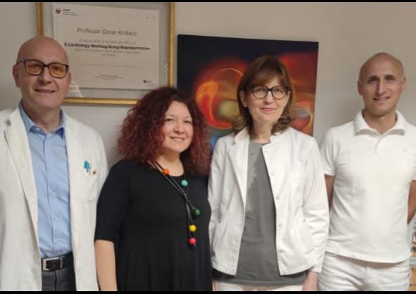
In recognition of these impressive outcomes, this integrated clinical care team was awarded Recognition of Achievement for the 2024 UNIVANTS of Healthcare Excellence awards. Congratulations to Andrea Snagić, Head of the Department of Medical Biochemistry and Laboratory Medicine, Goran Krstačić, Director of Institute for Cardiovascular Prevention and Rehabilitation, Ante Miljak, Clinical Care Coordinator, Nephrology, Sonja Frančula-Zaninović, Cardiologist.
To learn more about this best practice and others, please visit www.UnivantsHCE.com.
Pictured (from left to right): Goran Krstačić, Andrea Snagić, Sonja Frančula-Zaninović, Ante Miljak
Interview with Prof. Dr. Harjit Pal Bhattoa, New Chair of the Communications and Publications Division (CPD)
By Dra.BQF.María Pasquel-Moxley CPD Chair
Prof.
Dr. Harjit Pal Bhattoa, MD, PhD, MSc, DSc graduated from the Medical Faculty of the University of Debrecen, Hungary and is a Specialist in Laboratory Medicine and a Sub-Specialist in Laboratory Immunology and Hematology. He is the Head of the Endocrinology Unit at the Department of Laboratory Medicine at University of Debrecen. His major research interests are markers of bone turnover and vitamin D. He has been involved in numerous studies on primary and secondary osteoporosis. He has published over 100 peer-reviewed papers, authored one book and 9 book chapters. He serves as an Advisor to the European Commission in the field of medical devices and in vitro diagnostic medical devices. Apart from being the Chair of the IFCC Executive Committee of Communications and Publications Division (IFCC CPD), he is also a Consultant for the International Federation of Clinical Chemistry (IFCC) Scientific Division Committee on Bone Metabolism (IFCC C-BM). He is a Full-Member of the European Federation for Clinical Chemistry and Laboratory Medicine (EFLM) Communications Committee (EFLM C-C) and Secretary of the Committee: European Regulatory Affairs (EFLM C-ERA). He is the co-Editor-in-Chief of the Electronic Journal of the International Federation of Clinical Chemistry (eJIFCC) and the Editor of the EuroLabNews, the official newsletter of the EFLM, and is a regular reviewer for over 15 journals.
Prof. Dr. Harjit Pal Bhattoa says: “I have been with the IFCC Communications and Publications Division (CPD)
CPD group at IFCC WorldLab 2024 from left to right: Harjit Pal Bhattoa (HU), CPD Chair, Co-Editor eJIFCC ; Kannan Vaidyanathan (IN), Co-Editor eJIFCC; Maria Pasquel-Moxley (EC), C-PR Chair; Dr Tahir Pillay (SA), Past Chair CPD; Silvia Colli Lanzi, IFCC office; Deniz Topcu, C-IDC Chair, CPD Secretary; Dr. Raúl Girardi WG-IANT Chair; Colleen Strain, Corporate Member.
Working Group eJIFCC since 2015, as the Assistant Editor of the eJIFCC (Editor-in-Chief: Prof Gabor L Kovacs (2015-2017) and Prof Janos Kappelmayer (2018-2023) and am currently the eJIFCC co-Editor-in-Chief, along with Kannan Viadyanathan.
Having been on-board the IFCC CPD flagship under the exemplary Chairmanship of Prof Adeli Khosrow and later Prof Tahir Pillay, I gained in-depth insight into the unparalleled activities of the Committee. Under their leadership, I had the opportunity to recognize the importance of their mandate and the well-formulated terms of reference of the CPD.
The importance of transparent communication between the IFCC and all its interests cannot be over-emphasized. Efficient communication is paramount in forwarding Cont’d on page 20
the vision of the IFCC. Indicators of efficient communication are not necessarily based on the number of activities, but rather on the depth of penetration achieved by any message intended for dissemination.
Being an official platinum open-access Journal of the IFCC, the eJIFCC has strived to become a main stream Journal in the field. After successfully being indexed in PubMed and Scopus, we are striving for registration with the Web of Science, CrossRef, DOI, DOAJ and ICMJE. The introduction of the Editorial Manager platform for manuscript submission and iThenticate to screen plagiarism have made the Journal more attractive for potential contributors. eJIFCC, as a destination for future publications, is highlighted to the various IFCC Scientific Divisions and Emerging Technologies Division Functional Units.
The monthly IFCC eNews summarizes global activities in the field of Laboratory Medicine and we are striving for a more lively, casual and attractive layout.
The website is perhaps the main beacon of the IFCC. The importance of Internet and Digital Communication cannot be over-
Global MedLab Week 2025:
Get ready to participate!
The International Federation of Clinical Chemistry and Laboratory Medicine (IFCC) is pleased to invite you to celebrate the Global MedLab Week 2025 (GMLW 2025) from April 21-27.
This year’s theme “Labs Save Lives” underscores the vital impact of laboratory professionals and highlights the essential role they play in healthcare, often behind the scenes, but always at the forefront of patient care and medical innovations.
To join and support the Global MedLab Week campaign, please tweet, retweet, post and repost by tagging with our main hashtag #globalmedlabweek.
Visit the official Global MedLab Week 2025 website to access the original logos and icons, learn more about this important initiative, and submit your audio or video contribution: https://globalmedlabweek.org
emphasized and continual presence on all social media platforms is most desired. Although the accepted indicators of activity are usually the number of followers, likes, posts and re posts, nonetheless, quality content is our main goal.
One exemplary venture of the IFCC is the Global Med Lab Week (GMLW). The celebration of this festival has helped propel our profession into the limelight that it respectfully deserves. Apart from propagating the GMLW in the Latin world, the Working Group Ibero-America Nomenclature and Translation (WG-IANT) excels in making the IFCC activities available in Spanish and Portuguese, along with managing the electronic journal Diagnostico In Vitro (DIV), which is published with its new format every 4 months uninterrupted since 2015.
Being a global umbrella organization, sleek and polished public relations are mandatory for the IFCC. This is, perhaps, most efficiently achieved by delegating ambassadors to events held under the auspices of the IFCC, ensuring representation and showcasing its activities.
I feel privileged to lead the IFCC CPD Executive Committee with its deeply motivated and dedicated team”.
Follow the official network profiles:@globalmedlabweek (Facebook and Instagram)
Together, we will raise global awareness and highlight the significance of our work.
Let’s unite the global medical laboratory community in celebrating and promoting the indispensable role of laboratory professionals in healthcare.
Recognitions: This project is voluntary; there is no fee. Colleagues who submit their videos, audios or podcasts, will receive a certificate of participation from IFCC. Prizes will be awarded to the best videos and podcasts.
Executive Board Elections for the Term 2027-2029:
Report from the Nominations Committee
By Prof. Leslie C Lai, Chair, IFCC Nominations Committee
The current membership of the Nominations Committee is shown further below. We welcome new members to the Nominations Committee who began their first term of three years in 2024.
The following are changes to the regulations regarding election of Executive Board members that have been approved by IFCC Council members:
1. With the exception of the President and President Elect, members of the Executive Board are eligible for re-election only once for a given office.
2. No individual shall serve for more than six consecutive years on the Executive Board.
3. No individual shall serve for more than 6 years total in a given office.
4. No individual can be nominated for more than one position in the same year/same election cycle.
5. Elections for all IFCC Executive Board members will be held concurrently.
The election timeline of the next Executive Board for the term 2027 - 2029 is shown in the diagram below:
Medlab Middle East Rebranded as WHX Labs Dubai
Informa Markets (London, UK; www.informamarkets.com) has officially announced the rebranding of Medlab Middle East to WHX Labs Dubai, underscoring a transformative vision for connecting people, businesses, governments and innovators across borders. The name World Health Expo (WHX) unifies Informa’s healthcare events around the world to better facilitate connection and collaboration under a single brand.
Medlab Middle East 2025, held at the Dubai World Trade Centre (DWTC) under the theme ‘Empowering today’s medical labs for tomorrow’s future,’ brought together over 800 exhibitors and welcomed more than 20,000 visitors to explore the latest technology and updates for the laboratory industry. The event showcased the latest innovations and technologies across eight dynamic product categories, which include Disposables and Consumer Goods, Emergency Medicine, Imaging and Diagnostics, Healthcare and General Services, IT, Laboratory, Medical Equipment and Devices and Pharma and Nutrition. WHX Labs Dubai will build on Medlab Middle East’s 24 years of success, connecting more than half a million healthcare professionals in nine countries across four continents. By consolidating each event within the Informa Markets
Becton Dickinson to Spin Out Biosciences and Diagnostics Business
BHealthcare portfolio, WHX aims to amplify their impact, fostering greater innovation, stronger connections and transformative progress within the healthcare ecosystem.
Becton, Dickinson and Company (BD, Franklin Lakes, NJ, USA, www.bd.com), has announced that its board of directors has unanimously authorized BD management to pursue a plan to separate BD's Biosciences and Diagnostic Solutions business from the rest of BD to enhance strategic focus and growth-oriented investments and capital allocation for both BD and the separated business and enhance value creation for shareholders.
The separation decision was the result of a comprehensive business portfolio evaluation launched by BD in early 2024. The company believes that with the separation, the Biosciences and Diagnostic Solutions business will become a differentiated leader in Life Sciences Tools and Diagnostics. The Biosciences and Diagnostic Solutions business is expected to benefit from enhanced focus and investments as a pure-play Life Sciences Tools and Diagnostics leader. Biosciences is a leader in immunology and cancer research solutions and related clinical diagnostics, including flow cytometry instruments and reagents, and has innovative single-cell multiomics tools. Diagnostic Solutions is a leader in microbiology and infectious disease diagnostics, including molecular diagnostics, cervical cancer screening, microbiology automation and point-of-care offerings.
Bio-Rad to Acquire Digital PCR Developer in France
io-Rad Laboratories (Hercules, CA, USA; www.bio-rad.com) has entered into a binding offer to purchase all equity interests in Stilla Technologies (Villejuif, France; www.stillatechnologies. com). The acquisition remains subject to consultation with relevant employee representatives, regulatory approvals, and other customary closing conditions, and is expected to close by the end of the third quarter of 2025.
With operations in France and the U.S., Stilla develops and markets next-generation digital PCR instruments, consumables, and assays. The company’s Nio family of all-in-one digital PCR systems
Aaids the development of a wide range of genetic tests and molecular assays across multiple applications including liquid biopsy for oncology diagnostics, cell and gene therapy, organ transplant testing, infectious diseases, and food and environmental testing.
"Stilla’s next-generation digital PCR solutions would make a compelling and complementary addition to Bio-Rad’s best-in-class digital PCR portfolio,” said Norman Schwartz, CEO of Bio-Rad Laboratories. “Once closed, the acquisition will support our strategy to further expand our business into applied research and clinical diagnostics where customers expect a higher degree of automation and throughput capabilities.”
Siemens Licences Out Fast Track Diagnostics PCR Knowhow
dvanced Biological Laboratories (ABL, Luxembourg; www. ablsa.com) has signed a licensing and transfer agreement with Siemens Healthineers (Forchheim, Germany; www.siemenshealthineers.com) for the know-how and IP rights from Fast Track Diagnostics Luxembourg (FTD).
FTD was engaged in developing, manufacturing, and selling in-vitro diagnostic and research using only molecular testing products. As the owner of FTD, Siemens has licensed and transferred the know-how and IP rights to ABL and its affiliates (ABL Group) related to the design and
manufacturing of certain former FTD products. Based on this Know-How License and Transfer Agreement (Agreement), the ABL group intends to manufacture and commercialize its testing products equivalent to the former FTD products. The licensed know-how is related to real-time Polymerase Chain Reaction (PCR) tests, allowing for both singleplex and syndromic testing, to target conditions such as respiratory infections, gastroenteritis, meningitis, hepatitis, infections of the immunosuppressed, tropical diseases, sexually transmitted diseases, and early childhood diseases, and detect over 100 viruses, bacteria, parasites, and fungi.
Image: The unveiling of WHX Labs Dubai marks a new era for laboratory innovation (Photo courtesy of Informa Markets)
info@globetech.net
2025
MARCH
34th Annual Meeting of the Society of Virology (GfV). Mar 4-7; Hamburg, Germany; Web: virology-meeting.de
26th National Congress for Quality Assurance in Laboratory & EXPOQUÍM 2025. Mar 14-16; Zacatecas, Mexico; miconaquic.com
USCAP 113th Annual Meeting – United States and Canadian Academy of Pathology. Mar 22-27; Boston, MA, USA; Web: uscap.org
CACLP 2025 – 22nd China International In Vitro Diagnostic Expo. Mar 22-24; Hangzhou, China; Web: en.caclp.com
APRIL
ESCMID Global 2025. Apr 11-15; Vienna, Austria; Web: escmid.org
AMP Middle East 2025. Apr 12-13; Abu Dhabi, UAE; ampmiddleeast25.amp.org
Korea Lab 2025. Apr 22-25; Seoul, Korea; korealab.org
ECV 2025 – 9th European Congress of Virology. Apr 27-30; Dubrovnik, Croatia; eusv.eu
QICL 2025 - The 16th International & 22nd National Congress on Quality Improvement in Clinical Laboratories. Apr 29 – May 2; Tehran, Iran; iqctehran.ir
Expolab Monterrey 2025 – 25th National Congress of Clinical Chemistry and Laboratory Medica. Apr 30 –May 3; Monterrey, Mexico; fenacqc.org.mx
MAY
AZLTK & Lab Expo – 3rd International Azerbaijan Laboratory Medicine Congress & Lab Expo. May 1-3; Baku, Azerbaijan; azklmib.az
Immunology 2025 – Annual Meeting of the American
Association of Immunologists (AAI). May 3-7; Honolulu, HI, USA; Web: immunology2025.aai.org
ISLH 2025 Congress – International Society for Laboratory Hematology. May 7-9; Halifax, NS, Canada; Web: islh.org
22nd International Congress of Cytology. May 11-15; Florence, Italy; iccflorence2025.com
AACE Annual Meeting 2025 – American Association of Clinical Endocrinology. May 15-17; Orlando, FL, USA; am.aace.com
26th IFCC-EFLM EuroMedLab Congress of Clinical Chemistry and Laboratory Medicine. May 18-22; Brussels, Belgium; Web: euromedlab2025brussels.org
SLAS Europe 2025 Conference and Exhibition - Society of Laboratory Automation and Screening. May 20-22; Hamburg, Germany; slas.org
Hospitalar 2025. May 20-23; Sao Paulo, Brazil; hospitalar.com
ESHG 2025 – European Human Genetics Conference. May 24-27; Milan, Italy; 2025.eshg.org
ISBT Milan 2025 – 35th Regional Congress of the International Society of Blood Transfusion. May 31 - Jun 4; Milan, Italy; isbtweb.org
JUNE
WHX Labs Lagos 2025. Jun 2-4; Lagos, Nigeria; worldhealthexpo.com
LabMedUK25 – National Meeting of the Association for Laboratory Medicine (UK). Jun 9-11; Manchester, UK; Web: labmed.org.uk
WHX Miami 2025. Jun 11-13; Miami, FL, USA; Web: worldhealthexpo.com
108th Annual Meeting of the German Society for Pathology. Jun 12-14; Leipzig, Germany; Web: pathologie-dgp.de
EAACI 2025 – Annual Congress of the European Academy of Allergy & Clinical Immunology. Jun 13-16; Glasgow, Scotland; eaaci.org
50th CBAC – Congress of the Brazilian Society of Clini-
cal Analysis. Jun 15-18; Campinas, Brazil; sbac.org.br
ASM Microbe 2025 – American Society for Microbiology. Jun 19-23; Los Angeles, CA, USA; asm.org
ISTH 2025 Congress – International Society on Thrombosis and Haemostasis. Jun 21-25, Washington, DC, USA; isth2025.org
FOCIS 2025 – Annual Meeting of the Federation of Clinical Immunology Societies. Jun 24-27; Boston, MA, USA; focisnet.org
ESHRE 2025 – 41st Annual Meeting of the European Society of Human Reproduction and Embryology. Jun 29 - Jul 2; Paris, France; eshre.eu
JULY
FEBS 2025 – 49th Congress of the Federation of European Biochemical Societies. Jul 5-9; Istanbul, Turkey; 2025.febscongress.org
ASV 2025 – 44th Annual Meeting of the American Society for Virology (ASV). Jul 14-17; Montreal, Canada; asv.org
FEMS MICRO 2025 – Federation of European Microbiological Societies. Jul 14-17; Milan, Italy; femsmicro.org
WHX Labs Kuala Lumpur. Jul 16-18; Kuala Lumpur, Malaysia; Web: worldhealthexpo.com
IUIS 2025 – International Union of Immunological Societies. Aug 17-22; Vienna, Austria; iuis2025.org
ICBMBLM 2025 – Joint Conference of the Malaysian Association of Clinical Biochemists (MACB) & Malaysian Society for Biochemistry and Molecular Biology (MSBMB). Aug 25-27; Petaling Jaya, Malaysia; macb.org
SEPTEMBER
WHX Labs Cape Town 2025. Sep 2-4; Cape Town, South Africa; worldhealthexpo.com
Thailand Lab International 2025. Sep 3-5; Bangkok,
Thailand; thailandlab.com
ECP 2025 – 36th Congress of the European Society of Pathology. Sep 6-10; Vienna, Austria; esp-congress.org
CAP25 – Annual Meeting of the College of American Pathologists. Sep 13-16; Orlando, FL, USA; cap.org
EUROTOX 2025 – 59th Congress of the European Societies of Toxicology. Sep 14-17; Athens, Greece; eurotox2025.com
ESCV 2025 – 27th Annual Meeting of the European Society of Clinical Virology. Sep 17-20; Thessaloniki, Greece; escv2025.org
23rd International Congress of Therapeutic Drug Monitoring & Clinical Toxicology. Sep 21-24; Singapore; iatdmct2025.org
MASCL 2025 – Congress of the Association for Mass Spectrometry & Advances in Clinical Lab. Sep 21-26; Montreal, Canada; msacl.org OCTOBER
WHX Labs Nairobi 2025. Oct 6-8; Nairobi, Kenya; worldhealthexpo.com
ASHI 2025– 51st Annual Meeting of the American Society for Histocompatibility and Immunogenetics. Oct 6-10; Orlando, FL, USA; ashi-hla.org
JFBM 2025 – Journées Francophones de Biologie Médicale. Oct 8-10; Cannes, France; jfbm.fr
47th ISOBM Conference - International Society of Oncology and Biomarkers. Oct 13-15, Murnau am Staffelsee, Germany; isobm.org
62nd Annual Scientific Conference of the Australasian Association of Clinical Biochemistry and Laboratory Medicine (AACB). Oct 13-16; Auckland, New Zealand; aacb.asn.au
34th WASPALM & 51st ACBICON 2025 – World Congress of Association of Societies of Pathology and Association of Clinical Biochemists of India. Oct 13-16; Pune, India; waspalm2025.org
ASHG 2025 – Annual Meeting of the American Society of Human Genetics. Oct 14-18; Boston, MA, USA; ashg.org
17th National Conference of the Bulgarian Society of Clinical Laboratory. Sep 18-20; Golden Sands, Bulgaria; bsclconference.com
DKLM 2025 – Annual Congress of the German Society for Clinical Medicine and Laboratory Medicine (DGKL). Oct 23-24; Leipzig, Germany; laboratoriumsmedizin-kongress.de
ISBT Perth 2025 – Regional Congress of the International Society of Blood Transfusion. Oct 26-29; Perth, Australia; isbtweb.org
WSPID 2025 – 14th World Congress of the World Society for Pediatric Infectious Disease. Oct 28-31; Bangkok, Thailand; wspid2025.com
LMCE-KSLM 2025 – Laboratory Medicine Congress & Exhibition and 66th Annual Meeting of the Korean Society of Laboratory Medicine. Oct 29-31; Incheon, Korea; lmcekslm.org
NOVEMBER
APCCMI 2025 – 20th Asia Pacific Congress of Clinical Microbiology and Infection. Nov 2-4; Bangkok, Thailand; apccmi2025.com
57th SIBioC National Congress – Italian Society of
Clinical Biochemistry and Molecular Biology. Nov 5-7; Bologna, Italy; sibioc.it
73rd Annual Scientific Meeting of the American Society of Cytopathology (ASC). Nov 5-9; St. Louis, MO, USA; cytopathology.org
65th Annual Academic Assembly of the Japan Society of Clinical Chemistry (JSCC). Nov 7-9; Nagoya, Japan; jscc-jp.gr.jp
AMP 2025 – Association for Molecular Pathology Annual Meeting & Expo. Nov 11-15; Boston, MA, USA; amp25.amp.org
47th Annual Conference of the Association of Clinical Biochemists in Ireland (ACBI). Nov 14-15; Athlone, Ireland; acbi.ie
46th Annual Meeting of the American College of Toxicology (ACT). Nov 16-19; Phoenix, AZ, USA; actox.org
MEDICA 2025. Nov 17-20; Dusseldorf, Germany; medica-tradefair.com
ASCP 2025 – Annual Meeting of the American Society
for Clinical Pathology. Nov 17-20; Atlanta, GA, USA; ascp.org
DECEMBER
67th Annual Meeting & Exposition of the American Society of Hematology (ASH). Dec 6-9; Orlando, FL, USA; hematology.org
7th EFLM Conference on Preanalytical Phase. Dec 1213; Padua, Italy; eflmpreanalytical.org
2026
FEBRUARY
Labquality Days 2025 – International Congress on Quality in Laboratory Medicine. Feb 6-7; Helsinki, Finland; labqualitydays.fi
SLAS2026 International Congress & Exhibition. Feb 711; Boston, MA, USA; slas.org
WHX Labs Dubai 2025. Feb 9-12; Dubai, UAE; worldhealthexpo.com
Advanced Test Menu for Critical Care
References
The Modern Critical Care Profile
Prime Plus provides the most clinical value of any blood gas/critical care analyzer by adding essential tests for kidney function (BUN, Creatinine, eGFR), plasma volume (ePV), ionized magnesium (iMg) and MCHC.
Ionized Magnesium (iMg)
Hypomagnesemia is a frequent finding in critically ill patients.1 Magnesium therapy guided by real time ionized magnesium monitoring has been shown to improve outcome in these patients.2
Creatinine, eGFR, and BUN
Over 50% of patients admitted to the ICU develop some degree of acute kidney injury.3
Creatinine, eGFR, and BUN monitoring provides indication of changes in kidney function and helps guide therapy to prevent AKI.
Estimated Plasma Volume (ePV)
The plasma volume status of a patient is one of the top priorities in evaluating and treating critical illness including CHF, ARDS, AKI, and Sepsis.4-6
Mean Corpuscular Hemoglobin Concentration (MCHC)
Helps differentiate types of anemia.
1. Soliman HM. Development of ionized hypomagnesemia is associated with higher mortality rates. Crit Care Med 2003;31(4):1082-7.
2. Wilkes NJ et al. Correction of ionized plasma magnesium during cardiopulmonary bypass reduces the risk of postoperative cardiac arrhythmia. Anesth and Analg 2002;95(4) 828-834.
3. Mandelbaum T et al. Outcome of critically ill patients with acute kidney injury using the AKIN criteria. Crit Care Med 2011;39(12):2659-2664.
4. Kobayashi M et al. Prognostic Value of Estimated Plasma Volume in Heart Failure in Three Cohort Studies; Clin Res Cardiol 2019;108(5): 549-561.
5. Niedermeyer, et al. Calculated Plasma Volume Status Is Associated With Mortality in Acute Respiratory Distress Syndrome. Critical Care Explorations: September 2021, V3(9):1-9.
6. Kim HK et al. Prognostic Value of Estimated Plasma Volume Status in Patients with Sepsis. J Korea Med Sci 2020;9(37):1-10.